cardiobacterium hominis endocarditis: description of two patients
TRANSCRIPT
JOURNAL OF CLINICAL MICROBIOLOGY, Jan. 1977, P. 75-80Copyright © 1977 American Society for Microbiology
Vol. 5, No. 1Printed in U.S.A.
Cardiobacterium Hominis Endocarditis: Description of TwoPatients and Characterization of the Organism
DANIEL D. SAVAGE, ROBERT L. KAGAN, NATHANIEL A. YOUNG,* AND ANDREW E. HORVATH
Cardiology Branch, National Heart and Lung Institute, Bethesda, Maryland 20014; Clinical PathologyDepartment, Clinical Center, Laboratory of Pathology, National Cancer Institute, National Institutes ofHealth, Bethesda, Maryland 20014;* and Department ofPathology, Presbyterian Hospital, Albuquerque,
New Mexico
Received for publication 4 June 1976
Two cases of endocarditis caused by Cardiobacterium hominis are reported. Inboth instances infection was subacute and characterized by (i) implantation on
abnormal valves, (ii) chronic course lasting weeks to months before recognition,and (iii) rapid clinical and bacteriological response to penicillin, as well as otherantibiotics commonly used to treat infections caused by gram-negative bacilli.Our isolates of C. hominis are compared with strains in the National Institutesof Health culture collection. Optimal growth requires yeast extract and incuba-tion at 37°C with increased humidity and supplemental CO2. The production ofindole, a positive oxidase reaction, and characteristic sugar fermentations dis-tinguish C. hominis from other slow-growing, gram-negative bacilli.
Cardiobacterium hominis, a slow-growing,gram-negative bacillus formerly designated as"group II-D" (17), was described by Slotnick andDougherty in 1964 (12). Septicemia, usually as-sociated with endocarditis, is the only diseasestate with which the organism has been une-quivocally linked etiologically. This report de-scribes two patients with endocarditis causedby C. hominis and compares bacteriologicalcharacteristics of the isolates to strains in theNational Institutes of Health culture collection.
CASE STUDIESCase 1. A 62-year-old man with asymmetric
septal hypertrophy (ASH) was admitted to thehospital because of chest pain, chills, nightsweats, fatigue, orthopnea, and weight loss (25pounds) of 7-months duration. These symptomsfollowed a dental manipulation for which hereceived oral penicillin 3 days before and after-wards. Two months prior to admission he hadan influenza-like illness treated with a singleintramuscular injection of penicillin. He feltwell for 2 weeks before his symptoms returned.At admission, he appeared chronically ill anddyspneic. The heart rate was 95 beats/min, res-pirations 22 per min, blood pressure 100/64 mmof Hg, and temperature 38.5°C. A Roth spot waspresent in the left ocular fundus. The heart wasenlarged. A grade 5/6 apical holosystolic mur-mur and a grade 1/6 apical diastolic murmurwere present. The spleen was palpable. Thehematocrit was 28%, leukocyte count 8,200/mm3, and erythrocyte sedimentatioin rate 94
75
mm in 1 h. The rapid plasma reagin test forsyphilis was positive, but the fluorescent trepo-nemal antibody test was negative. Urinalysiswas unremarkable. A chest roentgenogramshowed increased pulmonary vascularity andenlargement of the left atrium and left ventri-cle. The electrocardiogram showed left bundlebranch block. All 14 blood cultures taken onhospital days 2 through 9 revealed a gram-negative rod, later identified as C. hominis.The patient was initially treated with genta-
micin and carbenicillin. After determinationthat the organism was sensitive to penicillin ata minimum inhibitory concentration of lessthan 4 ,ug/ml, treatment with 3 million U ofpenicillin G every 6 h intravenously was initi-ated and continued as the only antibiotic for 4weeks. After less than 1 week the patient be-came afebrile, the splenic size diminished, andhis exercise tolerance increased concomitantlywith the disappearance of chest pain and symp-toms of congestive heart failure.He was discharged after 46 days, but was
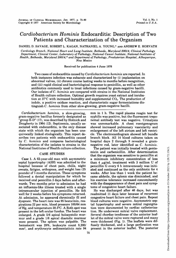
readmitted 11 days later because of recurrentcongestive heart failure and chest pain. Repeatblood cultures were negative. Asymmetric sep-tal hypertrophy and severe mitral regurgita-tion were documented by cardiac catheteriza-tion. He underwent mitral valve replacement.Several chordae tendineae of the anterior leaf-let of the mitral valve were ruptured and manywere thickened (Fig. 1). The leaflets were dif-fusely thickened, and a large perforation waspresent in the anterior leaflet. The posterior
Dow
nloa
ded
from
http
s://j
ourn
als.
asm
.org
/jour
nal/j
cm o
n 15
Oct
ober
202
1 by
128
.72.
156.
5.
76 SAVAGE ET AL.
FIG. 1. Opened excised valve from case 1 showing diffusely thickened leaflets, with a large perforation inthe anterior leaflet. Calcific nodules representing "healed vegetations" border the perforation.
leaflet was focally thickened. The aortic valvewas normal. The patient had an uneventfulrecovery and after 20 months remains in func-tional class I.Case 2. A 56-year-old man known to have a
precordial murmur since childhood developedincreasing dyspnea and palpitations after anupper respiratory infection. Examination dis-closed overt congestive heart failure. The mur-mur was believed to be due to congenital aorticvalvular disease. Four months later he sud-denly developed expressive and sensoryaphasia, which was attributed to a cerebralembolus. He recovered slowly, but, 5 monthsafter the cerebrovascular accident, he againdeveloped congestive heart failure. Because offever and splenomegaly he was admitted to thehospital. The heart rate was 110 beats/min,respirations 16 per min, blood pressure 100/60mm of Hg, and temperature 38.1°C. The heartwas enlarged. There was a grade 3/6 holosys-tolic murmur at the left sternal border andapex. Both the liver and spleen were palpable.The hematocrit was 31%, leukocyte count was6,500/mm' with a normal differential, and theerythrocyte sedimentation rate was 44 mm in 1h. Urinalysis revealed 4+ albumin and 20 leu-kocytes and 100 erythrocytes per high-powerfield. Gram-negative bacilli, later identified asC. hominis, were recovered from six of six bloodcultures. By disk sensitivity testing, the orga-nism was determined to be susceptible to peni-cillin, ampicillin, chloramphenicol, tetracy-cline, streptomycin, kanamycin, gentamicin,colistin, and carbenicillin. Serum agglutininswith a titer of 1:80 were demonstrated against
Brucella melitensis. Because of initial suspi-cion of brucella endocarditis, therapy was be-gun with tetracycline and streptomycin. Hepromptly became afebrile, but after 5 days thetherapy was changed to aqueous penicillin G,10 million U intravenously per day, for 18 days.Oral therapy was continued for another 3weeks with penicillin V, 5 g daily, plus probe-necid. The patient remained afebrile and re-covered uneventfully. Three months later heunderwent cardiac catheterization, which docu-mented a peak systolic gradient of 17 mm of Hgacross the aortic valve. Left ventricular angiog-raphy revealed a slightly calcified aortic valve.There was a marked reduction in overall my-ocardial contractility. The congestive heartfailure progressed, and he died 30 months laterwithout clinical or bacteriological evidence ofrecurrent infective endocarditis; however, noautopsy was done.
MATERIALS AND METHODSBlood culture technique. In case 1, 10 ml of ve-
nous blood collected by syringe and needle was inoc-ulated into each of two screw-capped vacuum bottlescontaining 100 ml of brain heart infusion (BHI)broth with sodium polyanetholsulfonate (SPS), p-aminobenzoic acid, and CO2 (BBL). Upon receipt inthe laboratory, one bottle was air-vented by a needleattached to a Swinney filter. This bottle was consid-ered aerobic, and the unvented bottle was consid-ered anaerobic. Both were incubated at 35°C andinspected macroscopically daily for evidence ofgrowth. Twenty-four-hour subcultures from bothanaerobic and aerobic bottles were made onto BYEagar (1) and incubated anaerobically or under 10%C02, respectively. In case 2, 5 ml of venous bloodwas inoculated into each of two vacuum bottles. One
J. CLIN. MICROBIOL.
Dow
nloa
ded
from
http
s://j
ourn
als.
asm
.org
/jour
nal/j
cm o
n 15
Oct
ober
202
1 by
128
.72.
156.
5.

C. HOMINIS ENDOCARDITIS 77
contained 100 ml of casein soy broth with SPS andCO2. The second contained 100 ml of thioglycolatebroth (without indicator) as well as SPS and CO2.Each bottle was incubated without venting at 35°C.Routine blind subcultures to chocolate agar slantswere made at 48 h and on day 10. Positive chocolateslants were sent to the National Institutes of Healthfor characterization. These subcultures were thenhandled as the subcultures from case 1. Organismsrecovered from blood cultures in case 1 and case 2were identified using the analysis outlined byWeaver et al. (19).
C. hominis bacterial strains. Three strains of C.hominis (NIH CPD no. 5, 7, and 8) were obtainedfrom the National Institutes of Health culture col-lection maintained by the Microbiology Service ofthe Clinical Pathology Department. Strain NIHCPD no. 8 was isolated by Irving Slotnick, Univer-sity of Florida, and has been suggested as a refer-ence strain for C. hominis (3). It corresponds toSlotnick's strain 6573.
Media. All C. hominis cultures were streaked on5% horse or sheep blood in Columbia base (Difco),phenylethyl alcohol medium, MacConkey agar(Difco), and two types of modified blood agars (1, 5).Growth at 22, 37, and 42°C was observed in Trypti-case soy broth (TSB, Difco) at 24 and 48 h. Motilitywas determined in semisolid motility medium(Difco) at 37 and 22°C.Growth requirements. Growth of C. hominis with
and without 5 to 10% CO2 was compared at 37°C. Amoisture chamber was used at 37°C to determinewhether increased humidity enhanced growth. Therequirement for accessory growth factors was as-sessed by placing paper strips containing X factor(heme), V factor (coenzyme NAD), and both X and Vfactors on a Columbia agar plate previously inocu-lated with the test organism.
Biochemical reactions. The methods for biochem-ical characterization followed standard microbiolog-ical techniques (16). Fermentation reactions weredetermined in phenol red broth base (Difco) contain-ing 1% carbohydrate. H2S production was tested bysuspending lead acetate-impregnated paper overTSI slants (Difco) and observing both the TSI buttand paper strip. Production of indole was tested inindole test broth (tryptone broth, Difco) by usingboth Kovacs' reagent and Ehrlich regent. A com-plete test for nitrate reduction was performed aftergrowth in nitrate beef extract broth (BBL). Thepresence of catalase was demonstrated by the evolu-tion of gas from 3% H202 placed directly on coloniesof the test organism on BHI agar. The oxidase reac-tion was demonstrated by the method of Kovacs aselaborated by Steel (15). The Voges-Proskauer testfor acetylmethylcarbinol and the methyl red testwere performed on a 48-h growth in MRVP medium(Difco). Simmons citrate agar (Difco) was employedto test the ability to utilize citrate as the sole sourceof carbon and energy. Urease production was deter-mined at 48 h on urea slants (Difco). Esculin hydrol-ysis in the presence of 40% bile was observed on bileesculin slants (Difco). Amino acid decarboxylase/dihydrolase activity was detected in Moeller me-dium (BBL) containing either L-arginine monohy-
drochloride, L-ornithine dihydrochloride, or L-lysinedihydrochloride. The tubes were inoculated by stab-bing deeply and then were overlayed with a 1-cmlayer of sterile mineral oil.
Antibiotic susceptibility. Susceptibility to anti-biotics of the isolates from cases 1 and 2 and thethree available strains of C. hominis was deter-mined by a serial dilution microtiter technique (7).The broth medium employed was one routinely usedfor testing hemophilus: 10% Fildes reagent, 0.5%yeast extract, and 2% IsoVitaleX in TSB base.
RESULTSC. hominis was recovered from all blood cul-
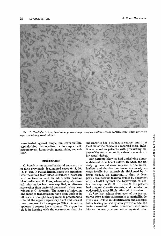
tures drawn in each patient, but its characteris-tically slow growth resulted in large numbers ofcultures being taken (14 in case 1 and 6 in case2) before growth was detected 6 days after ini-tial collection. Previous authors have empha-sized the pleomorphism of C. hominis and itsirregular staining (12, 13, 16). We confirmedthis when the organisms were grown on agarwithout yeast. However, when the organismswere grown on agar containing yeast extractthey appeared mainly as uniform gram-nega-tive rods (0.5 by 2 ,um) (Fig. 2). Both supple-mental CO2 and increased humidity, testedseparately, enhanced growth.
Biochemical characteristics of our isolatesare compared with reference strains in Table 1.C. hominis is reported to produce indole. How-ever, we found that indole production wasslight and could be missed unless concentratedby xylene extraction before adding Kovacs' orEhrlich reagent for detection. C. hominis mayresemble Actinobacillus actinomycetemcomi-tans or Haemophilus aphrophilus, but the lat-ter two are oxidase negative and reduce nitrateto nitrite. Other Haemophilus species requireV factor and may also require X factor. Pasteu-rella is distinguished by catalase positivity.Brucella and Bordetella bronchiseptica are alsocatalase positive and, moreover, differ from C.hominis in their ability to produce urease andreduce nitrates and in their failure to produceindole. Bordetella parapertussis, unlike C.hominis, is oxidase negative and catalase posi-tive and produces beta hemolysis on blood agar.Eikenella corrodens, a gram-negative rod re-cently recognized as a pathogen, has been im-plicated as a cause of endocarditis (4). Colonieshave a typical corroding morphology and do notproduce acid by fermentation or oxidation fromcarbohydrates (2).The C. hominis strains tested were uni-
formly susceptible to penicillin G as well asantibiotics commonly used to treat gram-nega-tive bacillary infections (Table 2). Minimal in-hibitory concentrations were less than 2 gg/mlwhen the two isolates and the reference strains
VOL. 5, 1977
Dow
nloa
ded
from
http
s://j
ourn
als.
asm
.org
/jour
nal/j
cm o
n 15
Oct
ober
202
1 by
128
.72.
156.
5.
78 SAVAGE ET AL.
I~~~4~~~d4
t~ ~~~\S/\' 3 'ji% < ,
yr~~~~Q
,@F,*, ow> ej4''6,iel,,,$i.~J
r
1444 A
FIG. 2. Cardiobacterium hominis organisms appearing as uniform gram-negative rods when grown onagar containing yeast extract.
f..A
were tested against ampicillin, carbenicillin,cephalothin, tetracycline, chloramphenicol,streptomycin, kanamycin, gentamicin, and col-istin.
DISCUSSIONC. hominis has caused bacterial endocarditis
in nine previously documented cases (6, 8, 10,14, 17, 20). In two additional cases the organismwas recovered from blood cultures: a newbornwith septicemia, and an adult with positiveblood cultures (17). Thus, where adequate clini-cal information has been supplied, no diseasestate other than bacterial endocarditis has beenrelated to C. hominis. The source of infectionand mode of transmission have been unclear inall cases, although the organism is presumed toinhabit the upper respiratory tract and feces ofmost humans of all age groups (13). C. hominisappears to possess low virulence. This hypothe-sis is in keeping with the observation that the
endocarditis has a subacute course, and in atleast six of the previously reported cases, infec-tion occurred in patients with preexisting dis-ease of the mitral or aortic valves or a ventricu-lar septal defect.Our patients likewise had underlying abnor-
malities of their heart valves. In ASH, the un-derlying heart disease in case 1, the mitralleaflets and chordae tendineae are nearly al-ways focally but extensively thickened by fi-brous tissue, an abnormality that at leastpartly results from trauma caused by abutmentof this leaflet against the hypertrophied ven-tricular septum (9, 10). In case 2, the patienthad congenital aortic stenosis, and the infectiveendocarditis most likely affected this valve.
C. hominis isolates from each of the two pa-tients were highly susceptible to penicillin de-rivatives. Delays in identification and suscepti-bility testing caused by slow growth of the bac-terium resulted in initial treatment with anti-biotics generally more active against other
J. CLIN. MICROBIOL.
I,.p
4.
It ..4 .l
'.. I
A.4
N .
f-.1
Av
." '11%4.21 ..c
..Io 'k-
Dow
nloa
ded
from
http
s://j
ourn
als.
asm
.org
/jour
nal/j
cm o
n 15
Oct
ober
202
1 by
128
.72.
156.
5.

C. HOMINIS ENDOCARDITIS 79
TABLE 1. Key biochemicala characteristics of fivestrains of C. hominis
NIH CPD strainDetermination Case 1 Case 2
5 7 8
Oxidase + +wb + +w +CatalaseDextrose A" A A A ALactoseSucrose A A A A AMaltose A A A A ADecarboxylase
reactionsIndole + + +w + +Growth10% C02 4+ 4+ 4+ 4+ 4+Aerobic 0 0 0 0 0Anaerobic 1+ 1+ 1+ 1+ 1+
Nitrate reduc- - - - - -tion
a The organisms grew poorly on triple sugar ironmedia and in MR-VP broth. They did not require Xand V factors, were nonmotile, and did not producegas from glucose.
b +w denotes a weakly positive reaction.' A denotes acidification.
TABLE 2. Antibiotic susceptibilities offive strains ofC. hominisa
NIH CPD strainAntibiotic Case 1 Case 2
5 7 8
Penicillin G <0.4 <0.4 <0.4 <0.4 <0.4Ampicillin <0.4 <0.4 <0.4 <0.4 <0.4Carbenicillin <1.6 <1.6 <1.6 <1.6 <1.6Cephalothin <0.4 <0.4 <0.4 <0.4 <0.4Tetracycline 1.6 0.8 0.8 0.8 <0.4Chloramphenicol 1.6 0.8 0.8 1.6 <0.4Streptomycin 0.8 1.6 0.8 1.6 0.8Kanamycin <0.4 0.8 <0.4 <0.4 0.8Gentamicin 0.8 0.8 0.2 0.4 0.8Colistin <0.4 <0.4 <0.4 <0.4 <0.4
a Numbers represent minimal inhibitory concen-tration (MIC) in micrograms per milliliter.
gram-negative bacilli. After identification ofthe organism, however, both patients receivedpenicillin in high doses intravenously, and bac-teriological cure resulted.The morphological characteristics of C. hom-
inis were dependent on whether or not yeastextract was incorporated into the growth me-dium. This organism has characteristicallybeen described as an irregularly straining,highly pleomorphic gram-negative rod. Wefound that on two types of modified blood agar,both containing yeast extract, the organism ap-pears homogeneously stained and of uniformdimensions with occasional irregular forms.These properties suggest to us that, in addition
to supplemental CO2 and increased humidity,yeast extract promotes optimal growth. With-out yeast extract highly pleomorphic, irregu-larly staining rods were seen. The biochemicalreactions of our two isolates are identical withSlotnick's reference strain 6573, as well as twoadditional strains of C. hominis available forcomparison. Among the most important bio-chemical reactions which distinguish C. hom-inis from other slow-growing, gram-negativebacilli with which it may be initially confusedare indole production, the oxidase reaction, andthe sugar fermentation pattern. The productionof indole, although slight, appears invariableand is best demonstrated by xylene extractionof the test medium.
ACKNOWLEDGMENTS
We wish to thank Vee J. Gill, who characterized theorganism identified in case 2, and William Roberts, Ste-phen Epstein, and James MacLowry, who reviewed thismanuscript.
LITERATURE CITED1. Barile, M. F., R. Yaguchi, and W. C. Eveland. 1958. A
simplified medium for the cultivation of the pleuro-pneumonia-like organisms and the L-forms of bacte-ria. Am. J. Clin. Pathol. 30:171-176.
2. Brooks, G. F., J. M. O'Donoghue, and J. P. Rissing.1974. Eikenella corrodens, a recently recognized path-ogen. Medicine 53:325-342.
3. Dorff, G. J., L. J. Jackson, and M. W. Rytel. 1974.Infections with Eikenella corrodens. Ann. Intern.Med. 80:305-309.
4. Dowell, V. R., Jr., and T. M. Hawkins. 1974. In Labora-tory methods in anaerobic bacteriology, p. 53. U. S.DHEW (CDC) 74-8272 CDC Laboratory Manual, At-lanta, G., p. 53.
5. Laguna, J., B. M. Derby, and R. Chase. 1975. Cerebralmycotic aneurysm. Arch. Neurol. 32:38-639.
6. Lapage, S. P. 1974. Genus Cardiobacterium Slotnickand Dougherty 1964, 271, p. 377-378. In R. E. Bu-chanan and N. E. Gibbons (ed.), Bergey's manual ofdeterminative bacteriology, 8th ed. The Williams &Wilkins Co., Baltimore.
7. MacLowry, J. D., M. J. Jaqua, and S. T. Selepak. 1970.Detailed methodology and implementation of a sem-iautomated serial dilution microtechnique for anti-microbial susceptibility testing. Appl. Microbiol. 21:46-53.
8. Midgley, J., S. P. Lapage, and B. A. G. Jenkins. 1970.Cardiobacterium hominis endocarditis. J. Med. Mi-crobiol. 3:91-98.
9. Morrow, A. G., W. C. Roberts, and J. Ross, Jr. 1968.Obstruction to left ventricular outflow. Current con-cepts of management and operative treatment. Ann.Intern. Med. 69:1255.
10. Perdue, C. D., Jr., E. R. Dorney, and F. Ferrier. 1975.Embolomycotic aneurysm associated with bacterialendocarditis due to Cardiobacterium hominis. Am.Surg. 34:901-904.
11. Roberts, W. C., and J. K. Perloff. 1972. Mitral valvulardisease. Ann. Intern. Med. 77:939-975.
12. Slotnick, I. J., and M. Dougherty. 1964. Further char-acterization of an unclassified group of bacteria caus-ing endocarditis in man: Cardiobacterium hominisgen. et sp. n. Antonie van Leeuwenhoek J. Microbiol.Serol. 30:261-272.
13. Slotnick, I. J., J. Mertz, and M. Dougherty. 1964. Flu-
VOL. 5, 1977
Dow
nloa
ded
from
http
s://j
ourn
als.
asm
.org
/jour
nal/j
cm o
n 15
Oct
ober
202
1 by
128
.72.
156.
5.

80 SAVAGE ET AL.
orescent antibody detection of human occurrrence ofan unclassified bacterial group causing endocarditis.J. Infect. Dis. 114:503-505.
14. Snyder, A. I., and P. D. Ellner. 1969. Cardiobacteriumhominis endocarditis. N.Y. State J. Med. 69:704-705.
15. Steel, K. J. 1961. The oxidase reaction as a taxonomictool. J. Gen. Microbiol. 25:297-306.
16. Tatum, M. W., W. H. Ewing, and R. E. Weaver. 1974.Miscellaneous gram-negative bacteria, p. 288-289. InE. H. Lennette, E. H. Spaulding, and J. P. Truant(ed.), Manual of clinical microbiology, 2nd ed. Amer-ican Society for Microbiology, Washington, D.C.
17. Tucker, D. N., I. J. Slotnick, and E. 0. King. 1962.
J. CLIN. MICROBIOL.
Endocarditis caused by a Pasteurella-like organism.Report of four cases. N. Engl. J. Med. 267:913-916.
18. Wang, K., F. L. Gobel, and D. F. Gleason. Bacterialendocarditis in idiopathic hypertrophic subaortic ste-nosis. Am. Heart J. 89:359-365.
19. Weaver, R. E., H. W. Tatum, and D. E. Hollis. 1972.The identification of unusual pathogenic gram nega-tive bacteria (Elizabeth 0. King). Center for DiseaseControl, Atlanta.
20. Weiner, M., S. Werthamer. 1975. Cardiobacteriumhominis endocarditis. Characterization of the unu-sual organisms and review of the literature. Am. J.Clin. Pathol. 63:131-134.
Dow
nloa
ded
from
http
s://j
ourn
als.
asm
.org
/jour
nal/j
cm o
n 15
Oct
ober
202
1 by
128
.72.
156.
5.