carcinoma of the oral cavity - leitlinienprogramm … · [email protected] ... prof. h. pistner,...
TRANSCRIPT
© German Guideline Program in Oncology | Guideline Oral Cavity Carcinoma | Short Version | November 2012
1
0
Short Version
Carcinoma of the Oral Cavity "Diagnosis and Management of Carcinoma of the Oral Cavity"
AWMF Registry Number 007/100OL
Version 2.0 - November 2012

2
Contents
1. About this Short Version ...................................................................... 4
1.1. Publisher 4
1.2. Coordinating association ....................................................................................................... 4
1.3. Guideline funding .................................................................................................................. 4
1.4. Contact 4
1.5. Special remarks ..................................................................................................................... 5
1.6. Additional guidance documents ............................................................................................. 5
1.7. Responsibilities ..................................................................................................................... 6 1.7.1. Authors of this guideline ................................................................................................ 6 1.7.2. Methodological support .................................................................................................. 6
1.8. Professional associations and organizations concerned .......................................................... 6
1.9. Grading of evidence according to SIGN .................................................................................. 8
1.10. Grading of recommendations ................................................................................................ 8
1.11. Abbreviations ........................................................................................................................ 9
2. Consensus-based, agreed Recommendations and Statements .............. 11
2.1. Risk factors ......................................................................................................................... 11
2.2. Screening and prevention .................................................................................................... 11
2.3. Primary diagnosis ................................................................................................................ 11
2.4. Imaging procedures and further diagnostics ........................................................................ 12
2.5. Imaging procedures and diagnostic methods for exclusion of synchronous secondary
tumors, distant metastases, unknown primary tumors (CUP) and recurrence ........................ 13
2.6. Biopsy and histopathology ................................................................................................... 13
2.7. Treatment recommendations ............................................................................................... 14
2.8. Surgery as treatment for primary cancer .............................................................................. 14
2.9. Cervical lymph node dissection............................................................................................ 15
2.10. Reconstruction .................................................................................................................... 16
2.11. Radiotherapy ....................................................................................................................... 17
© German Guideline Program in Oncology | Guideline Oral Cavity Carcinoma | Short Version | November 2012
3
2.12. Radiotherapy combined with chemotherapy ......................................................................... 17
2.13. Prevention and management of radiation-induced side effects ............................................. 18
2.14. Management of locoregional recurrence .............................................................................. 18
2.15. Palliative care and palliative medicine .................................................................................. 19
2.16. Follow-up and rehabilitation ............................................................................................... 19 2.16.1. Follow-up..................................................................................................................... 19 2.16.2. Functional masticatory rehabilitation ............................................................................ 19 2.16.3. Speech and swallowing rehabilitation ............................................................................ 20 2.16.4. Nutritional therapy ....................................................................................................... 20 2.16.5. Psychosocial counseling and support ............................................................................ 20
3. Quality Indicators ............................................................................... 21
4. References ......................................................................................... 23
© German Guideline Program in Oncology | Guideline Oral Cavity Carcinoma | Short Version | November 2012
1.1 Publisher 4
1. About this Short Version
1.1. Publisher
German Guideline Program in Oncology (GGPO)
of the Association of the Scientific Medical Societies in Germany, the German Cancer Society and German Cancer Aid
Office: c/o German Cancer Society Kuno-Fischer-Strasse 8
14057 Berlin
www.leitlinienprogramm-onkologie.de
1.2. Coordinating association German Society for Oral and Maxillofacial Surgery (DGMKG)
1.3. Guideline funding This guideline has been funded by German Cancer Aid under the auspices of the
German Guideline Program in Oncology
1.4. Contact Guideline coordinator:
Univ.-Prof. Dr. med. Dr. med. dent. Klaus-Dietrich Wolff Clinic and Polyclinic for Oral and Maxillofacial Surgery Rechts der Isar Hospital, Munich Technical University Ismaninger Str. 22
81675 Munich
Tel.: 004989 4140-2921
Fax: 004989 4140-4993
www.med.tum.de
© German Guideline Program in Oncology | Guideline Oral Cavity Carcinoma | Short Version | November 2012
1.5 Special remarks 5
1.5. Special remarks
Medical science is subject to continuing development, meaning that any information – particularly concerning diagnostic and therapeutic methods – can only reflect the standards of knowledge applicable at the time of this guideline going to print. Utmost care has been taken in the provision of recommendations for treatment and the selection as well as dosage of medicinal products. Nevertheless, users are asked to refer to the package leaflets and data sheets provided by manufacturers and to consult a specialist if they are in any doubt. In the general interests of all concerned, any discrepancies that come to light should please be notified to the editorial office of
the GGPO.
The user is responsible for all chosen diagnostic and therapeutic measures, medication and
dosage.
Registered trademarks (protected brand names) are not specifically acknowledged as such within this guideline. The absence of such acknowledgment does not imply, however, that the product
names are not protected.
This guideline is protected in its entirety by copyright. Utilization beyond the confines of copyright law without written permission from the editorial office of the GGPO is prohibited and punishable by law. No part of this document may be reproduced in any format without written permission from the editorial office of the GGPO. This applies in particular to the duplication, translation, microfilming and saving, utilization and exploitation of the document in electronic systems,
Intranets and the Internet.
1.6. Additional guidance documents The content of this short version is based on the full-length version of the S3 Guideline, "Diagnosis and Management of Carcinoma of the Oral Cavity", which can
be found at the following addresses:
http://www.awmf.org/leitlinien/aktuelle-leitlinien.html
http://www.leitlinienprogramm-onkologie.de/OL/leitlinien.html
http://www.krebsgesellschaft.de/wub_llevidenzbasiert,120884.html
http://www.dgmkg.de
The following documents accompany this short version:
− Guideline report
− Full version
− Patient guideline
All of these documents can likewise be found on the websites listed above.
© German Guideline Program in Oncology | Guideline Oral Cavity Carcinoma | Short Version | November 2012

1.7 Responsibilities 6
1.7. Responsibilities
1.7.1. Authors of this guideline Wolff K.-D., Bootz F., Beck J., Bikowski K., Böhme P., Budach W., Burkhardt A., Danker H., Eberhardt W., Engers R., Fietkau R., Frerich B., Gauler T., Germann G., Gittler-Hebestreit N., Grötz K., Horch R., Ihrler S., Keilholz U., Lell M., Lübbe A., Mantey W., Nusser-Müller-Busch R., Pistner H., Paradies K., Reichert T., Reinert S., Schliephake H.,
Schmitter M., Singer S., Westhofen M., Wirz S., Wittlinger M.
1.7.2. Methodological support
1. The German Guideline Program in Oncology: Prof. I. Kopp (AWMF), Marburg and Dr M. Follmann, MPH, MSC (DKG), Berlin
2. External consultants: Dr A. Nast, S. Rosumeck, Dr A. Sammain, and Prof. B. Rzany (Division of Evidence Based Medicine), Berlin
3. Guideline Officer of the coordinating association: Prof. H. Pistner, Erfurt
1.8. Professional associations and organizations concerned
Organizations Authors
German Society for Oral and Maxillofacial Surgery Wolff, K.-D., Grötz K., Reinert, S.
German/Austrian/Swiss Working Group on Maxillofacial Tumors (DÖSAK)
Frerich, B.
Study Group on Maxillofacial Surgery Reichert, T.
German Society of Dental, Oral & Craniomandibular Sciences
Schliephake, H.
German Society for Oto-Rhino-Laryngology Bootz F., Westhofen M.
German Society of Pathology Burkhardt A., Ihrler S.
German Society of Radiation Oncology Fietkau R., Budach W., Wittlinger M.
German Society of Hematology and Oncology Keilholz U., Gauler T., Eberhardt W.
German Society of Plastic and Reconstructive Surgery
Horch R., Germann G.
Working Group Head and Neck of the German Society of Radiology
Lell M.
© German Guideline Program in Oncology | Guideline Oral Cavity Carcinoma | Short Version | November 2012

1.8 Professional associations and organizations concerned 7
Organizations Authors
KOK Paradies K., Gittler-Hebestreit N.
Working Group for Supportive Care in Cancer, Rehabilitation and Social Medicine (ASORS)
Lübbe A.
AEK Engers K.
German Dental Association Boehme, P.
Federal Association of Panel Dentists Beck, J.
Working Group on Orofacial Pain of the German Association for the Study of Pain
Schmitter M.
Working Group on Tumor Pain of the German Association for the Study of Pain
Wirz S.
Patient representative Mantey W.
DVSG, Nat. Cancer Center Bikowski K.
German Logopedia Society Nusser-Müller-Busch R.
Working Group for Psycho-Oncology of the German Cancer Society (PSO)
Singer S., Danker H.
© German Guideline Program in Oncology | Guideline Oral Cavity Carcinoma | Short Version | November 2012
1.9 Grading of evidence according to SIGN 8
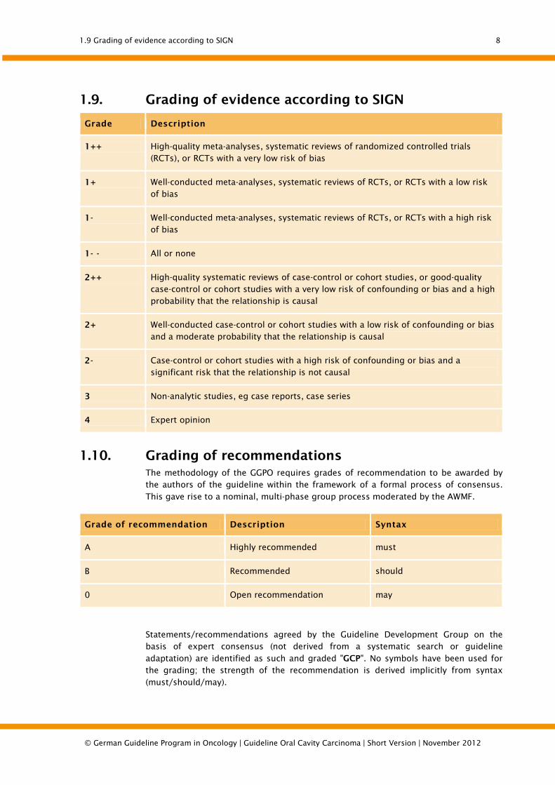
1.9. Grading of evidence according to SIGN
Grade Description
1++ High-quality meta-analyses, systematic reviews of randomized controlled trials (RCTs), or RCTs with a very low risk of bias
1+ Well-conducted meta-analyses, systematic reviews of RCTs, or RCTs with a low risk of bias
1- Well-conducted meta-analyses, systematic reviews of RCTs, or RCTs with a high risk of bias
1- - All or none
2++ High-quality systematic reviews of case-control or cohort studies, or good-quality case-control or cohort studies with a very low risk of confounding or bias and a high probability that the relationship is causal
2+ Well-conducted case-control or cohort studies with a low risk of confounding or bias and a moderate probability that the relationship is causal
2- Case-control or cohort studies with a high risk of confounding or bias and a significant risk that the relationship is not causal
3 Non-analytic studies, eg case reports, case series
4 Expert opinion
1.10. Grading of recommendations The methodology of the GGPO requires grades of recommendation to be awarded by the authors of the guideline within the framework of a formal process of consensus.
This gave rise to a nominal, multi-phase group process moderated by the AWMF.
Grade of recommendation Description Syntax
A Highly recommended must
B Recommended should
0 Open recommendation may
Statements/recommendations agreed by the Guideline Development Group on the basis of expert consensus (not derived from a systematic search or guideline
adaptation) are identified as such and graded "GCP". No symbols have been used for the grading; the strength of the recommendation is derived implicitly from syntax
(must/should/may).
© German Guideline Program in Oncology | Guideline Oral Cavity Carcinoma | Short Version | November 2012
1.11 Abbreviations 9
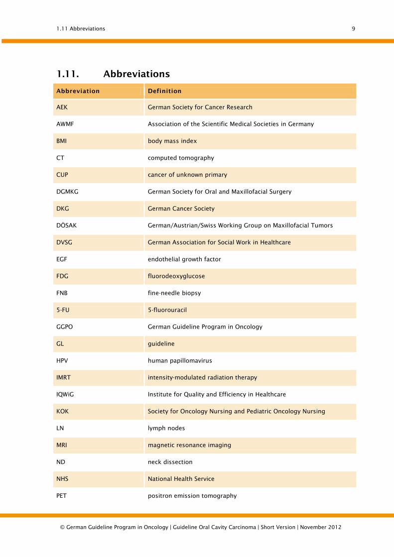
1.11. Abbreviations
Abbreviation Definition
AEK German Society for Cancer Research
AWMF Association of the Scientific Medical Societies in Germany
BMI body mass index
CT computed tomography
CUP cancer of unknown primary
DGMKG German Society for Oral and Maxillofacial Surgery
DKG German Cancer Society
DÖSAK German/Austrian/Swiss Working Group on Maxillofacial Tumors
DVSG German Association for Social Work in Healthcare
EGF endothelial growth factor
FDG fluorodeoxyglucose
FNB fine-needle biopsy
5-FU 5-fluorouracil
GGPO German Guideline Program in Oncology
GL guideline
HPV human papillomavirus
IMRT intensity-modulated radiation therapy
IQWiG Institute for Quality and Efficiency in Healthcare
KOK Society for Oncology Nursing and Pediatric Oncology Nursing
LN lymph nodes
MRI magnetic resonance imaging
ND neck dissection
NHS National Health Service
PET positron emission tomography
© German Guideline Program in Oncology | Guideline Oral Cavity Carcinoma | Short Version | November 2012
1.11 Abbreviations 10
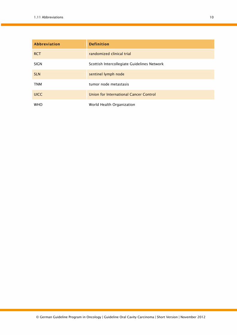
Abbreviation Definition
RCT randomized clinical trial
SIGN Scottish Intercollegiate Guidelines Network
SLN sentinel lymph node
TNM tumor node metastasis
UICC Union for International Cancer Control
WHO World Health Organization
© German Guideline Program in Oncology | Guideline Oral Cavity Carcinoma | Short Version | November 2012
2.1 Risk factors 11
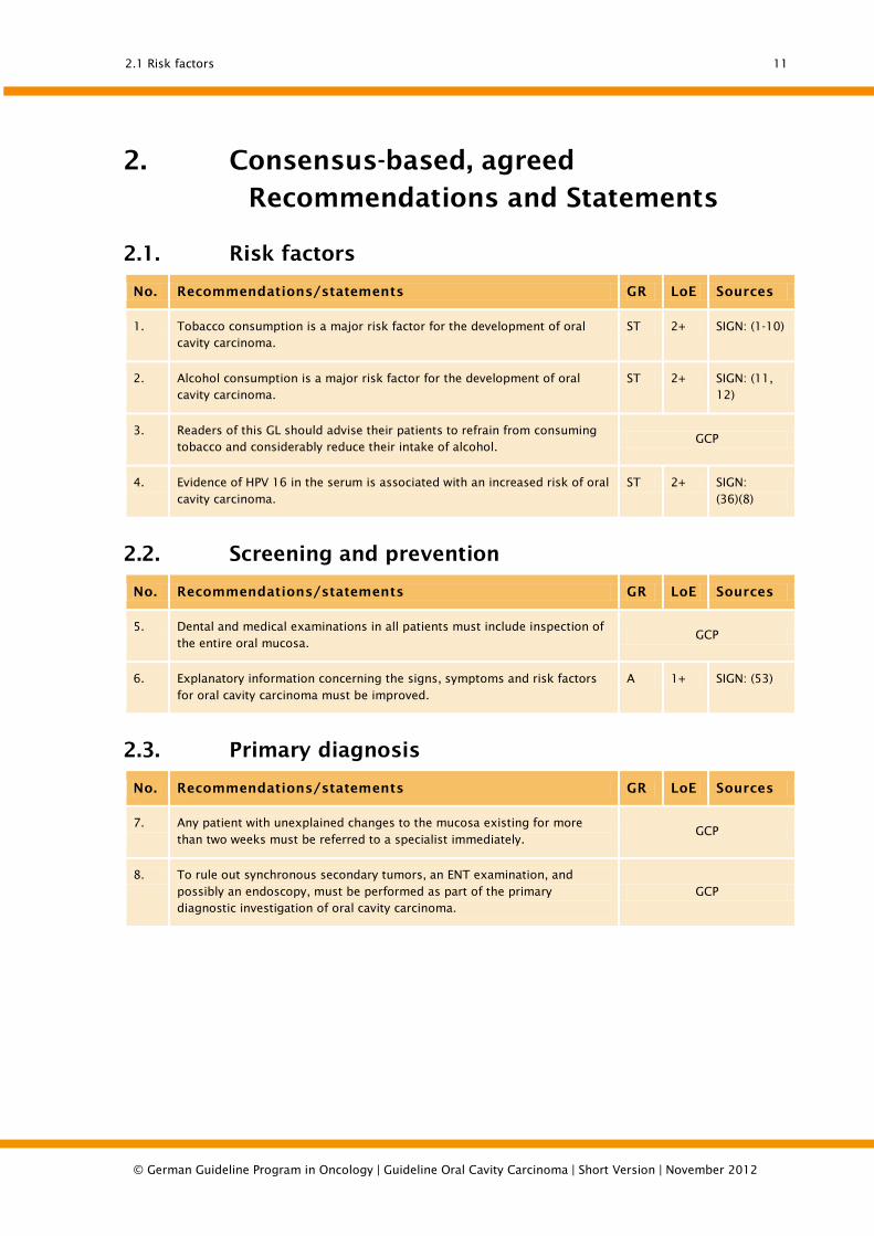
2. Consensus-based, agreed Recommendations and Statements
2.1. Risk factors
No. Recommendations/statements GR LoE Sources
1. Tobacco consumption is a major risk factor for the development of oral cavity carcinoma.
ST 2+ SIGN: (1-10)
2. Alcohol consumption is a major risk factor for the development of oral cavity carcinoma.
ST 2+ SIGN: (11, 12)
3. Readers of this GL should advise their patients to refrain from consuming tobacco and considerably reduce their intake of alcohol.
GCP
4. Evidence of HPV 16 in the serum is associated with an increased risk of oral cavity carcinoma.
ST 2+ SIGN: (36)(8)
2.2. Screening and prevention
No. Recommendations/statements GR LoE Sources
5. Dental and medical examinations in all patients must include inspection of the entire oral mucosa.
GCP
6. Explanatory information concerning the signs, symptoms and risk factors for oral cavity carcinoma must be improved.
A 1+ SIGN: (53)
2.3. Primary diagnosis
No. Recommendations/statements GR LoE Sources
7. Any patient with unexplained changes to the mucosa existing for more than two weeks must be referred to a specialist immediately.
GCP
8. To rule out synchronous secondary tumors, an ENT examination, and possibly an endoscopy, must be performed as part of the primary diagnostic investigation of oral cavity carcinoma.
GCP
© German Guideline Program in Oncology | Guideline Oral Cavity Carcinoma | Short Version | November 2012

2.4 Imaging procedures and further diagnostics 12
2.4. Imaging procedures and further diagnostics
No. Recommendations/statements GR LoE Sources
9. To determine the localization of oral cavity carcinoma, a CT or MRI scan should be performed.
B 3 SIGN: (13-15)
10. To prevent distorted contrast agent patterns in the primary tumor, the tumor biopsy should only be taken once cross-sectional imaging has been conducted.
GCP
11. Panoramic slicing is part of a basic dental diagnostic procedure, the results of which should be available prior to commencing specific cancer therapy.
GCP
12. If metal artifacts are likely to be revealed in the oral cavity, MRI should be given priority over CT for assessment of the primary tumor.
GCP
13 Evidence of the superiority of CT or MRI in the assessment of bone invasion from carcinomas of the oral mucosa is contradictory and unreliable.
ST 3 de novo: (16, 17)
14. Evidence of the superiority of CT or MRI in assessing the extent of the primary tumor is contradictory and unreliable.
ST 3 SIGN: (13, 14) de novo: (16, 18)
15. There is no sound evidence of a higher test quality or additional benefit from cone-beam CT (dental CT) as opposed to panoramic slicing for assessment of bone invasion in the mandible.
ST 3 de novo: (19)
16. PET/CT has no value in the primary diagnosis of the local spread of known oral cavity carcinoma.
ST 2+ de novo: (20-26)
17. To determine the N category, the entire region from the base of the skull to the upper thoracic aperture must be examined by CT or MRI.
A 2+ SIGN: (13, 27) de novo: (28-32)
18. The diagnostic specificity of lymph node staging of the neck may be improved by ultrasound-guided fine-needle biopsy.
ST 2++ SIGN: (33-35)
19. The diagnostic specificity and sensitivity of lymph node staging of the neck may be improved by FDG-PET/CT.
ST 2+ de novo: (21, 29, 30, 36-41)
20. There is no reliable evidence to suggest that SLN biopsy is a suitable method for avoiding elective cervical lymph node dissection.
ST 3 de novo: (42-47)
© German Guideline Program in Oncology | Guideline Oral Cavity Carcinoma | Short Version | November 2012
2.5 Imaging procedures and diagnostic methods for exclusion of synchronous secondary tumors, distant metastases, unknown primary tumors (CUP) and recurrence
13
2.5. Imaging procedures and diagnostic methods for exclusion of synchronous secondary tumors, distant metastases, unknown primary tumors (CUP) and recurrence
No. Recommendations/statements GR LoE Sources
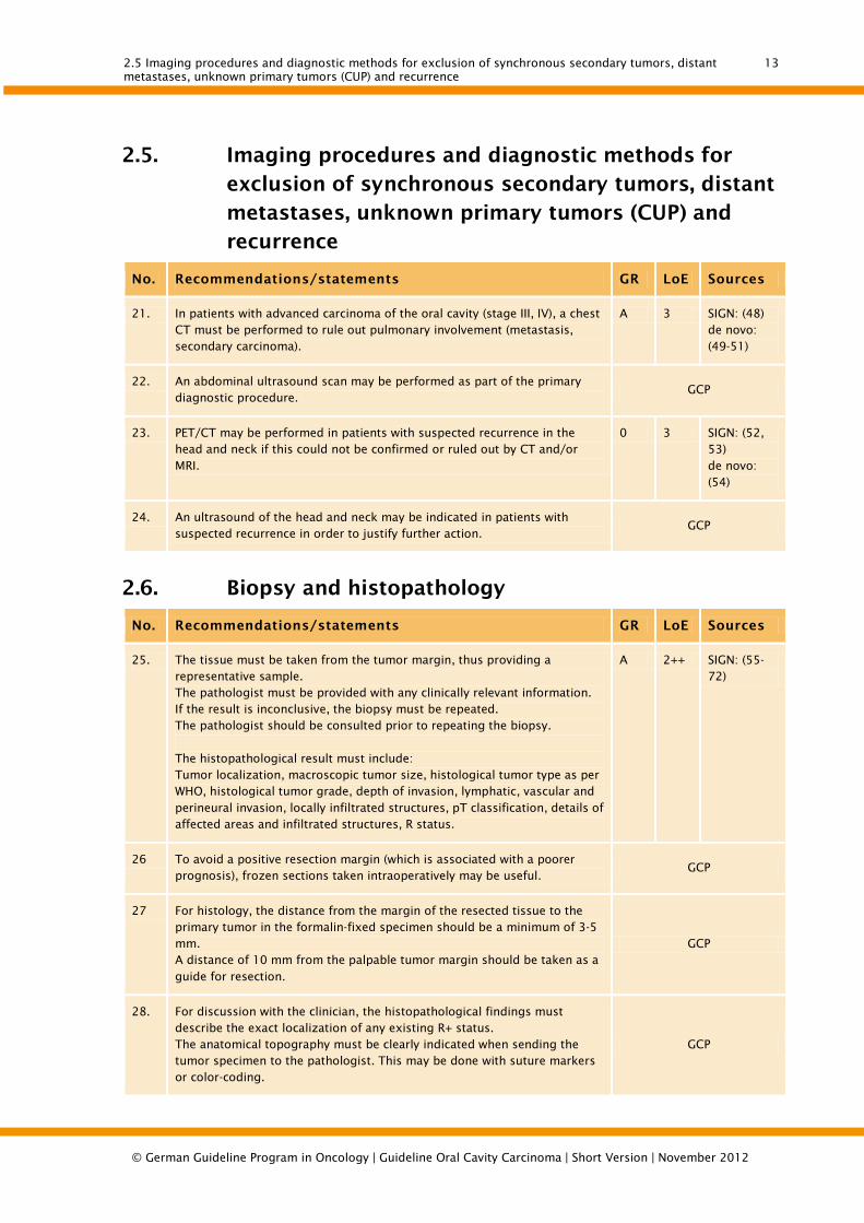
21. In patients with advanced carcinoma of the oral cavity (stage III, IV), a chest CT must be performed to rule out pulmonary involvement (metastasis, secondary carcinoma).
A 3 SIGN: (48) de novo: (49-51)
22. An abdominal ultrasound scan may be performed as part of the primary diagnostic procedure.
GCP
23. PET/CT may be performed in patients with suspected recurrence in the head and neck if this could not be confirmed or ruled out by CT and/or MRI.
0 3 SIGN: (52, 53) de novo: (54)
24. An ultrasound of the head and neck may be indicated in patients with suspected recurrence in order to justify further action.
GCP
2.6. Biopsy and histopathology
No. Recommendations/statements GR LoE Sources
25. The tissue must be taken from the tumor margin, thus providing a representative sample. The pathologist must be provided with any clinically relevant information. If the result is inconclusive, the biopsy must be repeated. The pathologist should be consulted prior to repeating the biopsy. The histopathological result must include: Tumor localization, macroscopic tumor size, histological tumor type as per WHO, histological tumor grade, depth of invasion, lymphatic, vascular and perineural invasion, locally infiltrated structures, pT classification, details of affected areas and infiltrated structures, R status.
A 2++ SIGN: (55-72)
26 To avoid a positive resection margin (which is associated with a poorer prognosis), frozen sections taken intraoperatively may be useful.
GCP
27 For histology, the distance from the margin of the resected tissue to the primary tumor in the formalin-fixed specimen should be a minimum of 3-5 mm. A distance of 10 mm from the palpable tumor margin should be taken as a guide for resection.
GCP
28. For discussion with the clinician, the histopathological findings must describe the exact localization of any existing R+ status. The anatomical topography must be clearly indicated when sending the tumor specimen to the pathologist. This may be done with suture markers or color-coding.
GCP
© German Guideline Program in Oncology | Guideline Oral Cavity Carcinoma | Short Version | November 2012

2.7 Treatment recommendations 14
No. Recommendations/statements GR LoE Sources
29. The histopathological findings from a neck dissection specimen must describe the side of the neck, type of neck dissection, eliminated levels, total number of lymph nodes plus number of lymph nodes affected, level of the affected lymph nodes, diameter of the largest affected lymph node, additionally removed structures and, if present, extracapsular spread.
A 2++ SIGN: (14, 56, 73-79)
2.7. Treatment recommendations
No. Recommendations/statements GR LoE Sources
30. Oral cavity carcinoma must be treated on an interdisciplinary basis after discussion of the case in question by a tumor board, comprising the specialist disciplines of oral and maxillofacial surgery, ENT, radiotherapy, oncology, pathology and radiology.
GCP
31. The patient must be kept fully informed about his condition, the treatment options and consequences.
GCP
32. Patients with carcinoma of the oral cavity should be examined by an experienced dental practitioner to ascertain their dental status prior to commencing treatment.
B 3 SIGN: (80, 81)
33. Provided the patient's general condition permits and the oral cavity carcinoma may be curatively resected, surgery should be performed and if possible combined with immediate reconstruction. Postoperative treatment should also be undertaken in advanced cancers.
B 3 SIGN: (82-85)
2.8. Surgery as treatment for primary cancer
No. Recommendations/statements GR LoE Sources
34. The treatment for oral cavity carcinoma must take the patient's individual situation into account. The decision to perform surgery must be made on the basis of the ability to achieve tumor-free resection margins and postoperative quality of life.
A 3 SIGN: (69, 86-92)
35. In case of a microscopic residual tumor (failed R0 resection), targeted follow-up resection should ensue with the aim of improving the patient's prognosis.
B 3 SIGN: (68)
36 Continuity of the mandible should be preserved on tumor resection, provided no radiological or intraoperative evidence has been found of tumor invasion of the bone.
B 3 SIGN: (93-95) de novo: (96-98)
© German Guideline Program in Oncology | Guideline Oral Cavity Carcinoma | Short Version | November 2012
2.9 Cervical lymph node dissection 15
2.9. Cervical lymph node dissection
No. Recommendations/statements GR LoE Sources
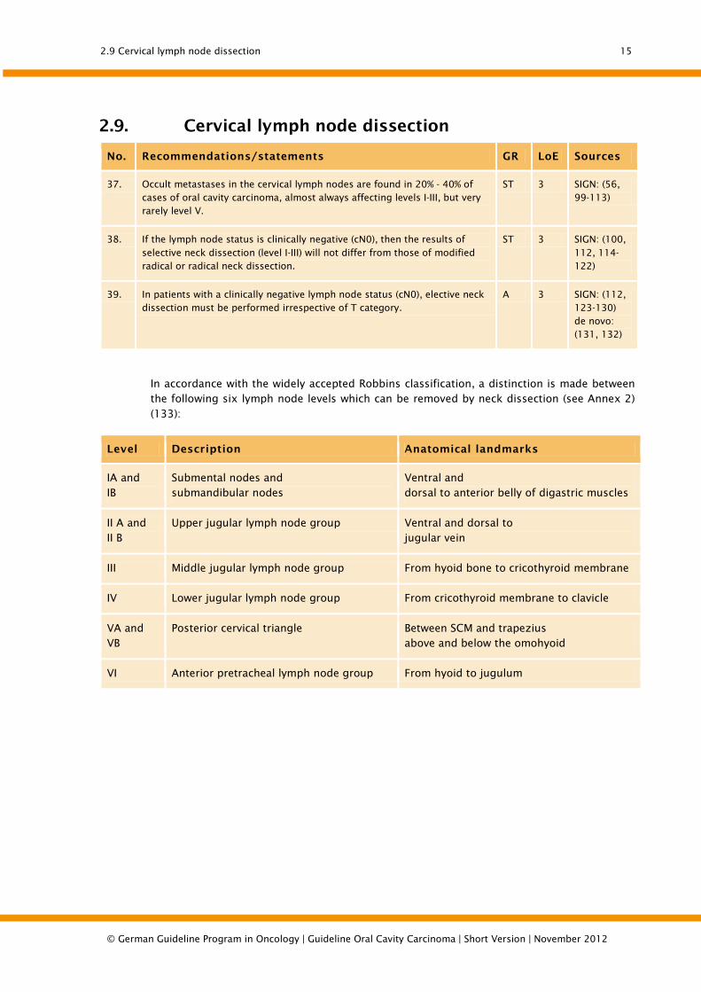
37. Occult metastases in the cervical lymph nodes are found in 20% - 40% of cases of oral cavity carcinoma, almost always affecting levels I-III, but very rarely level V.
ST 3 SIGN: (56, 99-113)
38. If the lymph node status is clinically negative (cN0), then the results of selective neck dissection (level I-III) will not differ from those of modified radical or radical neck dissection.
ST 3 SIGN: (100, 112, 114-122)
39. In patients with a clinically negative lymph node status (cN0), elective neck dissection must be performed irrespective of T category.
A 3 SIGN: (112, 123-130) de novo: (131, 132)
In accordance with the widely accepted Robbins classification, a distinction is made between the following six lymph node levels which can be removed by neck dissection (see Annex 2)
(133):
Level Description Anatomical landmarks
IA and IB
Submental nodes and submandibular nodes
Ventral and dorsal to anterior belly of digastric muscles
II A and II B
Upper jugular lymph node group Ventral and dorsal to jugular vein
III Middle jugular lymph node group From hyoid bone to cricothyroid membrane
IV Lower jugular lymph node group From cricothyroid membrane to clavicle
VA and VB
Posterior cervical triangle Between SCM and trapezius above and below the omohyoid
VI Anterior pretracheal lymph node group From hyoid to jugulum
© German Guideline Program in Oncology | Guideline Oral Cavity Carcinoma | Short Version | November 2012
2.10 Reconstruction 16
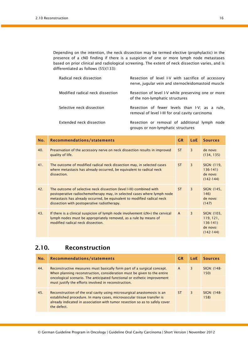
Depending on the intention, the neck dissection may be termed elective (prophylactic) in the presence of a cN0 finding if there is a suspicion of one or more lymph node metastases based on prior clinical and radiological screening. The extent of neck dissection varies, and is
differentiated as follows (55)(133):
Radical neck dissection Resection of level I-V with sacrifice of accessory
nerve, jugular vein and sternocleidomastoid muscle
Modified radical neck dissection Resection of level I-V while preserving one or more
of the non-lymphatic structures
Selective neck dissection Resection of fewer levels than I-V; as a rule,
removal of level I-III for oral cavity carcinoma
Extended neck dissection Resection or removal of additional lymph node
groups or non-lymphatic structures
No. Recommendations/statements GR LoE Sources
40. Preservation of the accessory nerve on neck dissection results in improved quality of life.
ST 3 de novo: (134, 135)
41. The outcome of modified radical neck dissection may, in selected cases where metastasis has already occurred, be equivalent to radical neck dissection.
ST 3 SIGN: (119, 136-141) de novo: (142-144)
42. The outcome of selective neck dissection (level I-III) combined with postoperative radiochemotherapy may, in selected cases where lymph node metastasis has already occurred, be equivalent to modified radical neck dissection with postoperative radiotherapy.
ST 3 SIGN: (145, 146) de novo: (147)
43. If there is a clinical suspicion of lymph node involvement (cN+) the cervical lymph nodes must be appropriately removed, as a rule by means of modified radical neck dissection.
A 3 SIGN: (103, 119, 121, 136-141) de novo: (142-144)
2.10. Reconstruction
No. Recommendations/statements GR LoE Sources
44. Reconstructive measures must basically form part of a surgical concept. When planning reconstruction, consideration must be given to the entire oncological scenario. The anticipated functional or esthetic improvement must justify the efforts involved in reconstruction.
A 3 SIGN: (148-150)
45. Reconstruction of the oral cavity using microsurgical anastomosis is an established procedure. In many cases, microvascular tissue transfer is already indicated in association with tumor resection so as to safely cover the defect.
ST 3 SIGN: (148-158)
© German Guideline Program in Oncology | Guideline Oral Cavity Carcinoma | Short Version | November 2012

2.11 Radiotherapy 17
2.11. Radiotherapy
No. Recommendations/statements GR LoE Sources
46. Interruption to radiotherapy will be detrimental to tumor control and so must be avoided.
A 2+ SIGN: (159-161)
47. If primary percutaneous irradiation is used alone, fractionation should be modified (hyperfractionation/acceleration).
GCP
2.12. Radiotherapy combined with chemotherapy
No. Recommendations/statements GR LoE Sources
48. Neoadjuvant or adjuvant chemotherapy for squamous cell carcinoma of the oral cavity, combined with surgery, does not have a positive effect.
ST 1++ SIGN: (162-164)
49. In concurrent primary radiochemotherapy, chemotherapy should include cisplatin or a combination containing cisplatin.
GCP
50. Patients with advanced, inoperable and non-metastatic oral cavity carcinoma, especially those aged 70 or under, must preferably be administered primary radiochemotherapy rather than radiotherapy alone.
A 1++ SIGN: (162, 163)
51. Radiochemotherapy must only be performed at facilities in which radiotherapy- or chemotherapy-induced acute toxicities can be diagnosed and adequately treated.
GCP
52. A combination of radiotherapy with cetuximab may be administered as an alternative to radiochemotherapy.
GCP
53. Postoperative radiotherapy or radiochemotherapy must be performed for advanced T categories (T3/T4), close or positive resection margins, perineural invasion, vascular invasion and/or lymph node involvement.
A 1++ SIGN: (117, 165-171)
54. Postoperative radiotherapy must be fractionated conventionally and constitute 54-60 Gy in 27-30 fractions over 5.5-6 weeks for an average risk, and 66 Gy in 33 fractions over 6.5 weeks for tumors with an increased risk of recurrence.
A 1++ SIGN: (169-172)
55. Postoperative radiotherapy should be commenced as early as possible and be completed within a maximum of 11 weeks after surgery.
B 2++ SIGN: (173, 174)
56. If radiotherapy is indicated, patients with increased histopathologic risk criteria for tumor recurrence (resection margin <5 mm and/or extracapsular tumor growth) after tumor resection should receive adjuvant treatment in the form of radiochemotherapy with cisplatin.
B 2++ SIGN: (169-171, 175-177)
57. Patients with small but accessible tumors (T1/T2) in the oral cavity may be treated in selected cases with interstitial brachytherapy.
0 3 SIGN: (178-181)
© German Guideline Program in Oncology | Guideline Oral Cavity Carcinoma | Short Version | November 2012

2.13 Prevention and management of radiation-induced side effects 18
2.13. Prevention and management of radiation-induced side effects
No. Recommendations/statements GR LoE Sources
58. There is evidence to suggest that the frequency and severity of radiation-induced xerostomia can be reduced by intensity-modulated radiotherapy (IMRT).
ST 3 SIGN: (182)
59. Patients undergoing irradiation for carcinoma of the oral cavity must be provided with optimal dental and oral health care.
GCP
60. Patients must undergo a dental examination and if necessary preservative and/or surgical restoration of the teeth prior to radio/radiochemotherapy of the oral cavity in order to avoid osteoradionecrosis.
GCP
61. When starting radiotherapy of the oral cavity a fluoride gel tray, and spacer if necessary, must be prepared.
GCP
62. Patients having undergone irradiation for carcinoma of the oral cavity should be offered oral pilocarpine three times daily if there is evidence of residual salivary gland function, provided there are no contraindications.
B 1+ SIGN: (183, 184)
2.14. Management of locoregional recurrence
No. Recommendations/statements GR LoE Sources
63. Salvage surgery should be considered in any patient with a resectable locoregional recurrence having previously undergone radiotherapy or surgery. The procedure should only be performed by an experienced surgical team with adequate experience of reconstructive techniques at a facility that offers suitable intensive care support.
B 3 SIGN: (185, 186)
64. Re-irradiation, possibly of a curative nature, should be considered in any patient with a non-resectable locoregional recurrence having already undergone irradiation. Irradiation should take place only at facilities with adequate expertise and ideally as part of a clinical therapeutic study.
B 3 SIGN: (187-192)
© German Guideline Program in Oncology | Guideline Oral Cavity Carcinoma | Short Version | November 2012
2.15 Palliative care and palliative medicine 19
2.15. Palliative care and palliative medicine
No. Recommendations/statements GR LoE Sources
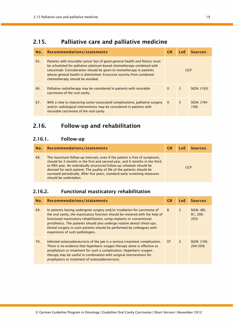
65. Patients with incurable cancer but of good general health and fitness must be scheduled for palliative platinum-based chemotherapy combined with cetuximab. Consideration should be given to monotherapy in patients whose general health is diminished. Excessive toxicity from combined chemotherapy should be avoided.
GCP
66. Palliative radiotherapy may be considered in patients with incurable carcinoma of the oral cavity.
0 3 SIGN: (193)
67. With a view to improving tumor-associated complications, palliative surgery and/or radiological interventions may be considered in patients with incurable carcinoma of the oral cavity.
0 3 SIGN: (194-199)
2.16. Follow-up and rehabilitation
2.16.1. Follow-up
No. Recommendations/statements GR LoE Sources
68. The maximum follow-up intervals, even if the patient is free of symptoms, should be 3 months in the first and second year, and 6 months in the third to fifth year. An individually structured follow-up schedule should be devised for each patient. The quality of life of the patients should be surveyed periodically. After five years, standard early screening measures should be undertaken.
GCP
2.16.2. Functional masticatory rehabilitation
No. Recommendations/statements GR LoE Sources
69. In patients having undergone surgery and/or irradiation for carcinoma of the oral cavity, the masticatory function should be restored with the help of functional masticatory rehabilitation, using implants or conventional prosthetics. The patients should also undergo routine dental check-ups. Dental surgery in such patients should be performed by colleagues with experience of such pathologies.
B 3 SIGN: (80, 81, 200-203)
70. Infected osteoradionecrosis of the jaw is a serious treatment complication. There is no evidence that hyperbaric oxygen therapy alone is effective as prophylaxis or treatment for such a complication. Hyperbaric oxygen therapy may be useful in combination with surgical interventions for prophylaxis or treatment of osteoradionecrosis.
ST 3 SIGN: (109, 204-209)
© German Guideline Program in Oncology | Guideline Oral Cavity Carcinoma | Short Version | November 2012

2.16 Follow-up and rehabilitation 20
2.16.3. Speech and swallowing rehabilitation
No. Recommendations/statements GR LoE Sources
71. Patients with difficulties chewing, speaking and swallowing should be provided with appropriate functional therapy. The patients should be introduced to suitably qualified therapists prior to commencing treatment if the scheduled surgical or conservative procedures are likely to cause difficulties with chewing, swallowing and/or speech.
B 2+ SIGN: (210-215)
72. Patients with dysphagia should undergo appropriate diagnostic procedures, eg high-frequency contrast-enhanced fluoroscopy or fiber-optic endoscopy.
B 2+ SIGN: (213, 214)
73. Patients having difficulty eating and speaking due to carcinoma of the oral cavity and/or undergoing radio/radiochemotherapy should have access to logopedists with experience of such pathologies before, during and after treatment.
B 2+ SIGN: (216)
2.16.4. Nutritional therapy
No. Recommendations/statements GR LoE Sources
74. Patients who due to the cancer or treatment are at risk of malnutrition should receive early professional dietary counseling and nutritional therapy.
B 2+ SIGN: (217-222)
2.16.5. Psychosocial counseling and support
No. Recommendations/statements GR LoE Sources
75. Patients with carcinoma of the oral cavity must be offered psychosocial support from a social worker.
GCP
76. To guarantee the continuity of psycho-oncological support after hospitalized treatment, patients with oral cavity carcinoma must be informed about the continued outpatient follow-up care available (cancer advisory bodies, practicing psychotherapists, self-help groups, social counseling).
GCP
© German Guideline Program in Oncology | Guideline Oral Cavity Carcinoma | Short Version | November 2012
Quality indicators 21
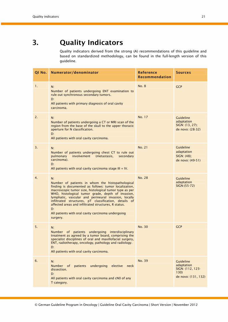
3. Quality Indicators Quality indicators derived from the strong (A) recommendations of this guideline and based on standardized methodology, can be found in the full-length version of this
guideline.
QI No. Numerator/denominator Reference Recommendation
Sources
1. N: Number of patients undergoing ENT examination to rule out synchronous secondary tumors. D: All patients with primary diagnosis of oral cavity carcinoma.
No. 8 GCP
2. N:
Number of patients undergoing a CT or MRI scan of the region from the base of the skull to the upper thoracic aperture for N classification.
D: All patients with oral cavity carcinoma.
No. 17 Guideline adaptation SIGN: (13, 27); de novo: (28-32)
3. N: Number of patients undergoing chest CT to rule out pulmonary involvement (metastasis, secondary carcinoma).
D: All patients with oral cavity carcinoma stage III + IV.
No. 21 Guideline adaptation SIGN: (48); de novo: (49-51)
4. N:
Number of patients in whom the histopathological finding is documented as follows: tumor localization, macroscopic tumor size, histological tumor type as per WHO, histological tumor grade, depth of invasion, lymphatic, vascular and perineural invasion, locally infiltrated structures, pT classification, details of affected areas and infiltrated structures, R status. D: All patients with oral cavity carcinoma undergoing surgery.
No. 28 Guideline adaptation SIGN:(55-72)
5. N:
Number of patients undergoing interdisciplinary treatment as agreed by a tumor board, comprising the specialist disciplines of oral and maxillofacial surgery, ENT, radiotherapy, oncology, pathology and radiology.
D: All patients with oral cavity carcinoma.
No. 30 GCP
6. N:
Number of patients undergoing elective neck dissection. D: All patients with oral cavity carcinoma and cN0 of any T category.
No. 39 Guideline adaptation SIGN: (112, 123-130) de novo: (131, 132)
© German Guideline Program in Oncology | Guideline Oral Cavity Carcinoma | Short Version | November 2012
Quality indicators 22
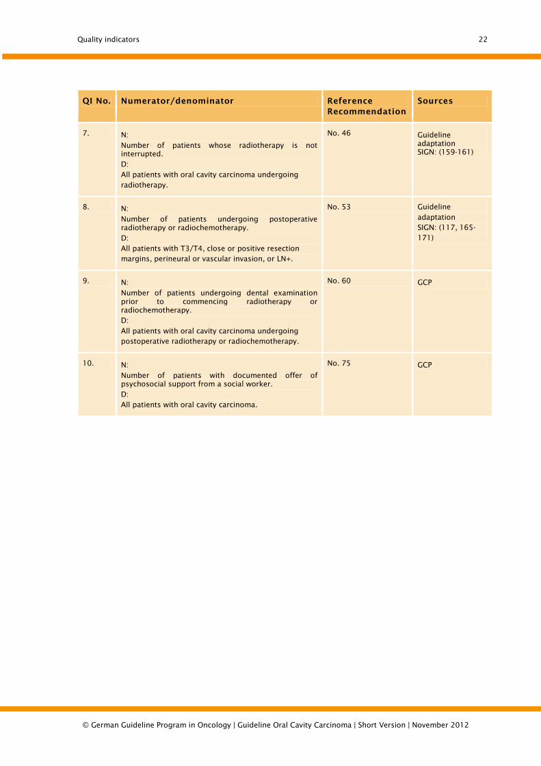
QI No. Numerator/denominator Reference Recommendation
Sources
7. N:
Number of patients whose radiotherapy is not interrupted. D: All patients with oral cavity carcinoma undergoing radiotherapy.
No. 46 Guideline adaptation SIGN: (159-161)
8. N:
Number of patients undergoing postoperative radiotherapy or radiochemotherapy. D: All patients with T3/T4, close or positive resection margins, perineural or vascular invasion, or LN+.
No. 53 Guideline adaptation SIGN: (117, 165-171)
9. N:
Number of patients undergoing dental examination prior to commencing radiotherapy or radiochemotherapy. D:
All patients with oral cavity carcinoma undergoing postoperative radiotherapy or radiochemotherapy.
No. 60 GCP
10. N:
Number of patients with documented offer of psychosocial support from a social worker. D: All patients with oral cavity carcinoma.
No. 75 GCP
© German Guideline Program in Oncology | Guideline Oral Cavity Carcinoma | Short Version | November 2012
References 23
4. References
1. Altieri A, Bosetti C, Talamini R, Gallus S, Franceschi S, Levi F, et al. Cessation of smoking and drinking and the risk of laryngeal cancer. British journal of cancer. 2002;87(11):1227-9.
2. Balaram P, Sridhar H, Rajkumar T, Vaccarella S, Herrero R, Nandakumar A, et al. Oral cancer in southern India: the influence of smoking, drinking, paan-chewing and oral hygiene. International journal of cancer Journal international du cancer. 2002;98(3):440-5.
3. Cheng YJ, Hildesheim A, Hsu MM, Chen IH, Brinton LA, Levine PH, et al. Cigarette smoking, alcohol consumption and risk of nasopharyngeal carcinoma in Taiwan. Cancer causes & control : CCC. 1999;10(3):201-7.
4. De Boer MF, Van den Borne B, Pruyn JF, Ryckman RM, Volovics L, Knegt PP, et al. Psychosocial and physical correlates of survival and recurrence in patients with head and neck carcinoma: results of a 6-year longitudinal study. Cancer. 1998;83(12):2567-79.
5. de Graeff A, de Leeuw JR, Ros WJ, Hordijk GJ, Blijham GH, Winnubst JA. Sociodemographic factors and quality of life as prognostic indicators in head and neck cancer. Eur J Cancer. 2001;37(3):332-9.
6. De Stefani E, Boffetta P, Oreggia F, Mendilaharsu M, Deneo-Pellegrini H. Smoking patterns and cancer of the oral cavity and pharynx: a case-control study in Uruguay. Oral oncology. 1998;34(5):340-6.
7. Franceschi S, Levi F, La Vecchia C, Conti E, Dal Maso L, Barzan L, et al. Comparison of the effect of smoking and alcohol drinking between oral and pharyngeal cancer. International journal of cancer Journal international du cancer. 1999;83(1):1-4.
8. Lissowska J, Pilarska A, Pilarski P, Samolczyk-Wanyura D, Piekarczyk J, Bardin-Mikollajczak A, et al. Smoking, alcohol, diet, dentition and sexual practices in the epidemiology of oral cancer in Poland. Eur J Cancer Prev. 2003;12(1):25-33.
9. Perea-Milla Lopez E, Minarro-Del Moral RM, Martinez-Garcia C, Zanetti R, Rosso S, Serrano S, et al. Lifestyles, environmental and phenotypic factors associated with lip cancer: a case-control study in southern Spain. British journal of cancer. 2003;88(11):1702-7.
10. Critchley JA, Unal B. Health effects associated with smokeless tobacco: a systematic review. Thorax. 2003;58(5):435-43. 11. Bagnardi V, Blangiardo M, La Vecchia C, Corrao G. Alcohol consumption and the risk of cancer: a meta-analysis. Alcohol
research & health : the journal of the National Institute on Alcohol Abuse and Alcoholism. 2001;25(4):263-70. 12. Corrao G, Bagnardi V, Zambon A, Arico S. Exploring the dose-response relationship between alcohol consumption and the
risk of several alcohol-related conditions: a meta-analysis. Addiction. 1999;94(10):1551-73. 13. Wilson JA, editor. Effective head and neck cancer management: consensus document. London: British Association of
Otorhinolaryngologists, Head and Neck Surgeons; 1998. 14. Leslie A, Fyfe E, Guest P, Goddard P, Kabala JE. Staging of squamous cell carcinoma of the oral cavity and oropharynx: a
comparison of MRI and CT in T- and N-staging. J Comput Assist Tomogr. 1999;23(1):43-9. 15. Kaanders JH, Hordijk GJ. Carcinoma of the larynx: the Dutch national guideline for diagnostics, treatment, supportive care
and rehabilitation. Radiother Oncol. 2002;63(3):299-307. 16. Imaizumi A, Yoshino N, Yamada I, Nagumo K, Amagasa T, Omura K, et al. A potential pitfall of MR imaging for assessing
mandibular invasion of squamous cell carcinoma in the oral cavity. AJNR Am J Neuroradiol. 2006;27(1):114-22. 17. Bolzoni A, Cappiello J, Piazza C, Peretti G, Maroldi R, Farina D, et al. Diagnostic accuracy of magnetic resonance imaging in
the assessment of mandibular involvement in oral-oropharyngeal squamous cell carcinoma: a prospective study. Arch Otolaryngol Head Neck Surg. 2004;130(7):837-43.
18. Van Cann EM, Koole R, Oyen WJG, de Rooy JWJ, de Wilde PC, Slootweg PJ, et al. Assessment of mandibular invasion of squamous cell carcinoma by various modes of imaging: constructing a diagnostic algorithm. Int J Oral Maxillofac Surg. 2008;37(6):535-41.
19. Hendrikx AWF, Maal T, Dieleman F, Van Cann EM, Merkx MAW. Cone-beam CT in the assessment of mandibular invasion by oral squamous cell carcinoma: results of the preliminary study. International Journal of Oral and Maxillofacial Surgery. 2010;39 (5):436-9.
20. Goerres GW, Schmid DT, Schuknecht B, Eyrich GK. Bone invasion in patients with oral cavity cancer: comparison of conventional CT with PET/CT and SPECT/CT.[Erratum appears in Radiology. 2006 Apr;239(1):303]. Radiology. 2005;237(1):281-7.
21. Yen T-C, Chang JT-C, Ng S-H, Chang Y-C, Chan S-C, Wang H-M, et al. Staging of untreated squamous cell carcinoma of buccal mucosa with 18F-FDG PET: comparison with head and neck CT/MRI and histopathology. J Nucl Med. 2005;46(5):775-81.
22. Hafidh MA, Lacy PD, Hughes JP, Duffy G, Timon CV. Evaluation of the impact of addition of PET to CT and MR scanning in the staging of patients with head and neck carcinomas. European Archives of Oto-Rhino-Laryngology. 2006;263(9):853-9.
23. Wax MK, Myers LL, Gona JM, Husain SS, Nabi HA. The role of positron emission tomography in the evaluation of the N-positive neck. Otolaryngology--head and neck surgery : official journal of American Academy of Otolaryngology-Head and Neck Surgery. 2003;129(3):163-7.
24. Krabbe CA, Dijkstra PU, Pruim J, van der Laan BFM, van der Wal JE, Gravendeel JP, et al. FDG PET in oral and oropharyngeal cancer. Value for confirmation of N0 neck and detection of occult metastases. Oral oncology. 2008;44(1):31-6.
25. Nahmias C, Carlson ER, Duncan LD, Blodgett TM, Kennedy J, Long MJ, et al. Positron emission tomography/computerized tomography (PET/CT) scanning for preoperative staging of patients with oral/head and neck cancer. J Oral Maxillofac Surg. 2007;65(12):2524-35.
26. Seitz O, Chambron-Pinho N, Middendorp M, Sader R, Mack M, Vogl TJ, et al. 18F-Fluorodeoxyglucose-PET/CT to evaluate tumor, nodal disease, and gross tumor volume of oropharyngeal and oral cavity cancer: comparison with MR imaging and validation with surgical specimen. Neuroradiology. 2009;51(10):677-86.
27. van den Brekel MW, Castelijns JA, Stel HV, Golding RP, Meyer CJ, Snow GB. Modern imaging techniques and ultrasound-guided aspiration cytology for the assessment of neck node metastases: a prospective comparative study. Eur Arch Otorhinolaryngol. 1993;250(1):11-7.
© German Guideline Program in Oncology | Guideline Oral Cavity Carcinoma | Short Version | November 2012

References 24
28. Dammann F, Horger M, Mueller-Berg M, Schlemmer H, Claussen CD, Hoffman J, et al. Rational diagnosis of squamous cell carcinoma of the head and neck region: comparative evaluation of CT, MRI, and 18FDG PET.[Erratum appears in AJR Am J Roentgenol. 2005 Jun;184(6):1968]. AJR Am J Roentgenol. 2005;184(4):1326-31.
29. Ng S-H, Yen T-C, Liao C-T, Chang JT-C, Chan S-C, Ko S-F, et al. 18F-FDG PET and CT/MRI in oral cavity squamous cell carcinoma: a prospective study of 124 patients with histologic correlation. J Nucl Med. 2005;46(7):1136-43.
30. Ng S-H, Yen T-C, Chang JT-C, Chan S-C, Ko S-F, Wang H-M, et al. Prospective study of [18F]fluorodeoxyglucose positron emission tomography and computed tomography and magnetic resonance imaging in oral cavity squamous cell carcinoma with palpably negative neck. J Clin Oncol. 2006;24(27):4371-6.
31. Sumi M, Kimura Y, Sumi T, Nakamura T. Diagnostic performance of MRI relative to CT for metastatic nodes of head and neck squamous cell carcinomas. J Magn Reson Imaging. 2007;26(6):1626-33.
32. Wiener E, Pautke C, Link TM, Neff A, Kolk A. Comparison of 16-slice MSCT and MRI in the assessment of squamous cell carcinoma of the oral cavity. Eur J Radiol. 2006;58(1):113-8.
33. Takes RP, Righi P, Meeuwis CA, Manni JJ, Knegt P, Marres HA, et al. The value of ultrasound with ultrasound-guided fine-needle aspiration biopsy compared to computed tomography in the detection of regional metastases in the clinically negative neck. Int J Radiat Oncol Biol Phys. 1998;40(5):1027-32.
34. Stokkel MP, ten Broek FW, Hordijk GJ, Koole R, van Rijk PP. Preoperative evaluation of patients with primary head and neck cancer using dual-head 18fluorodeoxyglucose positron emission tomography. Ann Surg. 2000;231(2):229-34.
35. Adams S, Baum RP, Stuckensen T, Bitter K, Hor G. Prospective comparison of 18F-FDG PET with conventional imaging modalities (CT, MRI, US) in lymph node staging of head and neck cancer. Eur J Nucl Med. 1998;25(9):1255-60.
36. Balogova S, Perie S, Kerrou K, Grahek D, Montravers F, Angelard B, et al. Prospective comparison of FDG and FET PET/CT in patients with head and neck squamous cell carcinoma. Mol Imaging Biol. 2008;10(6):364-73.
37. Jeong H-S, Baek C-H, Son Y-I, Ki Chung M, Kyung Lee D, Young Choi J, et al. Use of integrated 18F-FDG PET/CT to improve the accuracy of initial cervical nodal evaluation in patients with head and neck squamous cell carcinoma. Head Neck. 2007;29(3):203-10.
38. Kim SY, Roh JL, Kim JS, Ryu CH, Lee JH, Cho KJ, et al. Utility of FDG PET in patients with squamous cell carcinomas of the oral cavity. Eur J Surg Oncol. 2008;34(2):208-15.
39. Roh J-L, Yeo N-K, Kim JS, Lee JH, Cho K-J, Choi S-H, et al. Utility of 2-[18F] fluoro-2-deoxy-D-glucose positron emission tomography and positron emission tomography/computed tomography imaging in the preoperative staging of head and neck squamous cell carcinoma. Oral oncology. 2007;43(9):887-93.
40. Schwartz DL, Ford E, Rajendran J, Yueh B, Coltrera MD, Virgin J, et al. FDG-PET/CT imaging for preradiotherapy staging of head-and-neck squamous cell carcinoma. International Journal of Radiation Oncology, Biology, Physics. 2005;61(1):129-36.
41. Gencoglu U, Guneren E, Gozu A, Ozsoy Z, Vural G. The coherence between fine needle aspiration cytology and histopathology of palpable neck nodes in lower lip carcinoma patients. European Journal of Plastic Surgery. 2003;26 (2):82-4.
42. Civantos FJ, Zitsch RP, Schuller DE, Agrawal A, Smith RB, Nason R, et al. Sentinel lymph node biopsy accurately stages the regional lymph nodes for T1-T2 oral squamous cell carcinomas: results of a prospective multi-institutional trial. J Clin Oncol. 2010;28(8):1395-400.
43. Cammilleri S, Lussato D, Guelfucci B, Lief F, Bellot V, Chrestian M, et al. Ability of lymphoscintigraphy to direct sentinel node biopsy in the clinically NO check for patients with head and neck squamous cell carcinoma; a prospective study (preliminary results). Bull Cancer. 2004;91(4):E1-4.
44. Burns P, Foster A, Walshe P, O'Dwyer T. Sentinel lymph node biopsy in node-negative squamous cell carcinoma of the oral cavity and oropharynx. J Laryngol Otol. 2009;123(4):439-43.
45. Hart RD, Nasser JG, Trites JR, Taylor SM, Bullock M, Barnes D. Sentinel lymph node biopsy in N0 squamous cell carcinoma of the oral cavity and oropharynx. Arch Otolaryngol Head Neck Surg. 2005;131(1):34-8.
46. Hoft S, Maune S, Muhle C, Brenner W, Czech N, Kampen WU, et al. Sentinel lymph-node biopsy in head and neck cancer. British journal of cancer. 2004;91(1):124-8.
47. Keski-Santti H, Kontio R, Leivo I, Tornwall J, Matzke S, Makitie AA, et al. Sentinel lymph node biopsy as an alternative to wait and see policy in patients with small T1 oral cavity squamous cell carcinoma. Acta Otolaryngol (Stockh). 2008;128(1):98-102.
48. Arunachalam PS, Putnam G, Jennings P, Messersmith R, Robson AK. Role of computerized tomography (CT) scan of the chest in patients with newly diagnosed head and neck cancers. Clin Otolaryngol Allied Sci. 2002;27(12383307):409-11.
49. Andrle J, Schartinger VH, Schwentner I, Deibl M, Sprinzl GM. Initial staging examinations for head and neck squamous cell carcinoma: are they appropriate? J Laryngol Otol. 2009;123(8):885-8.
50. Ghosh SK, Roland NJ, Kumar A, Tandon S, Lancaster JL, Jackson SR, et al. Detection of synchronous lung tumors in patients presenting with squamous cell carcinoma of the head and neck. Head Neck. 2009;31(12):1563-70.
51. Loh KS, Brown DH, Baker JT, Gilbert RW, Gullane PJ, Irish JC. A rational approach to pulmonary screening in newly diagnosed head and neck cancer. Head Neck. 2005;27(11):990-4.
52. Bongers V, Hobbelink MG, van Rijk PP, Hordijk G-J. Cost-effectiveness of dual-head 18F-fluorodeoxyglucose PET for the detection of recurrent laryngeal cancer. Cancer Biother Radiopharm. 2002;17(12136522):303-6.
53. Lonneux M, Lawson G, Ide C, Bausart R, Remacle M, Pauwels S. Positron emission tomography with fluorodeoxyglucose for suspected head and neck tumor recurrence in the symptomatic patient. Laryngoscope. 2000;110(10983949):1493-7.
54. Nakamoto Y, Tamai K, Saga T, Higashi T, Hara T, Suga T, et al. Clinical value of image fusion from MR and PET in patients with head and neck cancer. Mol Imaging Biol. 2009;11(1):46-53.
55. Royal College of Pathologists. Standards and Datasets for Reporting Cancers: Datasets for histopathology reports on head and neck carcinomas and salivary neoplasms. 2nd Edition. London: The Royal College of Pathologists; 2005. [cited 11 August 2006]. Available from url: http://www.rcpath.org/resources/pdf/HeadNeckDatasetJun05.pdf.
56. Coatesworth AP, MacLennan K. Squamous cell carcinoma of the upper aerodigestive tract: the prevalence of microscopic extracapsular spread and soft tissue deposits in the clinically N0 neck. Head Neck. 2002;24(11891957):258-61.
57. Carinci F, Pelucchi S, Farina A, De Franciscis G, Calearo C. Extension as a prognostic factor in oropharyngeal cancer: largest mucosal dimension compared with number of (sub)sites involved. Br J Oral Maxillofac Surg. 1998;36(6):440-5.
58. Hirvikoski P, Kumpulainen E, Virtaniemi J, Pirinen R, Salmi L, Halonen P, et al. Enhanced apoptosis correlates with poor survival in patients with laryngeal cancer but not with cell proliferation, bcl-2 or p53 expression. Eur J Cancer. 1999;35(10448265):231-7.
© German Guideline Program in Oncology | Guideline Oral Cavity Carcinoma | Short Version | November 2012

References 25
59. Fortin A, Couture C, Doucet R, Albert M, Allard J, Tetu B. Does histologic grade have a role in the management of head and neck cancers? J Clin Oncol. 2001;19(11689578):4107-16.
60. Yamazaki H, Inoue T, Teshima T, Tanaka E, Koizumi M, Kagawa K, et al. Tongue cancer treated with brachytherapy: is thickness of tongue cancer a prognostic factor for regional control? Anticancer Res. 1998;18(2B):1261-5.
61. O'Brien CJ, Lauer CS, Fredricks S, Clifford AR, McNeil EB, Bagia JS, et al. Tumor thickness influences prognosis of T1 and T2 oral cavity cancer--but what thickness? Head Neck. 2003;25(11):937-45.
62. Nishimaki T, Kanda T, Nakagawa S, Kosugi S-i, Tanabe T, Hatakeyama K. Outcomes and prognostic factors after surgical resection of hypopharyngeal and cervical esophageal carcinomas. Int Surg. 2002;87(1):38-44.
63. Barnes L, Eveson JW, Reichart P, Sidransky D, editors. Pathology and Genetics of Head and Neck Tumours. Lyon: IARC Press; 2005. (WHO Classification of Tumours).
64. Ang KK, Berkey BA, Tu X, Zhang H-Z, Katz R, Hammond EH, et al. Impact of epidermal growth factor receptor expression on survival and pattern of relapse in patients with advanced head and neck carcinoma. Cancer Res. 2002;62(12499279):7350-6.
65. Spiro RH, Guillamondegui O, Paulino AF, Huvos AG. Pattern of invasion and margin assessment in patients with oral tongue cancer. Head Neck. 1999;21(5):408-13.
66. Wong RJ, Keel SB, Glynn RJ, Varvares MA. Histological pattern of mandibular invasion by oral squamous cell carcinoma. Laryngoscope. 2000;110(1):65-72.
67. Weijers M, Snow GB, Bezemer PD, van der Wal JE, van der Waal I. The clinical relevance of epithelial dysplasia in the surgical margins of tongue and floor of mouth squamous cell carcinoma: an analysis of 37 patients. J Oral Pathol Med. 2002;31(11896817):11-5.
68. Bailey JS, Blanchaert RH, Ord RA. Management of oral squamous cell carcinoma treated with inadequate excisional biopsy. J Oral Maxillofac Surg. 2001;59(11526566):1007-10.
69. McMahon J, O'Brien CJ, Pathak I, Hamill R, McNeil E, Hammersley N, et al. Influence of condition of surgical margins on local recurrence and disease-specific survival in oral and oropharyngeal cancer. Br J Oral Maxillofac Surg. 2003;41(4):224-31.
70. Slootweg PJ, Hordijk GJ, Schade Y, van Es RJJ, Koole R. Treatment failure and margin status in head and neck cancer. A critical view on the potential value of molecular pathology. Oral oncology. 2002;38(12110346):500-3.
71. Ribeiro NFF, Godden DRP, Wilson GE, Butterworth DM, Woodwards RTM. Do frozen sections help achieve adequate surgical margins in the resection of oral carcinoma? Int J Oral Maxillofac Surg. 2003;32(12729775):152-8.
72. DiNardo LJ, Lin J, Karageorge LS, Powers CN. Accuracy, utility, and cost of frozen section margins in head and neck cancer surgery. Laryngoscope. 2000;110(10 Pt 1):1773-6.
73. Brasilino de Carvalho M. Quantitative analysis of the extent of extracapsular invasion and its prognostic significance: a prospective study of 170 cases of carcinoma of the larynx and hypopharynx. Head Neck. 1998;20(1):16-21.
74. Tankere F, Camproux A, Barry B, Guedon C, Depondt J, Gehanno P. Prognostic value of lymph node involvement in oral cancers: a study of 137 cases. Laryngoscope. 2000;110(11129021):2061-5.
75. Woolgar JA, Rogers SN, Lowe D, Brown JS, Vaughan ED. Cervical lymph node metastasis in oral cancer: the importance of even microscopic extracapsular spread. Oral oncology. 2003;39(12509965):130-7.
76. Suoglu Y, Erdamar B, Katircioglu OS, Karatay MC, Sunay T. Extracapsular spread in ipsilateral neck and contralateral neck metastases in laryngeal cancer. Ann Otol Rhinol Laryngol. 2002;111(5 Pt 1):447-54.
77. Greenberg JS, Fowler R, Gomez J, Mo V, Roberts D, El Naggar AK, et al. Extent of extracapsular spread: a critical prognosticator in oral tongue cancer. Cancer. 2003;97(12627511):1464-70.
78. Esposito ED, Motta S, Cassiano B, Motta G. Occult lymph node metastases in supraglottic cancers of the larynx. Otolaryngology--head and neck surgery : official journal of American Academy of Otolaryngology-Head and Neck Surgery. 2001;124(3):253-7.
79. Enepekides DJ, Sultanem K, Nguyen C, Shenouda G, Black MJ, Rochon L. Occult cervical metastases: immunoperoxidase analysis of the pathologically negative neck. Otolaryngology--head and neck surgery : official journal of American Academy of Otolaryngology-Head and Neck Surgery. 1999;120(5):713-7.
80. Epstein JB, Lunn R, Le N, Stevenson-Moore P. Periodontal attachment loss in patients after head and neck radiation therapy. Oral surgery, oral medicine, oral pathology, oral radiology, and endodontics. 1998;86(6):673-7.
81. Denis F, Garaud P, Bardet E, Alfonsi M, Sire C, Germain T, et al. Late toxicity results of the GORTEC 94-01 randomized trial comparing radiotherapy with concomitant radiochemotherapy for advanced-stage oropharynx carcinoma: comparison of LENT/SOMA, RTOG/EORTC, and NCI-CTC scoring systems. Int J Radiat Oncol Biol Phys. 2003;55(1):93-8.
82. dos Santos CR, Goncalves Filho J, Magrin J, Johnson LF, Ferlito A, Kowalski LP. Involvement of level I neck lymph nodes in advanced squamous carcinoma of the larynx. Ann Otol Rhinol Laryngol. 2001;110(10):982-4.
83. Johansen LV, Grau C, Overgaard J. Hypopharyngeal squamous cell carcinoma--treatment results in 138 consecutively admitted patients. Acta Oncol. 2000;39(4):529-36.
84. Rodgers LW, Jr., Stringer SP, Mendenhall WM, Parsons JT, Cassisi NJ, Million RR. Management of squamous cell carcinoma of the floor of mouth. Head Neck. 1993;15(1):16-9.
85. Levendag PC, Nowak PJ, van der Sangen MJ, Jansen PP, Eijkenboom WM, Planting AS, et al. Local tumor control in radiation therapy of cancers in the head and neck. Am J Clin Oncol. 1996;19(5):469-77.
86. Parsons JT, Mendenhall WM, Stringer SP, Cassisi NJ, Million RR. An analysis of factors influencing the outcome of postoperative irradiation for squamous cell carcinoma of the oral cavity. Int J Radiat Oncol Biol Phys. 1997;39(1):137-48.
87. Sessions DG, Spector GJ, Lenox J, Parriott S, Haughey B, Chao C, et al. Analysis of treatment results for floor-of-mouth cancer. Laryngoscope. 2000;110(10):1764-72.
88. Kovacs AF. Relevance of positive margins in case of adjuvant therapy of oral cancer. Int J Oral Maxillofac Surg. 2004;33(5):447-53.
89. O'Brien CJ, Adams JR, McNeil EB, Taylor P, Laniewski P, Clifford A, et al. Influence of bone invasion and extent of mandibular resection on local control of cancers of the oral cavity and oropharynx. Int J Oral Maxillofac Surg. 2003;32(5):492-7.
90. Hicks WL, Loree TR, Garcia RI, Maamoun S, Marshall D, Orner JB, et al. Squamous cell carcinoma of the floor of mouth: a 20-year review. Head Neck. 1997;19(5):400-5.
91. Loree TR, Strong EW. Significance of positive margins in oral cavity squamous carcinoma. Am J Surg. 1990;160(4):410-4. 92. Sutton DN, Brown JS, Rogers SN, Vaughan ED, Woolgar JA. The prognostic implications of the surgical margin in oral
squamous cell carcinoma. Int J Oral Maxillofac Surg. 2003;32(1):30-4. 93. Brown JS, Kalavrezos N, D'Souza J, Lowe D, Magennis P, Woolgar JA. Factors that influence the method of mandibular
resection in the management of oral squamous cell carcinoma. Br J Oral Maxillofac Surg. 2002;40(4):275-84.
© German Guideline Program in Oncology | Guideline Oral Cavity Carcinoma | Short Version | November 2012

References 26
94. Wolff D, Hassfeld S, Hofele C. Influence of marginal and segmental mandibular resection on the survival rate in patients with squamous cell carcinoma of the inferior parts of the oral cavity. J Craniomaxillofac Surg. 2004;32(5):318-23.
95. Munoz Guerra MF, Naval Gias L, Campo FR, Perez JS. Marginal and segmental mandibulectomy in patients with oral cancer: a statistical analysis of 106 cases. J Oral Maxillofac Surg. 2003;61(11):1289-96.
96. Muscatello L, Lenzi R, Pellini R, Giudice M, Spriano G. Marginal mandibulectomy in oral cancer surgery: A 13-year experience. European Archives of Oto-Rhino-Laryngology. 2010;267 (5):759-64.
97. Abler A, Roser M, Weingart D. [On the indications for and morbidity of segmental resection of the mandible for squamous cell carcinoma in the lower oral cavity]. Mund Kiefer Gesichtschir. 2005;9(3):137-42.
98. Namaki S, Matsumoto M, Ohba H, Tanaka H, Koshikawa N, Shinohara M. Masticatory efficiency before and after surgery in oral cancer patients: comparative study of glossectomy, marginal mandibulectomy and segmental mandibulectomy. J Oral Sci. 2004;46(2):113-7.
99. Byers RM, El-Naggar AK, Lee YY, Rao B, Fornage B, Terry NH, et al. Can we detect or predict the presence of occult nodal metastases in patients with squamous carcinoma of the oral tongue? Head Neck. 1998;20(2):138-44.
100. Spiro RH, Morgan GJ, Strong EW, Shah JP. Supraomohyoid neck dissection. Am J Surg. 1996;172(8988669):650-3. 101. Nieuwenhuis EJC, Castelijns JA, Pijpers R, van den Brekel MWM, Brakenhoff RH, van der Waal I, et al. Wait-and-see policy for
the N0 neck in early-stage oral and oropharyngeal squamous cell carcinoma using ultrasonography-guided cytology: is there a role for identification of the sentinel node? Head Neck. 2002;24(11891961):282-9.
102. Shah JP, Candela FC, Poddar AK. The patterns of cervical lymph node metastases from squamous carcinoma of the oral cavity. Cancer. 1990;66(1):109-13.
103. Spiro JD, Spiro RH, Shah JP, Sessions RB, Strong EW. Critical assessment of supraomohyoid neck dissection. Am J Surg. 1988;156(4):286-9.
104. Byers RM, Wolf PF, Ballantyne AJ. Rationale for elective modified neck dissection. Head Neck Surg. 1988;10(3):160-7. 105. Smith GI, O'Brien CJ, Clark J, Shannon KF, Clifford AR, McNeil EB, et al. Management of the neck in patients with T1 and T2
cancer in the mouth. Br J Oral Maxillofac Surg. 2004;42(6):494-500. 106. Crean S-J, Hoffman A, Potts J, Fardy MJ. Reduction of occult metastatic disease by extension of the supraomohyoid neck
dissection to include level IV. Head Neck. 2003;25(12953312):758-62. 107. Hao S-P, Tsang NM. The role of supraomohyoid neck dissection in patients of oral cavity carcinoma(small star, filled). Oral
oncology. 2002;38(11978555):309-12. 108. O'Brien CJ, Traynor SJ, McNeil E, McMahon JD, Chaplin JM. The use of clinical criteria alone in the management of the
clinically negative neck among patients with squamous cell carcinoma of the oral cavity and oropharynx. Arch Otolaryngol Head Neck Surg. 2000;126(3):360-5.
109. Turner SL, Slevin NJ, Gupta NK, Swindell R. Radical external beam radiotherapy for 333 squamous carcinomas of the oral cavity--evaluation of late morbidity and a watch policy for the clinically negative neck. Radiother Oncol. 1996;41(1):21-9.
110. Henick DH, Silver CE, Heller KS, Shaha AR, El GH, Wolk DP. Supraomohyoid neck dissection as a staging procedure for squamous cell carcinomas of the oral cavity and oropharynx. Head Neck. 1995;17(2):119-23.
111. Cole I, Hughes L. The relationship of cervical lymph node metastases to primary sites of carcinoma of the upper aerodigestive tract: a pathological study. Aust N Z J Surg. 1997;67(12):860-5.
112. McGuirt WF, Johnson JT, Myers EN, Rothfield R, Wagner R. Floor of mouth carcinoma. The management of the clinically negative neck. Arch Otolaryngol Head Neck Surg. 1995;121(7873143):278-82.
113. Kligerman J, Lima RA, Soares JR, Prado L, Dias FL, Freitas EQ, et al. Supraomohyoid neck dissection in the treatment of T1/T2 squamous cell carcinoma of oral cavity. Am J Surg. 1994;168(7977957):391-4.
114. Johnson JT. Carcinoma of the larynx: selective approach to the management of cervical lymphatics. Ear Nose Throat J. 1994;73(5):303-5.
115. Manni JJ, van den Hoogen FJ. Supraomohyoid neck dissection with frozen section biopsy as a staging procedure in the clinically node-negative neck in carcinoma of the oral cavity. Am J Surg. 1991;162(4):373-6.
116. Leon X, Quer M, Orus C, Sancho FJ, Bague S, Burgues J. Selective dissection of levels II-III with intraoperative control of the upper and middle jugular nodes: a therapeutic option for the N0 neck. Head Neck. 2001;23(6):441-6.
117. Byers RM. Modified neck dissection. A study of 967 cases from 1970 to 1980. Am J Surg. 1985;150(4):414-21. 118. Bocca E, Pignataro O, Oldini C, Cappa C. Functional neck dissection: an evaluation and review of 843 cases. Laryngoscope.
1984;94(7):942-5. 119. Molinari R, Cantu G, Chiesa F, Grandi C. Retrospective comparison of conservative and radical neck dissection in laryngeal
cancer. Ann Otol Rhinol Laryngol. 1980;89(6 Pt 1):578-81. 120. Spiro RH, Gallo O, Shah JP. Selective jugular node dissection in patients with squamous carcinoma of the larynx or pharynx.
Am J Surg. 1993;166(4):399-402. 121. Bier J. Radical neck dissection versus conservative neck dissection for squamous cell carcinoma of the oral cavity. Recent
Results Cancer Res. 1994;134:57-62. 122. Results of a prospective trial on elective modified radical classical versus supraomohyoid neck dissection in the management
of oral squamous carcinoma. Brazilian Head and Neck Cancer Study Group. Am J Surg. 1998;176(5):422-7. 123. Fakih AR, Rao RS, Borges AM, Patel AR. Elective versus therapeutic neck dissection in early carcinoma of the oral tongue. Am
J Surg. 1989;158(4):309-13. 124. van den Brekel MW, Castelijns JA, Snow GB. Diagnostic evaluation of the neck. Otolaryngol Clin North Am. 1998;31(4):601-
20. 125. Ho CM, Lam KH, Wei WI, Lau WF. Treatment of neck nodes in oral cancer. Surg Oncol. 1992;1(1341238):73-8. 126. Lydiatt DD, Robbins KT, Byers RM, Wolf PF. Treatment of stage I and II oral tongue cancer. Head Neck. 1993;15(4):308-12. 127. Grandi C, Mingardo M, Guzzo M, Licitra L, Podrecca S, Molinari R. Salvage surgery of cervical recurrences after neck
dissection or radiotherapy. Head Neck. 1993;15(4):292-5. 128. Vandenbrouck C, Sancho-Garnier H, Chassagne D, Saravane D, Cachin Y, Micheau C. Elective versus therapeutic radical neck
dissection in epidermoid carcinoma of the oral cavity: results of a randomized clinical trial. Cancer. 1980;46(2):386-90. 129. Dias FL, Kligerman J, Matos de Sa G, Arcuri RA, Freitas EQ, Farias T, et al. Elective neck dissection versus observation in stage
I squamous cell carcinomas of the tongue and floor of the mouth. Otolaryngology--head and neck surgery : official journal of American Academy of Otolaryngology-Head and Neck Surgery. 2001;125(1):23-9.
130. Wolfensberger M, Zbaeren P, Dulguerov P, Muller W, Arnoux A, Schmid S. Surgical treatment of early oral carcinoma-results of a prospective controlled multicenter study. Head Neck. 2001;23(7):525-30.
© German Guideline Program in Oncology | Guideline Oral Cavity Carcinoma | Short Version | November 2012
References 27
131. D'Cruz AK, Siddachari RC, Walvekar RR, Pantvaidya GH, Chaukar DA, Deshpande MS, et al. Elective neck dissection for the management of the N0 neck in early cancer of the oral tongue: need for a randomized controlled trial. Head Neck. 2009;31(5):618-24.
132. Huang S-F, Kang C-J, Lin C-Y, Fan K-H, Yen T-C, Wang H-M, et al. Neck treatment of patients with early stage oral tongue cancer: comparison between observation, supraomohyoid dissection, and extended dissection. Cancer. 2008;112(5):1066-75.
133. Robbins KT, Clayman G, Levine PA, Medina J, Sessions R, Shaha A, et al. Neck dissection classification update: revisions proposed by the American Head and Neck Society and the American Academy of Otolaryngology-Head and Neck Surgery. Arch Otolaryngol Head Neck Surg. 2002;128(12117328):751-8.
134. Inoue H, Nibu K-I, Saito M, Otsuki N, Ishida H, Onitsuka T, et al. Quality of life after neck dissection. Arch Otolaryngol Head Neck Surg. 2006;132(6):662-6.
135. Laverick S, Lowe D, Brown JS, Vaughan ED, Rogers SN. The Impact of Neck Dissection on Health-Related Quality of Life. Archives of Otolaryngology - Head and Neck Surgery. 2004;130 (2):149-54.
136. Jesse RH, Ballantyne AJ, Larson D. Radical or modified neck dissection: a therapeutic dilemma. Am J Surg. 1978;136(707734):516-9.
137. Lingeman RE, Helmus C, Stephens R, Ulm J. Neck dissection: radical or conservative. Ann Otol Rhinol Laryngol. 1977;86(6 Pt 1):737-44.
138. Brandenburg JH, Lee CY. The eleventh nerve in radical neck surgery. Laryngoscope. 1981;91(11):1851-9. 139. Andersen PE, Shah JP, Cambronero E, Spiro RH. The role of comprehensive neck dissection with preservation of the spinal
accessory nerve in the clinically positive neck. Am J Surg. 1994;168(7977984):499-502. 140. Bocca E, Pignataro O. A conservation technique in radical neck dissection. Ann Otol Rhinol Laryngol. 1967;76(5):975-87. 141. Chu W, Strawitz JG. Results in suprahyoid, modified radical, and standard radical neck dissections for metastatic squamous
cell carcinoma: recurrence and survival. Am J Surg. 1978;136(707733):512-5. 142. Kohler HF, Cunha IWd, Kowalski LP. Impact of modified radical neck dissections on the number of retrieved nodes,
recurrence and survival. Rev Bras Otorrinolaringol (Engl Ed). 2010;76(3):374-7. 143. Patel RS, Clark JR, Gao K, O'Brien CJ. Effectiveness of selective neck dissection in the treatment of the clinically positive neck.
Head Neck. 2008;30(9):1231-6. 144. Rapoport A, Ortellado DK, Amar A, Lehn CN, Dedivitis RA, Perez RS, et al. Radical versus supraomohyoid neck dissection in
the treatment of squamous cell carcinoma of the inferior level of the mouth. Rev Bras Otorrinolaringol (Engl Ed). 2007;73(5):641-6.
145. Chepeha DB, Hoff PT, Taylor RJ, Bradford CR, Teknos TN, Esclamado RM. Selective neck dissection for the treatment of neck metastasis from squamous cell carcinoma of the head and neck. Laryngoscope. 2002;112(12148849):434-8.
146. Andersen PE, Warren F, Spiro J, Burningham A, Wong R, Wax MK, et al. Results of selective neck dissection in management of the node-positive neck. Arch Otolaryngol Head Neck Surg. 2002;128(12365890):1180-4.
147. Shepard PM, Olson J, Harari PM, Leverson G, Hartig GK. Therapeutic selective neck dissection outcomes. Otolaryngology - Head & Neck Surgery. 2010;142(5):741-6.
148. Villaret DB, Futran NA. The indications and outcomes in the use of osteocutaneous radial forearm free flap. Head Neck. 2003;25(6):475-81.
149. Suh JD, Sercarz JA, Abemayor E, Calcaterra TC, Rawnsley JD, Alam D, et al. Analysis of outcome and complications in 400 cases of microvascular head and neck reconstruction. Arch Otolaryngol Head Neck Surg. 2004;130(8):962-6.
150. Castelli ML, Pecorari G, Succo G, Bena A, Andreis M, Sartoris A. Pectoralis major myocutaneous flap: analysis of complications in difficult patients. Eur Arch Otorhinolaryngol. 2001;258(10):542-5.
151. Chiarini L, De Santis G, Bedogni A, Nocini PF. Lining the mouth floor with prelaminated fascio-mucosal free flaps: clinical experience. Microsurgery. 2002;22(5):177-86.
152. Azizzadeh B, Yafai S, Rawnsley JD, Abemayor E, Sercarz JA, Calcaterra TC, et al. Radial forearm free flap pharyngoesophageal reconstruction. Laryngoscope. 2001;111(5):807-10.
153. Jol JK, Quak JJ, de Bree R, Leemans CR. Larynx preservation surgery for advanced posterior pharyngeal wall carcinoma with free flap reconstruction: a critical appraisal. Oral oncology. 2003;39(6):552-8.
154. Disa JJ, Pusic AL, Hidalgo DA, Cordeiro PG. Microvascular reconstruction of the hypopharynx: defect classification, treatment algorithm, and functional outcome based on 165 consecutive cases. Plastic and reconstructive surgery. 2003;111(2):652-60; discussion 61-3.
155. Genden EM, Kaufman MR, Katz B, Vine A, Urken ML. Tubed gastro-omental free flap for pharyngoesophageal reconstruction. Arch Otolaryngol Head Neck Surg. 2001;127(7):847-53.
156. Hayden RE, Deschler DG. Lateral thigh free flap for head and neck reconstruction. Laryngoscope. 1999;109(9):1490-4. 157. Makitie AA, Beasley NJ, Neligan PC, Lipa J, Gullane PJ, Gilbert RW. Head and neck reconstruction with anterolateral thigh flap.
Otolaryngology--head and neck surgery : official journal of American Academy of Otolaryngology-Head and Neck Surgery. 2003;129(5):547-55.
158. Julieron M, Germain MA, Schwaab G, Marandas P, Bourgain JL, Wibault P, et al. Reconstruction with free jejunal autograft after circumferential pharyngolaryngectomy: eighty-three cases. Ann Otol Rhinol Laryngol. 1998;107(7):581-7.
159. Duncan W, MacDougall RH, Kerr GR, Downing D. Adverse effect of treatment gaps in the outcome of radiotherapy for laryngeal cancer. Radiother Oncol. 1996;41(3):203-7.
160. Robertson C, Robertson AG, Hendry JH, Roberts SA, Slevin NJ, Duncan WB, et al. Similar decreases in local tumor control are calculated for treatment protraction and for interruptions in the radiotherapy of carcinoma of the larynx in four centers. Int J Radiat Oncol Biol Phys. 1998;40(2):319-29.
161. Board of the Faculty of Clinical Oncology. Guidelines for the Management of the Unscheduled Interruption or Prolongation of a Radical Course of Radiotherapy. 2nd Edition. London: The Royal College of Radiologists; 2002. [cited 11 August 2006]. Available from url: http://www.rcr.ac.uk/docs/oncology/pdf/gaps.pdf.
162. Pignon JP, Bourhis J, Domenge C, Designe L. Chemotherapy added to locoregional treatment for head and neck squamous-cell carcinoma: three meta-analyses of updated individual data. MACH-NC Collaborative Group. Meta-Analysis of Chemotherapy on Head and Neck Cancer. Lancet. 2000;355(9208):949-55.
163. Bourhis J, Amand C, Pignon JP. Update of MACH-NC (Meta-Analysis of Chemotherapy in Head and Neck Cancer) database focused on concomitant chemoradiotherapy: 5505. J Clin Oncol 2004 ASCO Annual Meeting Proceedings (Post-Meeting Edition) 2004;22(14S (July 15 Supplement)).
© German Guideline Program in Oncology | Guideline Oral Cavity Carcinoma | Short Version | November 2012

References 28
164. Licitra L, Locati LD, Cavina R, Garassino I, Mattavelli F, Pizzi N, et al. Primary chemotherapy followed by anterior craniofacial resection and radiotherapy for paranasal cancer. Annals of oncology : official journal of the European Society for Medical Oncology / ESMO. 2003;14(3):367-72.
165. Vikram B, Strong EW, Shah JP, Spiro R. Failure in the neck following multimodality treatment for advanced head and neck cancer. Head Neck Surg. 1984;6(3):724-9.
166. Bartelink H, Breur K, Hart G, Annyas B, van Slooten E, Snow G. The value of postoperative radiotherapy as an adjuvant to radical neck dissection. Cancer. 1983;52(6):1008-13.
167. Jesse RH, Fletcher GH. Treatment of the neck in patients with squamous cell carcinoma of the head and neck. Cancer. 1977;39(Suppl 2):868-72.
168. Lundahl RE, Foote RL, Bonner JA, Suman VJ, Lewis JE, Kasperbauer JL, et al. Combined neck dissection and postoperative radiation therapy in the management of the high-risk neck: a matched-pair analysis. Int J Radiat Oncol Biol Phys. 1998;40(9486600):529-34.
169. Peters LJ, Goepfert H, Ang KK, Byers RM, Maor MH, Guillamondegui O, et al. Evaluation of the dose for postoperative radiation therapy of head and neck cancer: first report of a prospective randomized trial. Int J Radiat Oncol Biol Phys. 1993;26(1):3-11.
170. Bernier J, Domenge C, Ozsahin M, Matuszewska K, Lefebvre JL, Greiner RH, et al. Postoperative irradiation with or without concomitant chemotherapy for locally advanced head and neck cancer. N Engl J Med. 2004;350(19):1945-52.
171. Cooper JS, Pajak TF, Forastiere AA, Jacobs J, Campbell BH, Saxman SB, et al. Postoperative concurrent radiotherapy and chemotherapy for high-risk squamous-cell carcinoma of the head and neck. N Engl J Med. 2004;350(19):1937-44.
172. Sanguineti G, Richetti A, Bignardi M, Corvo R, Gabriele P, Sormani MP, et al. Accelerated versus conventional fractionated postoperative radiotherapy for advanced head and neck cancer: results of a multicenter Phase III study. Int J Radiat Oncol Biol Phys. 2005;61(3):762-71.
173. Ang KK, Trotti A, Brown BW, Garden AS, Foote RL, Morrison WH, et al. Randomized trial addressing risk features and time factors of surgery plus radiotherapy in advanced head-and-neck cancer. Int J Radiat Oncol Biol Phys. 2001;51(3):571-8.
174. Awwad HK, Lotayef M, Shouman T, Begg AC, Wilson G, Bentzen SM, et al. Accelerated hyperfractionation (AHF) compared to conventional fractionation (CF) in the postoperative radiotherapy of locally advanced head and neck cancer: influence of proliferation. British journal of cancer. 2002;86(4):517-23.
175. Bataini JP, Bernier J, Asselain B, Lave C, Jaulerry C, Brunin F, et al. Primary radiotherapy of squamous cell carcinoma of the oropharynx and pharyngolarynx: tentative multivariate modelling system to predict the radiocurability of neck nodes. Int J Radiat Oncol Biol Phys. 1988;14(3350718):635-42.
176. Bachaud JM, Cohen-Jonathan E, Alzieu C, David JM, Serrano E, Daly-Schveitzer N. Combined postoperative radiotherapy and weekly cisplatin infusion for locally advanced head and neck carcinoma: final report of a randomized trial. Int J Radiat Oncol Biol Phys. 1996;36(5):999-1004.
177. Bernier J, Cooper JS, Pajak TF, van Glabbeke M, Bourhis J, Forastiere A, et al. Defining risk levels in locally advanced head and neck cancers: a comparative analysis of concurrent postoperative radiation plus chemotherapy trials of the EORTC (#22931) and RTOG (# 9501). Head Neck. 2005;27(10):843-50.
178. Pernot M, Hoffstetter S, Peiffert D, Luporsi E, Marchal C, Kozminski P, et al. Epidermoid carcinomas of the floor of mouth treated by exclusive irradiation: statistical study of a series of 207 cases. Radiother Oncol. 1995;35(7480819):177-85.
179. Lefebvre JL, Coche-Dequeant B, Buisset E, Mirabel X, Van JT, Prevost B. Management of early oral cavity cancer. Experience of Centre Oscar Lambret. Eur J Cancer B Oral Oncol. 1994;30(3):216-20.
180. Hareyama M, Nishio M, Saito A, Kagami Y, Asano K, Oouchi A, et al. Results of cesium needle interstitial implantation for carcinoma of the oral tongue. Int J Radiat Oncol Biol Phys. 1993;25(1):29-34.
181. Pernot M, Malissard L, Hoffstetter S, Luporsi E, Peiffert D, Aletti P, et al. The study of tumoral, radiobiological, and general health factors that influence results and complications in a series of 448 oral tongue carcinomas treated exclusively by irradiation. Int J Radiat Oncol Biol Phys. 1994;29(4):673-9.
182. Chao KS, Deasy JO, Markman J, Haynie J, Perez CA, Purdy JA, et al. A prospective study of salivary function sparing in patients with head-and-neck cancers receiving intensity-modulated or three-dimensional radiation therapy: initial results. Int J Radiat Oncol Biol Phys. 2001;49(4):907-16.
183. Fisher J, Scott C, Scarantino CW, Leveque FG, White RL, Rotman M, et al. Phase III quality-of-life study results: impact on patients' quality of life to reducing xerostomia after radiotherapy for head-and-neck cancer--RTOG 97-09. Int J Radiat Oncol Biol Phys. 2003;56(3):832-6.
184. Cancer Care Ontario Practice Guidelines Initiative. Symptomatic treatment of radiation-induced xerostomia in head and neck cancer patients (revised). 2002. [cited 11 August 2006]. Available from url: http://www.cancercare.on.ca/pdf/pebc5-8s.pdf.
185. Goodwin WJ, Jr. Salvage surgery for patients with recurrent squamous cell carcinoma of the upper aerodigestive tract: when do the ends justify the means? Laryngoscope. 2000;110(3 Pt 2 Suppl 93):1-18.
186. Agra IM, Carvalho AL, Pontes E, Campos OD, Ulbrich FS, Magrin J, et al. Postoperative complications after en bloc salvage surgery for head and neck cancer. Arch Otolaryngol Head Neck Surg. 2003;129(12):1317-21.
187. Peiffert D, Pernot M, Malissard L, Aletti P, Hoffstetter S, Kozminski P, et al. Salvage irradiation by brachytherapy of velotonsillar squamous cell carcinoma in a previously irradiated field: results in 73 cases. Int J Radiat Oncol Biol Phys. 1994;29(4):681-6.
188. Mazeron JJ, Langlois D, Glaubiger D, Huart J, Martin M, Raynal M, et al. Salvage irradiation of oropharyngeal cancers using iridium 192 wire implants: 5-year results of 70 cases. Int J Radiat Oncol Biol Phys. 1987;13(7):957-62.
189. Haraf DJ, Weichselbaum RR, Vokes EE. Re-irradiation with concomitant chemotherapy of unresectable recurrent head and neck cancer: a potentially curable disease. Annals of oncology : official journal of the European Society for Medical Oncology / ESMO. 1996;7(9):913-8.
190. Pomp J, Levendag PC, van Putten WL. Reirradiation of recurrent tumors in the head and neck. Am J Clin Oncol. 1988;11(5):543-9.
191. Stevens KR, Jr., Britsch A, Moss WT. High-dose reirradiation of head and neck cancer with curative intent. Int J Radiat Oncol Biol Phys. 1994;29(4):687-98.
192. Emami B, Bignardi M, Spector GJ, Devineni VR, Hederman MA. Reirradiation of recurrent head and neck cancers. Laryngoscope. 1987;97(1):85-8.
193. Mohanti BK, Umapathy H, Bahadur S, Thakar A, Pathy S. Short course palliative radiotherapy of 20 Gy in 5 fractions for advanced and incurable head and neck cancer: AIIMS study. Radiother Oncol. 2004;71(3):275-80.
© German Guideline Program in Oncology | Guideline Oral Cavity Carcinoma | Short Version | November 2012

References 29
194. Laccourreye O, Lawson G, Muscatello L, Biacabe B, Laccourreye L, Brasnu D. Carbon dioxide laser debulking for obstructing endolaryngeal carcinoma: a 10-year experience. Ann Otol Rhinol Laryngol. 1999;108(5):490-4.
195. Wilner HI, Lazo A, Metes JJ, Beil KA, Nowack P, Jacobs J. Embolization in cataclysmal hemorrhage caused by squamous cell carcinomas of the head and neck. Radiology. 1987;163(3):759-62.
196. Citardi MJ, Chaloupka JC, Son YH, Ariyan S, Sasaki CT. Management of carotid artery rupture by monitored endovascular therapeutic occlusion (1988-1994). Laryngoscope. 1995;105(10):1086-92.
197. Morrissey DD, Andersen PE, Nesbit GM, Barnwell SL, Everts EC, Cohen JI. Endovascular management of hemorrhage in patients with head and neck cancer. Arch Otolaryngol Head Neck Surg. 1997;123(1):15-9.
198. Paiva MB, Blackwell KE, Saxton RE, Calcaterra TC, Ward PH, Soudant J, et al. Palliative laser therapy for recurrent head and neck cancer: a Phase II clinical study. Laryngoscope. 1998;108(9):1277-83.
199. Paleri V, Stafford FW, Sammut MS. Laser debulking in malignant upper airway obstruction. Head Neck. 2005;27(4):296-301. 200. Allison PJ, Locker D, Feine JS. The relationship between dental status and health-related quality of life in upper aerodigestive
tract cancer patients. Oral oncology. 1999;35(2):138-43. 201. Granstrom G. Radiotherapy, osseointegration and hyperbaric oxygen therapy. Periodontology 2000. 2003;33:145-62. 202. Mericske-Stern R, Perren R, Raveh J. Life table analysis and clinical evaluation of oral implants supporting prostheses after
resection of malignant tumors. The International journal of oral & maxillofacial implants. 1999;14(5):673-80. 203. McCord JF, Michelinakis G. Systematic review of the evidence supporting intra-oral maxillofacial prosthodontic care. The
European journal of prosthodontics and restorative dentistry. 2004;12(3):129-35. 204. Glanzmann C, Gratz KW. Radionecrosis of the mandibula: a retrospective analysis of the incidence and risk factors.
Radiother Oncol. 1995;36(2):94-9100. 205. Tong AC, Leung AC, Cheng JC, Sham J. Incidence of complicated healing and osteoradionecrosis following tooth extraction
in patients receiving radiotherapy for treatment of nasopharyngeal carcinoma. Australian dental journal. 1999;44(3):187-94. 206. Ang E, Black C, Irish J, Brown DH, Gullane P, O'Sullivan B, et al. Reconstructive options in the treatment of osteoradionecrosis
of the craniomaxillofacial skeleton. British journal of plastic surgery. 2003;56(2):92-9. 207. Annane D, Depondt J, Aubert P, Villart M, Gehanno P, Gajdos P, et al. Hyperbaric oxygen therapy for radionecrosis of the jaw:
a randomized, placebo-controlled, double-blind trial from the ORN96 study group. J Clin Oncol. 2004;22(24):4893-900. 208. Bennett MH, Feldmeier J, Hampson N, Smee R, Milross C. Hyperbaric oxygen therapy for late radiation tissue injury.
Cochrane Database Syst Rev. 2005(3):CD005005. 209. Coulthard P, Esposito M, Worthington HV, Jokstad A. Therapeutic use of hyperbaric oxygen for irradiated dental implant
patients: a systematic review. Journal of dental education. 2003;67(1):64-8. 210. Royal College of Speech and Language Therapists. Clinical Guidelines. London: RCSLT; 2004. [cited 11 August 2006].
Available from url: http://www.rcslt.org/resources/RCSLT_Clinical_Guidelines.pdf. 211. Sullivan P, Guildford A. Swallowing intervention in oncology. San Diego: Singular Publishing Group INC; 1999. 212. Kreuzer SH, Schima W, Schober E, Pokieser P, Kofler G, Lechner G, et al. Complications after laryngeal surgery:
videofluoroscopic evaluation of 120 patients. Clinical radiology. 2000;55(10):775-81. 213. Stenson KM, MacCracken E, List M, Haraf DJ, Brockstein B, Weichselbaum R, et al. Swallowing function in patients with head
and neck cancer prior to treatment. Arch Otolaryngol Head Neck Surg. 2000;126(3):371-7. 214. Aviv JE. Prospective, randomized outcome study of endoscopy versus modified barium swallow in patients with dysphagia.
Laryngoscope. 2000;110(4):563-74. 215. Furia CL, Kowalski LP, Latorre MR, Angelis EC, Martins NM, Barros AP, et al. Speech intelligibility after glossectomy and
speech rehabilitation. Arch Otolaryngol Head Neck Surg. 2001;127(7):877-83. 216. Eisbruch A, Lyden T, Bradford CR, Dawson LA, Haxer MJ, Miller AE, et al. Objective assessment of swallowing dysfunction
and aspiration after radiation concurrent with chemotherapy for head-and-neck cancer. Int J Radiat Oncol Biol Phys. 2002;53(1):23-8.
217. Clinical Standards: food, fluid and nutritional care in hospitals. Edinburgh: NHS Quality Improvement Scotland; 2003. [cited 1 March 2004]. Available from url: http://www.nhshealthquality.org.
218. Schweinfurth JM, Boger GN, Feustel PJ. Preoperative risk assessment for gastrostomy tube placement in head and neck cancer patients. Head Neck. 2001;23(5):376-82.
219. Deurloo EE, Schultze Kool LJ, Kroger R, van Coevorden F, Balm AJ. Percutaneous radiological gastrostomy in patients with head and neck cancer. Eur J Surg Oncol. 2001;27(1):94-7.
220. Beaver ME, Myers JN, Griffenberg L, Waugh K. Percutaneous fluoroscopic gastrostomy tube placement in patients with head and neck cancer. Arch Otolaryngol Head Neck Surg. 1998;124(10):1141-4.
221. Lee JH, Machtay M, Unger LD, Weinstein GS, Weber RS, Chalian AA, et al. Prophylactic gastrostomy tubes in patients undergoing intensive irradiation for cancer of the head and neck. Arch Otolaryngol Head Neck Surg. 1998;124(8):871-5.
222. Mekhail TM, Adelstein DJ, Rybicki LA, Larto MA, Saxton JP, Lavertu P. Enteral nutrition during the treatment of head and neck carcinoma: is a percutaneous endoscopic gastrostomy tube preferable to a nasogastric tube? Cancer. 2001;91(9):1785-90.
© German Guideline Program in Oncology | Guideline Oral Cavity Carcinoma | Short Version | November 2012