capture of at-rich chromatin by elys recruits pom121 and ndc1 to
TRANSCRIPT

Molecular Biology of the CellVol. 19, 3982–3996, September 2008
Capture of AT-rich Chromatin by ELYS Recruits POM121and NDC1 to Initiate Nuclear Pore AssemblyBeth A. Rasala,* Corinne Ramos,* Amnon Harel,*† and Douglass J. Forbes*
*Section of Cell and Developmental Biology, Division of Biological Sciences, University of California, SanDiego, La Jolla, CA 92093-0347; and †Department of Biology, Technion–Israel Institute of Technology, Haifa32000, Israel
Submitted January 8, 2008; Revised May 21, 2008; Accepted June 19, 2008Monitoring Editor: Karsten Weis
Assembly of the nuclear pore, gateway to the genome, from its component subunits is a complex process. In highereukaryotes, nuclear pore assembly begins with the binding of ELYS/MEL-28 to chromatin and recruitment of the largecritical Nup107-160 pore subunit. The choreography of steps that follow is largely speculative. Here, we set out tomolecularly define early steps in nuclear pore assembly, beginning with chromatin binding. Point mutation analysisindicates that pore assembly is exquisitely sensitive to the change of only two amino acids in the AT-hook motif of ELYS.The dependence on AT-rich chromatin for ELYS binding is borne out by the use of two DNA-binding antibiotics.AT-binding Distamycin A largely blocks nuclear pore assembly, whereas GC-binding Chromomycin A3 does not. Next,we find that recruitment of vesicles containing the key integral membrane pore proteins POM121 and NDC1 to theforming nucleus is dependent on chromatin-bound ELYS/Nup107-160 complex, whereas recruitment of gp210 vesicles isnot. Indeed, we reveal an interaction between the cytoplasmic domain of POM121 and the Nup107-160 complex. Our datathus suggest an order for nuclear pore assembly of 1) AT-rich chromatin sites, 2) ELYS, 3) the Nup107-160 complex, and4) POM121- and NDC1-containing membrane vesicles and/or sheets, followed by (5) assembly of the bulk of theremaining soluble pore subunits.
INTRODUCTION
The possession of a nuclear envelope (NE) that encompassesthe genome is the defining characteristic of all eukaryotes. Theenvelope consists of double nuclear membranes, hundredsto thousands of nuclear pore complexes (NPCs), and inhigher eukaryotes, a nuclear lamina. Bidirectional transportof protein and RNA molecules through the nuclear envelopeis mediated exclusively by NPCs, large structures �60–125MDa in size (Reichelt et al., 1990; Macara, 2001; Quimby andCorbett, 2001; Goldfarb et al., 2004; Pemberton and Paschal,2005; Patel et al., 2007).
In higher eukaryotes the nuclear envelope, including porecomplexes, disassembles at mitosis as a prelude to spindleassembly and chromosome segregation (Burke and Ellen-berg, 2002; Margalit et al., 2005; Prunuske et al., 2006). Thisdisassembly then necessitates nuclear envelope reformationaround each set of segregated chromosomes toward the endof mitosis, a process that involves both nuclear membranerecruitment and nuclear pore formation.
Analysis of the pore subunits produced by mitotic disas-sembly has provided the most useful clues to nearest neigh-bor interactions within the vertebrate pore. Vertebrate nu-clear pores are comprised of �30 different proteins ornucleoporins (Nups) in 8-32 copies each, to give a 500-1000protein structure (Cronshaw et al., 2002). At mitosis themassive vertebrate pore disassembles into �14 soluble sub-units, each with a distinct protein composition, whereas the
integral membrane pore proteins, POM121, NDC1, andgp210, segregate into endoplasmic reticulum (ER) sheetsand vesicles (Gerace et al., 1982; Wozniak et al., 1989; Greberet al., 1990; Hallberg et al., 1993; Ellenberg et al., 1997; Yanget al., 1997; Cotter et al., 1998; Daigle et al., 2001; Vasu andForbes, 2001; Liu et al., 2003; Suntharalingam and Wente,2003; De Souza et al., 2004; Hetzer et al., 2005; Schwartz, 2005;Lau et al., 2006; Madrid et al., 2006; Mansfeld et al., 2006;Stavru et al., 2006). Although the majority of soluble mitoticpore subunits consist of 1–3 nucleoporins, one key subunit isquite large: the Nup107-160 complex contains 9–10 differentproteins and is critical not only for pore structure and func-tion but, most relevant to this study, to the early steps ofnuclear pore assembly (Belgareh et al., 2001; Vasu et al., 2001;Harel et al., 2003b; Walther et al., 2003a).
Late in anaphase, nuclear pore assembly commences withthe soluble and integral membrane pore proteins comingtogether coincident with the newly forming nuclear mem-branes. Postmitotic NPC assembly is a stepwise process, butone only beginning to be understood. The end point isknown and consists of 1) the massive central scaffold of thepore with eight spoke-like elements, 2) eight cytoplasmicfilaments, and 3) eight shorter nuclear filaments that meld toform the nuclear pore basket. Certain nucleoporin subunitshave been classified by immunofluorescence on intact cellsinto early, mid-, or late-assembling, but the order of ass-embly of the majority of subunits has been unknown(Chaudhary and Courvalin, 1993; Bodoor et al., 1999; Hara-guchi et al., 2000; Belgareh et al., 2001; Daigle et al., 2001;Rabut et al., 2004; Rasala et al., 2006; Franz et al., 2007). Arecent study has made some headway on this order (Dultz etal., 2008) and confirmed that the Nup107-160 complex is avery early subunit.
This article was published online ahead of print in MBC in Press(http://www.molbiolcell.org/cgi/doi/10.1091/mbc.E08–01–0012)on July 2, 2008.
Address correspondence to: Douglass J. Forbes ([email protected]).
3982 © 2008 by The American Society for Cell Biology

An equally perplexing problem has been the timing androle of membrane assembly in the postmitotic assembly ofnuclear pores. Two major mechanistic models have beenproposed that differ substantially in regard to the role of thenuclear membranes in this process. One model, and consid-erable data, proposes that NPCs assemble within patches ofdouble nuclear membranes as soon as those membranesbegin to form on the surface of chromatin in late anaphase(Macaulay and Forbes, 1996; Goldberg et al., 1997; Harel etal., 2003a; Anderson and Hetzer, 2007; Baur et al., 2007; M.Hetzer, personal communication). In this model, becauseassembly occurs at a site on the double membranes, a dis-tinct and unique fusion event must occur between the innerand outer nuclear membranes for pore assembly to proceed.Indeed, precedent exists for an inner/outer nuclear mem-brane fusion event: this occurs during S-phase nuclear poreassembly in vertebrates (Maul et al., 1972) and in yeast, thatpossess an intact nucleus throughout the cell cycle (Mutveiet al., 1992; Winey et al., 1997). A second model for postmi-totic pore assembly proposes that most or all the solublesubunits are assembled on the chromatin in late mitosis andthe nuclear membranes then encircle and seal around thisstructure toward the end of the process (Sheehan et al., 1988;Burke and Ellenberg, 2002; Walther et al., 2003a; Burke, 2007;Antonin et al., 2008). Consistent with and important to bothmodels are the findings that a subset of targeting and initi-ating nucleoporins, i.e., ELYS and the Nup107-160 complex,can bind to chromatin even in the absence of membranes(Walther et al., 2003a,b; Baur et al., 2007; Franz et al., 2007;Gillespie et al., 2007). Clearly, important questions remainunanswered as to how the process of nuclear pore assemblyis initiated, ordered, and regulated.
The vertebrate protein ELYS has been shown to play theearliest known role in initiating and targeting nuclear poreassembly to the chromatin (Rasala et al., 2006; Franz et al.,2007). Mutations in MEL-28, the Caenorhabditis elegans ho-mologue of ELYS, show clear defects in nuclear envelopemorphology and function, consistent with this role (Fernan-dez and Piano, 2006; Galy et al., 2006). Vertebrate ELYS is alarge 270-kDa protein with putative nuclear localization sig-nal (NLSs), nuclear export signals (NESs), WD repeats andan AT-hook DNA-binding motif (Kimura et al., 2002). ELYSwas originally proposed to be a transcription factor involvedin murine embryonic hematopoiesis (Kimura et al., 2002).The subsequent finding that knockout mice lacking ELYSdie well before hematopoiesis, however, suggested an ear-lier and broader role for ELYS in the cell (Okita et al., 2004).
A link between ELYS and the vertebrate nuclear pore wasfirst identified in a mass spectrometry analysis of proteinsthat coimmunoprecipitate with the largest nuclear pore sub-unit, the Nup107-160 complex; ELYS was the most promi-nent protein discovered in this search (Rasala et al., 2006).When RNAi-mediated knockdown of ELYS was performedin human cultured cells, a large reduction of pore number inthe nuclear envelope was detected, together with an unex-pected increase in pore-containing membranes in the cyto-plasm known as annulate lamellae (AL; Rasala et al., 2006;Franz et al., 2007). These studies revealed that the most vitalrole of ELYS is to target pore assembly specifically to thechromatin periphery. In the absence of ELYS, nucleoporinassembly occurs within ER membranes to produce cytoplas-mic pores. Although both ELYS and the Nup107-160 com-plex have been shown to bind to chromatin in Xenopusnuclear reconstitution extracts early in pore assembly, ELYSis now known to target the Nup107-160 complex there(Franz et al., 2007). The immunodepletion of either ELYS orthe Nup107-160 complex in Xenopus nuclear reconstitution
studies results in nuclei that have intact nuclear membranes,but are devoid of nuclear pores (Harel et al., 2003b; Waltheret al., 2003a; Franz et al., 2007; Gillespie et al., 2007). The mostrecent study found that a 208-amino acid fragment of theELYS C-terminus that contains among other sequences pu-tative NLSs and an AT-hook motif (rATH) acts to preventendogenous ELYS from chromatin binding and, because ofthat prevents nuclear pore assembly, nuclear import, andultimately DNA replication (Gillespie et al., 2007).
In this study, we have dissected the molecular role ofELYS in the early steps of nuclear pore assembly through theuse of targeted deletion and point mutation analysis, se-quence-specific DNA-binding antibiotics, and analysis ofrecruitment of the soluble and integral membrane pore pro-teins. We find the chain of assembly involves AT-rich DNA,ELYS, the Nup107-160 complex, and POM121, which to-gether effectively mark the sites where pore assembly ini-tiates. The recruitment of the remaining soluble pore sub-units depends on the presence of the integral membranepore proteins and membrane vesicle fusion.
MATERIALS AND METHODS
Antibodies, Constructs, and Protein ExpressionTo generate the xELYS antiserum, Xenopus ELYS cDNA (LOC397707) waspurchased from ATCC (Manassas, VA). The extreme C-terminus of this clonewas PCR amplified using oligos 5�-CGGGATCCGAAATAAAGTTGATT-TCTCCTC-3� and 5�-ACGCGTCGACTCATCTCATCTTTCGCCGCGT-3� andsubcloned into pET28a. Recombinant, his-tagged protein was expressed inEscherichia coli BL21 expression cells, purified on Ni-NTA agarose (Qiagen,Valencia, CA), and used to immunize a rabbit. Anti-Xenopus ELYS antibodywas affinity purified as in Orjalo et al. (2006) and used for immunofluores-cence and immunoblotting. Because Xenopus ELYS is sensitive to degradation,Xenopus egg or cell lysates were prepared for immunoblotting by heating at60°C for 10 min in 1� sample buffer (62.5 mM Tris, pH 6.8, 10% glycerol, 2%SDS, 50 mM DTT, supplemented with bromophenol blue). Other antibodiesused in this study included anti-xNup160, anti-hNup133, anti-rat Nup98GLFG (Harel et al., 2003b); anti-hNup85, anti-Xenopus POM121, anti-gp210(Harel et al., 2003a); anti-xNup43, anti-xNup37 (Orjalo et al., 2006); anti-Nup93, anti-hNup205 (Miller and Forbes, 2000); anti-Tpr (Shah et al., 1998);anti-Xenopus importin � (Rasala et al., 2006); anti-xNup155 (S. Vasu and D. J.Forbes, unpublished data); anti-mNup53, anti-xNDC1 (V. Delmar and D. J.Forbes, unpublished data); anti-xNup50 (R. Sekhorn, unpublished data); anti-Orc2, anti-RCC1, anti-Mcm3 (generous gifts from Z. You, Washington Uni-versity, St. Louis, MO); anti-FG nucleoporin antibody mAb414 (used to probefor Nup358, Nup214, Nup153, and Nup62 by immunoblot) and anti-GST(Covance, Berkeley, CA); anti-human importin � (BD Transduction Labora-tories, Lexington, KY), anti-GAPDH (Calbiochem, San Diego, CA), and anti-ribophorin (Serotec, Oxford, United Kingdom).
To generate recombinant GST-�AT-hook, the above oligos and cDNA clonewere used and the PCR product was subcloned into pGEX-6P-3 (GE Health-care, Uppsala, Sweden). To generate recombinant GST-AT-hook�, oligos5�-CGGGATCCACCCAATATGTCTTCT-3� and 5�-ACGCGTCGACTCATCT-CATCTTTCGCCGCGT-3� were used and the PCR product was subclonedinto pGEX-6P-3. Stratagene’s QuickChange Site-directed Mutagenesis Kit(La Jolla, CA) was utilized to generate the GST-AT-hook 2R3A double pointmutant using mutagenesis oligos 5�-GTTCCGGCCTCAAAACCGGCAG-GCGCACCTCCAAAACACAAAGC-3� and 5�-GCTTTGTGTTTTGGAGGT-GCGCCTGCCGGTTTTGAGGCCGGAAC-3� and following the manufactur-er’s protocol. All recombinant, glutathione S-transferase (GST)-taggedproteins were expressed in E. coli BL21 expression cells and purified onglutathione Sepharose 4B beads (GE Healthcare).
RanQ69L was expressed, purified, and loaded with GTP as in Orjalo et al.(2006).
Nuclear and Annulate Lamellae Reconstitution ReactionsCytosolic and membrane vesicle fractions of Xenopus egg extracts were pre-pared as in Powers et al. (1995). Nuclei were reconstituted at room tempera-ture, by mixing Xenopus egg membrane vesicle and cytosolic fractions at a 1:20ratio with an ATP-regeneration system and sperm chromatin (Macaulay et al.,1995). Recombinant proteins (Figure 3) or buffer (0.35% ethanol), DistamycinA, or Chromomycin A3 (Sigma, St. Louis, MO; Figure 4) were added to thecytosol and membranes on ice at the specified concentrations before chroma-tin addition. Note that Chromomycin A3 is highly toxic and should behandled with care.
Initiating Nuclear Pore Assembly
Vol. 19, September 2008 3983

AL were assembled for 2 h at room temperature, by mixing Xenopus eggmembrane vesicle and cytosolic fractions at a ratio of 1:8, supplemented withglycogen as in Meier et al. (1995). Recombinant proteins, buffer (0.35% etha-nol) or Distamycin A, or 2 mM GTP�S were added to the reactions, asindicated. AL were diluted in 1� ELB (10 mM HEPES, pH 7.6, 50 mM KCl, 25mM MgCl2), and pelleted through a 30% sucrose cushion. The membranepellet was solubilized with SDS-containing sample buffer and subjected toimmunoblot analysis.
Nuclear ImportTo assay for nuclear import in the presence or absence of Distamycin A andChromomycin A3, green fluorescent protein (GFP)-M9 transport substrate (agenerous gift from A. Lachish-Zalait and M. Elbaum, Weizmann Institute,Rehovot, Israel) was added to reconstituted nuclei 60 min after the start ofassembly and fixed 5 min later in 3% paraformaldehyde. The DNA wasstained with Hoechst.
ImmunofluorescenceFor direct immunofluorescence, mAb414, affinity purified anti-POM121, oranti-xELYS were coupled to Alexa fluor dyes (Molecular Probes, Eugene, OR).To assay for the presence of nuclear pores or nucleoporins, nuclear reactionswere stopped on ice 1 h after the start of assembly. Directly labeled antibodieswere added to the reactions for at least 10 min. The nuclei were mounted onmounting media containing 3,3-dihexyloxacarbocyanine (DHCC) membranedye (green images) and Hoechst, or fixed with 3.2% formaldehyde, incubatedwith octadecyl rhodamine B chloride (R18, Molecular Probes) membrane dye(red images), and mounted on Vectashield with DAPI (Vector Laboratories,Burlingame, CA). Images were acquired using an Axiovert 200M (Carl Zeiss,Thornwood, NY) at a magnification of 63� using an oil objective (Carl Zeiss)with a 1.3 NA at 23°C and with Immersol 518F (Carl Zeiss) as the imagingmedium. Images were recorded using a Coolsnap HQ (Photometrics, Tucson,AZ) camera and Metavue software (Molecular Devices, Downingtown, PA).
POM121 PulldownHis-tagged Xenopus POM121 protein fragment aa 164-435 was expressed froma pET28a vector in E. coli BL21 expression cells and purified on Ni-NTAagarose (Qiagen). xPOM121 aa 164-435 was coupled to CnBr–Sepharose CL4Bbeads (GE Healthcare) prepared according to the manufacturer’s instructions.Beads (5 mg) containing POM121 fragment or the control His-GFP (25 �g)were incubated with membrane-free Xenopus egg cytosol that had been di-luted 1:20 in PBS with 1 mM PMSF and a protease inhibitor mixture (P8340;Sigma). This was incubated at room temperature with tumbling for 1 h. Thebeads were washed three times with PBS. Proteins were eluted with 100 mMglycine, pH 2.5, and neutralized with 100 mM Tris-HCl, pH 7.9. SDS-PAGEand immunoblotting were performed as in Shah et al. (1998).
Anchored Chromatin and Anchored Nuclei ReactionsProtocols were adapted from Macaulay and Forbes (1996). Crude nucleoplas-min was prepared by heating egg cytosol to 100°C for 5 min. The denaturedproteins were removed from nucleoplasmin by microcentrifugation at14,840 � g for 20 min. Demembranated sperm chromatin was decondensed byaddition of 2 volumes of crude nucleoplasmin for �10 min at room temper-ature. Decondensation state was monitored by fluorescence microscopy. De-condensed chromatin was diluted to 2500 sperm/�l in 1� ELB (10 mMHEPES, pH 7.6, 50 mM KCl, 25 mM MgCl2). Diluted decondensed spermchromatin, 50 �l, was allowed to settle by gravity onto poly-l-lysine–treatedcoverslips (12 mm; Fisher Scientific, Pittsburg, PA) for 2 h in a humidifiedchamber. The chromatin-coated coverslips were then washed with 1� ELBand blocked with 5% BSA/ELB for 20 min. For the anchored chromatinexperiments, membrane-free Xenopus egg cytosol (which was subjected to anadditional centrifugation at 14,840 � g for 20 min and designated as mem-brane-free by the absence of the integral membrane proteins ribophorin andgp210), an ATP-regenerating system, 25 �g/ml nocodazole, and recombinantproteins or antibiotics (where indicated) were combined on ice for a finalvolume of 30–40 �l and then added to the chromatin-coated coverslips.Chromatin-binding reactions were allowed to continue for 20–60 min. Cov-erslips were washed three times with 1� ELB-K (10 mM HEPES, pH 7.6, 125mM KCl, 25 mM MgCl2) to remove all unbound proteins. Chromatin-boundproteins were solubilized with SDS-containing sample buffer and subjected toimmunoblotting analysis.
Anchored nuclei reactions were conducted as above, by mixing Xenopusegg membrane vesicle and cytosolic fractions at a ratio of 1:10, an ATP-regenerating system, 25 �g/ml nocodazole, and recombinant proteins orantibiotics (where indicated) on ice for a final volume of 30–40 �l. Reactionswere incubated with chromatin-coated coverslips for 1 h at room tempera-ture. GTP�S, 2 mM, was included in the reaction, where indicated, to preventmembrane vesicle fusion.
RESULTS
Anti-ELYS Antibody Demonstrates That ELYS IsAbundant in Nuclear Pores, But Not AnnulateLamellae PoresTo investigate the molecular mechanism for ELYS functionin nuclear pore assembly, we utilized a nuclear reconstitu-tion system derived from Xenopus egg extract. The egg con-tains large stores of disassembled pore subunits for futurecell division; extracts of Xenopus eggs have been well char-acterized for studies of nuclear assembly and nuclear poreassembly (Forbes et al., 1983; Lohka and Masui, 1983; New-port, 1987). To study the role of ELYS, we generated anantibody to aa 2358-2408 of the Xenopus ELYS protein(LOC397707). This antibody recognized a protein of theexpected size (�270 kDa) in both Xenopus egg cytosol andXL177 cultured cell lysates (Figure 1B). Immunofluorescencerevealed that the antibody stains Xenopus in vitro–reconsti-tuted nuclei in a punctate nuclear rim pattern that colocal-izes, as expected, with known nucleoporins containing phe-nylalanine–glycine repeat domains (FG-Nups; Figure 1A).
The anti-xELYS antibody was next used to biochemicallyprobe for the presence of ELYS in nuclear pores and ALpores. AL are cytoplasmic stacks of membranes containingstructures identical to pores, typically found in rapidly di-viding cells such as gametes and tumor cells (Kessel, 1992;Meier et al., 1995). AL can be readily assembled in vitro inXenopus egg extracts in the absence of added chromatin(Dabauvalle et al., 1991; Meier et al., 1995; Miller and Forbes,2000). When immunoblots were probed with the anti-ELYSantibody, they showed that ELYS biochemically purifieswith reconstituted nuclei, but not with AL pore complexesassembled in vitro (Figure 1C, compare lanes 1 and 2). Thisdata authenticates the antibody and further supports theconclusion that ELYS acts to target pore assembly to thechromatin periphery (Rasala et al., 2006; Franz et al., 2007),rather than functioning as a structural component of thepore.
The ELYS C-Terminus Contains Both AT-Hook andnon-AT-Hook Chromatin-binding DomainsELYS has a putative AT-hook DNA-binding motif (Kimuraet al., 2002) and is indeed chromatin associated (Galy et al.,2006; Franz et al., 2007; Gillespie et al., 2007). To betterunderstand the interaction between chromatin and ELYS,we set out to specifically mutate the ELYS AT-hook motif totest whether it actually plays a role in the chromatin bindingof ELYS. This, though possibly assumed from recent work(Gillespie et al., 2007), has never been tested. We expressed aGST-tagged fragment corresponding to the C-terminal 128aa of Xenopus ELYS that contains the eight amino acid AT-hook motif, KPRGRPPK (AT-hook�, Figure 2A). We alsoexpressed an identical fragment, but one into which we hadintroduced two arginine (R)3alanine (A) point mutations inthe AT-hook motif to give KPAGAPPK (underscoring indi-cates mutated to alanine) (AT-hook-2R3A, Figure 2A).These arginine residues have been shown to be crucial forthe interaction between AT-hook motifs and DNA in pro-teins such as HMGA1/HMG-I(Y) and Taf1 (Huth et al., 1997;Metcalf and Wassarman, 2006).
To test the ability of the ELYS GST-AT-hook� and GST-AT-hook-2R3A fragments to bind to chromatin, we addeddecreasing concentrations of the fragments to egg cytosoland incubated this with “anchored chromatin” on coverslips(i.e., coverslips coated with decondensed Xenopus spermchromatin packets; Macaulay and Forbes, 1996). After 20min of incubation and subsequent washing, any chromatin-
B. A. Rasala et al.
Molecular Biology of the Cell3984

bound proteins were solubilized and analyzed. Immunoblotanalysis revealed that the ELYS GST-AT-hook� fragmentbound to chromatin (Figure 2B). However, we found thatELYS GST-AT-hook-2R3A also bound to chromatin withnear identical affinity (Figure 2B). This data suggested thateither the putative AT-hook motif does not contribute toELYS chromatin-binding ability or there might exist an ad-ditional chromatin-binding domain elsewhere in the C-ter-minus of ELYS.
To test for an additional chromatin binding domain, weexpressed a smaller fragment of the ELYS C-terminus, cor-responding to the last 51 aa (aa 2359-2408) and lacking theAT-hook motif (�AT-hook, Figure 2A). ELYS GST-�AT-hook was indeed able to bind to chromatin, albeit withapproximately a two- to threefold lower affinity compared
with the longer GST-AT-hook� (Figure 2C). GST, used as acontrol, did not bind to anchored chromatin (Figure 2C). Toconfirm that ELYS AT-hook motif indeed binds to chromatin,we tested ELYS aa 2281-2359, which contains the AT-hookmotif but lacks the second chromatin-binding domain, forchromatin binding. This smaller AT-hook� fragment bound tochromatin, but the 2R3A mutant of that fragment did not(data not shown). Together the data indicate that the C-termi-nal 128 amino acids of Xenopus ELYS contain two chromatin-binding domains, an AT-hook motif and a second domain.
The AT-Hook Motif Itself Is Required for the DominantNegative Effect of the ELYS C-Terminus on NuclearPore AssemblyThe ELYS GST-tagged recombinant protein fragments AT-hook�, AT-hook-2R3A, and �AT-hook are all capable ofchromatin binding, albeit with slightly varying affinities(Figure 2). We next tested whether these distinct ELYS frag-ments competed with endogenous ELYS for binding to chro-matin. The addition of 5 or 10 �M ELYS AT-hook� to eggcytosol readily blocked endogenous full-length ELYS bind-ing to anchored chromatin (Figure 3A, lane 3 and 6, topstrip). Higher concentrations (10 �M) of ELYS AT-hook-2R3A also blocked endogenous ELYS chromatin binding.However, in the presence of 5 �M AT-hook-2R3A, weobserved considerable ELYS chromatin binding (Figure 3A,lanes 4 and 7, top strip). The ELYS �AT-hook fragment didnot cause a major loss of endogenous ELYS chromatin bind-ing at either concentration (Figure 3A, lanes 5 and 8, topstrip). Thus, the ELYS fragment containing a functional AT-hook, AT-hook�, most efficiently outcompeted endogenousELYS for chromatin binding. (Lane 1 shows an aliquot oftotal cytosol run on the gel as a control for immunoblotting.)Addition of the ELYS AT-hook� fragment also efficientlyblocked the chromatin binding of the Nup107-160 complex(Figure 3A, lanes 3 and 6, second strip), reinforcing theconclusion that the binding of the Nup107-160 complex tochromatin is dependent on ELYS chromatin binding.
To compare the effects of ELYS AT-hook�, AT-hook-2R3A, and �AT-hook on nuclear pore assembly, we recon-stituted nuclei in vitro in the presence of the recombinantfragments and probed for nuclear pores using directly la-beled anti-FG-nucleoporin antibody (Alexa-568-mAb414).This antibody has been used extensively to indicate thepresence of mature nuclear pores in vivo and in reconsti-tuted nuclei (i.e., Harel et al., 2003a,b; D’Angelo et al., 2006;Franz et al., 2007). The addition of high concentrations (15�M) of ELYS fragments mutant in or deleted for the AT-hook, i.e., ELYS GST-AT-hook-2R3A or ELYS GST-�AT-hook, had no detrimental effects on nuclear pore assembly(Figure 3B, FG-Nups). Specifically, the rims of the assemblednuclei stained brightly with directly labeled anti-FG Nupantibody, mAb414, all along their length, typical of normalnuclear pore assembly. Moreover, nuclear membrane re-cruitment and fusion were normal, as determined by a con-tinuous and smooth nuclear rim stain with the membranedye DHCC (Figure 3B, DHCC). In contrast, the addition ofan equivalent concentration of ELYS GST-AT-hook� had noeffect on nuclear membrane fusion (Figure 3B, DHCC), butseverely inhibited nuclear pore assembly (Figure 3B, FG-Nups, second column). This ELYS GST-AT-hook� pheno-type of membrane-enclosed, but pore assembly–inhibitednuclei mimics the effects of ELYS immunodepletion (Franz etal., 2007). Although both the AT-hook� and AT-hook2R3A mutant give rise to smaller nuclei, the most distin-guishing difference between them is the absence of nuclearpores in the AT-hook� nuclei, demonstrating that nuclear
Figure 1. ELYS is abundant in nuclear pores, but not in annulatelamellae (AL) pores. (A) Xenopus reconstituted nuclei were stainedwith directly labeled anti-xELYS-AF568 (red) and mAb414-AF488(FG-Nups, green). A merge shows the two localized in an overlap-ping pattern at the nuclear rim. The DNA is stained with DAPI.Scale bar, 5 �m. (B) Anti-Xenopus ELYS antibody recognizes afull-sized protein band of the expected size of �270 kDa and asmaller band (�80 kDa) in Xenopus egg cytosol (lane 1) and XL177Xenopus cell lysates (lane 2). (C) Nuclear pores were assembled inthe presence of Xenopus egg membranes, cytosol, and sperm chro-matin (nuclei, lane 1). AL pores were assembled in the presence ofXenopus egg membranes and cytosol (AL, lane 2). Reactions con-taining cytosol only (lane 3) or membranes only (lane 4) were usedas controls. All reactions were spun through a 30% sucrose cushion,with the heavier components, including the nuclei (lane 1), AL (lane2), and membrane vesicles (lane 4) pelleting, whereas the solubleproteins (lane 3) remained in the supernatant. The pellets weresolubilized with SDS-containing sample buffer, and the presence ofELYS was determined by immunoblot. ELYS was enriched in thenuclear pore assembly reaction, whereas the rest of the solubleNups tested (Nup160, Nup133, Nup93, and Nup155) purified withboth nuclear and AL pores. Orc2, a nuclear protein not associatedwith pores, is enriched in the purified nuclei (lane 1). Riborphorin,an integral membrane ER protein, copurifies with the membranes.GAPDH, a cytosolic protein not associated with pores, is absent.
Initiating Nuclear Pore Assembly
Vol. 19, September 2008 3985

pore assembly is exquisitely sensitive to a change in theeight-amino acid AT-hook of ELYS.
Near the completion of this work, a study was publishedthat showed that a longer C-terminal recombinant fragment
of Xenopus ELYS, which the authors termed rATH, bound tochromatin, inhibited endogenous ELYS and members of theNup107-160 complex from binding to chromatin, andblocked NPC assembly (Gillespie et al., 2007). rATH (208 aa;
Figure 2. The ELYS C-terminus contains at leasttwo chromatin-binding domains. (A) Cartoonsrepresenting the xELYS C-terminal fragmentsused in this study. The red box represents theAT-hook motif, the black and white striped boxrepresents the arginine (R) to alanine (A) AT-hook motif double point mutant. (B and C) An-chored chromatin-binding assays in which chro-matin-coated coverslips were incubated withXenopus egg cytosol plus the indicated amountsof (B) GST-AT-hook�, GST-AT-hook 2R3A, or(C) GST, GST-AT-hook�, GST-�AT-hook. Im-munoblots were probed with anti-GST antibody.GST-AT-hook�, GST-AT-hook 2R3A, and GST-�AT-hook all bound to the anchored chromatinwith varying affinities, whereas GST did not.Dashes represent molecular-weight markers 55and 40 kDa (B) and 55, 40, 33, and 24 kDa (C).
Figure 3. The AT-hook motif itself isrequired for the dominant negative ef-fect of ELYS’ C-terminus on nuclearpore assembly. (A) Anchored chromatinbinding assay in which chromatin-coated coverslips were incubated withXenopus egg cytosol plus 10 �M GST, or5 or 10 �M GST-AT-hook�, GST-AT-hook 2R3A, or GST-�AT-hook. Immu-noblot analysis using revealed the rela-tive amounts of chromatin binding forendogenous ELYS, Nup107-160 com-plex members Nup160 and Nup133,Mcm2-7 complex member Mcm3, theRanGEF RCC1, and Orc2. (The apparentmobility shift of RCC1 was not repro-ducibly observed.) Each lane containsbound protein derived from an equalnumber of sperm chromatin immobi-lized on poly-l-lysine–treated cover-slips, except for the Cyt lane (3 �l ofXenopus egg cytosol diluted 1:10), whichis shown as a control for immunoblot-ting (lane 1). (B) Reconstituted nucleiwere assembled in the presence of 15�M GST, GST-AT-hook�, GST-AT-hook 2R3A, or GST-�AT-hook for 1 h.The presence of mature nuclear poreswas visualized by staining forthe FG-Nups using directly labeledmAb414-AF568 (red). Membrane fusionwas determined by continuous DHCCstain of the nuclear membranes (green).
DNA was visualized with DAPI (blue, merge). Scale bar, 10 �m. (C) Reconstituted nuclei were assembled in the presence of buffer orGST-AT-hook�, either in the presence or absence of 30 �M RanQ69L-GTP. The FG-Nups, representing mature nuclear pores, were stainedwith directly labeled mAb414-AF488 (green) and then fixed with 2% formaldehyde before being visualized by fluorescent microscopy. DNAwas stained with Hoechst (blue). Scale bar, 10 �m. (D) Annulate lamellae (AL) were assembled by combining purified Xenopus cytosol withmembranes in the presence of either 20 �M GST (lane 1) or GST-AT-hook� (lane 2) for 2 h. The addition of 2 mM GTP�S to reactionscontaining membranes and cytosol inhibits AL assembly and is shown for comparison (lane 3). All membranes, including membrane-associated proteins, were purified by high-speed centrifugation through a 30% sucrose cushion and were solubilized by SDS-containingsample buffer. Immunoblot analysis using mAb414 revealed that equal amounts of the soluble FG-nucleoporins, Nup358, Nup214, Nup153,and Nup62, assembled into AL supplemented with excess GST or GST-AT-hook�. Integral membrane protein ribophorin served as a loadingcontrol.
B. A. Rasala et al.
Molecular Biology of the Cell3986

aa 2200-2408) contains within it the smaller AT-hook� frag-ment of ELYS used here (128 aa; aa 2281-2408; Figure 2A),and the rATH data are consistent with our own AT-hook�data. However, that study did not in any way demonstratethat the AT-hook motif itself was the operationally impor-tant component of the 208 aa rATH fragment or whetherother distinct chromatin-binding domains were present. Ourdata demonstrate, for the first time, the importance of thespecific amino acids of the AT-hook motif to nuclear poreassembly.
During the above experiment, we also observed that ad-dition of the inhibitory AT-hook� fragment in the anchoredchromatin assay reduced the amount of chromatin-boundRCC1, the RanGEF, to some extent (Figure 3A, lane 3). Thefact that the nuclear membranes assemble well in the AT-hook� condition (Figure 3B) implies that there is adequateRCC1 and RanGTP present to promote nuclear assemblyand thus not the cause of the pore assembly defect. How-ever, to better demonstrate that the pore defect has nothingto do with Ran levels, either in our studies or those ofGillespie et al. (2007), we assembled nuclei in the presence of
ELYS GST-AT-hook� with or without excess Ran-GTP. In-deed, the nuclear pore defect induced by AT-hook� was notreversed by excess Ran-GTP, i.e., no nuclear pores were seenon the surface of chromatin (Figure 3C, right panel). Inter-estingly, abundant FG Nup-containing structures, typical ofAL, appeared in the cytoplasm under the Ran� conditions,further suggesting that the pore assembly defect is specificto pore assembly on the chromatin. Thus, the AT-hook motifitself is required for the dominant negative effect of the ELYSC-terminus on nuclear pore assembly.
The Dominant Negative Fragment of ELYS Does NotBlock the Assembly of Annulate LamellaeTo more definitively test whether GST-AT-hook� only actsat the chromatin level, rather than on another step in poreassembly, we tested this fragment on AL assembly in vitro.When interphase egg extract is incubated in the absence of asource of chromatin or DNA, AL containing cytoplasmicpores have been shown to readily form in vitro (Dabauvalleet al., 1991; Meier et al., 1995; Miller and Forbes, 2000).Annulate lamellae were assemble in the presence or absence
Figure 4. The antibiotic Distamycin A inhibitsnuclear pore assembly. (A) Reconstituted nucleiwere assembled in the presence of buffer, 10 �MDistamycin A (AT-binder), or 10 �M Chromomy-cin A3 (GC-binder). Nuclear pores were stainedwith directly labeled mAb414-AF568 (FG-Nups,red). High-magnification views of FG-stainingnuclear pores (red) are shown above the red FG-Nup panels. Membrane fusion was determinedby continuous DHCC stain of the nuclear mem-branes (green). DNA was stained with DAPI(blue, merge). Approximately 80% of the nucleiassembled in the presence of 10 �M Distamycincontained little to no visible nuclear pore stain-ing, whereas the majority of the nuclei assembledin the presence of buffer or 10 �M Chromomycindisplayed normal nuclear pore staining. Scalebar, 5 �m. (B) Anchored chromatin-binding assayin which chromatin-coated coverslips were incu-bated with Xenopus egg cytosol plus buffer (lane2), 10 �M Distamycin A (lane 3), or 10 �M Chro-momycin A3 (lane 4). Immunoblot analysis re-vealed the effect of each antibiotic on the relativeamounts of chromatin binding for endogenousELYS, Nup107-160 complex members Nup160and Nup133, Mcm2-7 complex member Mcm3,the RanGEF RCC1, and Orc2. Chromatin was notadded to the reaction in lane 1 to control fornonspecific binding to the BSA-treated cover-slips. In this experiment, each lane, with the ex-ception of lane 1, contains bound protein derivedfrom an equal number of sperm chromatin whichhad been immobilized on poly-l-lysine–treatedcoverslips. (C) To assess NPC function, nucleiwere assembled in control, Distamycin (10 �M),and Chromomycin A3 (10 �M) conditions for 1 h,before incubation with GFP-M9 transport sub-strate for 5 min. Reactions were stopped with 4%formaldehyde and assessed by fluorescence mi-croscopy for nuclear import. Chromatin was vi-sualized with Hoechst DNA dye. Representativenuclei are shown. (D) Annulate lamellae (AL)were assembled in vitro by incubating Xenopusegg membranes with cytosol, either in the pres-ence of buffer or 10 �M Distamycin A (Dst A; lanes 3 and 4). The purified membrane fractions were probed by immunoblotting (lanes 3–5).A reaction assembled in the presence of 2 mM GTP�S, which blocks membrane fusion and inhibits AL assembly (Meier et al., 1995), was usedas a negative control (lane 5). Distamycin A did not affect the assembly of soluble pore proteins Nup160, Nup133, Nup93, or Nup62 into AL.Mem, 3 �l of Xenopus egg membrane fraction diluted 1:20, and cyt, 3 �l of Xenopus egg cytosol diluted 1:10, are shown for comparison (lanes1 and 2). Ribophorin was used as a loading control.
Initiating Nuclear Pore Assembly
Vol. 19, September 2008 3987
of equimolar amounts of GST or ELYS GST-AT-hook� andthen isolated. Immunoblot analysis revealed that there wasno difference in the amounts of tested nucleoporins assem-bled into AL pore complexes in the presence of GST-AT-hook� compared with that with GST alone (Figure 3D, lanes1 and 2). An assembly reaction containing 2 mM GTP�S,known to inhibit AL formation (Meier et al., 1995), is shownfor comparison (Figure 3D, lane 3). Clearly, the ELYS AT-hook� fragment that blocked nuclear pore assembly did notblock AL pore assembly and thus acts at the surface of thechromatin to deny endogenous ELYS access.
The Antibiotic Distamycin A, Which Binds AT-rich DNA,Blocks Nuclear Pore AssemblyAT-hook motifs, found in a subset of DNA/chromatin-bind-ing proteins, such as the nonhistone chromosomal high mo-bility group HMG protein family, are known to bind specif-ically to the minor groove of DNA at AT-rich sequences(Reeves and Nissen, 1990; Aravind and Landsman, 1998;Reeves, 2001). The importance of the ELYS AT-hook motifthat we demonstrated above using the 2R3A point muta-tion implies that AT-rich DNA may play a role in NPCassembly. To analyze this further, we assembled reconsti-tuted nuclei in vitro in the presence of two sequence-specificantibiotics: 1) Distamycin A, which binds to the minorgroove of AT-rich regions of DNA, and 2) Chromomycin A3,which binds to the minor groove of GC-rich regions of DNA.These two antibiotics have been used previously to definethe binding specificities for certain DNA/chromatin-bindingproteins, including histone H1 (Kas et al., 1989), topoisom-erase II (Bell et al., 1997), and the nuclear envelope proteinLamin B (Rzepecki et al., 1998). Histone H1 and topoisom-erase II were prevented from DNA binding by Distamycin Ain vitro, whereas in vivo Lamin B was prevented fromchromatin binding by Chromomycin A3 and, to a lesserextent, Distamycin A. On addition to nuclear assembly re-actions, neither Distamycin nor Chromomycin affected nu-clear membrane recruitment or membrane vesicle fusion atany concentration tested; intact nuclear membranes assem-bled in both conditions (Figure 4A, DHCC), although in bothcases the nuclear were smaller.
In contrast, however, we found that the AT-rich DNAbinding antibiotic Distamycin A (10 �M) severely disruptednuclear pore assembly (Figure 4A, FG Nups). Many fewer(0–3) nuclear pores were observed per 5-�m linear run ofnuclear envelope than were seen in the same length ofnuclear envelope in control nuclei (Figure 4A, Buffer, FG-Nups). Importantly, addition of the GC-rich DNA bindingantibiotic Chromomycin A3 showed little effect on porenumber per linear run of nuclear envelope (Figure 4A, Chro-momycin, FG Nups). It should be noted, however, that atvery high concentrations of Chromomycin A3 (�50 �M; datanot shown), we did observe nuclear pore assembly defects,possibly resulting from some global alteration of chromatinstructure or composition such that ELYS was lost through amore nonspecific process.
To ascertain that Distamycin A blocks NPC assemblyspecifically through effect on DNA, rather than a nonspecificeffect of the antibiotic on pore proteins, we tested whetherDistamycin had an effect on the assembly of cytoplasmic ALpore complexes. We found that Distamycin A clearly had noeffect on AL pore assembly (Figure 4D).
Both Distamycin A and AT-hook motif proteins bind tothe minor groove of AT-rich DNA. The data above sug-gested that Distamycin A inhibits NPC assembly by pre-venting endogenous ELYS from binding to such AT-richsites. To test this conclusion, we asked whether Distamycin
blocked the binding of ELYS to chromatin. For this, ananchored chromatin assay was performed in the presence ofeither Distamycin A or Chromomycin A3. Immunoblot anal-ysis of the chromatin-bound proteins revealed that Distamy-cin A did indeed dramatically reduce the amount of ELYS,as well as the Nup107-160 complex, bound to chromatin(Figure 4B, lane 3). Chromomycin A3 affected ELYS and theNup107-160 complex to a significantly lesser extent (Figure4B, lane 4).
Interestingly, Mcm3, which was previously implicated tointeract with ELYS (Gillespie et al., 2007), was affected by theantibiotics in an opposite manner to that of ELYS and theNup107-160 complex (Figure 4B, lanes 3 and 4).
Finally, we assessed the antibiotic treated nuclei for NPCfunction by performing nuclear import assays with thetransportin substrate GFP-M9. Clearly, Chromomycin A3did not block the formation of functional nuclear pores,because GFP-M9 accumulated to high levels in the Chromo-mycin A3-treated nuclei (Figure 4C). In contrast, nuclei as-sembled in the presence of Distamycin imported very little(Figure 4C) or not at all (data not shown). Thus, the datapoints toward a mechanism in which ELYS initiates poreassembly specifically on AT-rich chromatin.
ELYS, the Nup107-160 Complex, and Nup153 Are the OnlySoluble Pore Subunits Found to Bind Chromatin in theAbsence of MembranesIn Xenopus, NPCs are assembled from 14 soluble subunits(Figure 5A) and three integral membrane proteins. Our data,together with others, demonstrates that NPC assembly be-gins with the chromatin binding of ELYS and is followed byrecruitment of the Nup107-160 complex (Franz et al., 2007;Gillespie et al., 2007). The next step in NPC assembly hasremained unknown.
We hypothesized that the step in NPC assembly thatimmediately follows the chromatin-binding of ELYS and theNup107-160 complex is either 1) the recruitment of addi-tional soluble pore subunits to the chromatin/ELYS/Nup107-160 precursor in a membrane-independent manneror 2) the recruitment of one or more integral membrane poreprotein(s) with its associated membrane vesicle or sheet. Toaddress the first possibility, we asked whether additionalsoluble pore subunits can bind to chromatin in the completeabsence of membranes. Only a fraction of the nucleoporinshave been previously tested for their ability to bind chroma-tin (Walther et al., 2003ab; Baur et al., 2007; Franz et al., 2007;Gillespie et al., 2007). Here we set out to perform a nearcomprehensive analysis. Previous chromatin-binding exper-iments were done by incubating chromatin and cytosol to-gether at room temperature, followed by fixation and puri-fication to remove any unbound soluble proteins, andidentification by immunofluorescence (Walther et al.,2003a,b; Franz et al., 2007). This type of analysis had threepotential problems: 1) few nucleoporins have been lookedat, 2) fixation before the separation of unbound solubleproteins from chromatin-bound proteins could well lead tofalse positives, and 3) any membrane contamination thatoccurred could induce full pore assembly in the membrane-containing regions and give the appearance in immunoflu-orescence of a false-positive association of nucleoporins withthe chromatin, especially in the presence of Ran, as shownby Baur et al. (2007).
In this study chromatin binding was assayed biochemi-cally by incubating anchored chromatin with cytosol, andbound proteins were detected by immunoblotting. A largenumber of nucleoporins was tested in a single experiment,while simultaneously monitoring for any membrane con-
B. A. Rasala et al.
Molecular Biology of the Cell3988

tamination. The cytosol used was shown in this manner tobe devoid of membrane proteins (Figure 5C, lanes 2 and 5–8;ribophorin and gp210).
When chromatin-coated coverslips were incubated withcytosol completely devoid of membranes in this assay, theonly pore subunits that bound to chromatin were found tobe ELYS and the Nup107-160 complex (Figure 5B, lane 7). Inthe positive control, where we assembled complete nuclei byaddition of membranes and cytosol to the anchored chro-matin templates, all of the tested soluble pore subunits (Fig-ure 5A, red) were found associated, except for Nup214,possibly because of the relatively low affinity of mAb414 forNup214 (Figure 5B, lane 3).
Next, when excess RanGTP was added to membrane-freecytosol, only one additional nucleoporin subunit was ob-served to bind to chromatin: Nup153 (Figure 5B, lane 8).Nup153 has previously been shown to bind to chromatin inthe presence of RanGTP and to biochemically interact withthe Nup107-160 complex (Vasu et al., 2001; Walther et al.,2003b). This data indicates that the majority of soluble nu-clear pore subunits do not bind to chromatin in the absenceof membranes, even when excess RanGTP is present, inthese experimental conditions.
Finally, when membranes and cytosol were added to an-chored chromatin in the presence of GTP�S, a compoundthat blocks membrane vesicle fusion (Macaulay and Forbes,1996), again only ELYS and the Nup107-160 complex boundto the chromatin templates without substantial binding ofother nucleoporin subunits (Figure 5B, lane 4). Thus, the invitro assembly of the bulk of the soluble nuclear pore sub-units requires the presence of membranes vesicles and im-portantly, requires membrane vesicle fusion.
POM121 Interacts with the Nup107-160 and Nup93-205Pore SubunitsThe data above suggested that the recruitment of mem-branes must follow the chromatin binding of ELYS and theNup107-160 complex. Three pore integral membrane pro-teins, POM121, NDC1, and gp210, exist in vertebrates.POM121 appears early in nuclear assembly, making its bind-ing partners in the nuclear pore of particular interest (Geraceet al., 1982; Chaudhary and Courvalin, 1993; Hallberg et al.,1993; Bodoor et al., 1999; Drummond and Wilson, 2002;Antonin et al., 2005; Dultz et al., 2008). Furthermore, RNAiknockdown of POM121 has, in many but not all cases,shown POM121 to be required for nuclear pore formation(Antonin et al., 2005; Mansfeld et al., 2006; Funakoshi et al.,2007).
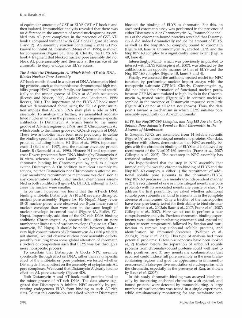
The �120-kDa POM121 protein in Xenopus and mammalsis a single transmembrane protein with the bulk of theprotein accessible for interaction with other nucleoporinsubunits (Figure 6A; Hallberg et al., 1993; Soderqvist andHallberg, 1994). However, the C-terminal third of POM121contains the FG repeat motifs present in a number of Nupsthat are thought to bind transport receptors such as importin� and transportin (Hallberg et al., 1993). A fragment ofPOM121, presumed to be available for interaction within thescaffold of the nuclear pore, but one that lacked FG repeats(in order to avoid transport receptor binding), was used tosearch for soluble nucleoporins that link to the criticalPOM121 protein. Using this fragment (aa 144-435, Figure6A), pulldowns were performed with Xenopus egg extracts.Nucleoporins that bound to the POM121 fragment in pull-downs were identified by immunoblotting (Figure 6B). No-tably, pore subunits containing Nup358, Nup214, Nup155,Nup62, and Nup53, showed no affinity for the POM121fragment (Figure 6B, lanes 3 and 5). Importin � and � bound,
Figure 5. Only ELYS and the Nup107-160 pore subunits bindchromatin in the absence of membranes and RanQ69L-GTP. (A) Adiagram representing the known soluble nucleoporin subunits(boxed). It should be noted that we were unable to probe for Aladinbecause of the lack of an anti-Xenopus Aladin antibody. Nucleopor-ins highlighted in red were assayed for their ability to bind toanchored chromatin by immunoblotting. (B and C) Anchored chro-matin binding assay in which chromatin-coated coverslips wereincubated with: Xenopus egg cytosol plus membranes to assembleanchored nuclei (lane 3); egg cytosol plus membranes plus 2 mMGTP�S to block membrane vesicle fusion (lane 4); buffer alone (lane6), membrane-free Xenopus egg cytosol (lane 7, arrowhead), or theidentical cytosol plus 30 �M RanQ69L-GTP (lane 8). A coverslip notcoated with chromatin was incubated with cytosol (lane 5) as acontrol for nonspecific protein binding. In this experiment, Orc2serves as a loading control. Mem, Xenopus egg membrane fractiondiluted 1:20, and cyt, Xenopus egg cytosol diluted 1:10, are shown asa control for immunoblotting with the various antibodies (lanes 1and 2). It should be noted that we did not detect chromatin bindingof Nup358 in the presence of excess RanGTP, as was previouslypublished (Walther et al., 2003b). (C) Immunoblots using antibodiesto the integral membrane proteins gp210 and ribophorin show thatmembrane vesicles are not present in the Xenopus egg cytosol (lanes2 and 5–8).
Initiating Nuclear Pore Assembly
Vol. 19, September 2008 3989
but were largely removed by RanQ69L-GTP (Figure 6B,compare lanes 3 and 5). The FG nucleoporin Nup153 alsobound to the POM121 beads, but it too was removed byRanQ69L-GTP, suggesting an indirect interaction, perhapsthrough its known binding to importin � (Shah and Forbes,1998; Shah et al., 1998; Ben-Efraim and Gerace, 2001; Waltheret al., 2003b).
Strikingly, the Nup107-160 complex bound strongly to thePOM121 fragment (Figure 6B, lane 3; see Nup160, Nup133,Nup85, Nup43, and Nup37). Its binding was unaffected byRanQ69L-GTP (Figure 6B, lane 5), indicating that the inter-
action is not mediated through importin � or �. A lesseramount of ELYS was also observed to bind to the POM121beads (data not shown) and may associate with POM121through its interaction with the Nup107-160 complex(Rasala et al., 2006). In addition, Nup93 and to a smallerextent Nup205, members of the Nup93-188-205 complex,also bound the POM121 beads (Figure 6B, lane 3 and 5).None of the above proteins bound to GFP negative controlbeads (Figure 6B, lane 2 and 4).
We conclude that the Nup107-160 complex and theNup93-188-205 complex, two key subunits of the nuclearpore’s central scaffold (Krull et al., 2004), bind to POM121 invitro. Because these two soluble pore subunits do notstrongly interact with one another in Xenopus egg extracts,even in the presence of RanGTP (Vasu et al., 2001 and Figure5B), they may bind to POM121 aa 144-435 independently.Notably, the very strong and specific interaction of theNup107-160 complex with POM121 implies that the recruit-ment of POM121 could be the next step in pore assemblyafter recruitment of ELYS and the Nup107-160 complex tochromatin.
ELYS and the Nup107-160 Complex Recruit POM121- andNDC1-containing Membrane VesiclesWe wanted to determine whether the recruitment ofPOM121-containing membrane vesicles is dependent onchromatin-bound ELYS/Nup107-160 or is found in nuclearmembranes independent of ELYS. To test this, we used thetools developed to produce ELYS-minus nuclei. Specifically,we assembled nuclei in the presence or absence of ELYSGST-AT-hook� or in the presence or absence of DistamycinA, and assayed for POM121. When nuclei were assembled inthe presence of GST or GST-AT-hook-2R3A, anti-POM121antibodies stained the nuclear rims in a normal punctatemanner (Figure 7A). In contrast, when nuclei were assem-bled in the presence of GST-AT-hook�, no POM121 staincould be visualized (Figure 7A, middle panel). This indicatesthat either POM121 was not recruited to ELYS-minus nucleior that it was recruited but could not oligomerize into adetectable entity in the absence of ELYS. When nuclei wereassembled in the presence of 10 �M Distamycin, againPOM121 antibodies failed to stain the nuclear membranes(Figure 7B). Thus, like the ELYS GST-AT-hook� fragment,Distamycin A prevents either the recruitment of POM121-containing membrane vesicles to nuclei or the assembly ofPOM121 into visible protein oligomers.
A recent study showed that POM121 and NDC1 are co-enriched in the same set of membrane vesicles in Xenopusegg extracts, but are separate from the membrane vesiclesthat contain gp210 (Antonin et al., 2005; Mansfeld et al.,2006). NDC1 is the only integral membrane pore proteinconserved in both yeast and higher eukaryotes and has beenshown to be essential for nuclear pore assembly (Chial et al.,1998; West et al., 1998; Lau et al., 2004; Lau et al., 2006;Madrid et al., 2006; Mansfeld et al., 2006; Stavru et al., 2006).
To determine whether the recruitment of NDC1 was de-pendent on chromatin-bound ELYS, ELYS-minus anchorednuclei were tested for the presence of NDC1 by immuno-blotting. This approach has the advantage over immunoflu-orescence of allowing one to determine the presence of apore membrane protein in nuclei, even if the protein is notoligomerized. Anchored nuclei were assembled in the pres-ence of GST, GST-AT-hook� or GST-�AT-hook and assayedfor the presence of NDC1 and gp210 by immunoblot anal-ysis (Figure 7C). We found that nuclei assembled in thepresence of GST and ELYS �AT-hook contained all thenucleoporins tested, including ELYS, Nup160, Nup133,
Figure 6. POM121 binds the Nup107-160 and the Nup93-205 com-plexes in the absence of FG Nups, Nup53, and Nup155. (A) A mapof POM121 and the fragment used. (B) The Xenopus POM121 frag-ment aa 144-435 (POM) was coupled to beads and mixed withXenopus egg cytosol in pulldown reactions, in the presence or theabsence of 10 �M RanQ69L-GTP. his-GFP beads were used as acontrol. The bound proteins were probed by immunoblotting withdifferent anti-nucleoporin and import factor antibodies, as shown inthe figure. Nup160, Nup133, Nup85, Nup43, and Nup37 of theNup107-160 complex, and Nup93 and Nup205 of the Nup93-205complex specifically bound to the POM121 fragment. Cyt, Xenopusegg cytosol diluted 1:10, is shown as a control for immunoblottingwith the various antibodies (lane 1).
B. A. Rasala et al.
Molecular Biology of the Cell3990

Figure 7. Chromatin-bound ELYS/Nup107-160 complex recruit POM121- and NDC1-containing membrane vesicles. (A) Reconstitutednuclei were assembled in the presence of 15 �M GST, GST-AT-hook�, or GST-AT-hook-2R3A. The presence of POM121 in the nuclearmembranes was probed for with directly labeled anti-POM121-AF488 (green). Membranes were stained with R18 membrane dye (red). DNAwas stained with Hoechst (blue). Nuclei were fixed before mounting onto slides. Scale bar, 10 �m. (B) Reconstituted nuclei were assembledin the presence of buffer or 10 �m Distamycin A and processed as in A. Scale bar, 10 �m. (C) Anchored nuclei were assembled by incubatingchromatin-coated coverslips with membranes and cytosol for 1 h, in the presence of either 15 �M GST, GST-AT-hook�, or GST-�AT-hook.A coverslip not treated with chromatin, but incubated with membranes, M, and cytosol, cyt, was used as a control for nonspecific binding(lane 4). The recruitment of integral membrane pore proteins NDC1 and gp210, in the presence (GST and GST-�AT-hook, lane 1 and 3) orabsence (GST-AT-hook�, lane 2) of chromatin-bound ELYS/Nup107-160 complex, was determined by immunoblot analysis.
Initiating Nuclear Pore Assembly
Vol. 19, September 2008 3991

Nup93, and the pore integral membrane proteins gp210 andNDC1 (Figure 7C, lanes 1 and 3), as expected from thenormal nuclear phenotype observed after addition of theseproteins in Figure 3B. However, anchored nuclei assembledin the presence of the AT-hook� fragment lacked not onlyELYS, Nup160, Nup133, and Nup93, but also lacked NDC1(Figure 7C, lane 2). In contrast, these nuclei did containgp210 and the ER/nuclear membrane protein ribophorin(Figure 7C, lane 2). We thus conclude that it is the recruit-ment of POM121/NDC1-containing membrane vesicles tonuclei that requires ELYS and the Nup107-160 complex to bepresent on chromatin, whereas the recruitment of gp210-containing vesicles does not.
DISCUSSION
The chromatin-binding protein ELYS targets nuclear poreassembly to the surface of chromosomes as nuclei form atthe end of mitosis. In this study we sought the molecularunderpinnings of the action of ELYS in the early steps ofnuclear pore assembly using an in vitro system. Emphasiz-ing that ELYS is the chromatin-targeting marker for nuclearpore assembly, we show that ELYS is not needed for ALpore formation (Figure 1). We found that the C-terminus ofELYS contains two chromatin-binding domains, an AT-hookmotif, and a downstream chromatin-binding domain (Fig-ures 2 and 3, and data not shown). Excess wild-type ELYSAT-hook� fragment blocks nuclear pore assembly, byblocking the recruitment of ELYS and the Nup107-160 com-plex, whereas a mutant in the AT-hook motif of this frag-ment does not (Figure 3). Distamycin A, an AT-rich DNA-binding antibiotic, phenocopies this inhibition of poreassembly, whereas Chromomycin A3, a GC-rich bindingantibiotic, does not (Figures 4 and 7). Although endogenousELYS and the Nup107-160 complex strongly bind to chro-matin in the absence of membranes, the remainder of the 14soluble subunits of the pore, with the possible exception ofNup153, requires membrane recruitment and fusion to as-sociate with chromatin-initiated nuclear intermediates (Fig-ure 5). By biochemical means, POM121 was found to bindthe Nup107-160 complex, arguing that POM121 membranerecruitment is the step that follows ELYS/Nup107-160 chro-matin binding (Figure 6). Importantly, the inhibition ofELYS chromatin binding also inhibits the recruitment ofPOM121 and NDC1 integral membrane proteins to formingnuclei (Figure 7).
A Model for the Early Steps in Nuclear Pore AssemblyThe data above support a model for the early steps innuclear pore assembly where the binding of ELYS to AT-rich
chromatin via the conserved AT-hook motif and an adjacentnon-AT-hook domain marks the sites of nuclear pore assem-bly (Figure 8). The binding of ELYS to AT-rich DNA tracts“seeds” the chromatin with pore initiation sites. Chromatin-bound ELYS recruits the Nup107-160 complex to nuclearpore initiation sites (Franz et al., 2007; Gillespie et al., 2007).The recruitment of pore membrane components NDC1 andPOM121 then occurs via an interaction between the Nup107-160 complex and the cytoplasmic domain of POM121. As-sembly of the remaining soluble pore subunits would thenfollow.
ELYS/Nup107-160 Recruit Key Pore Membrane ProteinsPOM121 and NDC1 to the Nuclear EnvelopeTargeting of pore membrane proteins POM121 and NDC1 toELYS-marked sites in vitro involves recruitment of a specificpool of vesicles (Figure 7). POM121 and NDC1 are known tobe coenriched in the same membrane vesicles in Xenopus eggextracts, while gp210 is contained in separate vesicles (An-tonin et al., 2005; Mansfeld et al., 2006). The in vitro systemused here allowed us to separate the binding of POM121/NDC1-containing membranes from the independent bind-ing of membranes involved in more general nuclear mem-brane assembly, which apparently includes gp210.Extrapolating from our data to in vivo conditions, one mustconsider that nuclear membrane assembly involves thebinding of ER membrane tubules that contain a mixture ofER and nuclear membrane proteins, including the integralmembrane pore proteins. Potentially, chromatin-boundELYS/Nup107-160 complex could, in vivo, attract POM121and NDC1 proteins, as they diffuse laterally in the forminginner nuclear membrane to place at least one copy directlyabove the pore-seeding points on chromatin.
Previous studies have indeed hypothesized a connectionbetween the Nup107-160 complex, or its yeast homologuethe Nup84 complex, and membranes (Heath et al., 1995; Li etal., 1995). Sec13 is a member of both the Nup107-160 com-plex and COPII vesicle-transport complexes (Siniossoglou etal., 1996; Fontoura et al., 1999; Harel et al., 2003b; Bickford etal., 2004; Loiodice et al., 2004). Other members of the Nup84complex also show similarity to proteins of the vesicular-transport complexes COPI, COPII, and clathrin (Devos et al.,2004). Indeed, the Nup107-160 complex member, Nup133,contains a membrane-curvature–sensing motif with the abil-ity to bind to liposomes (Drin et al., 2007). Lastly, the lateststructural model of the yeast NPC places the Nup84 com-plex adjacent to the nuclear membranes (Alber et al., 2007;Hsia et al., 2007). All these are consistent with our finding ofkey biochemical and functional interactions between theNup107-160 complex and POM121 and NDC1.
Figure 8. A model for the earlysteps in NPC assembly. On the basisof our findings, we propose a modelfor the early steps in NPC assembly.The first step in pore assembly is thebinding of ELYS (yellow) to AT-richchromatin sites, selected via the high-affinity AT-hook motif (black box)and strengthened by its second chro-matin binding domain. ELYS thenacts as a bridge between the chroma-
tin and the Nup107-160 complex (green; Rasala et al., 2006; Franz et al., 2007; Gillespie et al., 2007). Chromatin-bound ELYS/Nup107-160complex actively recruits the integral membrane pore proteins POM121 (red) and NDC1 (blue), likely through interaction with the cytosolicdomain of POM121. After membrane vesicle fusion, the rest of the soluble pore subunits assemble and a complete nuclear pore is formed.(Note, other vesicles/sheets bind to the chromatin via different linkages such as lamins to form the bulk of the nuclear membranes, but arenot shown here.)
B. A. Rasala et al.
Molecular Biology of the Cell3992

Yeast Lack a Direct ELYS HomologueAs essential as ELYS appears to be in metazoans, the yeastsSaccharomyces cerevisiae and Schizosaccharomyces pombe lack alarge canonical ELYS homologue. However, a small gene inS. pombe encoding 295 aa bears considerable homology to aa694–972 in the large human ELYS protein (2266 aa). We findno readily apparent S. cerevisiae homologue to either ELYSor the small S. pombe gene. Lacking the majority of ELYSstructure, nonmetazoans such as yeast may require a differ-ent initiation device for pore assembly in their intact nuclei.Considering that yeast do not undergo open mitosis andthus do not disassemble their NPCs, it is possible that atargeting protein such as ELYS is not required. However, ifyeast nuclear pores are indeed initiated from chromatinsites, then a prediction from the ELYS precedent would bethat any mechanism that acts to anchor the yeast Nup84complex to chromatin would be sufficient to initiate newpore assembly.
Nuclear Pores Are Initiated on AT-rich ChromatinTwo Xenopus ELYS isoforms have been published, one of2201 aa (Galy et al., 2006) and one of 2408 aa (Gillespie et al.,2007), the latter being that used here. Upstream of the AT-hook, however, we note that the larger isoform differs fromother vertebrate homologues in that it contains seven copiesof �31-aa repeat, which we observe to contain sequencesclosely related to AT-hook motifs, as defined by Huth et al.(1997). Xenopus, with its rapid cell division in early devel-opment where cell division takes place every 30 min for thefirst 12 divisions (Newport and Kirschner, 1982), might po-tentially use these excess AT-hook-like sequences of ELYS toaccomplish rapid nuclear pore assembly.
Our point mutation and antibiotic studies demonstratethat ELYS preferentially binds to and NPC assembly is pref-erentially initiated on AT-rich chromatin sites (Figures 3 and4). The best studied of the AT-hook–containing proteins isthe HMGA family of proteins, whose members function ingene transcription, DNA repair, and chromatin remodeling(Goodwin, 1998; Anand and Chada, 2000). Although firstgenerally described to bind DNA at any run of 5–6 AT basepairs, current studies on the HMGA family show that theseproteins likely bind to DNA with sequence specificity.HMGA2 was recently shown to bind to a 15-base pair con-sensus site: 5 AT-rich base pairs, 4–5 GC-rich base pairs, andthen 5–6 AT-rich base pairs (Cui and Leng, 2007). It wouldbe intriguing to determine whether ELYS binds to chromatinin a similar sequence-specific restricted manner.
A large body of evidence indicates that gene-poor/AT-rich regions of the genome are positioned at the nuclearperiphery and gene-rich/GC-rich regions are positioned inthe nuclear interior (Bernardi et al., 1985; Saccone et al., 1993)(Croft et al., 1999; Saccone et al., 2002; Zink et al., 2004; Fosterand Bridger, 2005). It is possible that the binding preferenceof ELYS might aid in the localization of AT-rich chromatin tothe nuclear periphery. However, a subset of newly activatedgenes move to the nuclear periphery after transcriptionalactivation, both in yeast and in vertebrates and is thought todo so through interaction with nuclear pore proteins (Caso-lari et al., 2005; Cabal et al., 2006; Brown and Silver, 2007).This movement could involve ELYS in vertebrates or, alter-natively, be an ELYS-independent pore localization event.
Nuclear Pore Assembly in S-PhaseOverall, the pore assembly model proposed here wouldrequire little alteration to function in the intact nuclear en-velope of an S phase cell, where pore number doubles.
Import of ELYS and the Nup107-160 complex into the intactnucleus, followed by their sequential binding to chromatin,could again form a platform on AT-rich sites. POM121 andNDC1, moving laterally through the inner nuclear mem-brane, could anchor and initiate pore assembly at theseplatforms. Consistent with this, both nuclear import in gen-eral and a nuclear pool of the Nup107-160 complex havebeen shown to be required for pore assembly in intact nuclei(Walther et al., 2003a; D’Angelo et al., 2006).
One possible extrapolation of our data is that there mightbe specific sites in the genome capable of initiating nuclearpore assembly. If so, in S-phase the pore initiation siteswould double as the DNA is replicated, which would con-trol the doubling in pore number previously observed (Maulet al., 1972). In order for this to occur, however, an intranu-clear pool of soluble ELYS/MEL-28 must exist. Indeed, sucha pool has already been described, both in HeLa cells (Rasalaet al., 2006) and in C. elegans (Galy et al., 2006). This intrigu-ing possibility awaits a more precise definition of whatconstitutes an ELYS-binding sequence on chromatin.
In a recent study, chromatin immunoprecipitation usingan Mcm3 antibody led Gillespie et al. (2007) to conclude thatELYS and the replication licensing complex, Mcm2-7, are inproximity on chromatin fragments, although the actual sizeof the average chromatin fragment in that study was notstated (Gillespie et al., 2007). Geminin, a protein that com-pletely blocks Mcm2-7 loading onto chromatin, caused adelay in ELYS and nucleoporin incorporation into reconsti-tuted nuclei. The authors proposed that the Mcm2-7 com-plex promotes ELYS binding to chromatin. In our study, wefound that a GC-binding antibiotic more strongly affectedMcm3 binding to chromatin, whereas an AT-binding anti-biotic more strongly inhibited ELYS binding to chromatin(Figure 4B). Further investigation will help to define thepotential relationship between replication licensing and nu-clear pore assembly.
SummaryIn summary, the binding of ELYS to chromatin, minimallyvia its key AT-hook motif and adjacent second chromatin-binding domain, initiates nuclear pore assembly on AT-richchromatin tracts. The subsequent recruitment of pore-spe-cific membrane vesicles containing POM121 and NDC1 isdependent on chromatin-bound ELYS and its binding part-ner, the Nup107-160 complex. This recruitment precedesfurther pore assembly. Indeed, assembly of the bulk of theremaining soluble pore subunits is dependent on the pres-ence of membranes and is sensitive to GTP�S. This latterdata also argue, in conjunction with the studies of Baur et al.(2007), that the majority of postmitotic nuclear pore assem-bly events occurs within a continuous double lipid bilayer.
ACKNOWLEDGMENTS
The authors thank the members of the Forbes laboratory and ZhongshengYou for many helpful discussions regarding this work. We especially thankValerie Delmar for critical reading of the manuscript and Corine Lau forreagents (Forbes lab). We also thank Zhongsheng You for the kind gift ofOrc2, RCC1, and Mcm3 antibodies and Aurelie Lachish-Zalait and MichaelElbaum for the generous gift of GFP-M9 import substrate. This work wassupported by a National Institute of General Medical Sciences Grant R01GM33279 to D.F. and a European Commission FP6 Marie Curie JRG grant031161 to A.H.
REFERENCES
Alber, F. et al. (2007). The molecular architecture of the nuclear pore complex.Nature 450, 695–701.
Initiating Nuclear Pore Assembly
Vol. 19, September 2008 3993

Anand, A., and Chada, K. (2000). In vivo modulation of Hmgic reducesobesity. Nat. Genet 24, 377–380.
Anderson, D. J., and Hetzer, M. W. (2007). Nuclear envelope formation bychromatin-mediated reorganization of the endoplasmic reticulum. Nat. CellBiol. 9, 1160–1166.
Antonin, W., Ellenberg, J., and Dultz, E. (2008). Nuclear pore complex assem-bly through the cell cycle: regulation and membrane organization. FEBS Lett.582, 2004–2016.
Antonin, W., Franz, C., Haselmann, U., Antony, C., and Mattaj, I. W. (2005).The integral membrane nucleoporin pom121 functionally links nuclear porecomplex assembly and nuclear envelope formation. Mol. Cell 17, 83–92.
Aravind, L., and Landsman, D. (1998). AT-hook motifs identified in a widevariety of DNA-binding proteins. Nucleic Acids Res. 26, 4413–4421.
Baur, T., Ramadan, K., Schlundt, A., Kartenbeck, J., and Meyer, H. H. (2007).NSF- and SNARE-mediated membrane fusion is required for nuclear enve-lope formation and completion of nuclear pore complex assembly in Xenopuslaevis egg extracts. J. Cell Sci. 120, 2895–2903.
Belgareh, N. et al. (2001). An evolutionarily conserved NPC subcomplex,which redistributes in part to kinetochores in mammalian cells. J. Cell Biol.154, 1147–1160.
Bell, A., Kittler, L., Lober, G., and Zimmer, C. (1997). DNA binding propertiesof minor groove binders and their influence on the topoisomerase II cleavagereaction. J. Mol. Recognit. 10, 245–255.
Ben-Efraim, I., and Gerace, L. (2001). Gradient of increasing affinity of impor-tin beta for nucleoporins along the pathway of nuclear import. J. Cell Biol. 152,411–417.
Bernardi, G., Olofsson, B., Filipski, J., Zerial, M., Salinas, J., Cuny, G., Meunier-Rotival, M., and Rodier, F. (1985). The mosaic genome of warm-bloodedvertebrates. Science 228, 953–958.
Bickford, L. C., Mossessova, E., and Goldberg, J. (2004). A structural view ofthe COPII vesicle coat. Curr. Opin. Struct. Biol. 14, 147–153.
Bodoor, K., Shaikh, S., Salina, D., Raharjo, W. H., Bastos, R., Lohka, M., andBurke, B. (1999). Sequential recruitment of NPC proteins to the nuclearperiphery at the end of mitosis. J. Cell Sci. 112(Pt 13), 2253–2264.
Brown, C. R., and Silver, P. A. (2007). Transcriptional regulation at the nuclearpore complex. Curr. Opin. Genet Dev. 17, 100–106.
Burke, B. (2007). Network news: complete nuclear coverage. Nat. Cell Biol. 9,1123–1124.
Burke, B., and Ellenberg, J. (2002). Remodelling the walls of the nucleus. Nat.Rev. Mol. Cell Biol. 3, 487–497.
Cabal, G. G. et al. (2006). SAGA interacting factors confine sub-diffusion oftranscribed genes to the nuclear envelope. Nature 441, 770–773.
Casolari, J. M., Brown, C. R., Drubin, D. A., Rando, O. J., and Silver, P. A.(2005). Developmentally induced changes in transcriptional program alterspatial organization across chromosomes. Genes Dev. 19, 1188–1198.
Chaudhary, N., and Courvalin, J. C. (1993). Stepwise reassembly of thenuclear envelope at the end of mitosis. J. Cell Biol. 122, 295–306.
Chial, H. J., Rout, M. P., Giddings, T. H., and Winey, M. (1998). Saccharomycescerevisiae Ndc1p is a shared component of nuclear pore complexes and spindlepole bodies. J. Cell Biol. 143, 1789–1800.
Cotter, L. A., Goldberg, M. W., and Allen, T. D. (1998). Nuclear pore complexdisassembly and nuclear envelope breakdown during mitosis may occur byboth nuclear envelope vesicularisation and dispersion throughout the endo-plasmic reticulum. Scanning 20, 250–251.
Croft, J. A., Bridger, J. M., Boyle, S., Perry, P., Teague, P., and Bickmore, W. A.(1999). Differences in the localization and morphology of chromosomes in thehuman nucleus. J. Cell Biol. 145, 1119–1131.
Cronshaw, J. M., Krutchinsky, A. N., Zhang, W., Chait, B. T., and Matunis,M. J. (2002). Proteomic analysis of the mammalian nuclear pore complex.J. Cell Biol. 158, 915–927.
Cui, T., and Leng, F. (2007). Specific recognition of AT-rich DNA sequences bythe mammalian high mobility group protein AT-hook 2, a SELEX study.Biochemistry 46, 13059–13066.
Dabauvalle, M. C., Loos, K., Merkert, H., and Scheer, U. (1991). Spontaneousassembly of pore complex-containing membranes (“annulate lamellae”) inXenopus egg extract in the absence of chromatin. J. Cell Biol. 112, 1073–1082.
Daigle, N., Beaudouin, J., Hartnell, L., Imreh, G., Hallberg, E., Lippincott-Schwartz, J., and Ellenberg, J. (2001). Nuclear pore complexes form immobilenetworks and have a very low turnover in live mammalian cells. J. Cell Biol.154, 71–84.
D’Angelo, M. A., Anderson, D. J., Richard, E., and Hetzer, M. W. (2006).Nuclear pores form de novo from both sides of the nuclear envelope. Science312, 440–443.
De Souza, C. P., Osmani, A. H., Hashmi, S. B., and Osmani, S. A. (2004).Partial nuclear pore complex disassembly during closed mitosis in Aspergillusnidulans. Curr. Biol. 14, 1973–1984.
Devos, D., Dokudovskaya, S., Alber, F., Williams, R., Chait, B. T., Sali, A., andRout, M. P. (2004). Components of coated vesicles and nuclear pore com-plexes share a common molecular architecture. PLoS Biol. 2, e380.
Drin, G., Casella, J. F., Gautier, R., Boehmer, T., Schwartz, T. U., and Antonny,B. (2007). A general amphipathic alpha-helical motif for sensing membranecurvature. Nat. Struct. Mol. Biol. 14, 138–146.
Drummond, S. P., and Wilson, K. L. (2002). Interference with the cytoplasmictail of gp210 disrupts “close apposition” of nuclear membranes and blocksnuclear pore dilation. J. Cell Biol. 158, 53–62.
Dultz, E., Zanin, E., Wurzenberger, C., Braun, M., Rabut, G., Sironi, L., andEllenberg, J. (2008). Systematic kinetic analysis of mitotic dis- and reassemblyof the nuclear pore in living cells. J. Cell Biol. 180, 857–865.
Ellenberg, J., Siggia, E. D., Moreira, J. E., Smith, C. L., Presley, J. F., Worman,H. J., and Lippincott-Schwartz, J. (1997). Nuclear membrane dynamics andreassembly in living cells: targeting of an inner nuclear membrane protein ininterphase and mitosis. J. Cell Biol. 138, 1193–1206.
Fernandez, A. G., and Piano, F. (2006). MEL-28 is downstream of the Rancycle and is required for nuclear-envelope function and chromatin mainte-nance. Curr. Biol. 16, 1757–1763.
Fontoura, B. M., Blobel, G., and Matunis, M. J. (1999). A conserved biogenesispathway for nucleoporins: proteolytic processing of a 186-kilodalton precur-sor generates Nup98 and the novel nucleoporin, Nup96. J. Cell Biol. 144,1097–1112.
Forbes, D. J., Kirschner, M. W., and Newport, J. W. (1983). Spontaneousformation of nucleus-like structures around bacteriophage DNA microin-jected into Xenopus eggs. Cell 34, 13–23.
Foster, H. A., and Bridger, J. M. (2005). The genome and the nucleus: amarriage made by evolution. Genome organisation and nuclear architecture.Chromosoma 114, 212–229.
Franz, C., Walczak, R., Yavuz, S., Santarella, R., Gentzel, M., Askjaer, P., Galy,V., Hetzer, M., Mattaj, I. W., and Antonin, W. (2007). MEL-28/ELYS isrequired for the recruitment of nucleoporins to chromatin and postmitoticnuclear pore complex assembly. EMBO Rep. 8, 165–172.
Funakoshi, T., Maeshima, K., Yahata, K., Sugano, S., Imamoto, F., andImamoto, N. (2007). Two distinct human POM121 genes: requirement for theformation of nuclear pore complexes. FEBS Lett. 581, 4910–4916.
Galy, V., Askjaer, P., Franz, C., Lopez-Iglesias, C., and Mattaj, I. W. (2006).MEL-28, a novel nuclear-envelope and kinetochore protein essential for zy-gotic nuclear-envelope assembly in C. elegans. Curr. Biol. 16, 1748–1756.
Gerace, L., Ottaviano, Y., and Kondor-Koch, C. (1982). Identification of amajor polypeptide of the nuclear pore complex. J. Cell Biol. 95, 826–837.
Gillespie, P. J., Khoudoli, G. A., Stewart, G., Swedlow, J. R., and Blow, J. J.(2007). ELYS/MEL-28 chromatin association coordinates nuclear pore com-plex assembly and replication licensing. Curr. Biol. 17, 1657–1662.
Goldberg, M. W., Wiese, C., Allen, T. D., and Wilson, K. L. (1997). Dimples,pores, star-rings, and thin rings on growing nuclear envelopes: evidence forstructural intermediates in nuclear pore complex assembly. J. Cell Sci. 110(Pt4), 409–420.
Goldfarb, D. S., Corbett, A. H., Mason, D. A., Harreman, M. T., and Adam,S. A. (2004). Importin alpha: a multipurpose nuclear-transport receptor.Trends Cell Biol. 14, 505–514.
Goodwin, G. (1998). The high mobility group protein, HMGI-C. Int. J. Bio-chem. Cell Biol. 30, 761–766.
Greber, U. F., Senior, A., and Gerace, L. (1990). A major glycoprotein of thenuclear pore complex is a membrane-spanning polypeptide with a largelumenal domain and a small cytoplasmic tail. EMBO J. 9, 1495–1502.
Hallberg, E., Wozniak, R. W., and Blobel, G. (1993). An integral membraneprotein of the pore membrane domain of the nuclear envelope contains anucleoporin-like region. J. Cell Biol. 122, 513–521.
Haraguchi, T., Koujin, T., Hayakawa, T., Kaneda, T., Tsutsumi, C., Imamoto,N., Akazawa, C., Sukegawa, J., Yoneda, Y., and Hiraoka, Y. (2000). Livefluorescence imaging reveals early recruitment of emerin, LBR, RanBP2, andNup153 to reforming functional nuclear envelopes. J. Cell Sci. 113(Pt 5),779–794.
B. A. Rasala et al.
Molecular Biology of the Cell3994

Harel, A., Chan, R. C., Lachish-Zalait, A., Zimmerman, E., Elbaum, M., andForbes, D. J. (2003a). Importin beta negatively regulates nuclear membranefusion and nuclear pore complex assembly. Mol. Biol. Cell 14, 4387–4396.
Harel, A., Orjalo, A. V., Vincent, T., Lachish-Zalait, A., Vasu, S., Shah, S.,Zimmerman, E., Elbaum, M., and Forbes, D. J. (2003b). Removal of a singlepore subcomplex results in vertebrate nuclei devoid of nuclear pores. Mol.Cell 11, 853–864.
Heath, C. V., Copeland, C. S., Amberg, D. C., Del Priore, V., Snyder, M., andCole, C. N. (1995). Nuclear pore complex clustering and nuclear accumulationof poly(A)� RNA associated with mutation of the Saccharomyces cerevisiaeRAT2/NUP120 gene. J. Cell Biol. 131, 1677–1697.
Hetzer, M., Walther, T. C., and Mattaj, I. W. (2005). Pushing the envelope:structure, function, and dynamics of the nuclear periphery. Annu. Rev. CellDev. Biol. 21, 347–380.
Hsia, K. C., Stavropoulos, P., Blobel, G., and Hoelz, A. (2007). Architecture ofa coat for the nuclear pore membrane. Cell 131, 1313–1326.
Huth, J. R., Bewley, C. A., Nissen, M. S., Evans, J. N., Reeves, R., Gronenborn,A. M., and Clore, G. M. (1997). The solution structure of an HMG-I(Y)-DNAcomplex defines a new architectural minor groove binding motif. Nat. Struct.Biol. 4, 657–665.
Kas, E., Izaurralde, E., and Laemmli, U. K. (1989). Specific inhibition of DNAbinding to nuclear scaffolds and histone H1 by distamycin. The role ofoligo(dA).oligo(dT) tracts. J. Mol. Biol. 210, 587–599.
Kessel, R. G. (1992). Annulate lamellae: a last frontier in cellular organelles.Int. Rev. Cytol. 133, 43–120.
Kimura, N., Takizawa, M., Okita, K., Natori, O., Igarashi, K., Ueno, M.,Nakashima, K., Nobuhisa, I., and Taga, T. (2002). Identification of a noveltranscription factor, ELYS, expressed predominantly in mouse foetal haema-topoietic tissues. Genes Cells 7, 435–446.
Krull, S., Thyberg, J., Bjorkroth, B., Rackwitz, H. R., and Cordes, V. C. (2004).Nucleoporins as components of the nuclear pore complex core structure andTpr as the architectural element of the nuclear basket. Mol. Biol. Cell 15,4261–4277.
Lau, C. K., Delmar, V. A., and Forbes, D. J. (2006). Topology of yeast Ndc1p:predictions for the human NDC1/NET3 homologue. Anat. Rec. A Discov.Mol. Cell Evol. Biol. 288, 681–694.
Lau, C. K., Giddings, T. H., Jr., and Winey, M. (2004). A novel allele ofSaccharomyces cerevisiae NDC1 reveals a potential role for the spindle polebody component Ndc1p in nuclear pore assembly. Eukaryot. Cell 3, 447–458.
Li, O., Heath, C. V., Amberg, D. C., Dockendorff, T. C., Copeland, C. S.,Snyder, M., and Cole, C. N. (1995). Mutation or deletion of the Saccharomycescerevisiae RAT3/NUP133 gene causes temperature-dependent nuclear accu-mulation of poly(A)� RNA and constitutive clustering of nuclear pore com-plexes. Mol. Biol. Cell 6, 401–417.
Liu, J., Prunuske, A. J., Fager, A. M., and Ullman, K. S. (2003). The COPIcomplex functions in nuclear envelope breakdown and is recruited by thenucleoporin Nup153. Dev. Cell 5, 487–498.
Lohka, M. J., and Masui, Y. (1983). Formation in vitro of sperm pronuclei andmitotic chromosomes induced by amphibian ooplasmic components. Science220, 719–721.
Loiodice, I., Alves, A., Rabut, G., Van Overbeek, M., Ellenberg, J., Sibarita,J. B., and Doye, V. (2004). The entire Nup107-160 complex, including threenew members, is targeted as one entity to kinetochores in mitosis. Mol. Biol.Cell 15, 3333–3344.
Macara, I. G. (2001). Transport into and out of the nucleus. Microbiol. Mol.Biol. Rev. 65, 570–594, table of contents.
Macaulay, C., and Forbes, D. J. (1996). Assembly of the nuclear pore: bio-chemically distinct steps revealed with NEM, GTP gamma S, and BAPTA. J.Cell Biol. 132, 5–20.
Macaulay, C., Meier, E., and Forbes, D. J. (1995). Differential mitotic phos-phorylation of proteins of the nuclear pore complex. J. Biol. Chem. 270,254–262.
Madrid, A. S., Mancuso, J., Cande, W. Z., and Weis, K. (2006). The role of theintegral membrane nucleoporins Ndc1p and Pom152p in nuclear pore com-plex assembly and function. J. Cell Biol. 173, 361–371.
Mansfeld, J. et al. (2006). The conserved transmembrane nucleoporin NDC1 isrequired for nuclear pore complex assembly in vertebrate cells. Mol. Cell 22,93–103.
Margalit, A., Vlcek, S., Gruenbaum, Y., and Foisner, R. (2005). Breaking andmaking of the nuclear envelope. J. Cell. Biochem. 95, 454–465.
Maul, G. G., Maul, H. M., Scogna, J. E., Lieberman, M. W., Stein, G. S., Hsu,B. Y., and Borun, T. W. (1972). Time sequence of nuclear pore formation in
phytohemagglutinin-stimulated lymphocytes and in HeLa cells during thecell cycle. J. Cell Biol. 55, 433–447.
Meier, E., Miller, B. R., and Forbes, D. J. (1995). Nuclear pore complexassembly studied with a biochemical assay for annulate lamellae formation.J. Cell Biol. 129, 1459–1472.
Metcalf, C. E., and Wassarman, D. A. (2006). DNA binding properties of TAF1isoforms with two AT-hooks. J. Biol. Chem. 281, 30015–30023.
Miller, B. R., and Forbes, D. J. (2000). Purification of the vertebrate nuclearpore complex by biochemical criteria. Traffic 1, 941–951.
Mutvei, A., Dihlmann, S., Herth, W., and Hurt, E. C. (1992). NSP1 depletionin yeast affects nuclear pore formation and nuclear accumulation. Eur. J. CellBiol. 59, 280–295.
Newport, J. (1987). Nuclear reconstitution in vitro: stages of assembly aroundprotein-free DNA. Cell 48, 205–217.
Newport, J., and Kirschner, M. (1982). A major developmental transition inearly Xenopus embryos: I. Characterization and timing of cellular changes atthe midblastula stage. Cell 30, 675–686.
Okita, K., Kiyonari, H., Nobuhisa, I., Kimura, N., Aizawa, S., and Taga, T.(2004). Targeted disruption of the mouse ELYS gene results in embryonicdeath at peri-implantation development. Genes Cells 9, 1083–1091.
Orjalo, A. V., Arnaoutov, A., Shen, Z., Boyarchuk, Y., Zeitlin, S. G., Fontoura,B., Briggs, S., Dasso, M., and Forbes, D. J. (2006). The Nup107-160 nucleoporincomplex is required for correct bipolar spindle assembly. Mol. Biol. Cell 17,3806–3818.
Patel, S. S., Belmont, B. J., Sante, J. M., and Rexach, M. F. (2007). Nativelyunfolded nucleoporins gate protein diffusion across the nuclear pore complex.Cell 129, 83–96.
Pemberton, L. F., and Paschal, B. M. (2005). Mechanisms of receptor-mediatednuclear import and nuclear export. Traffic 6, 187–198.
Powers, M. A., Macaulay, C., Masiarz, F. R., and Forbes, D. J. (1995). Recon-stituted nuclei depleted of a vertebrate GLFG nuclear pore protein, p97,import but are defective in nuclear growth and replication. J. Cell Biol. 128,721–736.
Prunuske, A. J., Liu, J., Elgort, S., Joseph, J., Dasso, M., and Ullman, K. S.(2006). Nuclear envelope breakdown is coordinated by both Nup358/RanBP2and Nup153, two nucleoporins with zinc finger modules. Mol. Biol. Cell 17,760–769.
Quimby, B. B., and Corbett, A. H. (2001). Nuclear transport mechanisms. CellMol. Life Sci. 58, 1766–1773.
Rabut, G., Doye, V., and Ellenberg, J. (2004). Mapping the dynamic organi-zation of the nuclear pore complex inside single living cells. Nat. Cell Biol. 6,1114–1121.
Rasala, B. A., Orjalo, A. V., Shen, Z., Briggs, S., and Forbes, D. J. (2006). ELYSis a dual nucleoporin/kinetochore protein required for nuclear pore assemblyand proper cell division. Proc. Natl. Acad. Sci. USA 103, 17801–17806.
Reeves, R. (2001). Molecular biology of HMGA proteins: hubs of nuclearfunction. Gene 277, 63–81.
Reeves, R., and Nissen, M. S. (1990). The AT-DNA-binding domain of mam-malian high mobility group I chromosomal proteins. A novel peptide motiffor recognizing DNA structure. J. Biol. Chem. 265, 8573–8582.
Reichelt, R., Holzenburg, A., Buhle, E. L., Jr., Jarnik, M., Engel, A., and Aebi,U. (1990). Correlation between structure and mass distribution of the nuclearpore complex and of distinct pore complex components. J. Cell Biol. 110,883–894.
Rzepecki, R., Bogachev, S. S., Kokoza, E., Stuurman, N., and Fisher, P. A.(1998). In vivo association of lamins with nucleic acids in Drosophila melano-gaster. J. Cell Sci. 111(Pt 1), 121–129.
Saccone, S., De Sario, A., Wiegant, J., Raap, A. K., Della Valle, G., andBernardi, G. (1993). Correlations between isochores and chromosomal bandsin the human genome. Proc. Natl. Acad. Sci. USA 90, 11929–11933.
Saccone, S., Federico, C., and Bernardi, G. (2002). Localization of the gene-richest and the gene-poorest isochores in the interphase nuclei of mammalsand birds. Gene 300, 169–178.
Schwartz, T. U. (2005). Modularity within the architecture of the nuclear porecomplex. Curr. Opin. Struct. Biol. 15, 221–226.
Shah, S., and Forbes, D. J. (1998). Separate nuclear import pathways convergeon the nucleoporin Nup153 and can be dissected with dominant-negativeinhibitors. Curr. Biol. 8, 1376–1386.
Shah, S., Tugendreich, S., and Forbes, D. (1998). Major binding sites for thenuclear import receptor are the internal nucleoporin Nup153 and the adjacentnuclear filament protein Tpr. J. Cell Biol. 141, 31–49.
Initiating Nuclear Pore Assembly
Vol. 19, September 2008 3995

Sheehan, M. A., Mills, A. D., Sleeman, A. M., Laskey, R. A., and Blow, J. J.(1988). Steps in the assembly of replication-competent nuclei in a cell-freesystem from Xenopus eggs. J. Cell Biol. 106, 1–12.
Siniossoglou, S., Wimmer, C., Rieger, M., Doye, V., Tekotte, H., Weise, C.,Emig, S., Segref, A., and Hurt, E. C. (1996). A novel complex of nucleoporins,which includes Sec13p and a Sec13p homolog, is essential for normal nuclearpores. Cell 84, 265–275.
Soderqvist, H., and Hallberg, E. (1994). The large C-terminal region of theintegral pore membrane protein, POM121, is facing the nuclear pore complex.Eur. J. Cell Biol. 64, 186–191.
Stavru, F., Hulsmann, B. B., Spang, A., Hartmann, E., Cordes, V. C., andGorlich, D. (2006). NDC1, a crucial membrane-integral nucleoporin of meta-zoan nuclear pore complexes. J. Cell Biol. 173, 509–519.
Suntharalingam, M., and Wente, S. R. (2003). Peering through the pore:nuclear pore complex structure, assembly, and function. Dev. Cell 4, 775–789.
Vasu, S., Shah, S., Orjalo, A., Park, M., Fischer, W. H., and Forbes, D. J. (2001).Novel vertebrate nucleoporins Nup133 and Nup160 play a role in mRNAexport. J. Cell Biol. 155, 339–354.
Vasu, S. K., and Forbes, D. J. (2001). Nuclear pores and nuclear assembly.Curr. Opin. Cell Biol. 13, 363–375.
Walther, T. C. et al. (2003a). The conserved Nup107-160 complex is critical fornuclear pore complex assembly. Cell 113, 195–206.
Walther, T. C., Askjaer, P., Gentzel, M., Habermann, A., Griffiths, G., Wilm,M., Mattaj, I. W., and Hetzer, M. (2003b). RanGTP mediates nuclear porecomplex assembly. Nature 424, 689–694.
West, R. R., Vaisberg, E. V., Ding, R., Nurse, P., and McIntosh, J. R. (1998).cut11(�): a gene required for cell cycle-dependent spindle pole body anchor-ing in the nuclear envelope and bipolar spindle formation in Schizosaccharo-myces pombe. Mol. Biol. Cell 9, 2839–2855.
Winey, M., Yarar, D., Giddings, T. H., Jr., and Mastronarde, D. N. (1997).Nuclear pore complex number and distribution throughout the Saccharomycescerevisiae cell cycle by three-dimensional reconstruction from electron micro-graphs of nuclear envelopes. Mol. Biol. Cell 8, 2119–2132.
Wozniak, R. W., Bartnik, E., and Blobel, G. (1989). Primary structure analysisof an integral membrane glycoprotein of the nuclear pore. J. Cell Biol. 108,2083–2092.
Yang, L., Guan, T., and Gerace, L. (1997). Integral membrane proteins of thenuclear envelope are dispersed throughout the endoplasmic reticulum duringmitosis. J. Cell Biol. 137, 1199–1210.
Zink, D., Fischer, A. H., and Nickerson, J. A. (2004). Nuclear structure incancer cells. Nat. Rev. Cancer 4, 677–687.
B. A. Rasala et al.
Molecular Biology of the Cell3996