canine leismaniasis
TRANSCRIPT

7/30/2019 Canine Leismaniasis
http://slidepdf.com/reader/full/canine-leismaniasis 1/60
CANINE
LEISMANIASIS
7/30/2019 Canine Leismaniasis
http://slidepdf.com/reader/full/canine-leismaniasis 2/60
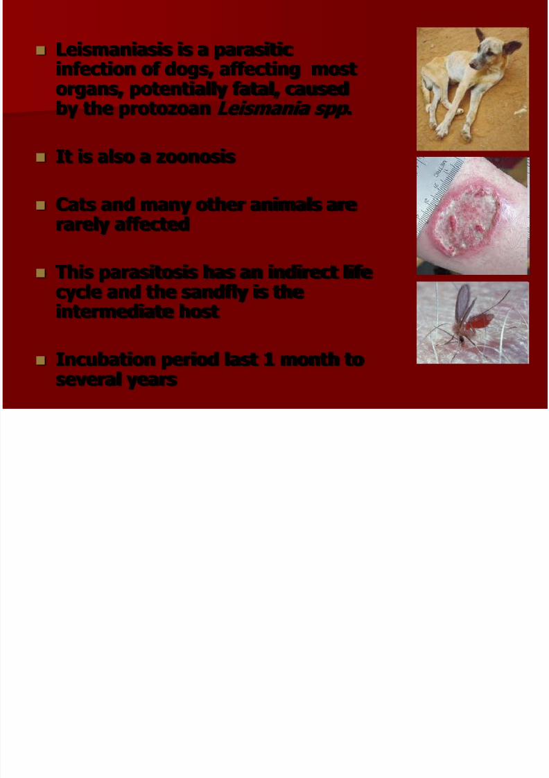
Leismaniasis is a parasiticinfection of dogs, affecting most
organs, potentially fatal, causedby the protozoan Leismania spp .
It is also a zoonosis
Cats and many other animals arerarely affected
This parasitosis has an indirect life
cycle and the sandfly is theintermediate host
Incubation period last 1 month to
several years

7/30/2019 Canine Leismaniasis
http://slidepdf.com/reader/full/canine-leismaniasis 3/60
Comments Infected dogs are the primary
reservoir for zoonotic visceral
leishmaniasis in endemic regionsand are the most significant risk factor predisposing humans toinfection
Dogs have a wide range of clinical
presentation caused by infectionwith L. infantum , ranging fromasymptomatic to fatal visceralizingdisease.
Host factors which determine clinical
outcome are poorly understood.
Immune status, the generalcondition, reinfections by theparasite, the saliva of sandfly andother factors play an important role
on the clinical status of the infecteddog
7/30/2019 Canine Leismaniasis
http://slidepdf.com/reader/full/canine-leismaniasis 4/60
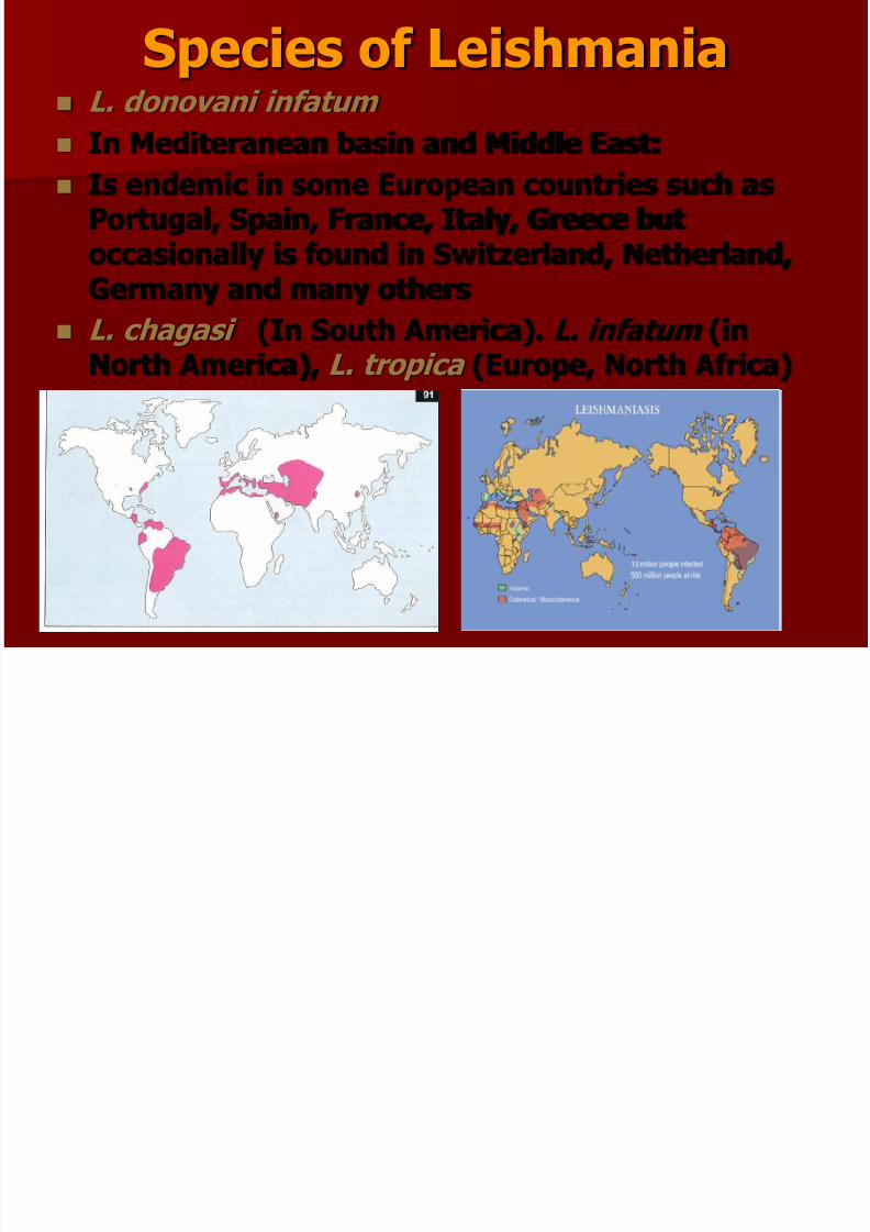
Species of Leishmania
L. donovani infatum
In Mediteranean basin and Middle East:
Is endemic in some European countries such asPortugal, Spain, France, Italy, Greece butoccasionally is found in Switzerland, Netherland,
Germany and many others
L. chagasi (In South America). L. infatum (inNorth America), L. tropica (Europe, North Africa)

7/30/2019 Canine Leismaniasis
http://slidepdf.com/reader/full/canine-leismaniasis 5/60
7/30/2019 Canine Leismaniasis
http://slidepdf.com/reader/full/canine-leismaniasis 6/60
7/30/2019 Canine Leismaniasis
http://slidepdf.com/reader/full/canine-leismaniasis 7/60
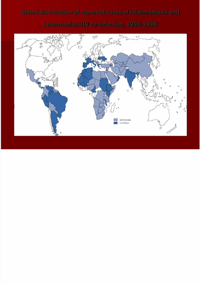
Global distribution of reported cases of leishmaniasis and
Leishmania/HIV co-infection, 1990-1998
7/30/2019 Canine Leismaniasis
http://slidepdf.com/reader/full/canine-leismaniasis 8/60
7/30/2019 Canine Leismaniasis
http://slidepdf.com/reader/full/canine-leismaniasis 9/60
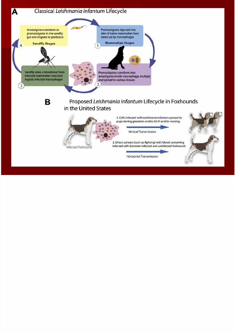
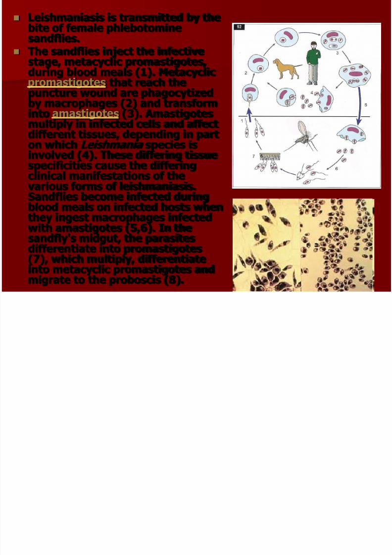
Leishmaniasis is transmitted by thebite of female phlebotominesandflies.
The sandflies inject the infectivestage, metacyclic promastigotes,during blood meals (1). Metacyclicpromastigotes that reach thepuncture wound are phagocytizedby macrophages (2) and transforminto amastigotes (3). Amastigotesmultiply in infected cells and affect
different tissues, depending in parton which Leishmania species isinvolved (4). These differing tissuespecificities cause the differingclinical manifestations of thevarious forms of leishmaniasis.Sandflies become infected during
blood meals on infected hosts whenthey ingest macrophages infectedwith amastigotes (5,6). In thesandfly's midgut, the parasitesdifferentiate into promastigotes(7), which multiply, differentiate
into metacyclic promastigotes andmigrate to the proboscis (8).

7/30/2019 Canine Leismaniasis
http://slidepdf.com/reader/full/canine-leismaniasis 10/60
Forms of Leishmania spp
Promastigotes
(In Sandflay)
Amastigotes
(In dog)

7/30/2019 Canine Leismaniasis
http://slidepdf.com/reader/full/canine-leismaniasis 11/60
SANDFLlIES
Europe (Phlebotomus spp)
America (Lutzomyia spp)

7/30/2019 Canine Leismaniasis
http://slidepdf.com/reader/full/canine-leismaniasis 12/60
Sand Flies
Sand flies belong to the fly familyPsychodidae , members of which arecharacterized by their densely hairywings which give them a moth-likeappearance.
Phlebotomines are distinguished fromother members of the family by the way
they hold their wings above the body ina vertical V.
There are about 700 species of phlebotomine sand flies of which about70 are considered to transmit diseases
to people. Sand flies (Phlebotominae) are blood
suckers and their larvae inhabit placeswhere there is high organic matter suchas in animal burrows, termite hills and
tree holes
7/30/2019 Canine Leismaniasis
http://slidepdf.com/reader/full/canine-leismaniasis 13/60
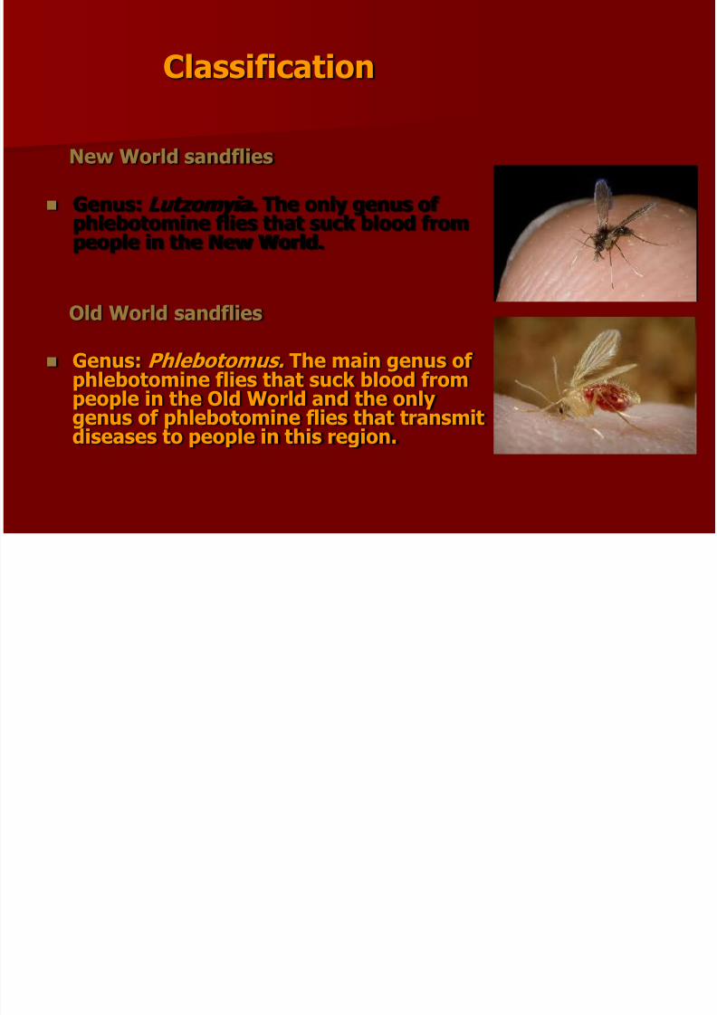
Classification
New World sandflies
Genus: Lutzomyia. The only genus of phlebotomine flies that suck blood from
people in the New World.
Old World sandflies
Genus: Phlebotomus. The main genus of
phlebotomine flies that suck blood frompeople in the Old World and the onlygenus of phlebotomine flies that transmitdiseases to people in this region.

7/30/2019 Canine Leismaniasis
http://slidepdf.com/reader/full/canine-leismaniasis 14/60
Life Cycle It is difficult studying the life cycle of
sand flies because the larvae are tinyand don't live in well defined places,like mosquito larvae. The entire lifecycle takes 20-40 days except indiapausing species (i.e. those thatstop developing when conditionsbecome too cold).
Eggs. The female lays 30-70 eggs byscattering them around a potentialbreeding site. They hatch within 1-2weeks.
Larvae. Larvae feed on dead organicmatter and are found in damp places
containing organic matter such ascracks in walls or rock, animalburrows and shelters, caves, or inleaf litter. In regions with coolwinters, larvae diapause in the fourth(final) instar.
Pupae. Pupal development takes 5-10 days.

7/30/2019 Canine Leismaniasis
http://slidepdf.com/reader/full/canine-leismaniasis 15/60
Adults.
Emerge from the pupae in darkness, often just before dawn.
Only the female sucks blood , the food being used for eggproduction. Both males and females feed on sugary secretionsfrom plants or from honeydew produced by homopteran bugs.Mating takes place at or near hosts: the males congregate inleks on or near the host and produce sex pheromones. Femaleshome in on hosts using both host odor and the odor produced by
the males. Vibration of the wings by males can be important inencouraging females to mate. Adults are mainly active in the early morning, evening and at
night although they can bite during the day if disturbed. Wheninactive, adult sand flies have habitat-specific resting sites thatare characteristic of particular species. One of the main ways inwhich entomologists study sand flies is by locating and studyingthem at their resting sites. Resting sites are often similar or nearto the larval breeding sites and are usually places that are cool,humid and dark. Sand flies are able to survive in dryenvironments by withdrawing to cool, humid resting sites duringthe day and then becoming active at night when ambienttemperatures drop and humidity increases.

7/30/2019 Canine Leismaniasis
http://slidepdf.com/reader/full/canine-leismaniasis 16/60
Seasonal activity of adultsis affected mainly bytemperature and rainfall.
Flies are very small andtheir temporary feeding onanimals may not benoticed.
These flies, lives thesummer time in temperateand Mediterranean areas.

7/30/2019 Canine Leismaniasis
http://slidepdf.com/reader/full/canine-leismaniasis 17/60
SANDFLlIES
Size
Sand flies are smaller thanmosquitoes but larger thanmidges, with a body length of 2-
3mm.
Colour
All sand flies are brownish indaylight but their bodies aredensely covered in oily hairs whichgive the insects a whitishappearance when illuminated

7/30/2019 Canine Leismaniasis
http://slidepdf.com/reader/full/canine-leismaniasis 18/60
V-shaped wings
This is perhaps the most distinctivefeature of the group. Phlebotominesat rest hold their wings in a raised “V” The wings are never closed or laid flatacross the body.
Flight
Phlebotomines have a weak, directflight and once on the host progressby a series of small hops. They do not hover round a host and as such areoften not recognised as a biting
nuisance.
7/30/2019 Canine Leismaniasis
http://slidepdf.com/reader/full/canine-leismaniasis 19/60
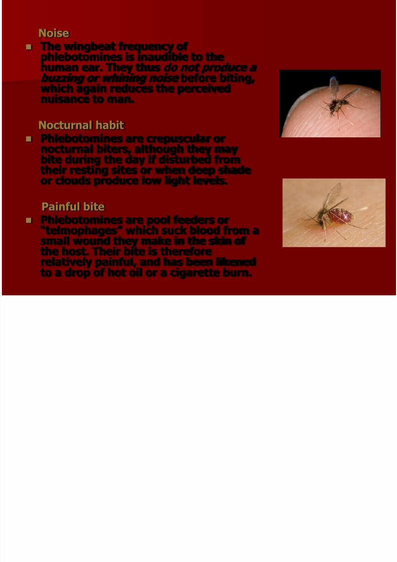
Noise
The wingbeat frequency of phlebotomines is inaudible to the
human ear. They thus do not produce a buzzing or whining noise before biting,which again reduces the perceivednuisance to man.
Nocturnal habit
Phlebotomines are crepuscular ornocturnal biters, although they maybite during the day if disturbed fromtheir resting sites or when deep shadeor clouds produce low light levels.
Painful bite Phlebotomines are pool feeders or
“telmophages” which suck blood from asmall wound they make in the skin of the host. Their bite is thereforerelatively painful, and has been likened
to a drop of hot oil or a cigarette burn.

7/30/2019 Canine Leismaniasis
http://slidepdf.com/reader/full/canine-leismaniasis 20/60
Life Cycle of Sandfly
Unlike in most biting Diptera, development of sand fliestakes place in terrestrial rather than aquatic microhabitats.
Although there have been relatively few successfulattempts to identify breeding sites in nature, eggs are laid
in soil rich in organic matter and the larvae pass throughfour instars before pupation and adult emergence.
The difficulty of finding breeding sites is an importantconstraint on vector control measures available to
leishmaniasis control programmes, application of larvicidesnot being a practical alternative.

7/30/2019 Canine Leismaniasis
http://slidepdf.com/reader/full/canine-leismaniasis 21/60
The eggs are elongated oval-shaped , pale atfirst and darkening on exposure to air witha single black “eye spot”.The larvae emerge through a J-shaped
fissure and are legless and whitish with adark head capsule. Those of the first instarcan be distinguished by the presence of twocaudal bristles, all subsequent instarsbearing four. Fourth instar larvae also havea prominent sclerite on the dorsum of the
penultimate segment.The pupae are golden brown and are affixedto the surface of the substrate in which theydeveloped by the final larval exuvium.Shortly before emergence the wings andeyes turn black.Male sand flies emerge about 24 h beforefemales, allowing their external genitaliatime to rotate 180° to the correct positionfor mating before females have emerged.The time from oviposition to adultemergence at ambient temperature isaround 4-6 weeks.
7/30/2019 Canine Leismaniasis
http://slidepdf.com/reader/full/canine-leismaniasis 22/60
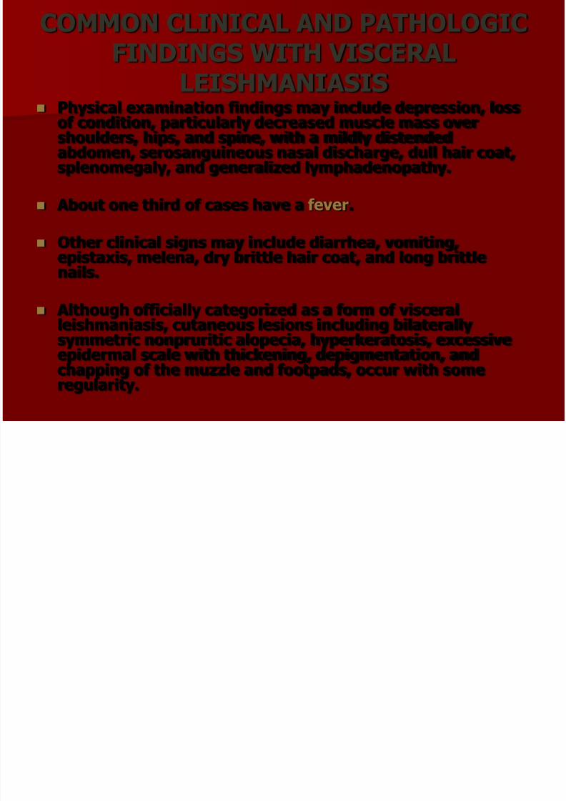
COMMON CLINICAL AND PATHOLOGICFINDINGS WITH VISCERAL
LEISHMANIASIS Physical examination findings may include depression, loss
of condition, particularly decreased muscle mass overshoulders, hips, and spine, with a mildly distendedabdomen, serosanguineous nasal discharge, dull hair coat,splenomegaly, and generalized lymphadenopathy.
About one third of cases have a fever.
Other clinical signs may include diarrhea, vomiting,epistaxis, melena, dry brittle hair coat, and long brittlenails.
Although officially categorized as a form of visceralleishmaniasis, cutaneous lesions including bilaterallysymmetric nonpruritic alopecia, hyperkeratosis, excessiveepidermal scale with thickening, depigmentation, andchapping of the muzzle and footpads, occur with someregularity.

7/30/2019 Canine Leismaniasis
http://slidepdf.com/reader/full/canine-leismaniasis 23/60
Abnormal clinical pathologic values often include decreasedhematocrit, thrombocytopenia, and signs of renal failureincluding azotemia, increased blood urea nitrogen andcreatinine, hyperphosphatemia, hypermagnesemia, andproteinuria.
Signs of hepatic compromise are also common includingelevated alkaline phosphatase (ALP), elevated alaninetransferase (ALT), and hypercholesterolemia.
Other common clinical chemistry abnormalities include
hyperproteinemia observed with hypergammaglobulinemiaand hypoalbuminemia.
7/30/2019 Canine Leismaniasis
http://slidepdf.com/reader/full/canine-leismaniasis 24/60
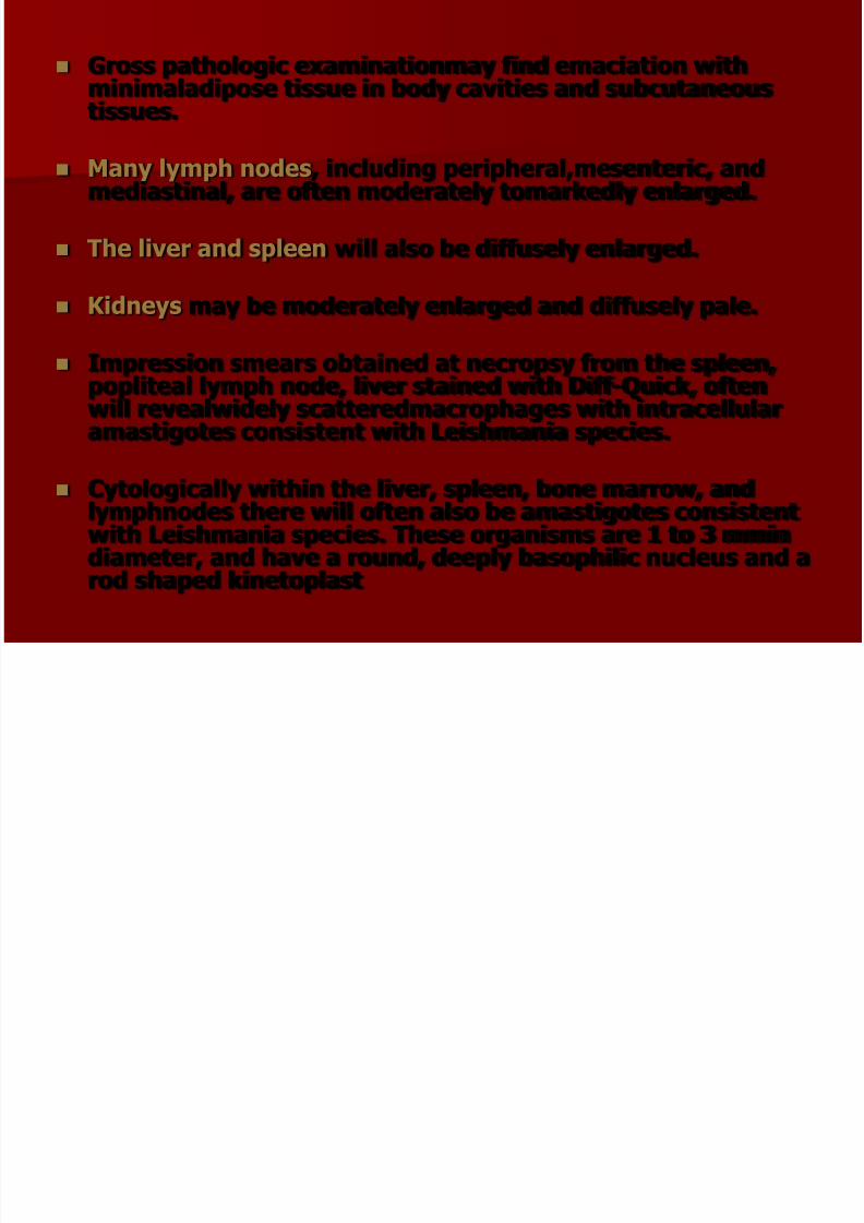
Gross pathologic examinationmay find emaciation withminimaladipose tissue in body cavities and subcutaneoustissues.
Many lymph nodes , including peripheral,mesenteric, andmediastinal, are often moderately tomarkedly enlarged.
The liver and spleen will also be diffusely enlarged.
Kidneys may be moderately enlarged and diffusely pale.
Impression smears obtained at necropsy from the spleen,popliteal lymph node, liver stained with Diff-Quick, oftenwill revealwidely scatteredmacrophages with intracellularamastigotes consistent with Leishmania species.
Cytologically within the liver, spleen, bone marrow, andlymphnodes there will often also be amastigotes consistentwith Leishmania species. These organisms are 1 to 3 mmindiameter, and have a round, deeply basophilic nucleus and arod shaped kinetoplast

7/30/2019 Canine Leismaniasis
http://slidepdf.com/reader/full/canine-leismaniasis 25/60
Dogs
90% of symptomatic also have cutaneousinvolvement
There is no sex or breed predilection
Cats
Cutaneous disease is rare
There is no sex or breed predilection
7/30/2019 Canine Leismaniasis
http://slidepdf.com/reader/full/canine-leismaniasis 26/60

7/30/2019 Canine Leismaniasis
http://slidepdf.com/reader/full/canine-leismaniasis 27/60

7/30/2019 Canine Leismaniasis
http://slidepdf.com/reader/full/canine-leismaniasis 28/60

7/30/2019 Canine Leismaniasis
http://slidepdf.com/reader/full/canine-leismaniasis 29/60

7/30/2019 Canine Leismaniasis
http://slidepdf.com/reader/full/canine-leismaniasis 30/60

7/30/2019 Canine Leismaniasis
http://slidepdf.com/reader/full/canine-leismaniasis 31/60

7/30/2019 Canine Leismaniasis
http://slidepdf.com/reader/full/canine-leismaniasis 32/60
7/30/2019 Canine Leismaniasis
http://slidepdf.com/reader/full/canine-leismaniasis 33/60

7/30/2019 Canine Leismaniasis
http://slidepdf.com/reader/full/canine-leismaniasis 34/60

7/30/2019 Canine Leismaniasis
http://slidepdf.com/reader/full/canine-leismaniasis 35/60
IMMUNE ALTERATION AND
7/30/2019 Canine Leismaniasis
http://slidepdf.com/reader/full/canine-leismaniasis 36/60
IMMUNE ALTERATION ANDPATHOGENESIS OF VISCERAL
LEISHMANIASIS Mammalian host responses which prevent progression to clinical VL has
been shown to be dependent on promoting T helper-1 IFN-g production-based immunity and parasiticidal activity within infected macrophages.
A key immunologic feature of late stage clinical VL in dogs is an inabilityto proliferate or to produce IFN-g in response to Leishmania antigen.
Pharmacologically-cured individuals are resistant to reinfection andmount antigen-specific IFN-g responses in vitro, indicating that there isnot an inherent defect in host CD41 T cell responses of clinical patientsonce they have reached this stage.
High levels of TNF-a have been proposed to stimulate production of regulatory cytokines, specifically IL-10, as a homeostatic response toprevent further inflammation-mediated pathology.
IL-10 can be produced by many cell types including T cells, B cells, andmacrophages. One of the proposed mechanisms of IL-10 promotion of VLis by conditioning macrophages for parasite growth and survival versuskilling of intracellular parasites.

7/30/2019 Canine Leismaniasis
http://slidepdf.com/reader/full/canine-leismaniasis 37/60
In surveillance studies, we have observed repeated caseswhere dogs do not show clinical signs of VL until there issecondary immunosuppression caused by pregnancy,concomitant Lyme disease, or other tick-borne illness.
This clinical shift toward disease consistently appears upona change from being seronegative to seropositive in thesedogs.
Further studies are required to determine the effects of immune alterations that lead to clinical disease in these
dogs. Congenital infection secondary to vertical transmission may
predispose to initial immune abnormalities, although by thetime clinical signs of disease and seroconversion haveappeared, evidence shows that CD41 T cells from thesedogs are able to respond normally to parasite antigen.
In advanced disease it is not unusual to seeimmunosuppression including T-cell changes, in terms of reduced CD41 T cell proliferation in response to whole Leinfantum antigen or routine canine vaccines and decreasedability of these cells to produceIFN-g in response to Leinfantum antigen.
GENETIC FACTORS RELATED TO
7/30/2019 Canine Leismaniasis
http://slidepdf.com/reader/full/canine-leismaniasis 38/60
GENETIC FACTORS RELATED TO VISCERAL LEISHMANIASIS DISEASE
SUSCEPTIBILITY
Although several genetic polymorphisms, includingalterations in TNF-a and solutecarrier family 11A1(SLC11A1, formerly NRAMP1) allelic expression, have beenindicated to predispose to disease, causative factors of
disease susceptibility in humans and dogs, specifically thoseassociated with heritability, remain elusive.
Breed type has also been shown to alter the response totherapy, suggesting that canine breed-related geneticfactors modulate disease progression and are thereforeprognostically significant

7/30/2019 Canine Leismaniasis
http://slidepdf.com/reader/full/canine-leismaniasis 39/60
DIFFERENTIAL DIAGNOSIS
Visceral - mycoses (blastomycosis,histoplasmosis), systemic lupus erythematosus,metastatic neoplasia, distember and vasculitis
Cutaneous – other causes of hyperceratosis:primary idiopathic seborrhea and nutritionaldermatoses (vitamin A responsive, zingresponsive): idiopathic nasodigitalhyperceratosis, lichenoid-psoriasiformdermatosis, mucocutaneous syndrome, pemfigus
foliaccus, epidermal dysplasia
Hyperglobulinemia need to differentiate fromchronic Erlichiosis and multiple myeloma

7/30/2019 Canine Leismaniasis
http://slidepdf.com/reader/full/canine-leismaniasis 40/60
DIAGNOSIS OF VISCERALLEISHMANIASIS
Cytology is the diagnostic method of
choice in order to indentify the typical
intracellular organisms from
Lymph tissues samples
Bone marrow samples
Spleen samples
Skin samples
By Immunohistochemical techniques
7/30/2019 Canine Leismaniasis
http://slidepdf.com/reader/full/canine-leismaniasis 41/60
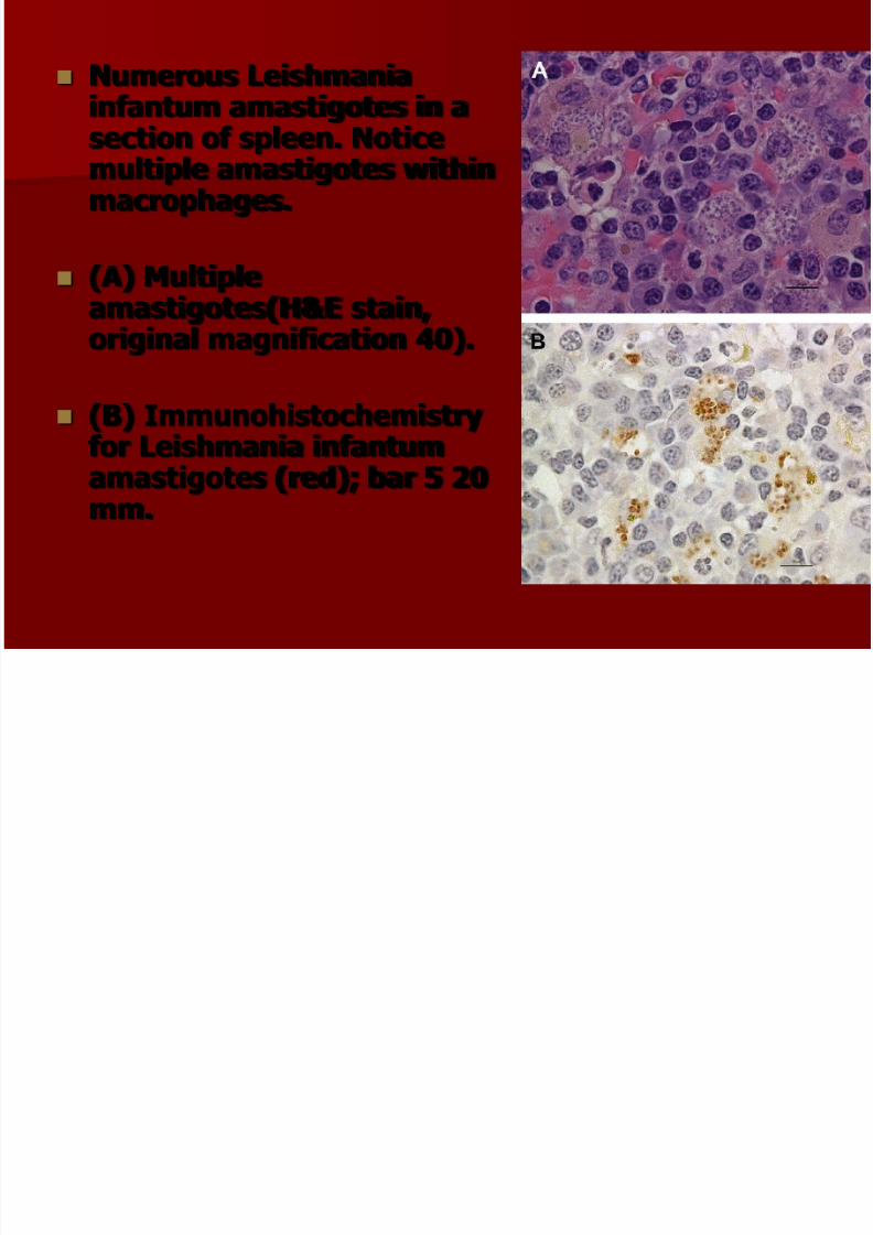
Numerous Leishmaniainfantum amastigotes in asection of spleen. Noticemultiple amastigotes withinmacrophages.
(A) Multiple
amastigotes(H&E stain,original magnification 40).
(B) Immunohistochemistryfor Leishmania infantumamastigotes (red); bar 5 20mm.

7/30/2019 Canine Leismaniasis
http://slidepdf.com/reader/full/canine-leismaniasis 42/60
Amastigotes are small round or oval bodies 1.5-3 X 2.5-3.5 μm in size and without a free flagellum.
The organisms has a relatively large nucleus anda kinetoplast
7/30/2019 Canine Leismaniasis
http://slidepdf.com/reader/full/canine-leismaniasis 43/60
DNA INDENTIFICATION BY PCR TECHNIQUE
PCR targeting Kinetoplast DNA increacesensitivity and specificity of Leishmania detection, use
Fresh and frozen bone marrow samples
Lymph node
Skin biopsy specimens
From the above samples the sensitivity of PCR approaches 95-100% however in peripheralblood samples is higher sortly after the infection(88%) and then decreases to 50-70%
SEROLOGY

7/30/2019 Canine Leismaniasis
http://slidepdf.com/reader/full/canine-leismaniasis 44/60
SEROLOGY
A wide variety of serological assays are available,utilizing indirect immunofluorescence (IFA),direct aglutination, conventional ELISA, dot-ELISA, competitive ELISA and western blotting
methodology. At present most tests employ crudeLeishmania antigen, although a recombinantLeishmania K39 ELISA has been recentlyvalidated.
Several rapid immunochromatographic test kitshave also been produced but their sensitivity andspecitifity varies from 35-76%
7/30/2019 Canine Leismaniasis
http://slidepdf.com/reader/full/canine-leismaniasis 45/60
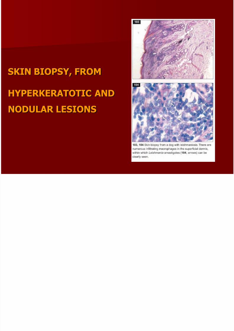
SKIN BIOPSY, FROM
HYPERKERATOTIC AND
NODULAR LESIONS

7/30/2019 Canine Leismaniasis
http://slidepdf.com/reader/full/canine-leismaniasis 46/60
TREATMENT/PROGNOSIS
Treatment of canine visceral leishmaniasis (CVL) is rarelycurative.
Prognosis for emaciated chronically infected animals isvery poor.
It is critical to advise the owner of potential zoonotictransmission of organisms from lesions to humans before
maintaining a Leishmania-infected dog in their household,particularly if there are immunosupressed people sharingthe household.
The owner should be informed that the organism will neverbe completely eliminated (ie, no sterile cure) and relapseoccurs very frequently requiring retreatment.
Treatment should be undertaken on an outpatient basis.Because of the chronic wasting that can occur withleishmaniasis, it is important to provide a good high-qualityprotein diet or a diet appropriate for renal insufficiency if this manifestation of leishmaniasis is present.
7/30/2019 Canine Leismaniasis
http://slidepdf.com/reader/full/canine-leismaniasis 47/60
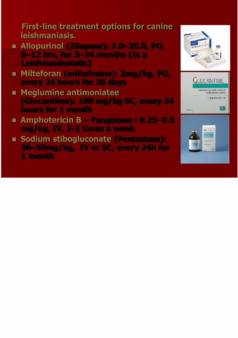
First-line treatment options for canineleishmaniasis.
Allopurinol (Zilapour): 7.0 –20.0, PO,8 –12 hrs, for 3 –24 months (Is aLeishmaniostatic)
Milteforan (miltefosine): 2mg/kg, PO,
every 24 hours for 28 days Meglumine antimoniatee
(Glucantime): 100 mg/kg SC, every 24hours for 1 month
Amphotericin B – Fungizone : 0.25 –0.5
mg/kg, IV, 2-3 times a week Sodium stibogluconate (Pentostam):
30 –50mg/kg, IV or SC, every 24h for1 month

7/30/2019 Canine Leismaniasis
http://slidepdf.com/reader/full/canine-leismaniasis 48/60
COMENTS Milteforan the only legal drug without side effects
Meglutamine antimoniate (Glucantime , Pfizer/Merial,France), which has some side effects.
Sodium stibogluconate (Pentostam, Wellcome FoundationLtd, U.K.), which requires daily injection and has severe side
effects,
Amphotericin B in the lipid emulsion or liposomal form isnon-nephrotoxic and is effective against the organism,although it is not thought to be superior to allopurinol as itis stillmore costly and more toxic.
Renal insufficiency must be treated before givingantimonial drugs or amphotericin B as prognosis isdependent on renal function at the onset of treatment.

7/30/2019 Canine Leismaniasis
http://slidepdf.com/reader/full/canine-leismaniasis 49/60
AFTER TREATMENT
Treatment efficacy is best monitored by clinicalimprovement and presence of organisms in biopsy or asmeasured by rigorously controlled qPCR.
Relapses occur a few months to a year after therapy, sodogs should be rechecked.
Relapses indetified by monitoring rise in blood globulins orreapepearance of clinical signs in a dog previously in
remission
Prognosis for a cure is very guarded, but therapydoesprovide infected dogs improved quality of life.

7/30/2019 Canine Leismaniasis
http://slidepdf.com/reader/full/canine-leismaniasis 50/60
Second-line drug, whichrequire further clinicalstudies to understand theirefficacy in dogs isparomomycin (Humantin).
Paromymycin has beenshown to have fewer sideeffects than other drugs in
humans. Use of this drughas been primarily targetedto the cutaneous versionsof Leishmania,less is knownabout its ability to remove
organ-based infection.

7/30/2019 Canine Leismaniasis
http://slidepdf.com/reader/full/canine-leismaniasis 51/60
Prevention measures
Sand fly vector control measures,including deltamethrin orpermethrin-impregnated collars areuseful todate to prevent disease.
Lemon repellents every day beforewalk (near head)
Avoid outdoor activities, especiallyfrom dusk to dawn, when sand flies
are the most active.
Keep dog in good hygiene conditions
Give diet high quality protein
Make special vaccines
C t l f S dfli

7/30/2019 Canine Leismaniasis
http://slidepdf.com/reader/full/canine-leismaniasis 52/60
Control of Sandflies
Spraying of residual insecticides on surfaces in the home.This has been the main way used for controlling sand fliesbut is obviously ineffective for those species which biteaway from the home such as those in South Americanforests. This control technique is also used for killing
Anopheles mosquitoes that transmit malaria and is some
regions it is effective in reducing both malaria andleishmaniasis. Killing of reservoir species. Certain species of mammals can
act as important reservoirs of Leishmania and by killing thereservoir species that are living near human habitation,disease rates can be decreased. For instance, rodenticideshave been used against the Great Gerbil Rhombomys opimus in Central Asia.
Insecticide spraying of larval habitat. This is usually notpossible because, usually, so little is known about wherethe larvae occur.

7/30/2019 Canine Leismaniasis
http://slidepdf.com/reader/full/canine-leismaniasis 53/60

7/30/2019 Canine Leismaniasis
http://slidepdf.com/reader/full/canine-leismaniasis 54/60

7/30/2019 Canine Leismaniasis
http://slidepdf.com/reader/full/canine-leismaniasis 55/60

7/30/2019 Canine Leismaniasis
http://slidepdf.com/reader/full/canine-leismaniasis 56/60

7/30/2019 Canine Leismaniasis
http://slidepdf.com/reader/full/canine-leismaniasis 57/60

7/30/2019 Canine Leismaniasis
http://slidepdf.com/reader/full/canine-leismaniasis 58/60
SUMMARY FOR ROMANIA
At present Leishmaniasis is an important diseaseof dogs (and humans)
But probably, climatic conditions will changedramatically in few years
Always you must collect information about thepossibility of dog transmission in endemic areasor origin of dog, specially in this case that clinicalsymptoms are in differential diagnosis
Also you must recommend your clients witch willtravel with their dogs in endemic areas to takeprevention measures

7/30/2019 Canine Leismaniasis
http://slidepdf.com/reader/full/canine-leismaniasis 59/60
Canine leishmaniasis has clinical symptoms frommany organs but the more commons are,coetaneous lesions, enlarged lymph modes andglomerulonephritis
Special test kits are useful in the beginning, forthe diagnosis but cytology is the diagnosticmethod of choice.
Alopourinole is a cheap drug wich you can sugestfor prevention (dose 20mg/kg daily for a week) just after travel
For the treatment, combination of Miltefosine and
Alopourinole is very effective (without sideefects)
Vaccines at this time are very effective, but don’tfully protect dogs

7/30/2019 Canine Leismaniasis
http://slidepdf.com/reader/full/canine-leismaniasis 60/60