candida glabrata: review of epidemiology, pathogenesis ... · nosocomial c. glabrata have been...
TRANSCRIPT

CLINICAL MICROBIOLOGY REVIEWS,0893-8512/99/$04.0010
Jan. 1999, p. 80–96 Vol. 12, No. 1
Copyright © 1999, American Society for Microbiology. All Rights Reserved.
Candida glabrata: Review of Epidemiology, Pathogenesis, andClinical Disease with Comparison to C. albicans
PAUL L. FIDEL, JR.,1* JOSE A. VAZQUEZ,2 AND JACK D. SOBEL2
Department of Microbiology, Immunology, and Parasitology, Louisiana State University Medical Center,New Orleans, Louisiana,1 and Division of Infectious Diseases, Wayne State University
School of Medicine, Detroit, Michigan2
INTRODUCTION .........................................................................................................................................................80BIOLOGY ......................................................................................................................................................................81EPIDEMIOLOGY.........................................................................................................................................................82PATHOGENESIS..........................................................................................................................................................83
Virulence ....................................................................................................................................................................83Host Defense..............................................................................................................................................................84Animal Models ..........................................................................................................................................................85
CLINICAL SPECTRUM OF INFECTION ...............................................................................................................87Superficial Infections................................................................................................................................................87
Oropharyngeal.......................................................................................................................................................88(i) Clinical manifestations...............................................................................................................................88(ii) Management ...............................................................................................................................................88
Esophageal .............................................................................................................................................................88(i) Clinical manifestations...............................................................................................................................88(ii) Management ...............................................................................................................................................89
Vulvovaginal...........................................................................................................................................................89(i) Clinical manifestations...............................................................................................................................89(ii) Management ...............................................................................................................................................89
Urinary tract..........................................................................................................................................................90(i) Clinical manifestations...............................................................................................................................90(ii) Management ...............................................................................................................................................90
Systemic Infections ...................................................................................................................................................90Clinical manifestations ........................................................................................................................................91Management ..........................................................................................................................................................91
ANTIFUNGAL RESISTANCE ....................................................................................................................................91Classification .............................................................................................................................................................91Evidence for Clinical and In Vitro Resistance .....................................................................................................91Mechanisms of Resistance.......................................................................................................................................92Clinical Relevance.....................................................................................................................................................92
CONCLUSION..............................................................................................................................................................93REFERENCES ..............................................................................................................................................................93
INTRODUCTION
Historically, Candida glabrata has been considered a rela-tively nonpathogenic saprophyte of the normal flora of healthyindividuals, rarely causing serious infection in humans (57,163). However, following the widespread and increased use ofimmunosuppressive therapy together with broad-spectrum an-timycotic therapy, the frequency of mucosal and systemic in-fections caused by C. glabrata has increased significantly (65,86, 90, 120, 143, 166, 179, 184). In fact, depending on the siteof infection, C. glabrata is often the second or third mostcommon cause of candidiasis after C. albicans. C. glabratainfections can be mucosal or systemic and are common inabnormal hosts (e.g., immunocompromised persons or thosewith diabetes mellitus) (53, 148, 149, 182). In contrast to otherCandida species, C. glabrata is not dimorphic; consequently, it
is found as blastoconidia both as a commensal and as a patho-gen. C. glabrata infections are difficult to treat and are oftenresistant to many azole antifungal agents, especially flucon-azole (65, 90, 167, 179). Consequently, C. glabrata infectionshave a high mortality rate in compromised, at-risk hospitalizedpatients.
Unfortunately, there have been relatively few investigationsof C. glabrata compared to other Candida species. Althoughthis infection is second or third in frequency after C. albicans,difficult to treat, and associated with a high mortality rate,publications to date on C. glabrata account for only a smallpercentage of published studies on medically important fungalinfections. Very little is known about the virulence of C. gla-brata, and virtually nothing is known about the host defensesdirected against the organism. There are only two establishedanimal models of experimental C. glabrata infections (systemicand vaginal) (24, 41). Therefore, studies to understand thepathogenesis of C. glabrata infections are sorely needed. Thisreview discusses what is currently known about C. glabratainfections and includes specific comparisons to C. albicanswherever possible. Specific topics discussed include its biology,
* Corresponding author. Mailing address: Department of Microbi-ology, Immunology, and Parasitology, Louisiana State University Med-ical Center, 1901 Perdido St., New Orleans, LA 70112. Phone: (504)568-4066. Fax: (504) 568-4066. E-mail: [email protected].
80
on January 16, 2020 by guesthttp://cm
r.asm.org/
Dow
nloaded from

epidemiology, pathogenesis, clinical perspectives, treatment,and antifungal resistance.
BIOLOGY
C. glabrata, together with other Candida species, belongs tothe class Fungi Imperfecti, the order Moniliales, and the familyCryptococcaceae (91, 148). C. glabrata is a nondimorphic yeastthat exists as small blastoconidia under all environmental con-ditions as a pathogen. In fact, C. glabrata is the only Candida
species that does not form pseudohyphae at temperaturesabove 37°C. Figure 1 shows wet-mount preparations of C.glabrata and C. albicans at similar magnifications. It is clearthat C. glabrata blastoconidia (1 to 4 mm) are considerablysmaller than C. albicans blastoconidia (4 to 6 mm). On Sab-ouraud dextrose agar, C. glabrata forms glistening, smooth,cream-colored colonies which are relatively indistinguishablefrom those of other Candida species except for their relativesize, which is quite small. On Chromagar, a relatively new agarthat distinguishes different Candida species by color as a result
FIG. 1. Size differential of C. glabrata and C. albicans. Shown are wet-mount slide preparations of C. glabrata (A) and C. albicans (B) on a hemocytometer.Magnification, 3400.
VOL. 12, 1999 CANDIDA GLABRATA INFECTIONS 81
on January 16, 2020 by guesthttp://cm
r.asm.org/
Dow
nloaded from

of biochemical reactions, C. glabrata colonies appear pink topurple, in contrast to C. albicans colonies, which appear greento blue-green. A critical distinguishing characteristic of C. gla-brata is its haploid genome, in contrast to the diploid genomeof C. albicans and several other non-albicans Candida species(176). Finally, C. glabrata is distinguishable from C. albicans byits small-subunit rRNA (4).
Most medically important Candida species can be easilydifferentiated from one another by either established commer-cially available biochemical tests or molecular biology tech-niques. With the advent of molecular genetics, newer identifi-cation methods have emerged. These methods usecomparative analysis of chromosomal DNA to identify Can-dida species from each other and also to delineate differentstrains within a species. These newer methods include restric-tion fragment length polymorphisms, pulsed-field gel electro-phoresis, randomly amplified polymorphic DNA, and DNAprobes (77, 79, 95, 170). By using contour-clamped homoge-neous electric field gel electrophoresis (CHEF), a form ofpulsed-field gel electrophoresis, chromosomal DNA from C.glabrata can be separated based on the different chromosomalmolecular weights and thus can be subjected to electrophoretickaryotyping (EK). The EK pattern of C. glabrata generallyproduces 10 to 13 bands (79, 170). Depending on the EKpatterns, C. glabrata can be classified into several differentstrain types. To date, 28 strain types have been formally de-scribed (170), although more than 70 different strains havebeen identified (168). In contrast, CHEF usually separates C.albicans chromosomal DNA into eight chromosomal bands,with more than 90 different strain types identified to date(168). Figure 2 shows the CHEF-derived DNA-banding pat-terns characteristic of C. glabrata and C. albicans.
The biochemical reactions of C. glabrata are also quite dis-tinct. In contrast to C. albicans, which ferments and/or assim-ilates a number of sugars, C. glabrata ferments and assimilatesonly glucose and trehalose (91). In fact, this repertoire of sugarutilization is unique compared to the majority of Candidaspecies and is used by several commercially available kits (API20C, Uni-Yeast-Tek, and YeastIdent) to identify yeast to thelevel of genus and species.
Historically, C. glabrata was classified in the genus Torulopsis(91). The genus Torulopsis was described in 1894, while thegenus Candida was not named until 1913. C. glabrata was
originally placed in the genus Torulopsis due to its lack ofpseudohypha production. However, in 1978, it was determinedthat the ability to produce pseudohyphae was not a reliabledistinguishing factor for members of the genus Candida and itwas proposed that T. glabrata be classified in the genus Can-dida (91). The incorporation of T. glabrata into the genusCandida required that the description relative to pseudohy-phae for the genus Candida be changed from “pseudomycelial”to “pseudohyphae: absent, rudimentary, or well developed”(91). This change in nomenclature has taken considerable timeto gain acceptance by the medical mycology community, andseveral publications still refer to C. glabrata as T. glabrata.Wherever possible, efforts should be made to use the contem-porary nomenclature.
EPIDEMIOLOGYCandida species are ubiquitous organisms (115). An increas-
ing incidence of fungal infections with Candida species hasbeen noted in immunocompromised patients such as intensive-care, postsurgical, and neutropenic patients (7, 11, 14, 67, 90,175). Candida species are most frequently isolated from theoral cavity and are detected in approximately 31 to 55% ofhealthy individuals (115). Colonization rates increase with se-verity of illness and duration of hospitalization (115, 170, 175).Historically, C. albicans accounted for 70 to 80% of the isolatesrecovered from infected patients. C. glabrata and C. tropicaliseach accounted for approximately 5 to 8% of isolates, whileother non-albicans Candida species occur only rarely (3, 7).However, more recent epidemiological data reveal a mycolog-ical shift from C. albicans to the non-albicans Candida speciessuch as C. glabrata, C. tropicalis, C. parapsilosis, and C. krusei(7, 90, 107, 180, 183, 184).
The changing patterns and the increasing incidence of dis-seminated Candida infection are also evident in a large autopsyseries (11). The high mortality rate associated with bacterialinfections has declined with the early administration of empir-ical antibiotics, while systemic fungal infections have becomeincreasingly important in causing morbidity and mortality inimmunocompromised patients. Candida is now the fourth mostcommon organism recovered from blood cultures in hospital-ized patients (7). C. glabrata has recently emerged as an im-portant nosocomial pathogen, yet little is known about itsepidemiology. Although C. albicans is the most common fungalspecies isolated from blood, C. glabrata currently ranks fourthamong Candida species (third in patients who have undergonesurgery) and is associated with an equally high mortality rate(51, 90, 181, 184). C. glabrata is of special importance becauseof its innately increased resistance to antifungal agents, specif-ically the azoles (49, 61, 174, 181, 184). The current epidemi-ological data for C. glabrata is summarized in Table 1.
A clear understanding of the epidemiology of Candida in-fection and colonization has been difficult because of a lack of
FIG. 2. CHEF of genomic DNA from representative isolates of C. albicansand C. glabrata. Lanes 1 to 3 and 5 are similar strains of C. albicans; lanes 4 and6 are strains of C. glabrata.
TABLE 1. Epidemiology of C. glabrata infection
Predominantly nosocomial (except vaginal)Immunocompromised or debilitated hostSpecific risk factors:
Prolonged hospitalizationPrior antibiotic useUse of fluconazole
General use in hospitalPatient exposure
Hand carriage by hospital personnelOften mixed fungal infection
82 FIDEL ET AL. CLIN. MICROBIOL. REV.
on January 16, 2020 by guesthttp://cm
r.asm.org/
Dow
nloaded from

reliable typing systems to evaluate strain homology. Previoustyping systems have relied on phenotypic differences within aCandida species, which may not reflect true strain differences(26, 71, 106). However, recent advances in the use of moleculartechniques have enabled investigators to develop a typing sys-tem with greater sensitivity (26, 34, 70, 71, 106, 169, 172).Molecular typing of Candida by DNA fingerprinting involvingvarious molecular techniques (restriction fragment lengthpolymorphism, CHEF, and randomly amplified polymorphicDNA), has the capability to differentiate closely related strainswhich may have phenotypic similarities (26, 70, 79, 161, 169,172).
Based upon epidemiological studies, it is apparent that hu-mans are exposed repeatedly to Candida in food and othersources. However, the natural history of this commensal “nor-mal” colonization over weeks, months, and years is poorlyunderstood. Nevertheless, one may reasonably conclude thatCandida colonization is almost universal. A feature common tocolonized individuals is that the most frequent species are stillC. albicans, and so far no unique strains of C. albicans or anynon-albicans Candida species with specific gastrointestinaltract tropism have been identified. DNA typing of Candidastrains obtained from AIDS patients with oral and esophagealcandidiasis indicate an identical distribution frequency to thoseof isolates present in healthy subjects (12). This suggests thatAIDS-associated candidiasis is not caused by unique or partic-ularly virulent strains but probably results from defects in hostdefense mechanisms.
Until recently, most reports describing the epidemiology ofnosocomial C. glabrata have been retrospective, and few stud-ies have evaluated independent risk factors associated withnosocomial C. glabrata acquisition and subsequent infection.Knowledge of the epidemiology of fungal nosocomial coloni-zation and infection with C. glabrata is, however, essential forthe prevention of further spread as well as of nosocomialinfection. In a recent study by Vazquez and colleagues (170),multivariate prospective case-control analysis along with mo-lecular analysis of C. glabrata demonstrated that patients withnew acquisition of C. glabrata had a longer duration of hospi-talization (18.8 and 7.6 days, respectively; P , 0.001) and morefrequent prior antimicrobial use (100 and 65%, respectively;P , 0.001) compared to patients from whom Candida specieswere not recovered during the study. These results are similarto the findings noted in earlier epidemiological studies of C.albicans, C. lusitaniae, and C. parapsilosis (138, 139, 172). Littleis known about the hospital reservoirs of C. glabrata, but, aswith C. albicans, probable sources include a complex interac-tion of environmental and human reservoirs (72, 172). Theunique role of the hospital environment as a potential reservoirfor Candida species is further suggested by findings in a recentstudy in which identical strains of C. glabrata were isolatedfrom the environment before being newly acquired by patientsadmitted into a Bone Marrow Transplant Unit (170). Fungalorganisms isolated from the inanimate hospital environmentwere previously considered to contribute little to nosocomialfungal infection. Although infecting strains can be culturedfrom environmental surfaces, it is believed that the environ-ment becomes passively contaminated by organisms from pa-tients (170, 172). Two studies have implicated carriage on thehands of hospital personnel as a possible source of an outbreak(75, 172). Thus, C. glabrata may be similar to C. albicans andother nosocomial pathogens that are acquired directly or indi-rectly from contaminated environmental surfaces. Previous un-derstanding of the pathogenesis of C. glabrata colonization andinfection assumed that the organisms responsible for disease
were endogenously acquired exclusively from the patients’ ownflora.
The role of carriage by personnel in dissemination of C.glabrata remains to be clarified. Although C. glabrata is notfrequently recovered from the hands of hospital personnel,transient carriage is suggested by its isolation on environmen-tal surfaces in contact with hands (170). Perhaps more frequentculturing of the hands of personnel or the use of liquid mediato recover yeasts may have improved the detection rates of C.glabrata. Proximity to a patient with infection or colonizationincreases the risk of nosocomial acquisition (170). As in earlierstudies (124, 172), the results of longitudinal cultures showedthat 75% of patients generally carried the same strain type ofC. glabrata over time (170), with minimal strain diversityamong individual patients. This finding is significantly differentfrom the results described for the nosocomial acquisition of C.albicans, in which there was considerable strain diversity (172).Moreover, in this study, 71% of patients with positive C. gla-brata cultures had more than one Candida species isolated.The most frequent combination was C. glabrata and C. albi-cans, which was found in approximately 70% of the patients.This again is in contrast to the findings previously described forC. albicans, which showed that only 39% of patients with C.albicans had more than one Candida species identified (175).Finally, unlike C. albicans, C. glabrata has not been recoveredfrom the food provided to hospitalized patients, potentiallycontributing to the lack of identifiable C. glabrata strain diver-sity.
In conclusion, these studies suggest that nosocomial acqui-sition of C. glabrata is not uncommon and may be due toexogenous acquisition. In addition, two major risk factors as-sociated with C. glabrata colonization are prolonged durationof hospitalization and prior antimicrobial use. Further pro-spective studies are sorely needed to define more clearly thereservoirs of infection, as well as the mode of transfer andmeasures for preventing the spread of infection.
PATHOGENESIS
In this section, although very little has been studied, wediscuss what is currently known about virulence factors of C.glabrata, host defense against this organism, and establishedexperimental animal models of C. glabrata infections.
Virulence
The relatively nonpathogenic nature of C. glabrata in animalmodels (24, 41, 145) suggests that it has few virulence at-tributes. However, the high mortality rate and the rapidity ofthe spread of disease would argue to the contrary. The fact isthat few studies have been conducted on virulence of C. gla-brata. In contrast, C. albicans has several known virulencefactors contributing to its pathogenicity that include adherenceto epithelial and endothelial cells, proteinase production (17,135), hypha and pseudohypha formation (114, 154), pheno-typic switching (156), phospholipase production (5, 73), andantigenic modulation as a result of pseudohypha formation(25). If C. glabrata is low in virulence, the lack of hypha for-mation may be a contributing factor. Indeed, hypha formationis a recognized means of increased adherence and tissue inva-sion by C. albicans as well as a means of increasing proteolyticenzyme elaboration and antigen modulation (114).
Proteinase production by C. albicans is associated withpathogenicity (17, 135). For example, virulent C. albicans iso-lates often produce aspartyl proteinase. These isolates aremore pathogenic in a variety of animal models of experimental
VOL. 12, 1999 CANDIDA GLABRATA INFECTIONS 83
on January 16, 2020 by guesthttp://cm
r.asm.org/
Dow
nloaded from

Candida infections (17, 23). Although little is known of pro-teinase production by C. glabrata, a single study has shown thatisolates of C. glabrata are at least capable of proteinase pro-duction, but the type of proteinase was not specified (19).
Adherence is an extremely important virulence factor, al-though the actual adherence property may be compounded byother virulence properties. For example, cell surface hydro-phobicity (CSH), which is affected by environmental factors,can affect specific adherence based upon interaction of adhesinreceptors. In a study with limited numbers of C. glabrata iso-lates tested, C. glabrata was shown to have comparable CSH toC. albicans (85). Interestingly, however, while the CSH of C.albicans was extremely sensitive to specific growth conditions,numerous isolates of C. glabrata were relatively insensitive tothose same growth conditions (60), suggesting that C. glabratais not as sensitive or as influenced by environmental factors. Incomparative in vitro assays of adherence to vascular endothe-lium, while C. albicans was by far the most adherent species, C.glabrata was the least adherent, alone with C. parapsilosis andC. kefyr, behind C. tropicalis and C. krusei (84). Moreover,while C. albicans is recognized avidly by monoclonal antibodiesto b2 integrins (adhesin receptors), binding to C. glabrata bythe same antibodies was undetectable, as was binding to C.parapsilosis and C. krusei. These results suggest that C. glabratamay not express these specific adhesins and thus would have adisadvantage in adherence (8). The presence of fibronectin,and laminin receptors, fibrinogen-binding proteins, and man-noprotein adhesins are also considered important means ofadhesion to endothelial and/or epithelial cells (reviewed inreference 69). While extensive work has been performed onsurface ligands of C. albicans, nothing is known about thesereceptors and proteins on C. glabrata. It will be important toreexamine many of the parameters from earlier studies, to-gether with a number of new parameters, by using currentclinical C. glabrata isolates obtained from patients with fulmi-nant candidiasis.
Extracellular membrane-damaging phopholipases are con-sidered virulence factors for C. albicans (5, 73). Although theseenzymes have not been studied extensively, phospholipase Aand B and lysophospholipase-transacylase are produced byvirulent but not avirulent (commensal) strains of C. albicans.These phospholipase-producing strains also adhered moststrongly to epithelial cells. Furthermore, the production ofthese phospholipases by clinical isolates correlated with patho-genicity and was predictive of mortality in animal models (5,73). Phospholipase activity has not been studied in C. glabrata.
Another virulence factor of C. albicans is specific phenotypicinstability, which allows strains to switch colony phenotypewithout affecting the identifiable genotype; this is termed “phe-notypic switching” (155, 156). Although phenotypic switchingwas studied largely as an in vitro phenomenon, there is someevidence of in vivo phenotype switching and an association ofswitched phenotypes with virulence. Switching of phenotypesin clinical C. albicans isolates from women with recurrent C.albicans vaginitis has been reported (158). Recently, it wasdetermined that phenotype switching does occur in C. glabrata(157). It is interesting that such a phenomenon would occur innondimorphic organisms as well as in haploid organisms. Al-though the relationship of this C. glabrata phenotype switchingto virulence is unknown, it may enhance virulence and play arole in causing symptomatic infection.
Host Defense
Little is known about host defense against C. glabrata. Incontrast, considerable work has been described on host de-
fenses against C. albicans. As a result, we now have a fairlycomprehensive understanding of the dominant host defenseand protective mechanisms against invasive C. albicans infec-tion, both superficial and systemic, but we know little about C.glabrata infection. With respect to defense against systemic C.albicans infections, clinical observations and experimentalstudies suggest that polymorphonuclear leukocytes are the pre-dominant cell type that protects against candidemia and sys-temic candidiasis (32, 35, 66, 114). Clinically, this is supportedby the fact that neutropenic patients are particularly suscepti-ble to systemic C. albicans infections. In addition, it has beenshown in an animal model that T cells may be of some signif-icance against systemic C. albicans infections. Specifically,studies in mice have shown that a Th1-type response charac-terized by the cytokines interleukin-2 (IL-2), gamma inter-feron, and IL-12 is associated with protection against systemicinfection whereas Th2-type responses characterized by the cy-tokines IL-4, IL-5, and IL-10 and antibody production (immu-noglobulin A [IgA] and IgE) is associated with susceptibility tosystemic infection (134). T cells and cell-mediated immunity(CMI), on the other hand, form the predominant host defensemechanism against mucosal C. albicans infection. This comesfrom both clinical observations (a high incidence of mucosalcandidiasis in patients with reduced CMI) and clinical andexperimental studies showing the critical role of T cells inprotection against C. albicans mucosal infections (i.e., chronicmucocutaneous candidiasis and gastrointestinal candidiasis)(10, 15, 81, 82, 114). Historically, vaginal infections were in-cluded in the mucosal infections affected by T-cell host defensemechanisms. However, recent studies suggest that if T cells areindeed important, it is the local rather than the systemic T-cellresponse that is protective against vaginal C. albicans infection.This conclusion is based in part on studies in an experimentalanimal model of vaginitis as well as on clinical studies inwomen with recurrent vulvovaginal candidiasis (40, 43–46). Inaddition, although controversy abounds, properly controlledclinical studies suggest that Candida vaginitis is not more com-mon in human immunodeficiency virus (HIV)-infected womenand, if observed, does not correlate with decreased CD4 cellcounts (20, 74, 131, 177). Recent studies suggest that innateresistance may also be critical for protection against vaginal C.albicans infections (160). Although antibodies are readily in-duced from exposure to C. albicans, it remains unclear if theyplay a protective role against C. albicans infections. Althoughseveral authors have concluded that they are nonprotective(101, 133), there are reports showing that specific antibodiesprotect against experimental systemic or vaginal C. albicansinfections (58, 102, 103). Clinical experience, however, showsthat individuals with B-cell deficiencies do not have increasedsusceptibility to C. albicans infection (133).
Since C. glabrata is a commensal organism similar to C.albicans, there are likely to be normal host mechanisms thateffectively control C. glabrata, holding it in check and suppress-ing the expression of its pathogenic properties, thereby pre-venting infection. However, the relatively low pathogenicity ofC. glabrata compared to C. albicans in animal models (re-viewed below) suggests that control of C. glabrata may notrequire mechanisms that are as stringent as that required tohold C. albicans in check. Nevertheless, the increased preva-lence of C. glabrata infections in immunocompromised indi-viduals indicates that some level of host defense does indeedexist. The interaction of Candida species with endothelial andepithelial cells has recently taken an immunological twist inaddition to a simple adherence phenomenon. We recentlyshowed that epithelial cells inhibit the growth of C. albicans invitro (160), and Filler et al. have shown that endothelial cells
84 FIDEL ET AL. CLIN. MICROBIOL. REV.
on January 16, 2020 by guesthttp://cm
r.asm.org/
Dow
nloaded from

phagocytize C. albicans (47). Unfortunately, C. glabrata did notinduce endothelial-cell phagocytosis (47), suggesting that thisendothelial-cell activity may be species specific or restricted toC. albicans alone. However, it remains possible that both con-ventional and unconventional immune cells play some role ininnate and/or acquired host defense against C. glabrata infec-tion.
There has been only one formal clinical study that examinedhost defenses in patients with C. glabrata infections (105). Inthis German study, humoral and innate cellular defenses wereexamined in women with either C. glabrata or C. albicansvaginitis. A total of 14 women with C. glabrata vaginitis and 20and 42 women with acute or chronic C. albicans vaginitis,respectively, were tested. The responses were compared tothose in 77 control women. For each woman, secretory IgA(sIgA), IgA, and numbers of granulocytes and macrophages invaginal secretions and IgA in blood were tested. For eachparameter, few differences were detected with respect to thecontrols. In fact, the only difference in the entire study was inwomen with C. glabrata vaginitis, who showed a slight, butsignificantly lower level of sIgA in vaginal secretions (105).However, it is unclear what proportion of the IgA measuredwas C. glabrata or Candida specific. Also noted in the womenwith C. glabrata vaginitis was a lack of inflammation comparedto those with C. albicans vaginitis. While no clear pattern oflocal or systemic innate or humoral immune deficiency wasobserved in women with C. glabrata vaginitis and althoughlocal or systemic T-cell function in response to C. glabrata wasnot tested, it would appear that identification of immunologi-cal deficiencies and dysfunctions in C. glabrata-infected womenmay prove to be as difficult as it has been for those with C.albicans vaginitis (42, 44, 48, 105).
In the absence of other formal studies, there have beenclinical observations that provide some indication of what maybe important for host defense against mucosal or systemic C.glabrata infections. The incidence of C. glabrata mucosal orsystemic infections in cancer patients (182), transplant recipi-ents (184), and AIDS patients (37, 140, 179), in whom T-cellfunction is impaired, suggests that T cells may be important forprotection of at least some tissues against C. glabrata infection.Additionally, histological examination of tissues infected withC. glabrata has shown relatively mild infiltrates of lymphocytes,macrophages, and neutrophils (61) compared to that observedin C. albicans infection. In contrast, there are no known reportsof increased C. glabrata infections in patients with B-cell defi-ciencies, again suggesting that antibodies are not critical toprotection against C. glabrata infections.
In studies comparing antigens of C. glabrata to those foundin other Candida species, specific antigens appear to be com-mon across several Candida species (13, 109). Certain antibod-ies produced against C. albicans recognize C. glabrata as well asother Candida species. Specifically, antibodies reacting withantigen 6 of C. albicans serotype A react with C. glabrata aswell, suggesting that antigen 6 is conserved between the twospecies (109). Additionally, Cutler and coworkers (13) havereported an antibody produced against C. glabrata that alsocross-reacts with other Candida species. These results suggestthat protective immunity against Candida species, specificallyC. albicans, may be capable of providing a level of protectionagainst C. glabrata infections as well. This could potentiallyinclude any form of innate resistance (polymorphonuclear leu-kocytes, macrophages, and natural killer cells) or acquiredCMI (T cells) in addition to humoral responses (B cells andantibodies).
Our laboratory has performed a limited number of experi-ments involving immune system reactivity to C. glabrata. In a
limited number of tests performed with human peripheralblood lymphocytes, we recently found that human peripheralblood lymphocytes respond in vitro to heat-killed C. glabrata ina manner similar (approximately 80 to 85% in magnitude) tothat observed for C. albicans (38). Thus, normal healthy adultsappear to be sensitized to C. glabrata with demonstrable cell-mediated responsiveness, although we recognize that such re-sponses may be the result of cross-reactive antigens on C.glabrata recognized by C. albicans-specific cells. In an animalmodel, we found that nonobese diabetic (NOD) mice infectedvaginally with C. glabrata did not respond by developing de-layed-type hypersensitivity to C. albicans culture filtrate anti-gen (38) whereas mice used in the experimental C. albicansvaginitis model (CBA/J mice) readily respond to C. albicansculture filtrate antigen by developing delayed-type hypersensi-tivity (39). This data suggests that a vaginal C. glabrata infec-tion does not induce a systemic CMI response that is cross-reactive or responsive to C. albicans antigen. However, it is notknown whether this is due to the lack of cross-reactivity be-tween C. glabrata and C. albicans, the lack of induction of C.glabrata-specific CMI, or the inability of NOD mice to mountan effective T-cell response. There have been inconsistent re-sults with the NOD mice regarding in vitro T-cell reactivity. Inone study, draining lymph node cells from NOD mice infectedvaginally with C. glabrata responded to both heat-killed C.glabrata and heat-killed C. albicans as detected by lymphocyteproliferation, whereas in another study, the lymph node cellsdid not respond to either particulate antigen (38). Althoughadditional studies should be performed, if indeed C. glabrata-infected mice do generate Candida-specific T-cell responses inthe draining lymph nodes, there appears to be some level ofcross-reactivity between the responses to C. glabrata and C.albicans. However, the critical experiments involving the lymphnode responses to C. glabrata in C. albicans-infected mice havenot been performed. The predominant response of draininglymph node cells in such infected mice to C. albicans antigen isa Th1-type response characterized by the production of IL-2and gamma interferon (110). Finally, understanding the im-portant host defenses against C. glabrata will require controlledstudies conducted in animal models of systemic and mucosal C.glabrata infections.
Animal Models
Historically, there has been little interest in developing an-imal models of C. glabrata infection. Even now, despite theemergence of both systemic and mucosal C. glabrata infections,there are still only a few established animals models. Therelative lack of pathogenicity of C. glabrata may have ham-pered the development of such models, and it continues to doso. Currently, there are two established murine models of C.glabrata infections, systemic and vaginal (24, 41). For eachmodel, steps have had to be taken to either manipulate themice or identify a strain of mouse particularly susceptible toinfection. In the systemic model, with several clinical isolates ofC. glabrata, mice had to immunosuppressed with 5-fluorouracil(150 mg/kg) intravenously or subjected to gamma irradiationwith 450 to 550 rads to achieve a sustained infection for 7 days(24). The smallest inoculum required to achieve an infection inthese mice was 108 blastoconidia. This is approximately 3 to 4log units higher than that which is lethal for immunocompetentmice inoculated systemically with C. albicans. In infected mice,a C. glabrata organ burden was detectable in the kidneys andspleen 7 days after inoculation. Since the focus of the study wasto test various antimycotic treatment regimens during thecourse of a vigorous infection, a kinetic study of the organ
VOL. 12, 1999 CANDIDA GLABRATA INFECTIONS 85
on January 16, 2020 by guesthttp://cm
r.asm.org/
Dow
nloaded from

fungal burden was not performed although the authors statedthat lethality was not observed. Thus, survival was obviouslynot a parameter for consideration in the studies. In any event,the kidney and spleen fungal burden was quite high in manyanimals (104 to 108 CFU/organ), although the range of CFUper organ within a group of animals was large. Thus, the organburden in C. glabrata-infected mice was comparable to thatdetected in C. albicans-infected mice (43); however, oneshould recall that the C. glabrata-infected mice were immuno-suppressed. Moreover, it is notable that experimental C. gla-brata infections are generally not lethal in animals. From this,one can appreciate the differences in relative pathogenicitybetween C. albicans and C. glabrata. While lack of lethality inexperimental studies does not match the high mortality oftenseen in clinical cases of C. glabrata infection, one must recog-nize that the clinical experience is a reflection of the advancedstate of debilitation of patients who become infected with C.glabrata. Clearly, more studies of the kinetics of the modelmust be performed to better understand the progression ofinfection. Although a section in this review is devoted to treat-ment of C. glabrata infections, the results of this systemic-infection model are consistent with clinical experience, in thatamphotericin was most efficacious while fluconazole was gen-erally ineffective. Moreover, a lack of correlation between invitro susceptibility tests and in vivo efficacy was often evident(24, 41).
A recent report describing an increase in C. glabrata vaginalinfections (151) emphasized the need to develop a vaginalmodel of C. glabrata infection. In particular, models of C.glabrata mucosal infections had been difficult to establish. Inone report, an oral C. glabrata infection in rats could not beachieved (145). Our laboratory attempted to develop an ex-perimental model of vaginal C. glabrata infection to comple-ment our model of vaginal C. albicans infection (39). This alsoproved difficult. Preliminary experiments with the mouse strainused for C. albicans vaginal infection (immunocompetentCBA/J mice) showed no detectable C. glabrata vaginal burdenas early as 6 days following an intravaginal inoculum in spite ofusing multiple clinical C. glabrata isolates and pseudoestrusconditions (required to achieve a vaginal C. albicans infection)(41). Similarly, a low detectable vaginal fungal burden was
observed in DBA/2 mice, which are highly susceptible to sys-temic C. albicans infection (99). In contrast, nonobese diabetic(NOD/Lt) mice were susceptible to C. glabrata vaginal infec-tion (Fig. 3) (41). In comparison to C. albicans infections,although a higher inoculum of C. glabrata was routinely used(1 3 107 blastoconidia) than that of C. albicans (5 3 105
blastoconidia), inocula as low as 5 3 105 blastoconidia werecapable of establishing C. glabrata infections. The infection wassustained for 14 days at high titers and became resolved inmost animals by 21 days. The vaginal titers of C. glabrata at 6to 14 days postinoculum (.106 CFU) were higher than thosecommonly observed in C. albicans-infected mice (104 to 105
CFU) (39) and persist in pseudoestrus-treated mice for 8weeks or more (39). We next examined how NOD mice couldsupport vaginal infections caused by other fungal species,namely, C. albicans (highly virulent) and Saccharomyces cerevi-siae (low virulence). Intravaginal inoculation with C. albicansresulted in extremely high titers of C. albicans (.106 CFU) anda surprising 20% mortality rate, although no dissemination ofthe organism could be detected (kidney dysfunction was sus-pected as the cause of death). Animals inoculated with S.cerevisiae had low but detectable titers of vaginal fungal burden(,103 CFU) early postinoculum (days 6 to 10), with the ma-jority of animals resolving the infection by 14 days (Fig. 4).Another interesting feature of the C. glabrata vaginal infectionin NOD mice was the relative lack of a requirement forpseudoestrus to acquire a sustained vaginal infection with ei-ther C. glabrata or C. albicans. Although the rates of infectionwere generally greater in pseudoestrus-treated mice, the vag-inal fungal burdens were comparable in pseudoestrus-treatedor and nontreated mice. This observation is in keeping with aclinical observation of C. glabrata being frequent in postmeno-pausal women developing Candida vaginitis (150).
Since it is difficult in animal models of vaginitis to determinewhether a state of colonization or infectivity is achieved in theabsence of measurable signs and symptoms of inflammation(and more difficult for the non-hypha-producing C. glabrata),there is nevertheless considerable evidence that the C. gla-brata-inoculated animals were indeed infected. First, NODmice had high titers of vaginal fungal burden whereas othermurine strains did not. Second, there was a lymphoid cell-like
FIG. 3. Experimental C. glabrata infections in mice with intermediate(CBA/J) and high (DBA/2) susceptibilities to C. albicans systemic infection andin NOD/Lt mice. Data points represent mean CFU 6 standard errors of themean (SEM) in animals with positive cultures only (the percentage of animalswith positive cultures is shown). Reprinted from reference 41 with permission ofthe publisher.
FIG. 4. Comparative analysis of C. glabrata, C. albicans, and S. cerevisiaevaginal fungal burden in NOD mice. Data points represent mean CFU 6 SEMfor animals with positive cultures (the percentage of animals with positive cul-tures is shown) following intravaginal inoculation with 1 3 107 blastoconidia ofC. glabrata or S. cerevisiae or 5 3 105 blastoconidia of C. albicans. Reprinted fromreference 41 with permission of the publisher.
86 FIDEL ET AL. CLIN. MICROBIOL. REV.
on January 16, 2020 by guesthttp://cm
r.asm.org/
Dow
nloaded from
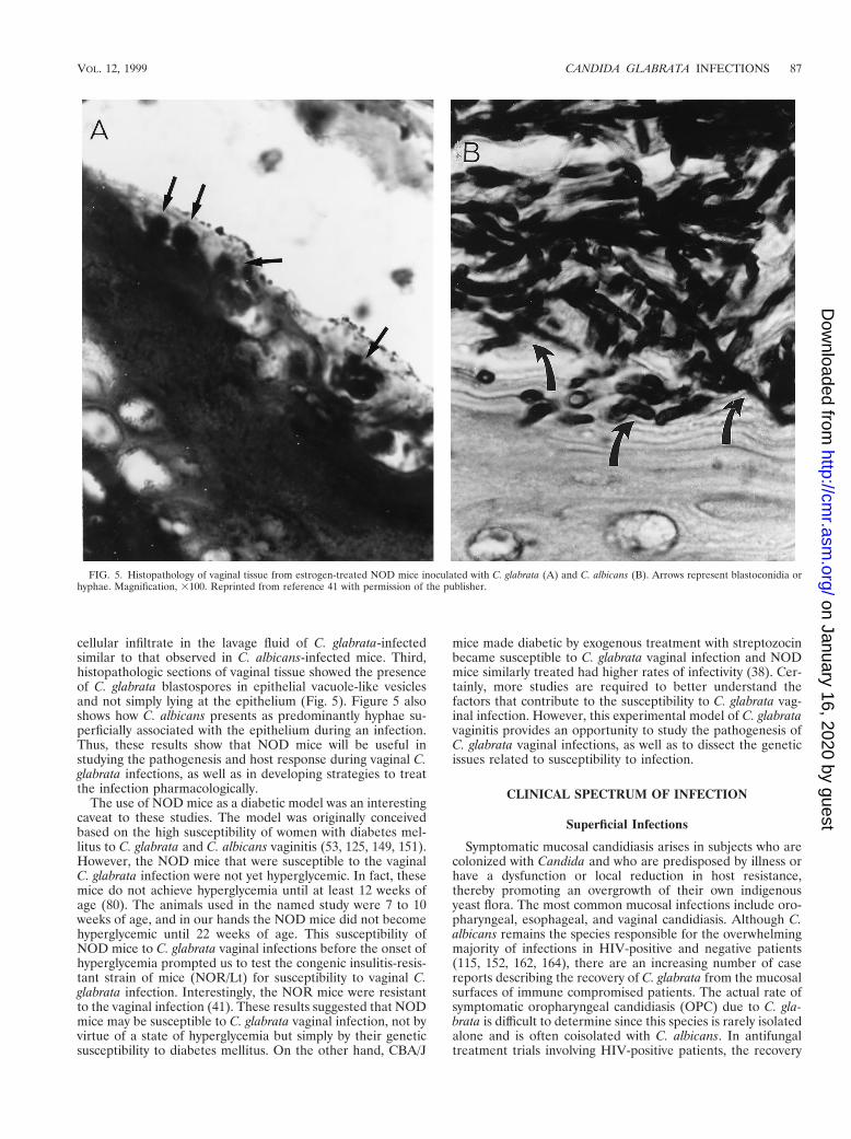
cellular infiltrate in the lavage fluid of C. glabrata-infectedsimilar to that observed in C. albicans-infected mice. Third,histopathologic sections of vaginal tissue showed the presenceof C. glabrata blastospores in epithelial vacuole-like vesiclesand not simply lying at the epithelium (Fig. 5). Figure 5 alsoshows how C. albicans presents as predominantly hyphae su-perficially associated with the epithelium during an infection.Thus, these results show that NOD mice will be useful instudying the pathogenesis and host response during vaginal C.glabrata infections, as well as in developing strategies to treatthe infection pharmacologically.
The use of NOD mice as a diabetic model was an interestingcaveat to these studies. The model was originally conceivedbased on the high susceptibility of women with diabetes mel-litus to C. glabrata and C. albicans vaginitis (53, 125, 149, 151).However, the NOD mice that were susceptible to the vaginalC. glabrata infection were not yet hyperglycemic. In fact, thesemice do not achieve hyperglycemia until at least 12 weeks ofage (80). The animals used in the named study were 7 to 10weeks of age, and in our hands the NOD mice did not becomehyperglycemic until 22 weeks of age. This susceptibility ofNOD mice to C. glabrata vaginal infections before the onset ofhyperglycemia prompted us to test the congenic insulitis-resis-tant strain of mice (NOR/Lt) for susceptibility to vaginal C.glabrata infection. Interestingly, the NOR mice were resistantto the vaginal infection (41). These results suggested that NODmice may be susceptible to C. glabrata vaginal infection, not byvirtue of a state of hyperglycemia but simply by their geneticsusceptibility to diabetes mellitus. On the other hand, CBA/J
mice made diabetic by exogenous treatment with streptozocinbecame susceptible to C. glabrata vaginal infection and NODmice similarly treated had higher rates of infectivity (38). Cer-tainly, more studies are required to better understand thefactors that contribute to the susceptibility to C. glabrata vag-inal infection. However, this experimental model of C. glabratavaginitis provides an opportunity to study the pathogenesis ofC. glabrata vaginal infections, as well as to dissect the geneticissues related to susceptibility to infection.
CLINICAL SPECTRUM OF INFECTION
Superficial Infections
Symptomatic mucosal candidiasis arises in subjects who arecolonized with Candida and who are predisposed by illness orhave a dysfunction or local reduction in host resistance,thereby promoting an overgrowth of their own indigenousyeast flora. The most common mucosal infections include oro-pharyngeal, esophageal, and vaginal candidiasis. Although C.albicans remains the species responsible for the overwhelmingmajority of infections in HIV-positive and negative patients(115, 152, 162, 164), there are an increasing number of casereports describing the recovery of C. glabrata from the mucosalsurfaces of immune compromised patients. The actual rate ofsymptomatic oropharyngeal candidiasis (OPC) due to C. gla-brata is difficult to determine since this species is rarely isolatedalone and is often coisolated with C. albicans. In antifungaltreatment trials involving HIV-positive patients, the recovery
FIG. 5. Histopathology of vaginal tissue from estrogen-treated NOD mice inoculated with C. glabrata (A) and C. albicans (B). Arrows represent blastoconidia orhyphae. Magnification, 3100. Reprinted from reference 41 with permission of the publisher.
VOL. 12, 1999 CANDIDA GLABRATA INFECTIONS 87
on January 16, 2020 by guesthttp://cm
r.asm.org/
Dow
nloaded from
of non-albicans Candida species is generally less than 10% ofall isolates recovered, with C. glabrata making up less than 5%(55, 122, 173). However, subjects in these treatment studies areoften selected, while patients with advanced disease, who arelikely to be infected with resistant strains, are excluded, result-ing in an underestimation of the frequency of C. glabrata in-fection. Moreover, in several of the antifungal treatment trialsfor fluconazole-refractory OPC in AIDS patients, the inci-dence of C. glabrata producing OPC was less than 10% (16,121). In the HIV-seronegative population, the occurrence ofOPC and esophageal candidiasis due to C. glabrata is rare.Data are still incomplete, because only a few small studies haveattempted to investigate the incidence of non-albicans Candidaspecies as a cause of OPC and esophageal candidiasis (49,140). At present, it is unclear why the incidence of mucosalcandidiasis due to C. glabrata is so low. Perhaps studies eval-uating the virulence factors of C. glabrata involved in the at-tachment and colonization of mucosal surfaces would shedsome light on this important issue.
There continues to be considerable controversy aboutwhether C. glabrata, as part of a mixed fungal culture withcoexistent C. albicans, actually contributes to the developmentof OPC. Many investigators consider that C. glabrata functionsas an innocent bystander only and that therapy should be basedupon susceptibility of the coexistent C. albicans (140). While C.albicans is undoubtedly the more virulent, frequent, and dom-inant pathogen, C. glabrata is occasionally found as the singleand only clinical species isolated in AIDS patients with OPC.Accordingly, while directing therapy against C. albicans inmixed infections, especially those not responding to appropri-ate therapy, it is prudent not to ignore C. glabrata in mixedinfections.
Oropharyngeal. (i) Clinical manifestations. Several clinicalforms of OPC exist; the most common and widely recognizedis acute pseudomembranous candidiasis, commonly referred toas thrush. OPC can also occur in an erythematous form that isoften asymptomatic. OPC is often the first manifestation ofHIV infection (21, 56, 147), with approximately 80 to 90% ofpatients with AIDS ultimately developing OPC at some stageduring their disease progression (28).
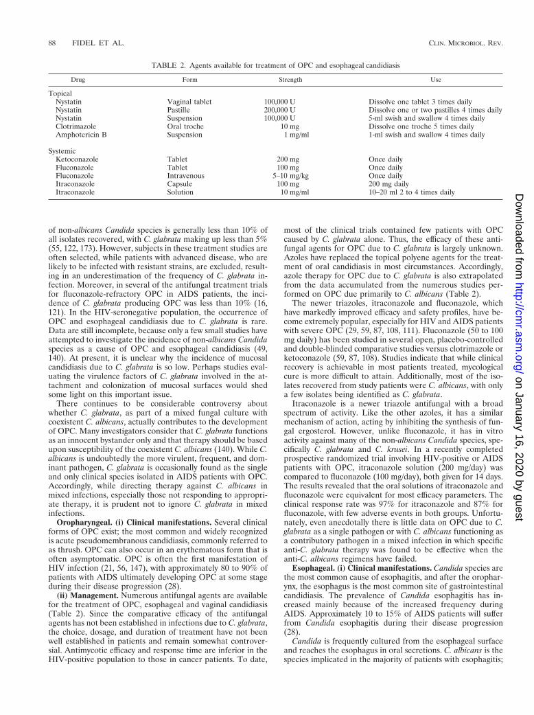
(ii) Management. Numerous antifungal agents are availablefor the treatment of OPC, esophageal and vaginal candidiasis(Table 2). Since the comparative efficacy of the antifungalagents has not been established in infections due to C. glabrata,the choice, dosage, and duration of treatment have not beenwell established in patients and remain somewhat controver-sial. Antimycotic efficacy and response time are inferior in theHIV-positive population to those in cancer patients. To date,
most of the clinical trials contained few patients with OPCcaused by C. glabrata alone. Thus, the efficacy of these anti-fungal agents for OPC due to C. glabrata is largely unknown.Azoles have replaced the topical polyene agents for the treat-ment of oral candidiasis in most circumstances. Accordingly,azole therapy for OPC due to C. glabrata is also extrapolatedfrom the data accumulated from the numerous studies per-formed on OPC due primarily to C. albicans (Table 2).
The newer triazoles, itraconazole and fluconazole, whichhave markedly improved efficacy and safety profiles, have be-come extremely popular, especially for HIV and AIDS patientswith severe OPC (29, 59, 87, 108, 111). Fluconazole (50 to 100mg daily) has been studied in several open, placebo-controlledand double-blinded comparative studies versus clotrimazole orketoconazole (59, 87, 108). Studies indicate that while clinicalrecovery is achievable in most patients treated, mycologicalcure is more difficult to attain. Additionally, most of the iso-lates recovered from study patients were C. albicans, with onlya few isolates being identified as C. glabrata.
Itraconazole is a newer triazole antifungal with a broadspectrum of activity. Like the other azoles, it has a similarmechanism of action, acting by inhibiting the synthesis of fun-gal ergosterol. However, unlike fluconazole, it has in vitroactivity against many of the non-albicans Candida species, spe-cifically C. glabrata and C. krusei. In a recently completedprospective randomized trial involving HIV-positive or AIDSpatients with OPC, itraconazole solution (200 mg/day) wascompared to fluconazole (100 mg/day), both given for 14 days.The results revealed that the oral solutions of itraconazole andfluconazole were equivalent for most efficacy parameters. Theclinical response rate was 97% for itraconazole and 87% forfluconazole, with few adverse events in both groups. Unfortu-nately, even anecdotally there is little data on OPC due to C.glabrata as a single pathogen or with C. albicans functioning asa contributory pathogen in a mixed infection in which specificanti-C. glabrata therapy was found to be effective when theanti-C. albicans regimens have failed.
Esophageal. (i) Clinical manifestations. Candida species arethe most common cause of esophagitis, and after the orophar-ynx, the esophagus is the most common site of gastrointestinalcandidiasis. The prevalence of Candida esophagitis has in-creased mainly because of the increased frequency duringAIDS. Approximately 10 to 15% of AIDS patients will sufferfrom Candida esophagitis during their disease progression(28).
Candida is frequently cultured from the esophageal surfaceand reaches the esophagus in oral secretions. C. albicans is thespecies implicated in the majority of patients with esophagitis;
TABLE 2. Agents available for treatment of OPC and esophageal candidiasis
Drug Form Strength Use
TopicalNystatin Vaginal tablet 100,000 U Dissolve one tablet 3 times dailyNystatin Pastille 200,000 U Dissolve one or two pastilles 4 times dailyNystatin Suspension 100,000 U 5-ml swish and swallow 4 times dailyClotrimazole Oral troche 10 mg Dissolve one troche 5 times dailyAmphotericin B Suspension 1 mg/ml 1-ml swish and swallow 4 times daily
SystemicKetoconazole Tablet 200 mg Once dailyFluconazole Tablet 100 mg Once dailyFluconazole Intravenous 5–10 mg/kg Once dailyItraconazole Capsule 100 mg 200 mg dailyItraconazole Solution 10 mg/ml 10–20 ml 2 to 4 times daily
88 FIDEL ET AL. CLIN. MICROBIOL. REV.
on January 16, 2020 by guesthttp://cm
r.asm.org/
Dow
nloaded from
rarely is C. glabrata or any other Candida species recoveredfrom esophageal samples. As with OPC, any C. glabrata strainrecovered from esophageal surfaces is generally coisolatedwith C. albicans. However, in contrast to oral candidiasis, evenless is known about host and yeast factors operative in thepathogenesis of esophageal candidiasis, and experimentalmodels have not been established. Esophageal candidiasis inHIV-positive patients may be the first manifestation of frankAIDS.
(ii) Management. As stated above, all of the clinical efficacystudies evaluating antifungal agents for esophageal infectionwere performed on C. albicans. Therefore, as with most strat-egies used to treat infections due to C. glabrata, we tend toextrapolate the data acquired from studies involving C. albi-cans.
Oral and intravenous fluconazole treatments have now be-come an integral part of the management of Candida esoph-agitis. Oral fluconazole enjoys a superior safety profile com-pared to ketoconazole and has superior gastric absorption;when necessary, fluconazole can be given intravenously.
In a recently published trial by Wilcox et al., patients treatedwith oral itraconazole solution at a dose of 200 mg/day had arate of clinical response comparable to that of patients treatedwith 100 mg of fluconazole per day (94 and 91%, respectively)(178). The mycological cure rates for this study was also sim-ilar, 92% for itraconazole and 78% for fluconazole.
Although used extensively in the pre-azole era for the moresevere forms of esophagitis, therapy with an intravenous solu-tion of amphotericin B is now used primarily in the azole-refractory cases. Low-dose intravenous amphotericin B, usingeither 0.15 to 0.3 mg/kg/day or 10 to 20 mg/day for 10 days, isoften sufficient for moderate disease caused by C. albicans (9,104), but with azole-refractory esophagitis, higher doses (0.5 to0.7 mg/kg/day) are necessary.
Vulvovaginal. (i) Clinical manifestations. The majority ofwomen with Candida vaginitis suffer from uncomplicated vag-initis characterized by sporadic attacks of mild to moderateseverity due to C. albicans, and these attacks occur in healthyadult women without any predisposing factors (152). In con-trast, approximately 10% of women suffer from complicatedCandida vaginitis, in which attacks either are more severe,occur on a recurrent basis, or are due to non-albicans Candidaspecies. Patients with complicated Candida vaginitis frequentlyhave predisposing factors in the form of uncontrolled diabetesor other immunosuppressive conditions. Accordingly, vaginitiscaused by C. glabrata represents a complicated form of disease.
Most clinical series have found that C. albicans is responsiblefor approximately 90% of episodes of Candida vaginitis. In thelast decade, there have been increasing reports of vaginitis dueto non-albicans Candida species. In these patients, C. glabratais the most common organism isolated (18, 151). Whetherthere is a real, absolute increase in vaginitis episodes caused byC. glabrata or whether the reported incidents reflect an in-creased awareness resulting in more frequent cultures taken, asopposed to routine microscopy, is unclear. Unfortunately, ep-idemiological studies do not include sentinel screening sites,but depend on data obtained from tertiary-care centers, whichreflect a major acquisition bias in the overall prevalence anddistribution of Candida species. The apparent increase in vag-initis caused by non-albicans Candida species is thought toreflect the increased use of short courses of both topical andoral azole antimycotic regimens. Other theories include thewidespread use and abuse of topical over-the-counter antifun-gal agents. Finally, some investigators have postulated that C.glabrata infections emerge as breakthrough vaginal infectionsin women receiving long-term maintenance low-dose flucon-
azole prophylactic regimens. In prospective longitudinal stud-ies performed by Fidel and coworkers, the emergence of non-albicans Candida species causing breakthrough Candidavaginitis in women already receiving maintenance azole ther-apy was not apparent in studies performed over many years(97). In contrast, HIV-positive women treated with fluconazole(200 mg) once weekly as long-term suppressive maintenancechemoprophylaxis showed a moderate shift in vaginal myco-flora while demonstrating effective reduction in episodes ofCandida vaginitis (141). The vaginal flora in women receivingfluconazole shifted to an increase in absolute isolation rates ofC. glabrata, but with a low attack rate of clinical vaginitis.
Although it was postulated that HIV infection would beassociated with an increased prevalence of vaginal non-albi-cans Candida species in a manner similar to the emergence ofOPC caused by non-albicans Candida species, no such datahave emerged to date in HIV seropositive women. In a wom-en’s cohort study (142) (HIV Epidemiological Research Study[HERS]), both baseline and follow-up studies failed to identifyan increased colonization rate as well as vaginitis caused bynon-albicans Candida species in HIV-positive women. Simi-larly, in contrast to OPC, non-albicans Candida species as wellas C. albicans did not emerge with increased frequency inwomen with low CD4 counts (142).
In small clinical studies, a variety of risk factors haveemerged for C. glabrata vaginitis. These include older patients,underlying medical conditions such as uncontrolled diabetesmellitus, and douching (53). Given the small number of pa-tients with C. glabrata vaginitis, no large-scale studies havedescribed the clinical characteristics of vaginitis caused by C.glabrata. It is widely assumed that clinical symptoms would beidentical. Geiger et al., however, have reported subtle differ-ences in the clinical presentation of C. glabrata vaginitis (53).In a study of 80 patients, an abnormal discharge was lessfrequently reported in women with symptomatic vaginitis dueto C. glabrata in comparison to C. albicans. This may reflect theeffects of lack of hypha formation by the C. glabrata blasto-conidia. In general, vaginitis due to C. glabrata was reported tobe more indolent with reduced inflammation and hence lessdyspareunia. In addition, patients with C. glabrata vaginitisfrequently reported a burning sensation as an alternative toitch. Clinical findings of the inflammatory reaction in the vulvaand vestibule were similar to those associated with C. albicans.In contrast, speculum examination of the vagina, although re-vealing diffuse erythema, rarely revealed a caseous discharge inthe presence of C. glabrata.
Diagnosis of C. glabrata vaginitis is more difficult than that oftypical Candida vaginitis. This is because of the failure of theC. glabrata organisms to form pseudohyphae and hyphae invivo. Accordingly, on saline and KOH microscopy, numerousbudding yeasts are seen but hypha elements are absent. Thereis some evidence that vaginitis with C. glabrata often occurs ata somewhat higher vaginal pH, usually at the upper limit ofnormal. Not infrequently, C. glabrata vaginitis coexists withbacterial vaginosis, and the higher pH of the latter may repre-sent the link between the two entities.
(ii) Management. There is scant information on guidelinesfor management of vaginitis due to C. glabrata. In virtually allclinical studies of yeast vaginitis, patients with vaginitis due toC. glabrata were excluded or the numbers were not largeenough that any variable response rate was detectable, even inlarge studies. Accordingly, the clinical response of patientswith C. glabrata vaginitis to conventional topical or oral ther-apy is largely unknown. Published experience in the manage-ment of C. glabrata patients reflects a biased view of patientsreferred to specialized clinics only after they have failed to
VOL. 12, 1999 CANDIDA GLABRATA INFECTIONS 89
on January 16, 2020 by guesthttp://cm
r.asm.org/
Dow
nloaded from

respond to a large number of topical and oral azole agents (53,151, 159). The percentage of patients with C. glabrata vaginitis,seen by primary care practitioners, who respond to initialcourses of azole therapy is therefore unknown.
In vitro studies reveal that the MICs of all available azolesfor C. glabrata are higher than that for most C. albicans isolates(96). The increase in MICs varies, however, with the specificazole. Butoconazole shows excellent in vitro activity, as domiconazole and clotrimazole. Terconazole, itraconazole, andketoconazole show moderate activity. Fluconazole shows rela-tively poor in vitro activity, and, not infrequently, there is frankresistance. Published studies, of which there are few, revealthat in spite of in vitro activity, azole therapy does not predict-ably eradicate C. glabrata in vivo (125, 151). If an attempt is tobe made to treat C. glabrata with either oral or topical azoletherapy, fluconazole should not be the drug of choice, and allthe other azoles agents should not be prescribed as shortcourse regimens, i.e., single-dose or 1- to 3-day regimens. Ac-cordingly, in a previously untreated patient, it is not unreason-able to use nonfluconazole azoles for 7 to 14 days.
Sobel et al. recently reported on the successful use of boricacid vaginal capsules in the treatment of C. glabrata vaginitis inwomen who had failed several courses of azole therapy (151).Boric acid, 600 mg in gelatin capsules, was administered intra-vaginally once a day for 14 days. In uncontrolled studies, thesuccess rate measured by mycological eradication of the or-ganism approximated 70%. Approximately 30% of the patientsremained culture positive, and many of these returned within ashort period with recurrence of vulvovaginal symptoms. Thesepatients were then retreated with boric acid and given a main-tenance regimen of boric acid prescribed several times a weekfor an additional period. However, the safety of the latterregimen is unknown, and, given the potential systemic toxicityof boric acid, it should not be undertaken lightly. As an alter-native to boric acid maintenance therapy, nystatin vaginal sup-positories (100,000 U daily) can be used as a maintenanceregimen following the initial clinical and mycological successfultherapy with boric acid. For patients who fail to respond toboric acid or for whom the boric acid or nystatin maintenancetherapy becomes ineffective, topical flucytosine prescribedonce a day for 14 days is generally recommended. A mainte-nance regimen with flucytosine is not available because of localtoxicity, expense, and the potential for development of resis-tance. Most patients who receive flucytosine do extremely well,since C. glabrata is highly sensitive to this drug. For patientswho fail to respond to both boric acid and flucytosine regi-mens, combination regimens including a topical antifungalsuch as boric acid, flucytosine, and nystatin can be coadminis-tered with oral itraconazole. Although the value of oral itra-conazole as definitive therapy is largely unknown, itraconazoledemonstrates considerable in vitro activity (96). Based on diskagar diffusion susceptibility testing, terconazole has been con-sidered to be highly active against C. glabrata; however, clinicalexperience with terconazole does not indicate any advantageover any of the other topical agents (151).
To date, it is unclear whether recurrent vaginitis due to C.glabrata is due to the same pathogenic mechanisms as recur-rent vaginitis due to C. albicans. With C. albicans, a host factorrather than the lack of susceptibility of a microorganism totherapy is postulated to be responsible for recurrent disease(40, 149). In contrast, the additional element contributing torecurrence of C. glabrata infection is likely to be the resistanceof the organisms to antifungal agents rather than a host factor.Nevertheless, in some patients, both components may be ac-tive. The treatment of C. glabrata vaginitis in HIV-positive
women follows the same principles, and there is no evidence ofhigher failure rates.
Urinary tract. (i) Clinical manifestations. Urinary tract in-fections due to Candida species have markedly increased in thelast two decades (132). Candida species are now responsiblefor approximately 10% of urinary tract infections in hospital-ized patients (185). In contrast to OPC and vaginal candidiasis,approximately 50% of urinary isolates of Candida are due tonon-albicans Candida species, the most common of which is C.glabrata. In a recent large multicenter study, C. glabrata wasresponsible for 20% of the Candida urinary tract infections(153). Not infrequently, C. glabrata is part of a polymicrobialinfection, including either bacterial uropathogens or a secondCandida species, usually C. albicans.
No unique epidemiological risk factors for C. glabrata uri-nary tract infections have been reported, although underlyingdiabetes mellitus is by no means an infrequently associatedfactor. Similar to C. albicans urinary tract infections, the ma-jority of C. glabrata urinary tract infections occur in elderlyhospitalized, debilitated, and catheterized patients who haverecently received antibacterial agents.
The clinical spectrum of C. glabrata urinary tract infectionsappears identical to that caused by other species of Candida.The majority of patients are asymptomatic. Rarely do lowerurinary tract symptoms develop, especially in catheterized pa-tients. The risk of an ascending infection with involvement ofthe kidneys is rare and occurs mostly in patients with foreignbodies or stents and in the presence of obstruction. Rarelydoes C. glabrata fungemia complicate ascending Candida pye-lonephritis. To complete the picture, candiduria caused by C.glabrata rarely complicates hematogenous candidiasis, in whichrenal candidiasis occurs with subsequent seeding of the urine.The diagnosis of C. glabrata urinary tract infection, althoughconfirmed on culture, is usually suggested by the presence ofbudding yeast without hypha formation on microscopy of urinesamples. The finding of C. glabrata, even in large numbers, inthe urine, while indicative of urinary tract infection, does notlocalize the anatomical site of infection, which requires clinicalcorrelation. Identifying the site of infection forms the basis forsuccessful management.
(ii) Management. Asymptomatic candiduria is generally nottreated. The natural history of asymptomatic candiduria is suchthat the candiduria often resolves spontaneously, especiallywhen catheterization is changed or discontinued. Moreover,ascending infections resulting in sepsis are infrequent. Asymp-tomatic candiduria should be treated following renal trans-plantation, in neutropenic patients, and before attemptingelective instrumentation or surgery of the urinary tract.
Symptomatic urinary tract infection caused by C. glabrata,although often successfully treated with amphotericin B bloodirrigation or washout, may be effectively treated by systemictherapy with either amphotericin B or fluconazole. In a recentstudy of a large number of patients with asymptomatic candi-duria, C. glabrata urinary tract infection appeared to respondto fluconazole therapy (200 mg/day) for 14 days at the samerate as did C. albicans infection. In a logistic regression anal-ysis, C. glabrata species did not emerge as a factor influencingthe outcome of antifungal therapy (2).
Systemic Infections
Advances in medical technology have had a major effect inreducing the morbidity and mortality of previously fatal dis-eases. With these benefits has come an increase in nosocomialfungal infections, primarily due to Candida species (3, 7, 31).Candidal infections may involve any anatomical structure and
90 FIDEL ET AL. CLIN. MICROBIOL. REV.
on January 16, 2020 by guesthttp://cm
r.asm.org/
Dow
nloaded from

are the cause of more fatalities than are any other systemicmycosis (115). A myriad of predisposing factors for systemiccandidal infection have been previously identified (14, 90).Although few studies have evaluated specific risk factors forsystemic C. glabrata infection, the risk factors leading to infec-tion are similar to those from C. albicans infections. In oneprospective epidemiological study evaluating C. glabrata colo-nization in medical intensive care units and in bone marrowtransplant patients, the significant risk factors for nosocomialcolonization with C. glabrata were prolonged hospitalizationand prior antimicrobial use (170). A more recent concern,however, has been the numerous reports describing the in-creasing incidence of colonization and infection by non-albi-cans Candida species (specifically C. glabrata and C. krusei) inimmunocompromised hosts (113, 180–182, 184). The increasein the infections by non-albicans Candida species is postulatedto be associated with the increasing use of antifungal agents.According to several investigators, the increase in the fre-quency of C. glabrata infections has paralleled the increase useof fluconazole in some hospitals (1, 181–184). In a more recentstudy, however, investigators described the association be-tween C. glabrata infection and amphotericin B use rather thanfluconazole (112a). C. glabrata is of special importance becauseof its reduced susceptibility to antifungal agents (100, 129).
Clinical manifestations. Candida may involve any organ sys-tem, and candidemia has a diverse clinical picture, rangingfrom low-grade fever to fulminant septic shock. There are nocharacteristic signs and symptoms in disseminated candidiasis.Similarly, no unique clinical features are associated with C.glabrata. Often, the only manifestation is persistent fever in apatient whose condition is deteriorating and who is unrespon-sive to antimicrobial agents and has negative blood cultures. C.glabrata fungemia has been associated with a higher mortalityrate than C. albicans. In fact, Komshian et al. reported a 100%mortality in 12 patients with C. glabrata fungemia (90). Thehigher mortality rate described by some investigators may notsignify increased virulence but may reflect the more advancedstate of debilitation in patients who acquire C. glabrata infec-tion. In a more recent study, Fraser et al. found no differencein mortality rates between C. albicans and C. glabrata (51).
Management. Amphotericin B has been the “gold standard”in systemic fungal infections including candidemia, despitehaving a high adverse effect profile. Recently, prospective ran-domized clinical studies concluded that fluconazole, at a min-imum dose of 400 mg/day, is as effective as amphotericin B inthe management of candidemia in neutropenic and nonneu-tropenic patients. In addition, fluconazole is better toleratedand has fewer adverse effects (2, 130). Unfortunately, as inprevious clinical antifungal trials, the majority of patientstreated in these studies were infected with C. albicans and fewhad non-albicans Candida species, including C. glabrata. Ac-cordingly, a Candida species-specific subanalysis and conclu-sion was not possible. Physicians are left to extrapolate thedata obtained from the clinical trials treating C. albicans tomanaging infections due to C. glabrata. All antifungal agentshave higher MICs for C. glabrata strains than for C. albicans(129). Thus, until more data are available, many clinicians treatC. glabrata fungemia with high-dose amphotericin B (0.6 to 1.0mg/kg/day) or fluconazole (10 to 15 mg/kg/day) until the invitro susceptibility data indicate that the clinical isolate is sus-ceptible to fluconazole. After resolution of fungemia, the treat-ment course may be completed with oral fluconazole (30).Amphotericin B is more likely to be chosen to treat the hemo-dynamically unstable and septic patient.
In the past, many patients with life-threatening candidiasisdied without receiving antifungal therapy. Clinicians are fre-
quently required to act definitively and early on the basis of ahigh index of suspicion. To be effective, any therapy must begiven early and, regrettably empirically, in the febrile high-riskpatient. Empirical therapy with amphotericin B is especiallyindicated in the granulocytopenic patient with persistent feverafter 3 to 7 days of antibiotic therapy, even in the absence ofmicrobiological confirmation. Amphotericin B has been thedrug of choice in this setting. This choice is especially justifiedsince several investigators have documented the increase in theisolation of C. glabrata and C. krusei in neutropenic patients(181–184). There are no data on the role of the new lipidformulation of amphotericin B in treating C. glabrata funge-mia. Although these new formulations result in higher-doseamphotericin B administration, superior success rates have notbeen determined.
ANTIFUNGAL RESISTANCE
Classification
Antifungal resistance can be divided into two categories:clinical resistance and in vitro resistance. Clinical resistancesignifies a lack of a clinical response to the antifungal agentused. More often than not, clinical failure is due to low levelsof the drug in serum and/or tissues for numerous reasons, mostnotably noncompliance with the medication regimen. Finally,one significant reason for clinical failure or resistance in AIDSpatients is the presence of a severely immunosuppressive state,where the antifungal agents alone, including high-dose fungi-cidal agents, are unable to eradicate the fungi from the host.
In vitro resistance can be subdivided into primary resistanceand secondary resistance. Primary resistance is also known asintrinsic or innate resistance and occurs when the organism isnaturally resistant to the antifungal agent (e.g., C. krusei, whichis known to be universally resistant to fluconazole) (183). Onthe other hand, secondary or acquired resistance is describedwhen the isolate producing infection becomes resistant to theantifungal agent. This form of resistance, which was rare in thepast, is now the most frequently reported form in AIDS pa-tients who suffer from recurrent azole-resistant oropharyngealor esophageal candidiasis (36, 49, 76, 83).
Evidence for Clinical and In Vitro Resistance
Antifungal resistance in Candida species was virtually non-existent until the arrival of HIV infection. In the past, evenwhen resistance was described, it was generally associated withthe imidazole class of antifungal agents and was usually dis-covered in patients with chronic mucocutaneous candidiasis,who were being given chronic ketoconazole therapy (68). How-ever, there are now numerous reports of oral thrush andesophageal candidiasis that are clinically refractory to all azoleand polyene antifungal agents (28, 49, 78, 83, 98, 137). Underthe selective pressure of numerous antifungal agents, popula-tions of resistant or relatively resistant yeasts have emerged.There are numerous case reports describing the colonizationand infection of compromised patients taking long-term oralantifungal agents, from whom C. krusei and C. glabrata withdocumented in vitro antifungal resistance have been recovered(181–184). Even amphotericin B-resistant C. albicans, C. guil-liermondii, and Cryptococcus neoformans, a rare phenomenonin the past, have recently been reported (6, 173). These resis-tant yeasts are capable of producing debilitating and invasivefungal disease that is more difficult to eradicate (6, 78, 123).Overall, compared to other Candida species, especially C. al-bicans, C. glabrata isolates tend to be associated with higher
VOL. 12, 1999 CANDIDA GLABRATA INFECTIONS 91
on January 16, 2020 by guesthttp://cm
r.asm.org/
Dow
nloaded from

MICs of all azoles and are innately less susceptible to allantifungal agents including amphotericin B (170, 171). Thefrequency or prevalence of azole-resistant C. glabrata is un-known. Fluconazole-resistant isolates have been found pre-dominantly in AIDS patients with OPC and esophageal can-didiasis. In addition, resistant isolates have been found infungemic patients and among vaginal isolates. In some cases,primary in vitro resistance to fluconazole has been reported(128, 129). By far, however, secondary in vitro resistance is themost common form of resistance in C. glabrata (181, 182, 184)and is most often seen for fluconazole. The reason for thisrapid development of secondary antifungal resistance is un-known, but the haploid state of C. glabrata is thought to be acontributing factor. In contrast, in vitro resistance of C. gla-brata and C. albicans to ketoconazole and itraconazole is some-what less common (,15%) yet still significant. Several clinicalstudies have documented the selection of C. glabrata in pa-tients treated with fluconazole for prolonged periods (128,129), whereas C. albicans resistance to fluconazole had beenrare. Accordingly, the use of intermittent versus continuouslong-term azole therapy needs to be compared, as does theneed to establish the minimum effective dose which will notselect for resistant strains of C. albicans or the selection ofmore resistant Candida species, including C. glabrata (94).
An emerging dilemma is the development of multi-azolecross-resistance in Candida isolates recovered from AIDS pa-tients with fluconazole-refractory OPC (112, 170). In onestudy, 45 isolates from 41 patients who failed to respond to atleast 400 mg of fluconazole per day underwent in vitro testing.Twenty-seven C. albicans isolates had fluconazole MICs of.20 mg/ml; 41% of these isolates were also cross-resistant toclotrimazole, while another 11% were cross-resistant to itra-conazole and ketoconazole (112). In another recent study, theauthors evaluated 25 C. glabrata isolates recovered from pa-tients with fluconazole-refractory OPC. Of these C. glabrataisolates, 68% were fluconazole resistant, and of these, 94%were also cross-resistant to ketoconazole and 88% were alsocross-resistant to clotrimazole (170). In contrast, in the samestudy, 60 C. albicans isolates were recovered from similar pa-tients, and while 78% were resistant to fluconazole, only 7%were cross-resistant to itraconazole, 11% were cross-resistantto ketoconazole, and 41% were cross-resistant to clotrimazole.
Fortunately, clinically significant amphotericin B resistanceis still very uncommon among most Candida species except forC. lusitaniae and C. guilliermondii. Similarly, amphotericin Bresistance has not been described in C. glabrata (52), althoughthe MICs are higher than those seen for C. albicans.
Flucytosine resistance has been described extensively in C.albicans. Primary resistance rates vary from 5 to 50% depend-ing on the species of Candida and the technique used to per-form the susceptibility studies (54, 144, 173). Flucytosine re-sistance is also very common in C. tropicalis, C. krusei, and C.parapsilosis, many isolates of which have greater primary resis-tance rates than do C. albicans isolates (50, 118, 119, 144). Incontrast, the majority of C. glabrata isolates are exquisitelysusceptible to flucytosine. Flucytosine has not been widely usedin C. glabrata infections but may be useful in the future.
Mechanisms of Resistance
The specific mechanisms of antifungal resistance to theazole class of antifungal agents are not yet fully understood. Ithas been suggested, however, that the sterol composition of thefungal plasma membrane is altered, thus reducing the uptakeof the antifungal agent into the cell (146). Recent studies withseveral different azoles evaluating C. albicans, C. glabrata, and
S. cerevisiae have demonstrated at least three known mecha-nisms of resistance: (i) changes in the P-450 lanosterol demeth-ylase enzyme, (ii) changes in D5–6-sterol desaturase, and, morerecently, (iii) an energy-dependent drug efflux mechanism (63,64, 116, 117). In C. glabrata, several mechanisms of azole re-sistance have been identified: increased P-450-dependent er-gosterol synthesis and an energy-dependent efflux pump offluconazole, possibly via a multidrug resistance-type trans-porter (117, 165, 166, 173).
Clinical Relevance
The clinical effects of antifungal resistance in the AIDSpopulation were recently demonstrated by Koletar et al. (88).The authors evaluated AIDS patients who failed to respond tostandard antifungal therapy for OPC and reported a mediansurvival of 184 days after the onset of fluconazole-resistantthrush and only 83 days after the onset of clinical resistance toamphotericin B. Although mucosal candidiasis does not resultin death directly, clinical antifungal failure is most probably acomorbidity factor in the rapid demise of these patients. Theestimated frequency of azole resistance is still unknown; it ispostulated that 4 to 6% of C. albicans isolates recovered frompersons with AIDS are resistant to antifungal agents (27). Incontrast, the frequency of resistance in C. glabrata is relativelyunknown and difficult to predict, since few studies have ad-dressed the issue. In those that have, few reports have de-scribed the incidence of azole resistance among any of thenon-albicans Candida species, including C. glabrata (126, 127).
The management of fluconazole-resistant mucosal candidi-asis is frequently unsatisfactory or short-lived, with periodicand rapid recurrences. Some patients will respond to a dou-bling of the dose of fluconazole. For example, if they fail torespond to 200 mg/day, an increase to 400 mg/day will fre-quently produce a clinical response for a while. However, theimprovement is generally transient, and the infection recursrapidly once this stage of the disease is reached. Several recentstudies of the itraconazole oral solution have demonstratedpromising results in AIDS patients who have not responded tofluconazole at 200 mg/day (22, 33, 121). These studies havedemonstrated clinical cure and improvement in 55 to 70% ofpatients entered into the study. As expected, mycological curerates were very low (,30%) and relapses were rapid (usuallywithin 14 days) once the itraconazole solution was terminated.The recent approval of amphotericin B oral suspension is anew therapeutic option in these patients with azole-unrespon-sive mucosal candidiasis (113). In several small studies, theclinical improvement rates varied from 50 to 75%, but as withall these patients, the relapse rate is high and usually occurredwithin 4 weeks (113, 173).
Several new antifungal compounds are currently in variousphases of development, and the results appear encouraging inearly in vitro trials. Two new azoles, voriconazole and SCH56592, have excellent in vitro activity against fluconazole-resis-tant C. albicans and C. glabrata isolates (91, 93, 136). In addi-tion to the azoles, a new group of antifungal agents, the pneu-mocandins, are being evaluated in clinical trials. MK-911, anew parenteral pneumocandin, is currently in clinical trials inthe United States. In vitro results with this new antifungal drugare very promising for many Candida species, including flucon-azole-resistant C. albicans. In addition, the in vitro activityagainst C. glabrata and C. krusei is excellent (173).
92 FIDEL ET AL. CLIN. MICROBIOL. REV.
on January 16, 2020 by guesthttp://cm
r.asm.org/
Dow
nloaded from

CONCLUSION
Candida glabrata is emerging as a major pathogen in the1990s. Previously largely ignored, this organism received littleattention; therefore, not surprisingly, our knowledge of it is notonly incomplete but also significantly lacking. We now have themolecular tools to study the epidemiology of C. glabrata, andinvestigations are needed. With C. glabrata increasingly beingrecognized as a problem pathogen in superficial and systemiccandidiasis, host risk factors need additional study, as do im-portant virulence factors. A major issue is the management ofC. glabrata infections. Symptomatic infection is more difficultto eradicate with all of the available antifungal drugs. Theazole antifungal agents that have proven so successful againstC. albicans have been woefully inadequate against C. glabratavaginitis, although these azoles appear adequate for C. glabratafungemia. Understanding the mechanism of innate and ac-quired resistance may facilitate the development of new targetsfor novel antifungal agents. In any event, if C. glabrata infec-tions are to be adequately controlled in the future, compre-hensive studies of their epidemiology, pathogenesis, and resis-tance must be performed.
REFERENCES
1. Abi-Said, D. E., O. Uzon, I. Raad, H. Pinzcowski, and O. Vartivarian. 1997.The epidemiology of hematogenous candidiasis caused by different Can-dida species. Clin. Infect. Dis. 24:1122–1128.
2. Anaissie, E. J., R. O. Darouiche, D. Abi-Said, O. Uzum, J. Mera, L. O.Gentry, T. Williams, D. P. Kontoyiannis, C. L. Karl, and G. P. Bodey. 1996.Management of invasive candidial infections: result of a prospective, ran-domized, multicenter study of fluconazole versus amphotericin B and re-view of the literature. Clin. Infect. Dis. 23:964–972.
3. Banerjee, S. N., T. G. Emori, D. H. Culver, R. P. Gaynes, W. R. Jarvis, andT. Horan. 1991. Secular trends in nosocomial primary blood stream infec-tions in the United States. Am. J. Med. 91:86S–89S.
4. Barns, S. M., D. J. Lane, M. L. Sogin, C. Bibeau, and W. G. Weisburg. 1991.Evolutionary relationships among pathogenic Candida species and rela-tives. J. Bacteriol. 173:2250–2255.
5. Barret-Bee, K., Y. Hayes, R. G. Wilson, and J. F. Ryley. 1985. A comparisonof phospholipase activity, cellular adherence and pathogenicity of yeasts.J. Gen. Microbiol. 131:1217–1221.
6. Bastide, M., M. Mallie, B. Montes, and J. M. Bastide. 1989. The epidemi-ology of hematogenous candidiasis caused by different Candida species.Pathol. Biol. 37:694–699.
7. Beck-Sague, C. M., and T. R. Jarvis. 1993. Secular trends in the epidemi-ology of nosocomial fungal infections in the United States. J. Infect. Dis.167:1247–1251.
8. Bendel, C. M., and M. K. Hostetter. 1991. Correlation of adhesion andpathogenic potential in yeast. Pediatr. Res. 29:167A. (Abstract.)
9. Bennett, J. E. 1978. Diagnosis and management of candidiasis in the im-munocompromised host. Scand. J. Infect. Dis. Suppl. 16:83–86.
10. Bennett, J. E. 1990. Antimicrobial agents: antifungal agents, p. 1165–1181.A. G. Gilman, T. W. Rall, A. S. Nies, and P. Taylor (ed.), Goodman andGilman’s the pharmacological basis of therapeutics. Pergamon Press, Inc.,Elmsford, N.Y.
11. Bodey, G. P. 1986. Candidiasis in cancer patients. Am. J. Med. 77:13–19.12. Boerlin, P., F. Boerlin-Petzold, J. Goudet, C. Durussel, J. Pagani, J. Chave,
and J. Bille. 1996. Typing Candida albicans oral isolates from human im-munodeficiency virus-infected patients by multilocus enzyme electrophore-sis and DNA fingerprinting. J. Clin. Microbiol. 35:1235–1248.
13. Brawner, D. L., and J. E. Cutler. 1984. Variability in expression of a cellsurface determinant on Candida albicans as evidenced by an agglutinatingmonoclonal antibody. Infect. Immun. 43:966.
14. Bross, J., G. H. Talbot, G. Maislin, S. Hurwitz, and B. L. Strom. 1989. Riskfactors for nosocomial candidemia: a case control study in adults withoutleukemia. Am. J. Med. 87:614–620.
15. Cantorna, M. T., and E. Balish. 1990. Mucosal and systemic candidiasis incongenitally immunodeficient mice. Infect. Immun. 58:1093–1100.
16. Cartledge, J. D., J. Midgley, and B. G. Gazzard. 1997. Itraconazole cyclo-dextrin solution: the role of in vitro susceptibility testing in predictingsuccessful treatment of HIV-related fluconazole-resistant and fluconazole-susceptible oral candidosis. AIDS 11:163–168.
17. Cassone, A., F. DeBernardis, F. Mondello, T. Ceddia, and L. Agatensi.1987. Evidence for a correlation between proteinase secretion and vulvo-vaginal candidosis. J. Infect. Dis. 156:777–783.
18. Cauwenbergh, G. 1990. Vaginal candidiasis: evolving trends in the inci-
dence and treatment of non-Candida albicans infections. Curr. Probl. Ob-stet. Gynecol. Fertil. 8:241–245.
19. Chakrabarti, A., N. Nayak, and P. Talwar. 1991. In vitro proteinase pro-duction by Candida species. Mycopathologia 114:163–168.
20. Clark, R. A., S. A. Blakley, J. Rice, and W. Brandon. 1995. Predictors ofHIV progression in women. J. Acquired Immune Defic. Syndr. Hum. Ret-rovirol. 9:43–50.
21. Coker, R. J., M. Fisher, and D. R. Tomlinson. 1995. Management ofmycoses associated with HIV disease. Int. J. STD AIDS 6:408–412.
22. Coppola, S., G. Angarano, M. T. Montagna, P. Congedo, L. Monno, A.Bellisario, and G. Pastore. 1995. Efficacy of itraconazole in treating AIDS-associated infections due to Candida krusei. Eur. J. Epidemiol. 11:243–244.
23. DeBernardis, F., L. Agatensi, I. K. Ross, G. W. Emerson, R. Lorenzini, P. A.Sullivan, and A. Cassone. 1990. Evidence for a role for secreted aspartateproteinase of Candida albicans in vulvovaginal candidiasis. J. Infect. Dis.161:1276–1283.
24. DeBernardis, F., P. Chiani, M. Ciccozzi, G. Pellegrini, T. Ceddia, G.D’Offizzi, I. Quinti, P. Sullivan, and A. Cassone. 1996. Elevated asparticproteinase secretion and experimental pathogenicity of Candida albicansisolates from oral cavities of subjects infected with human immunodefi-ciency virus. Infect. Immun. 64:466–471.
25. DeBernardis, F., A. Molinari, M. Boccanera, A. Stringaro, R. Robert, J. M.Senet, G. Arancia, and A. Cassone. 1994. Modulation of cell surface-asso-ciated mannoprotein antigen expression in experimental candidal vaginitis.Infect. Immun. 62:509–519.
26. Dembry, L. M., J. A. Vazquez, and M. J. Zervos. 1994. DNA analysis in thestudy of the epidemiology of nosocomial candidiasis. Infect. Control. Hosp.Epidemiol. 5:48–53.
27. Denning, D. W. 1994. Fluconazole resistance in Candida in patients withAIDS: a therapeutic approach. J. Infect. 26:117–125.
28. Diamond, R. D. 1991. The growing problem of mycoses in patients infectedwith the human immunodeficiency virus. Rev. Infect. Dis. 13:480–486.
29. Dupont, B., and C. Drouhet. 1988. Fluconazole in the management oforopharyngeal candidiasis in a predominantly HIV antibody-positive groupof patients. J. Med. Vet. Mycol. 26:67–71.
30. Edwards, J. E., Jr., G. P. Bodey, R. A. Bowden, T. Buchner, B. E. DePauw,S. G. Filler, M. A. Ghannoum, M. Glauser, R. Herbrecht, C. A. Kauffman,S. Kohno, P. Martino, F. Meunier, T. Mori, M. A. Pfaller, J. H. Rex, T. R.Rogers, R. H. Rubin, J. Solomkin, C. Viscoli, T. J. Walsh, and M. White.1997. International Conference for the Development of a Consensus on theManagement and Prevention of Severe Candidal Infections. Clin. Infect.Dis. 25:161–163.
31. Edwards, J. E., Jr. 1991. Invasive Candida infections-evolution of a fungalpathogen. N. Engl. J. Med. 324:1060–1062.
32. Ehrensaft, D. V., R. B. Epstien, S. Sarpel, and B. R. Andersen. 1979.Disseminated candidias in leukopenic dogs. Proc. Soc. Exp. Biol. Med.160:6–10.
33. Eichel, M., G. Just-Nubling, E. B. Helm, and W. Stille. 1996. Itraconazolesuspension in the treatment of HIV-infected patients suffering from flu-conazole-resistant oropharyngeal and esophageal candidosis. Mycoses 39:102–106.
34. Eisenstein, B. I., and N. C. Engleberg. 1986. Applied molecular genetics:new tools for microbiologists and clinicians. J. Infect. Dis. 153:416–430.
35. Elin, R. J., J. B. Edelin, and S. M. Wolff. 1974. Infection and immunoglob-ulin concentrations in Chediak-Higashi mice. Infect. Immun. 10:88–91.
36. Fan-Havard, P., D. Capano, S. M. Smith, A. Mangia, and R. H. Eng. 1991.Development of resistance in Candida isolates from patients receiving pro-longed antifungal therapy. Antimicrob. Agents Chemother. 35:2302–2305.
37. Fichtenbaum, C. J., and W. Powderly. 1998. Refractory mucosal candidiasisin patients with human immunodeficiency virus infection. Clin. Infect. Dis.26:556–565.
38. Fidel, P. L., Jr. 1998. Unpublished observations.39. Fidel, P. L., Jr., M. E. Lynch, and J. D. Sobel. 1993. Candida-specific
cell-mediated immunity is demonstrable in mice with experimental vaginalcandidiasis. Infect. Immun. 61:1990–1995.
40. Fidel, P. L., Jr., and J. D. Sobel. 1996. Immunopathogenesis of recurrentvulvovaginal candidiasis. Clin. Microbiol. Rev. 9:335–348.
41. Fidel, P. L., Jr., J. L. Cutright, L. Tait, and J. D. Sobel. 1996. A murinemodel of Candida glabrata vaginitis. J. Infect. Dis. 173:425–431.
42. Fidel, P. L., Jr., K. A. Ginsburg, J. L. Cutright, N. A. Wolf, D. Leaman, K.Dunlap, and J. D. Sobel. 1997. Vaginal-associated immunity in women withrecurrent vulvovaginal candidiasis: evidence for vaginal Th1-type responsesfollowing intravaginal challenge with Candida antigen. J. Infect. Dis. 176:728–739.
43. Fidel, P. L., Jr., M. E. Lynch, D. H. Conaway, L. Tait, and J. D. Sobel. 1995.Mice immunized by primary vaginal C. albicans infection develop acquiredvaginal mucosal immunity. Infect. Immun. 63:547–553.
44. Fidel, P. L., Jr., M. E. Lynch, V. Redondo-Lopez, J. D. Sobel, and R.Robinson. 1993. Systemic cell-mediated immune reactivity in women withrecurrent vulvovaginal candidiasis (RVVC). J. Infect. Dis. 168:1458–1465.
45. Fidel, P. L., Jr., M. E. Lynch, and J. D. Sobel. 1994. Effects of pre-inducedCandida-specific systemic cell-mediated immunity on experimental vaginal
VOL. 12, 1999 CANDIDA GLABRATA INFECTIONS 93
on January 16, 2020 by guesthttp://cm
r.asm.org/
Dow
nloaded from

candidiasis. Infect. Immun. 62:1032–1038.46. Fidel, P. L., Jr., M. E. Lynch, and J. D. Sobel. 1995. Circulating CD4 and
CD8 T cells have little impact on host defense against experimental vaginalcandidiasis. Infect. Immun. 63:2403–2408.
47. Filler, S. G., J. N. Swerdloff, C. Hobbs, and P. M. Luckett. 1995. Penetrationand damage of endothelial cells by Candida albicans. Infect. Immun. 63:976–983.
48. Fong, I. W., P. McCleary, and S. Read. 1992. Cellular immunity of patientswith recurrent or refractory vulvovaginal moniliasis. Am. J. Obstet. Gy-necol. 166:887–890.
49. Fox, R. I., K. R. Neal, C. L. S. Leen, M. E. Ellis, and B. K. Mandal. 1991.Fluconazole resistant Candida in AIDS. J. Infect. Dis. 22:201–204.
50. Francis, P., and T. J. Walsh. 1992. Evolving role of flucytosine in immu-nocompromised patients: new insights into safety, pharmacokinetics andantifungal therapy. Clin. Infect. Dis. 15:1003–1018.
51. Fraser, V. J., J. Dunkel, S. Storfer, G. Medoff, and W. C. Dunagan. 1992.Candidemia in a tertiary care hospital: epidemiology, risk factors, andpredictors of mortality. Clin. Infect. Dis. 15:414–421.
52. Gallis, H. A., R. H. Drew, and W. W. Pickard. 1990. Amphotericin B: 30years of clinical experience. Rev. Infect. Dis. 12:308–328.
53. Geiger, A. M., B. Foxman, and J. D. Sobel. 1995. Chronic vulvovaginalcandidiasis: characteristics of women with Candida albicans, Candida gla-brata, and no Candida. Genitourin. Med. 71:304–307.
54. Ghannoum, M. A., J. R. Rex, and J. N. Galgiani. 1996. Susceptibility testingof fungi: current status of correlation of in vitro data with clinical outcome.J. Clin. Microbiol. 34:489–495.
55. Graybill, J. R., J. Vazquez, R. O. Darouiche, R. Morhart, D. Greenspan, C.Tuazon, L. J. Wheat, J. Carey, I. H. R. G. Leviton, R. R. Macgregor, W.Valenti, M. Restrepo, and B. L. Moskovitz. 1998. Randomized trial ofitraconazole oral solution for oropharyngeal candidiasis in HIV/AIDS pa-tients. Am. J. Med. 104:33–39.
56. Greenspan, D. 1994. Treatment of oropharyngeal candidiasis in HIV-pos-itive patients. J. Am. Acad. Dermatol. 31:S51–S55.
57. Haley, L. D. 1961. Yeasts of medical importance. Am. J. Clin. Pathol.36:227–234.
58. Han, Y., and J. E. Cutler. 1995. Antibody response that protects againstdisseminated candidiasis. Infect. Immun. 63:2714–2719.
59. Hay, R. J. 1990. Overview of studies of fluconazole in oropharyngeal can-didiasis. Rev. Infect. Dis. 12:334–337.
60. Hazen, K. C., B. J. Plotkin, and D. M. Klima. 1986. Influence of growthconditions on cell surface hydrophobicity of Candida albicans and Candidaglabrata. Infect. Immun. 54:269–271.
61. Hickey, W. F., L. H. Sommerville, and F. J. Schoen. 1983. DisseminatedCandida glabrata: report of a uniquely severe infection and a literaturereview. Am. J. Clin. Pathol. 80:724–727.
62. Reference deleted.63. Hitchcock, C. A. 1993. Resistance of Candida albicans to azole antifungal
agents. Biochem. Soc. Trans. 21:1039–1047.64. Hitchcock, C. A., K. J. Barrett-Bee, and N. J. Russell. 1987. Inhibition of 14
a-sterol demethylase activity in Candida albicans Darlington does not cor-relate with resistance to azole. J. Med. Vet. Mycol. 25:329–333.
65. Hitchcock, C. A., G. W. Pye, P. F. Troke, E. M. Johnson, and D. W.Warnock. 1993. Fluconazole resistance in Candida glabrata. Antimicrob.Agents Chemother. 37:1962–1965.
66. Holm, H. W., and R. M. Marwin. 1967. Effects of surface active agents onthe susceptibility of Swiss mice to Candida albicans. Mycopathol. Mycol.Appl. 33:186–192.
67. Horn, R., B. Wong, T. E. Kiehn, and D. Armstrong. 1985. Fungemia in acancer hospital: changing frequency, earlier onset, and results of therapy.Rev. Infect. Dis. 7:646–655.
68. Horsburgh, C. R., Jr., and C. H. Kirkpatrick. 1983. Long-term therapy ofchronic mucocutaneous candidiasis with ketoconazole: Experience with 21patients. Am. J. Med. 74:23–29.
69. Hostetter, M. K. 1994. Adhesins and ligands involved in the interaction ofCandida spp. with epithelial and endothelial surfaces. Clin. Microbiol. Rev.7:29–42.
70. Howell, S. A., R. M. Anthony, and E. Power. 1996. Application of RAPDand restriction enzyme analysis to the study of oral carriage of Candidaalbicans. Lett. Appl. Microbiol. 22:125–128.
71. Howell, S. A., and W. C. Noble. 1990. Typing tools for the investigation ofepidemic fungal infection. Epidemiol. Infect. 105:1–9.
72. Hunter, P. R., G. A. J. Harrison, and C. A. M. Fraser. 1990. Cross infectionand diversity of Candida albicans strain carriage in patients and nursing staffon an intensive care unit. J. Med. Vet. Mycol. 28:317–325.
73. Ibrahim, A. S., F. Mirbod, S. G. Filler, B. Yoshiko, G. Cole, Y. Kitajima,J. E. Edwards, Jr., Y. Nosawa, and M. A. Ghannoum. 1995. Evidenceimplicating phospholipase as a virulence factor of Candida albicans. Infect.Immun. 63:1993–1998.
74. Imam, N., C. C. J. Carpenter, K. H. Mayer, A. Fisher, M. Stein, and S. B.Danforth. 1990. Hierarchical pattern of mucosal Candida infections inHIV-seropositive women. Am. J. Med. 89:142–146.
75. Isenberg, H. D., V. Tucci, F. Cintron, C. Singer, G. S. Weinstein, and D.
Tyras. 1989. Single source outbreak of Candida tropicalis complicatingcoronary bypass surgery. J. Clin. Microbiol. 27:2426–2428.
76. Johnson, E. M., D. W. Warnock, J. Luker, S. R. Porter, and C. Scully. 1995.Emergence of azole drug resistance in Candida species from HIV-infectedpatients receiving prolonged fluconazole therapy for oral candidiasis. J.Antimicrob. Chemother. 35:103–114.
77. Jordan, J. A. 1994. PCR identification of four medically important Candidaspecies by using a single primer pair. J. Clin. Microbiol. 32:2962–2967.
78. Just, G., D. Steinheimer, M. Schnellbach, C. Bottinger, E. B. Helm, and W.Stille. 1989. Change of causative organisms under antifungal treatment inimmunosuppressed patients with HIV infection. Mycoses 32:47–51.
79. Khattak, M. N., J. P. Burnie, R. C. Matthews, and B. A. Oppenheim. 1992.Clamped homogenous electric field gel electrophoresis typing of Torulopsisglabrata isolates causing nosocomial infections. J. Clin. Microbiol. 30:2211–2215.
80. Kikutani, H., and S. Makino. 1992. The murine autoimmune diabetesmodel: NOD and related strains. Adv. Immunol. 51:285–322.
81. Kirkpatrick, C. H. 1989. Chronic mucocutaneous candidiasis. Eur. J. Clin.Microbiol. Infect. Dis. 8:448–456.
82. Kirkpatrick, C. H., R. R. Rich, and J. E. Bennett. 1971. Chronic mucocu-taneous candidiasis: model building in cellular immunity. Ann. Intern. Med.75:955–978.
83. Kitchen, V. S., M. Savage, and J. R. Harris. 1995. Candida albicans resis-tance in AIDS. J. Infect. Dis. 22:204–205.
84. Klotz, S. A., D. Drutz, J. L. Harrison, and M. Huppert. 1983. Adherenceand penetration of vascular endothelium by Candida yeasts. Infect. Immun.42:374–384.
85. Klotz, S. A., D. Drutz, and J. E. Zajic. 1985. Factors governing adherenceof Candida species to plastic surfaces. Infect. Immun. 50:97–101.
86. Knoke, M., K. Schulz, and H. Bernhardt. 1997. Dynamics of Candidaisolations from humans from 1992–1995 in Greifswald, Germany. Mycoses40:105–110.
87. Koletar, S. L., J. A. Russell, R. J. Fass, and J. F. Plouffe. 1990. Comparisonof oral fluconazole and clotrimazole troches as treatment for oral candidi-asis in patients infected with human immunodeficiency virus. Antimicrob.Agents. Chemother. 34:2267–2268.
88. Koletar, S. L., H. G. Raimundo, and M. B. Raimundo. 1994. Thrush unre-sponsive to treatment with fluconazole in HIV-positive patients, abstr. 35,p. 61. In Proceedings of the First National Conference on Human Retro-viruses.
89. Reference deleted.90. Komshian, S. V., A. K. Uwaydah, and J. D. Sobel. 1989. Fungemia caused
by Candida species and Torulopsis glabrata in the hospitalized patient:frequency, characteristics, and evaluation of factors influencing outcome.Rev. Infect. Dis. 11:379–390.
91. Kwon-Chung, K. J., and J. E. Bennett. 1992. Candidiasis (moniliasis,thrush, Candida paronychia, Candida endocarditis, bronchomycosis, my-cotic vulvovaginitis, candiosis), p. 280–336. In C. Cann (ed.), Medical my-cology. Lea & Febiger, Philadelphia, Pa.
92. Reference deleted.93. Law, D., C. B. Moore, and D. W. Denning. 1997. Activity of SCH 56592
compared with those of fluconazole and itraconazole against Candida spp.Antimicrob. Agents. Chemother. 41:2310–2311.
94. Leen, C. L. S., E. M. Dunbar, and M. E. Ellis. 1990. Once weekly flucon-azole to prevent recurrence of oropharyngeal candidiasis in patients withAIDS and AIDS-related complex: a double-blind placebo-controlled study.J. Infect. 21:55–60.
95. Lockhart, S. R., S. Joly, C. Pujol, J. D. Sobel, M. A. Pfaller, and D. R. Soll.1997. Development and verification of fingerprinting probes for Candidaglabrata. Microbiology 143:3733–3746.
96. Lynch, M. E., and J. D. Sobel. 1994. Comparative in vitro activity ofantimycotic agents against pathogenic vaginal yeast isolates. J. Med. Vet.Mycol. 32:267–274.
97. Lynch, M. E., J. D. Sobel, and P. L. Fidel, Jr. 1996. Role of antifungal drugresistance in the pathogenesis of recurrent vulvovaginal candidasis. J. Med.Vet. Mycol. 34:337–339.
98. Maenza, J. R., J. C. Keruly, R. D. Moore, R. E. Chaisson, W. G. Merz, andJ. E. Gallant. 1996. Risk factors for fluconazole-resistant candidiasis inhuman immunodeficiency virus-infected patients. J. Infect. Dis. 173:219–225.
99. Marquis, G., S. Montplaisir, M. Pelletier, S. Moussean, and P. Auger. 1986.Strain-dependent differences in susceptibility of mice to experimental can-didosis. J. Infect. Dis. 154:906–909.
100. Marriot, M. S., and K. Richardson. 1987. The discovery and mode of actionof fluconazole. J. R. Prous Science Publishers, Barcelona, Spain.
101. Mathur, S., G. Virella, J. Koistinen, E. O. Horger, T. A. Mahvi, and H. H.Fudenberg. 1977. Humoral immunity in vaginal candidiasis. Infect. Immun.15:287–294.
102. Matthews, R. C., J. P. Burnie, D. Howat, T. Rowland, and F. Walton. 1991.Autoantibody to heat-shock protein 90 can mediate protection againstsystemic candidosis. Immunology 74:20–24.
103. Matthews, R. C., J. P. Burnie, and S. Tabaqchali. 1984. Immunoblot anal-
94 FIDEL ET AL. CLIN. MICROBIOL. REV.
on January 16, 2020 by guesthttp://cm
r.asm.org/
Dow
nloaded from

ysis of the serological response in systemic candidosis. Lancet ii:1415–1418.104. Medoff, G., W. E. Dismukes, R. H. I. Meade, and J. M. Moses. 1972. A new
therapeutic approach to Candida infections, a preliminary report. Arch.Intern. Med. 130:241–245.
105. Mendling, W., and U. Koldovsky. 1996. Immunological investigations invaginal mycoses. Mycoses 39:177–183.
106. Merz, W. G. 1986. Candida albicans strain delineation. Clin. Microbiol.Rev. 3:321–334.
107. Merz, W. G., J. E. Karp, D. Schron, and R. Saral. 1986. Increased incidenceof fungemia caused by Candida krusei. J. Clin. Microbiol. 24:581–584.
108. Meunier, F. M., M. Paesmans, and P. Autier. 1994. Value of antifungalprophylaxis with antifungal drugs against oropharyngeal candidiasis in can-cer patients. Eur. J. Cancer 30B:196–199.
109. Miyakawa, Y., K. Kagaya, Y. Fukazawa, and S. Soe. 1986. Production andcharacterization of agglutinating monoclonal antibodies against predomi-nant antigenic factors for Candida albicans. J. Clin. Microbiol. 23:881.
110. Mosman, T. R., and R. L. Coffman. 1989. Th1 and Th2 cells: differentpatterns of lymphokine secretion lead to different functional properties.Annu. Rev. Immunol. 7:145–173.
111. Murray, P. A., S. L. Koletar, I. Mallegol, J. Wu, and B. L. Moskovitz. 1997.Itraconazole oral solution versus clotrimazole troches for the treatment oforopharyngeal candidiasis in immunocompromised patients. Clin. Ther.19:1–10.
112. Neuman, S. L., T. P. Flanigan, A. Fisher, M. G. Rinaldi, M. Stein, and K.Vigilante. 1994. Clinically significant mucosal candidiasis resistant to flu-conazole treatment in patients with AIDS. Clin. Infect. Dis. 19:684–686.
112a.Nguyen, M. H., J. E. Peacock, A. J. Morris, D. C. Tanner, M. L. Nguyen, D.R. Snydman, M. M. Wagener, M. G. Rinaldi, and V. L. Yu. 1996. Thechanging face of candidemia: emergence of non-Candida albicans speciesand antifungal resistance. Am. J. Med. 100:617–623.
113. Nguyen, M. T., P. J. Weiss, and LaBarre. 1996. Orally administered am-photericin B in the treatment of oral candidiasis in HIV-infected patientscaused by azole-resistant Candida albicans. AIDS 10:1745–1747.
114. Odds, F. C. 1988. Chronic mucocutaneous candidiosis, p. 104–110. In Can-dida and candidosis. University Park Press, Baltimore, Md.
115. Odds, F. C. 1988. Ecology and epidemiology of candidiasis, p. 89. In Can-dida and candidosis. University Park Press, Baltimore, Md.
116. Odds, F. C. 1993. Resistance to azole-derivative antifungals. J. Antimicrob.Chemother. 31:463–471.
117. Parkinson, T., D. J. Falconer, and C. A. Hitchcock. 1995. Fluconazoleresistance due to energy dependent drug efflux in Candida glabrata. Anti-microb. Agents Chemother. 39:1696–1699.
118. Pawlik, B., and J. Barylak. 1978. Phenotypes of resistance to fluoropyri-dines and the occurrence of 5-fluorocytosine-resistant mutants in Candidaand Torulopsis yeast. Mykosen 21:377–381.
119. Pawlik, B., Z. Liber, and E. Jurkowska. 1989. Occurrence and the sensitivityto drugs of fungi other than Candida albicans isolated from the vagina.Med. Dosw. Mikrobiol. 41:60–66.
120. Pfaller, M. A. 1996. Nosocomial candidiasis: emerging species, reservoirs,and modes. Clin. Infect. Dis. 22:S89–S94.
121. Phillips, P., J. Zemcov, and W. Mahmood. 1996. Itraconazole cyclodextrinsolution for fluconazole-refractory oropharyngeal candidiasis in AIDS: cor-relation of clinical response with in vitro susceptibility. AIDS 10:1369–1376.
122. Pons, V., D. Greenspan, and M. Debruin. 1993. Therapy for oropharyngealcandidiasis in HIV-infected patients: A randomized prospective multi-center study of oral fluconazole versus clotrimazole troches. J. AcquiredImmune Defic. Syndr. 6:1311–1316.
123. Powderly, W. G., G. S. Kobayashi, G. P. Herzig, and G. Medoff. 1988.Amphotericin B-resistant yeast infection in severely immunocompromisedpatients. Am. J. Med. 84:826–832.
124. Reagan, D. R., M. A. Pfaller, R. J. Hollis, and R. P. Wenzel. 1990. Char-acterization of the sequence of colonization and nosocomial candidemiausing DNA fingerprinting and a DNA probe. J. Clin. Microbiol. 28:2733–2738.
125. Redondo-Lopez, V., M. Lynch, C. Schmitt, R. Cook, and J. D. Sobel. 1990.Torulopsis glabrata vaginitis: clinical aspects and susceptibility to antifungalagents. Obstet. Gynecol. 76:651–655.
126. Revankar, S. G., O. Dib, W. R. Kirkpatrick, R. K. McAtee, A. W. Fothergill,M. G. Rinaldi, S. W. Redding, and T. F. Patterson. 1998. Clinical evaluationand microbiology of oropharyngeal infection due to fluconazole-resistantCandida in human immunodeficiency virus-infected patients. Clin. Infect.Dis. 26:960–963.
127. Revankar, S. G., W. R. Kirkpatrick, R. K. McAtee, O. Dib, A. W. Fothergill,S. W. Redding, M. G. Rinaldi, and T. F. Patterson. 1996. Detection andsignificance of fluconazole resistance in oropharyngeal candidiasis in hu-man immunodeficiency virus-infected patients. J. Infect. Dis. 174:821–827.
128. Rex, J. H., M. A. Pfaller, L. A. Barry, P. W. Nelson, and C. D. Webb. 1995.Antifungal susceptibility testing of isolates from a randomized, multicentertrial of fluconazole versus amphotericin B as treatment of nonneutropenicpatients with candidemia. Antimicrob. Agents Chemother. 39:40–44.
129. Rex, J. H., M. G. Rinaldi, and M. A. Pfaller. 1995. Resistance of Candidaspecies to fluconazole. Antimicrob. Agents Chemother. 39:1–8.
130. Rex, J. H., and A. M. Sugar. 1994. A randomized trial comparing flucon-azole with amphotericin B for the treatment of candidemia in patientswithout neutropenia. N. Engl. J. Med. 331:1325–1330.
131. Rhoads, J. L., D. C. Wright, R. R. Redfield, and D. S. Burke. 1987. Chronicvaginal candidiasis in women with human immunodeficiency virus infection.JAMA 257:3105–3107.
132. Rivett, A. G., J. A. Perry, and J. Cohen. 1986. Urinary candidiasis: aprospective study in hospitalized patients. Urol. Res. 14:183–186.
133. Rogers, T. J., and E. Balish. 1980. Immunity to Candida albicans. Micro-biol. Rev. 44:660–682.
134. Romani, L., P. Puccetti, and F. Bistoni. 1996. Biological role of Th cellsubsets in candidiasis, p. 114–137. In S. Romagnani (ed.), Th1 and Th2 cellsin health and diseases. Karger, Farmington, Conn.
135. Ross, I. K., F. DeBernardis, G. W. Emerson, A. Cassone, and P. A. Sullivan.1990. The secreted aspartate proteinase of Candida albicans: physiology ofsecretion and virulence of a proteinase-deficient mutant. J. Gen. Microbiol.136:687–694.
136. Ruhnke, M., A. Schmidt-Westhausen, and M. Trautmann. 1997. In vitroactivities of voriconazole (UK-109,496) against fluconazole-susceptibilityand resistant Candida albicans isolates from oral cavities of patients withhuman immunodeficiency virus infection. Antimicrob. Agents Chemother.41:575–577.
137. Ryley, J. F., R. G. Wilson, and K. J. Barrett-Bee. 1984. Azole resistance inCandida albicans. Sabouraudia 22:53–63.
138. Sanchez, V., J. A. Vazquez, D. Barth-Jones, J. D. Sobel, and M. J. Zervos.1993. Nosocomial acquisitions of Candida parapsilosis: an epidemiologicalstudy. Am. J. Med. 94:577–582.
139. Sanchez, V. T., J. A. Vazquez, D. Jones, L. M. Dembry, J. D. Sobel, andM. J. Zervos. 1992. Epidemiology of nosocomial infection with Candidalusitaniae. J. Clin. Microbiol. 30:3005–3008.
140. Sangeorzan, J. A., S. F. Bradley, X. He, L. T. Zarins, G. L. Ridenour, R. N.Tiballli, and C. A. Kauffman. 1994. Epidemiology of oral candidiasis inHIV-infected patients: colonization, infection, treatment, and emergence offluconazole resistance. Am. J. Med. 97:339–346.
141. Schuman, P., L. Capps, and G. Peng. 1997. Weekly fluconazole for thetreatment of mucosal candidiasis in women with HIV infection. A random-ized, double blind, placebo controlled trial. Ann. Intern. Med. 126:689–696.
142. Schuman, P., J. D. Sobel, S. Ohmit, K. H. Mayer, A. Rompalo, A. Duerr,D. K. Smith, D. Warren, and R. S. Klein. Mucosal Candida colonizationand candidiasis in women with or without at-risk for HIV infection. Clin.Infect. Dis., in press.
143. Schwab, U., F. Chernomas, L. Larcom, and J. Weems. 1997. Moleculartyping and fluconazole susceptibility of urinary Candida glabrata isolateshospitalized patients. Diagn. Microbiol. Infect. Dis. 29:11–17.
144. Shadomy, S., C. B. Kirchoff, and A. Espinel-Ingroff. 1973. In vitro activityof 5-fluorocytosine against Candida and Torulopsis species. Antimicrob.Agents Chemother. 3:9–14.
145. Shakir, B. S., M. V. Martin, and C. J. Smith. 1983. Relative effectiveness ofvarious yeasts, Candida spp. and Torulopsis glabrata, for inducing palatalinfection in the Wistar rat. Arch. Oral Biol. 28:1069–1071.
146. Shepherd, M. G., R. T. M. Poulter, and P. A. Sullivan. 1985. Candidaalbicans: biology, genetics, and pathogenicity. Annu. Rev. Microbiol. 39:579–614.
147. Silverman, S. J., W. Gallo, M. L. McKnight, P. Mayer, S. deSanz, and M.Tan. 1996. Clinical characteristics and management responses in 85 HIV-infected patients with oral candidiasis. Oral Surg. Oral Med. Oral Pathol.Oral Radiol. Endod. 82:402–407.
148. Sinnott, J. T., IV. 1987. Candida (Torulopsis) glabrata. Infect. Control.8:334–336.
149. Sobel, J. D. 1988. Pathogenesis and epidemiology of vulvovaginal candidi-asis. Ann. N. Y. Acad. Sci. 544:547–557.
150. Sobel, J. D. 1998. Unpublished observations.151. Sobel, J. D., and W. Chaim. 1997. Treatment of Torulopsis glabrata vaginitis:
retrospective review of boric acid therapy. Clin. Infect. Dis. 24:649–652.152. Sobel, J. D., S. Faro, R. Force, B. Foxman, W. J. Ledger, P. R. Nyirjesy,
B. D. Reed, and P. R. Summers. 1998. Vulvovaginal candidiasis: epidemi-ologic, diagnostic, and therapeutic considerations. Am. J. Obstet. Gynecol.178:203–211.
153. Sobel, J. D., C. A. Kaufman, D. McKinsey, and M. Zervos. Candiduria—arandomized, double-blind study of treatment with fluconazole. Submittedfor publication.
154. Sobel, J. D., G. Muller, and H. R. Buckley. 1984. Critical role of germ tubeformation in the pathogenesis of candidal vaginitis. Infect. Immun. 44:576–580.
155. Soll, D. R. 1988. High frequency switching in Candida albicans and itsrelations to vaginal candidiasis. Am. J. Obstet. Gynecol. 158:997–1001.
156. Soll, D. R. 1992. High-frequency switching in Candida albicans. Clin. Mi-crobiol. Rev. 5:183–203.
157. Soll, D. R. 1998. Unpublished observations.158. Soll, D. R., R. Galask, S. Isley, et al. 1989. Switching of Candida albicans
during successive episodes of recurrent vaginitis. J. Clin. Microbiol. 27:681–690.
VOL. 12, 1999 CANDIDA GLABRATA INFECTIONS 95
on January 16, 2020 by guesthttp://cm
r.asm.org/
Dow
nloaded from

159. Spinillo, A., E. Capuzzo, T. Egbe, F. Baltaro, S. Nicola, and G. Piazzi. 1995.Torulopsis glabrata vaginitis. Obstet. Gynecol. 85:993–998.
160. Steele, C., H. Ozenci, W. Luo, M. Scott, and P. L. Fidel Jr. Growth inhi-bition of Candida albicans by vaginal cells from native mice. Submitted forpublication.
161. Steffan, P., D. Boikov, C. Xu, J. D. Sobel, R. A. Akins, and J. A. Vazquez.1997. Identification of Candida species by randomly amplified polymorphicDNA fingerprinting of colony lysates. J. Clin. Microbiol. 35:2031–2039.
162. Stenderup, A. 1990. Oral mycology. Acta Odontol. Scand. 48:3–10.163. Stenderup, A., and G. T. Pederson. 1962. Yeasts of human origin. Acta
Pathol. Microbiol. Scand. 54:462–472.164. Stenderup, A., and H. Schonheyder. 1984. Mycoses complicating AIDS.
Microbiol. Sci. 1:219–223.165. Vanden-Bossche, H., D. W. Warnock, and B. Dupont. 1994. Mechanisms
and clinical impact of antifungal resistance. J. Med. Vet. Mycol. 32:189–202.166. Vanden-Bossche, H., P. Marichal, F. C. Odds, L. LeJeune, and M. C.
Coene. 1992. Characterization of an azole-resistant Candida glabrata iso-late. Antimicrob. Agents Chemother. 36:2602–2610.
167. Reference deleted.168. Vazquez, J. 1998. Unpublished observations.169. Vazquez, J. A., A. Beckley, J. D. Sobel, and M. J. Zervos. 1991. Comparison
of restriction enzyme analysis versus pulse-field gradient gel electrophoresisas a typing system for Candida albicans. J. Clin. Microbiol. 29:962–967.
170. Vazquez, J. A., L. M. Dembry, V. Sanchez, M. A. Vazquez, J. D. Sobel, C.Dmuchowski, and M. J. Zervos. 1998. Nosocomial Candida glabrata colo-nization: an epidemiologic study. J. Clin. Microbiol. 36:421–426.
171. Vazquez, J. A., C. Kauffman, J. D. Sobel, M. B. Perri, and M. J. Zervos.1991. A comparative study of in vitro susceptibility of Candida species, p.178. In Program and Abstracts of the 31st Interscience Conference onAntimicrobial Agents and Chemotherapy. American Society for Microbi-ology, Washington, D.C.
172. Vazquez, J. A., C. Sanchez, C. Dmuchowski, L. Dembry, J. D. Sobel, andM. J. Zervos. 1993. Nosocomial acquisition of Candida albicans: an epide-miologic study. J. Infect. Dis. 168:195–201.
173. Vazquez, J. A., and J. D. Sobel. 1997. Epidemiologic overview of resistance
to oral antifungal agents in the immunocompromised host. Excerpta. Med.Int. Congr. Ser. 1997:1–11.
174. Warnock, D., J. Burke, N. J. Cope, E. Johnson, N. Von Fraunhoffer, and E.Williams. 1988. Fluconazole resistance in Candida glabrata. Lancet ii:1310.(Letter.)
175. Wey, S. B., M. Motomi, M. A. Pfaller, R. F. Wolsen, and R. P. Wenzel. 1989.Risk factors for hospital-acquired candidemia: a matched case controlstudy. Arch. Intern. Med. 149:2249–2253.
176. Whelan, W. L., S. Simon, E. S. Beneke, and A. L. Rogers. 1984. Auxotrophicvariants of Torulopsis glabrata. FEMS Microbiol. Lett. 24:1–4.
177. White, M. H. 1996. Is vulvovaginal candidiasis an AIDS-related illness. Clin.Infect. Dis. 22(Suppl. 2):S124–S127.
178. Wilcox, C. M., R. O. Darouiche, L. Laine, B. L. Moskovitz, I. Mallegol, andJ. Wu. 1997. A randomized double-blind comparison of itraconazole oralsolution and fluconazole tablets in the treatment of esophageal candidiasis.J. Infect. Dis. 176:227–232.
179. Willocks, L., C. L. Leen, R. P. Brettle, D. Urquhart, T. B. Russell, and L. J.Milne. 1991. Fluconazole resistance in AIDS patients. Antimicrob. AgentsChemother. 28:939.
180. Wingard, J., W. Merz, and R. Saral. 1979. Candida tropicalis: a majorpathogen in immunocompromised patients. Ann. Intern. Med. 91:529–543.
181. Wingard, J. R. 1994. Infections due to resistant Candida species in patientswith cancer who are receiving chemotherapy. Clin. Infect. Dis. 19:S49–S53.
182. Wingard, J. R. 1995. Importance of Candida species other than C. albicansas pathogens in oncology patients. Clin. Infect. Dis. 20:115–125.
183. Wingard, J. R., W. G. Merz, M. G. Rinaldi, T. R. Johnson, J. E. Karp, andR. Saral. 1991. Increase in Candida krusei infection among patients withbone marrow transplantation and neutropenia treated prophylactically withfluconazole. N. Engl. J. Med. 18:1274–1277.
184. Wingard, J. R., W. G. Merz, M. G. Rinaldi, C. B. Miller, J. E. Karp, and R.Saral. 1993. Association of Torulopsis glabrata infections with fluconazoleprophylaxis in neutropenic bone marrow transplant patients. Antimicrob.Agents Chemother. 37:1847–1849.
185. Wise, G. J., and D. A. Silver. 1993. Fungal infections of the genitourinarysystem. J. Urol. 149:1377–1388.
96 FIDEL ET AL. CLIN. MICROBIOL. REV.
on January 16, 2020 by guesthttp://cm
r.asm.org/
Dow
nloaded from