cancer stem cell assays dr. serdar sivgin september 2010 kayseri
TRANSCRIPT

Cancer stem cell assays
Dr. Serdar SivginSeptember 2010
Kayseri

Cancer stem cells
Rare cells within tumors with the ability to self-renew and give rise to the phenotypically diverse
tumor cell population to drive tumorigenesis
Normal stem cells
Rare cells within organs with the ability to self-renew and give rise to all types of cells within the
organ to drive organogenesis
Maria M. (Marj) Peña

In 1963: Robert Bruce and colleagues used the spleen colony-forming assay (CFU-S) to show that only a small subset of primary transplanted mouse lymphoma cells formed colonies in spleens of recipient animals.
Demonstration of cancer stem cells

Normal and Cancer stem cells
– Self renewal• Tissue-specific normal stem cells must self-
renew throughout the lifetime of the animal to maintain specific organs
• Cancer stem cells undergo self-renewal to maintain tumor growth
– Differentiation into phenotypically diverse mature cell types• Give rise to a heterogeneous population of cells
that compose the organ or the tumor but lack the ability for unlimited proliferation (hierarchical arrangement of cells)
Maria M. (Marj) Peña

• Regulated by similar pathways
– Pathways that regulate self-renewal in normal stem cells are dys-regulated in cancer stem cells
Normal and Cancer stem cells
Developmental signaling pathways involved in HSC selfrenewal. Nature 453, 306-313 (15 May 2008)

What controls self-renewal?Intrinsic control Extrinsic control
Self-renewal is driven intrinsically by gene expression in a cell type specific manner and is modulated through interactions with extrinsic cues from the environment, the stem cell niche. The self-renewal property of CSCs may be regulated by the CSC niche.

Functional comparison between NSCs, pCSCs and CSCs
Self- Renewal
Multi-potency ofdifferentiation
Genomicinstability
Differentiation inducedcelldeath (DICD)
Piwil2 Tumourigenesis in recipients
Benign MalignNSCs + + - - - - -pCSCs + + + +(MIN) + +++ SCID but not
BMR mice
CSCs + - + +/++? (MIN/CIN?)
–/+? ++ SCID & IC mice
NSCs: Normal ctem cellspCSCs:Pre-cancerous stem cellsCSCs:Cancer stem cellsMIN: micro-satellite instabilityCIN: chromosomal instability.
Gao JX. Cancer stem cells: the lessons from pre-cancerous stem cells. J Cell Mol Med. 2008;12(1):67-96.
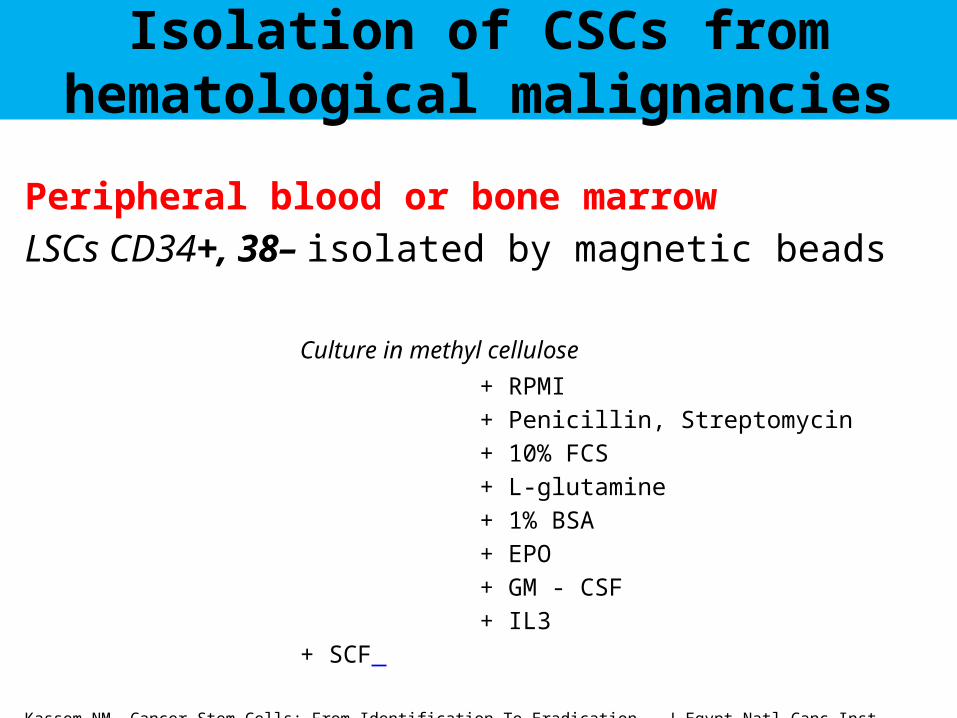
Isolation of CSCs from hematological malignancies
Peripheral blood or bone marrow
LSCs CD34+, 38– isolated by magnetic beads
Culture in methyl cellulose
+ RPMI
+ Penicillin, Streptomycin
+ 10% FCS
+ L-glutamine
+ 1% BSA
+ EPO
+ GM - CSF
+ IL3
+ SCF
Kassem NM. Cancer Stem Cells: From Identification To Eradication . J Egypt Natl Canc Inst. 2008;20(3):209-15.

Isolation of CSCs from solid tumors
Tumor tissue
Washed then subjected to enzymatic dissociation CSCs CD44+, 24– isolated by magnetic beads
Culture in growth factor cocktail
+ EGF
+ bFGF
+ FBS
+ LIF
Kassem NM. Cancer Stem Cells: From Identification To Eradication . J Egypt Natl Canc Inst. 2008;20(3):209-15.
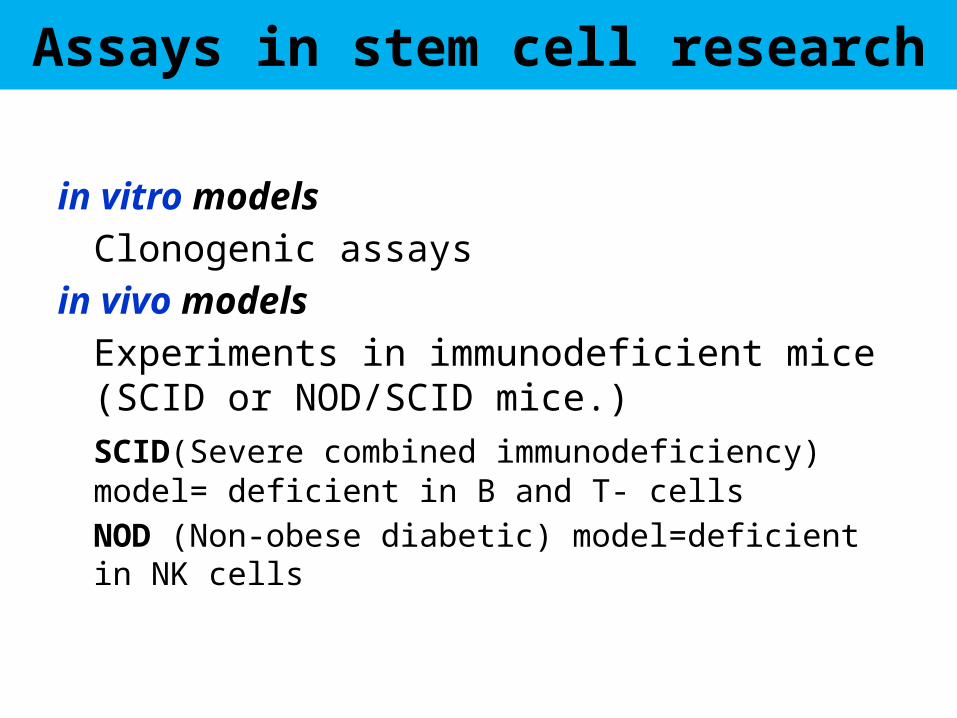
Assays in stem cell research
in vitro models
Clonogenic assays
in vivo models
Experiments in immunodeficient mice (SCID or NOD/SCID mice.)
SCID(Severe combined immunodeficiency) model= deficient in B and T- cells
NOD (Non-obese diabetic) model=deficient in NK cells

Clonogenic assays in stem cell research in vitro model
Neurosphere formation in brain tumor= clonogenic potential
Galli R et al Isolation and characterization of tumorigenic, stem-like neural precursors from human glioblastoma. Cancer Res. 2004;64:7011-7021Singh SK, Hawkins C, Clarke ID, Squire JA, Bayani J, Hide T, Henkelman RM, Cusimano MD, Dirks PB. Identification of human brain tumour initiating cells. Nature. 2004;432:396-401
Normal neural stem cells
Cancer neurospheres
Long-term self-renewing
+ +
Multi-lineage-differentiating
+ +
In vivo tumorigenicity
- +
CD133 - +
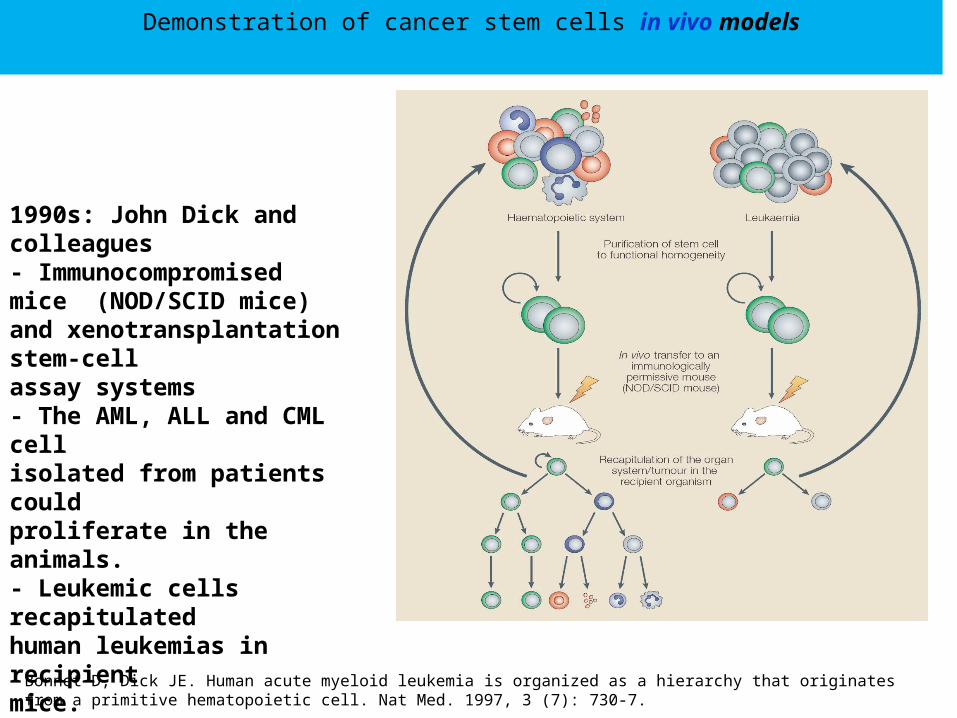
Demonstration of cancer stem cells in vivo models
1990s: John Dick and colleagues- Immunocompromised mice (NOD/SCID mice) and xenotransplantation stem-cellassay systems- The AML, ALL and CML cellisolated from patients couldproliferate in the animals.- Leukemic cells recapitulatedhuman leukemias in recipientmice.
Bonnet D, Dick JE. Human acute myeloid leukemia is organized as a hierarchy that originates from a primitive hematopoietic cell. Nat Med. 1997, 3 (7): 730-7.

in vivo tumorigenic potentialof selected CD133+ tumor cells
Bao S, Wu Q, McLendon RE, Hao Y, Shi Q, Hjelmeland AB, Dewhirst MW, Bigner DD, Rich JN. Glioma stem cells promote radioresistance by preferential activation of the DNA damage response. Nature. 2006 Dec 7;444(7120):756-60.

Verification of macroscopic and microscopic metastases by fluorescence histology
Maria M. (Marj) Peña

Properties of CSCs
• Tumourigenesis in recipients• CSC specific antigens• Genomic instability• Drug resistance• Radiation resistance
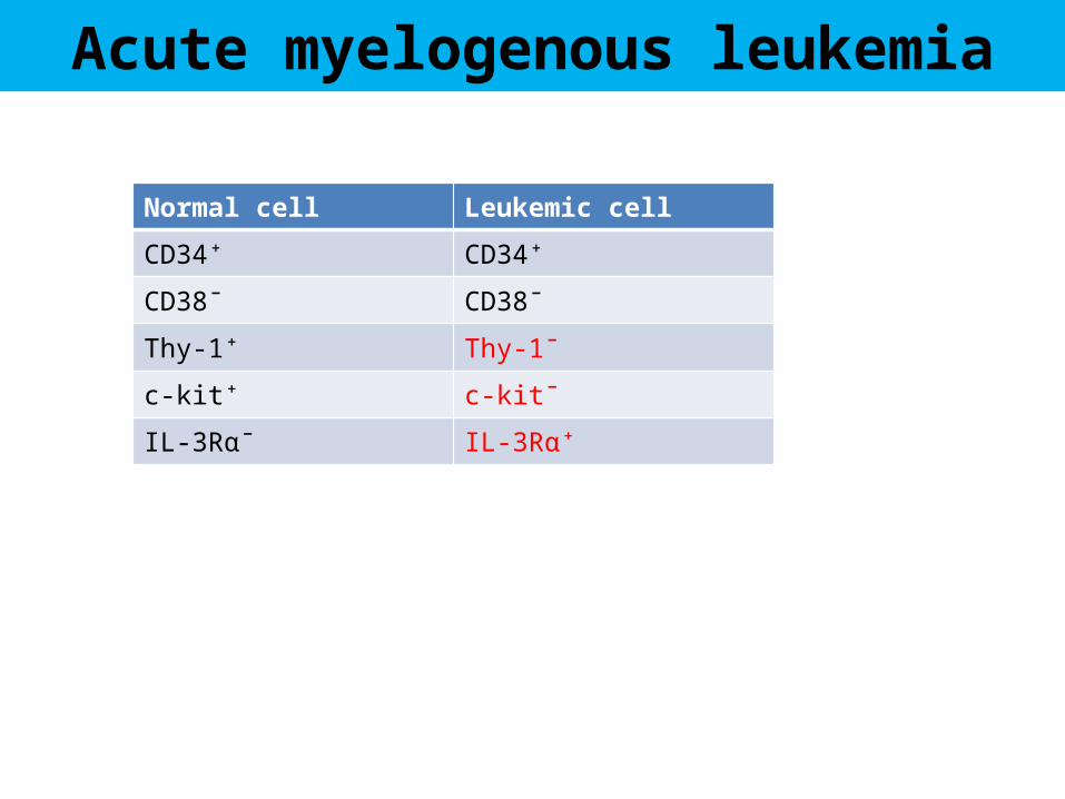
Acute myelogenous leukemia
Normal cell Leukemic cell
CD34⁺ CD34⁺
CD38¯ CD38¯
Thy-1⁺ Thy-1¯
c-kit⁺ c-kit¯
IL-3Rα¯ IL-3Rα⁺

Acute myelogenous leukemia
Normal cell Breast cancer cell Breast cancer stem cell
CD44− CD44+ CD44+
CD24+ CD24−/low CD24−/low
CXCR4 BMI-1
CCR7 ESA/ Flotilin2
EpCam Sca-1/ Ly6
ErbB2/ HER2
ErbB2/ HER2
NCAM-L-1/ CD171
Breast Cancer
• CD44+ enhances invasion and metastasis• Piwil 2 enhances CD44+expresion in fibroblasts

1. The magnetic activated cell sorting system (MACS)
2. Immunocytochemistry
3. FACS
4. Immunomagnetic beads and other beads- based detection methods
5. Epitelial immunospot technique
Detection and characterization of CSCs

Minn, A. J. et al. J. Clin. Invest. 2005;115:44-55
SCPs from MDA-MB-231 cells have a poor-prognosis gene expression signature
Maria M. (Marj) Peña

Technical challenges in CSCs
• Tumors from different patients are different (morphology, cell surface markers, genetic lesions, response to therapy)
• Within an individual tumor, all cancer cells are not equal (variation in genetic and epigenetic abnormalities, distinct proliferative and differentiative capacity
*In patients in remission, the AML1-ETO transcripts were found in a fraction of normal HSCs in the marrow, indicating that the translocation occurred originally in normal HSC and that additional mutations subsequently lead to leukaemia.
(CD34 + Cd38 - Thy-1 + → CD34 + Cd38 - Thy-1 - )
• Absence of specific microenvironment• The method for prospective isolation of CSCs from various tumors
(identification of markers). • The assays to demonstrate the populations are truly CSCs
*Miyamoto, T et al, Proc . Natl. Acad. Sci. USA 97: 7521-7526, 2000