cancer cell report - brainlife.org · cancer cell report accelerated metastasis after short-term...
TRANSCRIPT
Cancer Cell
Report
Accelerated Metastasis after Short-Term Treatmentwith a Potent Inhibitor of Tumor AngiogenesisJohn M.L. Ebos,1,2 Christina R. Lee,1 William Cruz-Munoz,1 Georg A. Bjarnason,3 James G. Christensen,4
and Robert S. Kerbel1,2,*1Molecular and Cellular Biology Research, Sunnybrook Health Sciences Centre, Toronto, ON M4N 3M5, Canada2Department of Medical Biophysics, University of Toronto, Toronto, ON M5G 2M9, Canada3Sunnybrook Odette Cancer Centre, Toronto, ON M5G 2M9, Canada4Pfizer Global Research and Development, La Jolla Labs, La Jolla, CA 92121, USA
*Correspondence: [email protected]
DOI 10.1016/j.ccr.2009.01.021
SUMMARY
Herein we report that the VEGFR/PDGFR kinase inhibitor sunitinib/SU11248 can accelerate metastatic tumorgrowth and decrease overall survival in mice receiving short-term therapy in various metastasis assays,including after intravenous injection of tumor cells or after removal of primary orthotopically grown tumors.Acceleration of metastasis was also observed in mice receiving sunitinib prior to intravenous implantation oftumor cells, suggesting possible ‘‘metastatic conditioning’’ in multiple organs. Similar findings with additionalVEGF receptor tyrosine kinase inhibitors implicate a class-specific effect for such agents. Importantly, theseobservations of metastatic acceleration were in contrast to the demonstrable antitumor benefits obtainedwhen the same human breast cancer cells, as well as mouse or human melanoma cells, were grown ortho-topically as primary tumors and subjected to identical sunitinib treatments.
INTRODUCTION
Multiple strategies for inhibiting the VEGF pathway have been
shown in numerous preclinical studies to hinder tumor growth,
and the recent approval of a VEGF-neutralizing antibody (beva-
cizumab) and VEGF receptor tyrosine kinase inhibitors (RTKIs)
(sorafenib and sunitinib) has clinically validated targeting VEGF
or its receptors (particularly VEGFR2) as an anticancer treatment
(Folkman, 2007). The purpose of the present study was to test
whether a VEGF RTKI, when administered daily for short periods,
can influence the growth of experimental and spontaneous
metastasis in mice. The rationale for this experimental design
was based on a number of recent clinical and preclinical obser-
vations. First, while sorafenib and sunitinib have been approved
for the treatment of renal cell carcinoma, as well as hepatocel-
lular carcinoma (sorafenib only) and gastrointestinal stromal
tumor (sunitinib only), enduring clinical responses are rare. More-
232 Cancer Cell 15, 232–239, March 3, 2009 ª2009 Elsevier Inc.
over, single-agent use of such drugs, including bevacizumab,
has not led to meaningful beneficial activity in many cases.
Second, when such agents are administered on a discontinuous
schedule, such as with sunitinib (4 weeks on/2 weeks off), tumor
regrowth has sometimes been observed during drug-free break
periods (Burstein et al., 2008) or when treatment is discontinued
(Johannsen et al., 2008). Third, rapid tumor revascularization has
been reported when therapy is stopped in preclinical studies
(Mancuso et al., 2006). Finally, we recently reported that
a number of changes in proangiogenic plasma proteins
observed in patients after sunitinib treatment could be recapitu-
lated in non-tumor-bearing mice, and moreover, in a dose-
dependent manner (Ebos et al., 2007). Together, these results
suggest a systemic host response to VEGF inhibition that could
play a role in regrowth of both tumor and vasculature in eventual
evasion of response during continued antiangiogenic treatment
(Bergers and Hanahan, 2008; Casanovas et al., 2005) as well
SIGNIFICANCE
The use of VEGF pathway inhibitors to impair angiogenesis now represents a clinically validated anticancer treatmentstrategy. However, the benefits of VEGF-targeted agents in the treatment of late-stage cancers can be transitory, resultingin eventual drug resistance, tumor growth and/or regrowth, and rapid vascular recovery when therapy is stopped. Our find-ings here demonstrate that angiogenesis inhibition in mice can lead to opposing effects on tumor growth and metastasisdepending on tumor stage and treatment duration. These observations could have clinical implications with respect tooptimal dose, treatment schedule, and therapy in the adjuvant/neoadjuvant setting and highlight the importance of testingadditional drugs in combination as a possible approach to abrogate this effect.
Cancer Cell
Brief Antiangiogenic Therapy Increases Metastasis
as potentially influencing the progression of secondary (meta-
static) disease—a possibility that could impact the outcome of
neoadjuvant and adjuvant administration of such agents (von
Mehren, 2008; Shuch et al., 2008). In this study, we examined
the impact of sunitinib on metastasis, with the choice of sunitinib
based not only on our aforementioned work but also on the
discontinuous administration schedule of this agent currently
used in clinical practice (Motzer et al., 2006).
RESULTS AND DISCUSSION
Our first experiments tested the effect of short-term sunitinib
treatment in a model of experimental metastasis. Human meta-
B
C
A
0 5
0
10
20
30
40
50
60
70
80
90
100
25 35 45 55 65 75
Group AGroup BGroup C
** ***
Days Postimplantation
Survival (%
)
-7 0 7 14 21 28 35 42
106
107
108
Group AGroup BGroup C
120 mg/kg
Vehicle
120 mg/kg
i.v. tumor implantation(LM2-4LUC+ Cells)
Days Postimplantation
Tu
mo
r B
urd
en
(Pho
tons
/s)
Group A Group B Group C
71
27Day
s A
fter
Tum
or Im
plan
tatio
n
Photons/s
107
106
105
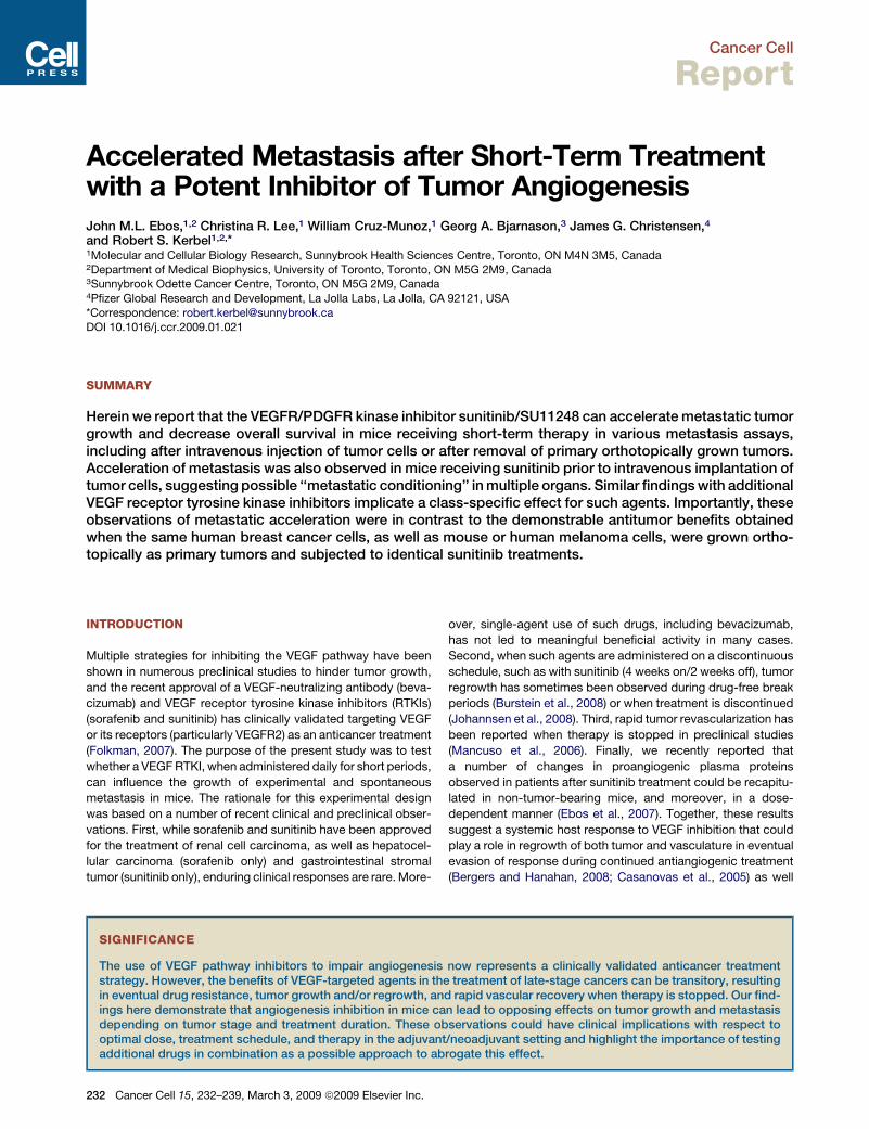
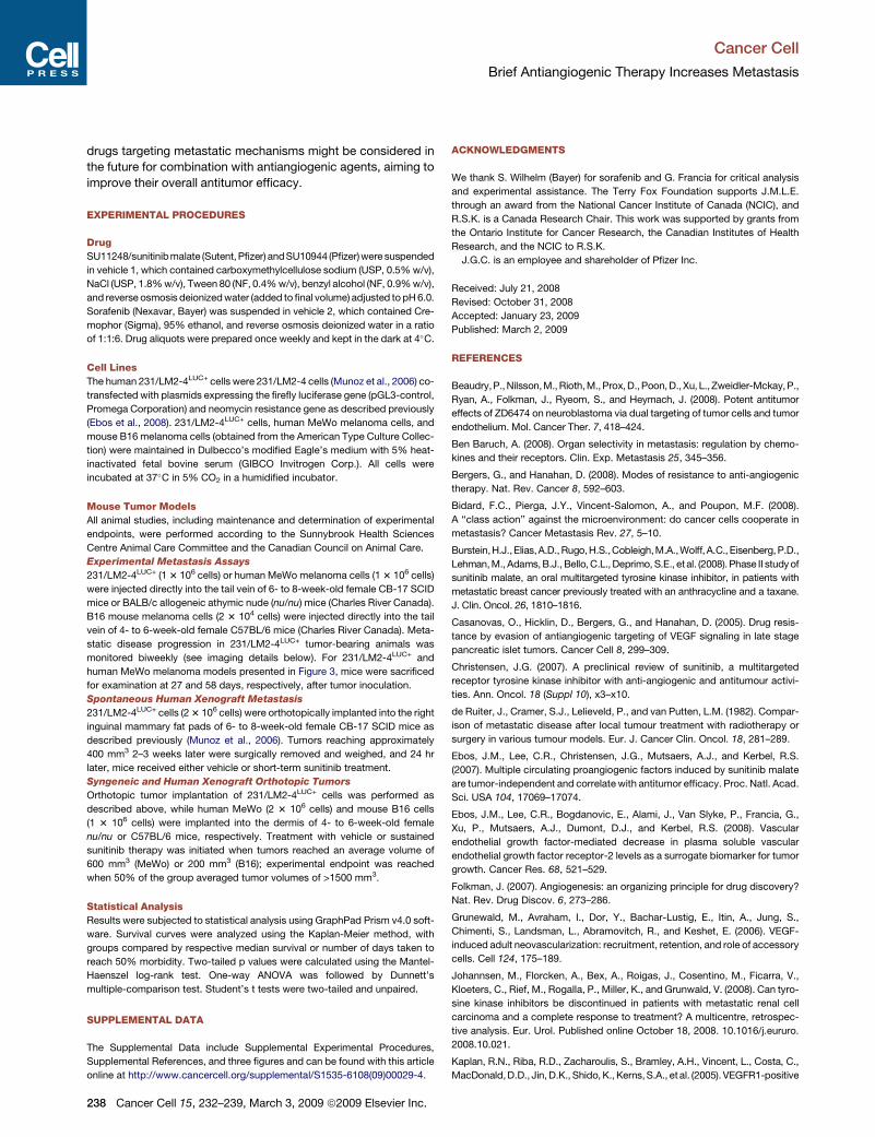
Figure 1. Accelerated Experimental Metastasis and
Decreased Survival after Short-Term Sunitinib Treat-
ment before and after Intravenous Tumor Inoculation
(A) 231/LM2-4LUC+ cells were injected into the tail vein of SCID
mice that received vehicle (group A) or short-term sunitinib
treatment daily for 7 days either before (group B) or after tumor
inoculation (group C). Quantification of bioluminescence
showed accelerated tumor growth in groups B and C
compared with controls. A representative experiment is
shown. Group A, n = 10; group B, n = 5; group C, n = 10.
Data are presented as mean ± SD.
(B) Kaplan-Meier survival curve shows significantly decreased
median survival of mice in group B (log-rank test, p = 0.0055)
and group C (log-rank test, p < 0.0001) compared with
group A. Data represent a summary of multiple experiments.
Group A, n = 31; group B, n = 11; group C, n = 19. 0.001 <
**p < 0.01; ***p < 0.001.
(C) Representative images for each group taken 1, 7, and
27 days following tumor implantation, with increased metas-
tasis visible in sunitinib-treated mice. Sunitinib dose and
treatment schedule were performed as illustrated in (A).
static breast cancer 231/LM2-4LUC+ cells express-
ing luciferase were injected into the tail vein of
severe combined immunodeficiency (SCID) mice,
which then received vehicle treatment or a short-
term sunitinib therapy regimen (120 mg/kg/daily
for 7 days) administered by gavage either before
or after tumor cell inoculation (groups A, B, and C,
respectively; Figure 1A). Short-term sunitinib treat-
ment administered either before or after tumor cell
injection resulted in accelerated experimental
metastasis as measured by bioluminescence (Fig-
ure 1A) and significantly reduced median survival
compared to vehicle-treated controls (Figure 1B).
Representative images showing increased metas-
tasis in sunitinib-treated mice are shown in
Figure 1C. The choice of 120 mg/kg daily sunitinib
for a 7-day period was based on prior preclinical
studies that had demonstrated this short-term
regimen to maximize the aforementioned multiple
host-derived changes in proangiogenic proteins in
mouse plasma. A sustained sunitinib therapy
regimen of 60 mg/kg/day given continuously
(described below) had previously been shown to
result in optimal tumor inhibition with minimal
toxicity after long-term therapy (Ebos et al., 2007). The treatment
for group B was stopped 24 hr prior to injection of tumor cells in
order to minimize any potential direct drug effect on tumor cells,
as blood concentrations of sunitinib are significantly reduced
24 hr after treatment cessation, and to maximize the aforemen-
tioned sunitinib-induced host molecular changes, which were
shown to be reversed within 2–5 days after stopping therapy
(Ebos et al., 2007). Importantly, similar results were obtained
when nu/nu mice were treated with other VEGF RTKIs including
sorafenib (150 mg/kg/day) and SU10944 (225 mg/kg/day)
7 days prior to tumor cell inoculation (see Figures S1A–S1C avail-
able online). These results suggest that a host response to multi-
targeted angiogenic kinase inhibition can result in conditions that
Cancer Cell 15, 232–239, March 3, 2009 ª2009 Elsevier Inc. 233
Cancer Cell
Brief Antiangiogenic Therapy Increases Metastasis
allow for increased tumor initiation even after drug has been
removed.
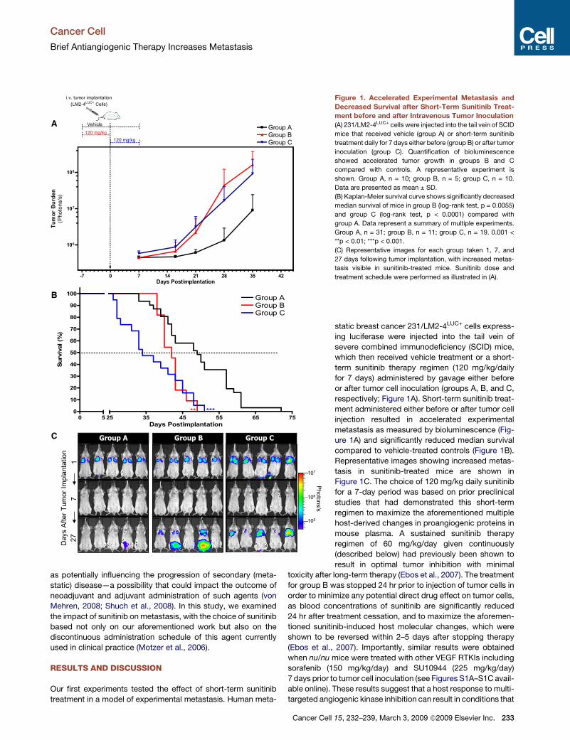
We next tested the effect of short-term sunitinib treatment on
distant spontaneous metastasis generated after primary tumor
removal using the protocol illustrated in Figure 2A. Mice receiving
short-term adjuvant sunitinib therapy showed increased sponta-
neous metastatic tumor burden as measured by biolumines-
cence (Figures 2A and 2B), which corresponded with decreased
overall survival (Figure 2C). To ensure equal tumor burden
between groups prior to drug treatment, resected tumors were
weighed prior to sorting into groups A and B (Figure 2D).
We have previously reported that surgical resection of highly
metastatic orthotopicallygrown 231/LM2-4 tumors leads to spon-
taneous metastasis in the lungs, liver, and lymph nodes, with the
primary determinant for euthanasia being extensive visceral and
peripheral metastasis (Man et al., 2007). In the spontaneous and
experimental metastasis studies described herein, we similarly
considered visceral and peripheral disease as the primary reason
for mouse sacrifice (data not shown). To test for differences in
5B
efor
e30
Day
s A
fter
Tum
or R
esec
tion
Group A Group B
Group A Group B
0.0
0.1
0.2
0.3
0.4
0.5
0.6
0.7
Tu
mo
r W
eig
ht a
t R
es
ec
tio
n (g
)
0
0
10
20
30
40
50
60
70
80
90
100
Group AGroup B
25 35 45 55 65 75 85 95 105 115
**
Days Postresection
Su
rv
iva
l (%
)
A
B
C D
Photons/s
107
106
105
0 7 14 21 28 35 42 49
105
106
107
108
109
Group AGroup B120 mg/kg
Mammary fat pad orthotopictumor implantation(LM2-4LUC+ Cells)
VehicleTumors grownuntil 400 mm3
Primary tumor resection
Days Postresection
Tu
mo
r B
urd
en
(Pho
tons
/s)
Figure 2. Short-Term Sunitinib Treatment
Increases Spontaneous Metastasis and
Decreases Survival after Removal of
Primary Human Xenograft Tumors
(A) Orthotopically grown 231/LM2-4LUC+ tumors
were surgically removed, and SCID mice were
treated daily with vehicle (group A) or short-term
sunitinib therapy (group B). Biweekly quantifica-
tion of bioluminescence showed accelerated
tumor growth and increased spontaneous metas-
tasis in group B compared with group A. Data are
presented as mean ± SD.
(B) Representative bioluminescence images visu-
alizing tumor cells before and after primary tumor
resection (days 5 and 30 after resection).
(C) Kaplan-Meier survival curves of the corre-
sponding mice show significantly decreased
median survival in group B (log-rank test, p =
0.0024) compared with group A. 0.001 < **p <
0.01.
(D) Resected tumors were weighed prior to sorting
into groups A and B to ensure equal tumor burden
between groups. Sunitinib dose and treatment
schedule were performed as illustrated in (A).
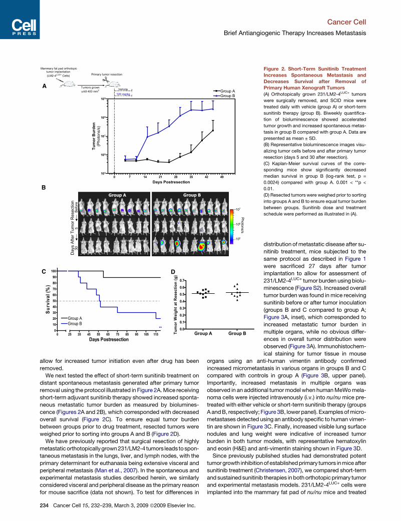
distribution of metastatic disease after su-
nitinib treatment, mice subjected to the
same protocol as described in Figure 1
were sacrificed 27 days after tumor
implantation to allow for assessment of
231/LM2-4LUC+ tumor burden using biolu-
minescence (Figure S2). Increased overall
tumor burden was found in mice receiving
sunitinib before or after tumor inoculation
(groups B and C compared to group A;
Figure 3A, inset), which corresponded to
increased metastatic tumor burden in
multiple organs, while no obvious differ-
ences in overall tumor distribution were
observed (Figure 3A). Immunohistochem-
ical staining for tumor tissue in mouse
organs using an anti-human vimentin antibody confirmed
increased micrometastasis in various organs in groups B and C
compared with controls in group A (Figure 3B, upper panel).
Importantly, increased metastasis in multiple organs was
observed in an additional tumor model when human MeWo mela-
noma cells were injected intravenously (i.v.) into nu/nu mice pre-
treated with either vehicle or short-term sunitinib therapy (groups
A and B, respectively; Figure 3B, lower panel). Examples of micro-
metastases detected using an antibody specific to human vimen-
tin are shown in Figure 3C. Finally, increased visible lung surface
nodules and lung weight were indicative of increased tumor
burden in both tumor models, with representative hematoxylin
and eosin (H&E) and anti-vimentin staining shown in Figure 3D.
Since previously published studies had demonstrated potent
tumor growth inhibition of established primary tumors in mice after
sunitinib treatment (Christensen, 2007), we compared short-term
and sustained sunitinib therapies in both orthotopic primary tumor
and experimental metastasis models. 231/LM2-4LUC+ cells were
implanted into the mammary fat pad of nu/nu mice and treated
234 Cancer Cell 15, 232–239, March 3, 2009 ª2009 Elsevier Inc.
Cancer Cell
Brief Antiangiogenic Therapy Increases Metastasis
Liver Kidney BrainSpleen
0 1 5 10 15 20 25
Carcass
Brain
Heart
Liver
Kidneys
Lung
Spleen
Intestines
Femur
Group CGroup BGroup A
*
*
Photons/s
(Fold change of control)
10 20 30 40 50 601
*
Tot
al T
umor
Bur
den
Spleen Liver Kidney Femurs Brain
Group A 1/10 3/10 0/10 3/10 2/10
Group B 6/10 1/10 0/10 3/10 2/10
Group C 5/10 3/10 2/10 1/10 4/10
Group A 0/5 1/5 1/5 NT 0/5
Group B 1/5 2/5 1/5 NT 3/5
LM
2-4
LU
C+
Me
Wo
100µm100µm100µm100µm
100µm 100µm 100µm 100µmB
>150>150>150>150>150
1.241.230.691.450.22
A5
183963110
0.170.170.160.230.19
12345
12345
Group
B
A
C
H&E
Mouse
Lung
Nodule
sLung
Weig
ht
Lung α-Vimentin
MeW
oL
M2-4
LU
C+
11244710134145
0.140.160.150.200.150.200.140.180.180.14
1456
1023343755136
0.170.150.180.130.180.430.140.170.350.39
0001225202230
0.180.140.140.160.140.160.170.160.200.16
12345678910
12345678910
12345678910
Immunostaining
1 cm
1 cm
1 cm
1 cm
1 cm
100µm
100µm400µm
400µm 400µm
400µm 400µm
400µm 400µm
400µm
A
B
C
D
LM
2-4
LU
C+
Me
Wo
Figure 3. Increased Multiorgan Metastasis in Mice after Short-Term Sunitinib Treatment
(A) Following the same experimental design as described in Figure 1A, SCID mice were sacrificed at day 27 to compare increases in overall tumor burden after
short-term sunitinib treatment (inset) and corresponding increased bioluminescence in multiple organs (groups A, B, and C). Data are presented as mean ± SEM.
Instances of highly divergent bioluminescence values did not permit statistical significance to be reached in all groups. 0.01 < *p < 0.05 by one-way ANOVA.
(B) Micrometastases were confirmed by immunostaining for human vimentin in organs of the 231/LM2-4LUC+ tumor model in (A) (upper panel) or in nu/nu mice
receiving vehicle (group A) or short-term sunitinib therapy (group B) prior to intravenous (i.v.) inoculation with human MeWo melanoma cells (lower panel). Tissue
sections were scored as positive or negative based on the presence or absence of detectable micrometastases. NT = not tested.
(C) Representative examples of micrometastases in spleen, liver, kidney, and brain shown using human-specific vimentin antibodies.
(D) Excised lungs from 231/LM2-4LUC+ and MeWo tumor models were scored visually for surface tumor nodules, with confirmation of macrometastasis by hema-
toxylin and eosin (H&E) and anti-vimentin immunostaining (representative images shown). For groups A, B, and C in the 231/LM2-4LUC+ tumor model, n = 10 per
group. For groups A and B in the MeWo tumor model, n = 5 per group.
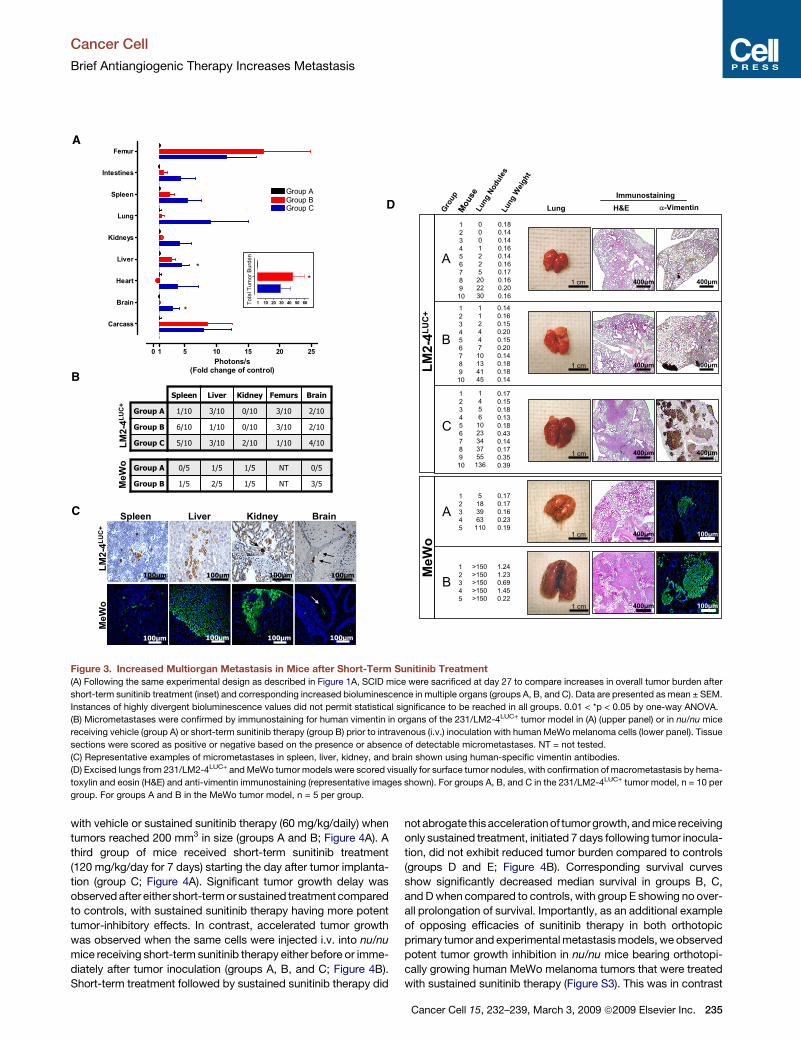
with vehicle or sustained sunitinib therapy (60 mg/kg/daily) when
tumors reached 200 mm3 in size (groups A and B; Figure 4A). A
third group of mice received short-term sunitinib treatment
(120 mg/kg/day for 7 days) starting the day after tumor implanta-
tion (group C; Figure 4A). Significant tumor growth delay was
observed after either short-term or sustained treatment compared
to controls, with sustained sunitinib therapy having more potent
tumor-inhibitory effects. In contrast, accelerated tumor growth
was observed when the same cells were injected i.v. into nu/nu
mice receiving short-term sunitinib therapy either before or imme-
diately after tumor inoculation (groups A, B, and C; Figure 4B).
Short-term treatment followed by sustained sunitinib therapy did
not abrogate thisacceleration of tumorgrowth, and mice receiving
only sustained treatment, initiated 7 days following tumor inocula-
tion, did not exhibit reduced tumor burden compared to controls
(groups D and E; Figure 4B). Corresponding survival curves
show significantly decreased median survival in groups B, C,
and D when compared to controls, with group E showing no over-
all prolongation of survival. Importantly, as an additional example
of opposing efficacies of sunitinib therapy in both orthotopic
primary tumor and experimental metastasis models, we observed
potent tumor growth inhibition in nu/nu mice bearing orthotopi-
cally growing human MeWo melanoma tumors that were treated
with sustained sunitinib therapy (Figure S3). This was in contrast
Cancer Cell 15, 232–239, March 3, 2009 ª2009 Elsevier Inc. 235

Cancer Cell
Brief Antiangiogenic Therapy Increases Metastasis
B
C
A
-7 0 7 14 21 28 35 42
106
107
108
Group AGroup BGroup CGroup D
60 mg/kg
120 mg/kg
Vehicle
Group E
120 mg/kg
120 mg/kg
60 mg/kg
i.v. tumor implantation(LM2-4LUC+ Cells)
Days Postimplantation
Tu
mo
r B
urd
en
(Pho
tons
/s)
0
0
10
20
30
40
50
60
70
80
90
100
110
Group AGroup BGroup CGroup DGroup E
30 40 50 60 70 80
** ** *
Days Postimplantation
Survival (%
)
0 7 14 21 28 35 42 49 56 63
0
250
500
750
1000
1250
1500
1750
2000
Group AGroup BGroup C
60 mg/kg
Vehicle
120 mg/kg
Mammary fat pad orthotopictumor implantation(LM2-4LUC+ Cells)
**,***
}
Days Postimplantation
Tu
mo
r V
olu
me (m
m3)
-7 0 7 14 21 28 35 42 49
0
10
20
30
40
50
60
70
80
90
100
Group AGroup BGroup C
Group EGroup D60 mg/kg
120 mg/kg
Vehicle
120 mg/kg
120 mg/kg60 mg/kg
**
**
i.v. tumor implantation(B16 Cells)
Days Postimplantation
Su
rv
iva
l (%
)
0 7 14 21 28
0
250
500
750
1000
1250
1500
1750
2000
2250
Group AGroup B
60 mg/kg
Vehicle
Subdermal orthotopictumor implantation
(B16 cells)
Days Postimplantation
Tu
mo
r V
olu
me
(m
m3)
E
D
Figure 4. Differentiating Opposing Efficacies of Short-Term and Sustained Sunitinib Treatment in Primary and Metastatic Disease
(A) nu/nu mice bearing orthotopically grown 231/LM2-4LUC+ tumors received either vehicle (group A) or sustained sunitinib therapy (group B) when tumors
reached an average volume of 200 mm3. A third group received short-term sunitinib therapy (group C) starting the day after tumor implantation. Group A reached
tumor volume endpoint (1500 mm3) at 27 days, with comparative tumor volume significantly reduced in group B (p = 0.0002 by Student’s t test) and group C
(p = 0.0027 by Student’s t test) at the same time point. Mice receiving sustained sunitinib therapy showed greater tumor growth inhibition compared to mice
receiving short-term therapy (41 to 62 days to endpoint, respectively). n = 5 for all groups.
(B) nu/numice injected i.v. with231/LM2-4LUC+ cells were treated daily with vehicle (groupA) or receivedshort-termsunitinib therapy for7 days eitherbefore (group B)
or after tumor inoculation (group C). Quantification of bioluminescence showed accelerated metastatic tumor growth in groups B and C after short-term sunitinib
therapy. Mice in groups D and E received sustained sunitinib therapy starting on day 8, with group D also receiving short-term sunitinib therapy similar to group C.
(C) Corresponding Kaplan-Meier survival curves show that median survival was significantly decreased in groups B, C, and D (p = 0.0011, p = 0.0022, and p = 0.0365
by log-rank test, respectively) and was not significantly different in group E (p = 0.4485 by log-rank test) compared to control mice in group A. For (B) and (C): group A,
n = 7; group B, n = 7; group C, n = 7; group D, n = 5; group E, n = 5.
236 Cancer Cell 15, 232–239, March 3, 2009 ª2009 Elsevier Inc.

Cancer Cell
Brief Antiangiogenic Therapy Increases Metastasis
to the increased metastasis observed in the lungs of mice 58 days
after i.v. inoculation of tumor cells immediately following short-
term sunitinib therapy (Figure 3D).
We next tested whether similar results would be observed in
a syngeneic mouse tumor model following the same treatment
protocol as in Figure 4A. Mouse B16 melanoma cells were
implanted subdermally into the flanks of C57BL/6 mice and, as
in the aforementioned human xenograft experiments, primary
tumor growth was found to be delayed in mice receiving sus-
tained sunitinib treatment compared to controls (groups A and
B; Figure 4D). In contrast, and similar to Figure 4C, C57BL/6
mice receiving short-term sunitinib treatment for 7 days prior to
i.v. tumor inoculation showed decreased survival compared to
controls, whereas sustained sunitinib therapy, initiated 8 days
after inoculation, showed no survival advantage (groups A, B,
and E; Figure 4E). Interestingly, mice receiving short-term suniti-
nib therapy immediately following tumor inoculation, which was
then either stopped after 7 days (group C) or followed by sus-
tained sunitinib therapy (group D), exhibited a survival advantage
in some instances. Group D mice showed a significant prolonga-
tion of survival compared to control mice, while mice in group C
had biphasic effects, with about half of the mice progressing
with accelerated metastasis and the remainder showing a prolon-
gation in survival, even after cessation of treatment (Figure 4E).
One potential explanation for these results could stem from
possible direct antitumor activity of sunitinib therapy against
B16 cells—something that has been demonstrated previously
with other VEGF RTKIs (Beaudry et al., 2008).
Taken together, our results show that VEGF receptor tyrosine
kinase inhibition by the same drug, administered in different
schedules and doses using three different tumor cell models,
can have opposing effects on tumor growth. Sustained sunitinib
treatment of preestablished tumors led to significantgrowth inhibi-
tion in both orthotopically grown primary human xenograft and
syngeneic tumors. In contrast, short-term treatment prior to i.v.
inoculationwith the samecellsproducedanaccelerationofmetas-
tasis and corresponding significant reduction in median survival.
Furthermore, sustained treatment in both 231/LM2-4LUC+ and
B16 experimental metastasis models, initiated 7 days after tumor
cell inoculation, did not produce a survival advantage compared to
controls. Conversely, short-term sunitinib treatment immediately
following tumor inoculation was shown to accelerate metastatic
disease (in the 231/LM2-4LUC+ model) or produce a biphasic
response (in the B16 model).
Our results complement those of Paez-Ribes et al. (2009) in this
issue of Cancer Cell. Importantly, in both studies, antiangiogenic
drug treatment was shown to have potent inhibitory effects in
localized tumors. Furthermore, Paez-Ribes et al. show that treat-
ment of tumor-bearing mice with antiangiogenic drugs, including
the VEGFR2-blocking monoclonal antibody DC101 and VEGF
RTKIs such as sunitinib and SU10944, leads to increased local
tumor cell invasion and enhanced distant metastasis after pro-
longed treatment or, in the case of DC101, after only short-term
treatment. As Paez-Ribes et al. demonstrate, these effects
appear to be an adaptive/evasive response by the tumor cells
themselves triggered by a disruption of the tumor vasculature.
Our results present a second possibility independent of adapta-
tions by an established tumor that involves microenvironmental
changes in mouse organs that are ‘‘conditioned’’ to be more
permissive to tumor extravasation. But how might such a meta-
static ‘‘conditioning’’ effect occur? A number of potential mech-
anisms alone or in combination could play a role. One is the
aforementioned induced upregulation of multiple circulating
proangiogenic cytokines and growth factors in response to treat-
ment, including osteopontin, G-CSF, and SDF1a (Ebos et al.,
2007)—all of which have been implicated in angiogenesis and/
or metastasis (McAllister et al., 2008; Ben Baruch, 2008; Wai
and Kuo, 2008; Natori et al., 2002; Zhang et al., 2000). Second,
and likely related to such molecular changes, the mobilization
of bone marrow-derived cells may facilitate an enhanced ‘‘pre-
metastatic niche,’’ including circulating endothelial (Okazaki
et al., 2006) and myeloid progenitors (Shojaei et al., 2008),
CXCR4+ recruited bone marrow circulating cells (Grunewald
et al., 2006), and circulating VEGFR1+ bone marrow cells (Kaplan
et al., 2005). Finally, the well-recognized target promiscuity of
RTKIs such as sunitinib and sorafenib (Karaman et al., 2008)
may produce a plethora of broad host microenvironmental
responses to cellular inhibition/injury. These responses in turn
may promote tumor extravasation, similar in principal to the
enhanced metastasis observed after treatment with radiation
and numerous chemotherapeutic drugs (van Putten et al.,
1975; Vollmer and Conley, 1984; de Ruiter et al., 1982)—all of
which may involve various proinflammatory responses (Noonan
et al., 2008) or alterations in the endothelial microenvironment.
Collectively, such effects could create a more favorable meta-
static niche (Bidard et al., 2008). Importantly, however, unlike
chemotherapy and radiation treatments, which act in large part
via direct tumor cytotoxicity by nonspecifically targeting prolifer-
ating cells and are administered for defined periods, antiangio-
genic agents act in large part against host tumor support
processes, thus indirectly prohibiting tumor growth, and are
meant (at least theoretically) to be administered indefinitely.
Regardless of the actual mechanisms involved, our results may
be pertinent to the consideration of several prominent issues in
cancer therapeutics, including the relative benefits of discontin-
uous versus continuous treatment schedules of antiangiogenic
drugs (such as sunitinib), duration of treatment, use of such drugs
in the neoadjuvant and adjuvant setting, and the prospect that
(D) In a syngeneic tumor model, C57BL/6 mice bearing mouse B16 melanoma tumors grown orthotopically showed delayed primary tumor growth after sustained
sunitinib therapy compared with control mice (groups A and B, with time to endpoint 17 and 28 days, respectively). Group A, n = 4; group B, n = 4.
(E) C57BL/6 mice receiving short-term sunitinib therapy prior to i.v. inoculation with the same mouse B16 melanoma tumor cells showed accelerated experimental
metastasis and decreased survival compared to controls (group B; p = 0.0014 by log-rank test). Delayed metastasis and increased survival were observed in mice
receiving short-term sunitinib treatment followed by sustained sunitinib treatment (group D; p = 0.0047 by log-rank test). Mice receiving sustained sunitinib therapy
7 days after tumor implantationshowedno difference in survival compared tovehicle-treated controlmice (groupE; p = 0.6368by log-rank test).Mice receivingshort-
term sunitinib therapy following tumor implantation exhibited a bimodal response that included either accelerated metastasis and reduced survival or extended
survival (group C; p = 0.3391 by log-rank test). Group A, n = 10; group B, n = 10; group C, n = 9; group D, n = 5; group E, n = 5. Sunitinib dose and treatment schedule
were performed as illustrated.
Data are presented as mean ± SD. 0.01 < *p < 0.05; 0.001 < **p < 0.01; ***p < 0.001.
Cancer Cell 15, 232–239, March 3, 2009 ª2009 Elsevier Inc. 237
Cancer Cell
Brief Antiangiogenic Therapy Increases Metastasis
drugs targeting metastatic mechanisms might be considered in
the future for combination with antiangiogenic agents, aiming to
improve their overall antitumor efficacy.
EXPERIMENTAL PROCEDURES
Drug
SU11248/sunitinibmalate (Sutent,Pfizer) andSU10944 (Pfizer)were suspended
in vehicle 1, which contained carboxymethylcellulose sodium (USP, 0.5% w/v),
NaCl (USP, 1.8% w/v), Tween 80 (NF, 0.4% w/v), benzyl alcohol (NF, 0.9% w/v),
and reverse osmosis deionized water (added to final volume) adjusted to pH 6.0.
Sorafenib (Nexavar, Bayer) was suspended in vehicle 2, which contained Cre-
mophor (Sigma), 95% ethanol, and reverse osmosis deionized water in a ratio
of 1:1:6. Drug aliquots were prepared once weekly and kept in the dark at 4�C.
Cell Lines
The human 231/LM2-4LUC+ cells were 231/LM2-4 cells (Munoz et al., 2006) co-
transfected with plasmids expressing the firefly luciferase gene (pGL3-control,
Promega Corporation) and neomycin resistance gene as described previously
(Ebos et al., 2008). 231/LM2-4LUC+ cells, human MeWo melanoma cells, and
mouse B16 melanoma cells (obtained from the American Type Culture Collec-
tion) were maintained in Dulbecco’s modified Eagle’s medium with 5% heat-
inactivated fetal bovine serum (GIBCO Invitrogen Corp.). All cells were
incubated at 37�C in 5% CO2 in a humidified incubator.
Mouse Tumor Models
All animal studies, including maintenance and determination of experimental
endpoints, were performed according to the Sunnybrook Health Sciences
Centre Animal Care Committee and the Canadian Council on Animal Care.
Experimental Metastasis Assays
231/LM2-4LUC+ (1 3 106 cells) or human MeWo melanoma cells (1 3 106 cells)
were injected directly into the tail vein of 6- to 8-week-old female CB-17 SCID
mice or BALB/c allogeneic athymic nude (nu/nu) mice (Charles River Canada).
B16 mouse melanoma cells (2 3 104 cells) were injected directly into the tail
vein of 4- to 6-week-old female C57BL/6 mice (Charles River Canada). Meta-
static disease progression in 231/LM2-4LUC+ tumor-bearing animals was
monitored biweekly (see imaging details below). For 231/LM2-4LUC+ and
human MeWo melanoma models presented in Figure 3, mice were sacrificed
for examination at 27 and 58 days, respectively, after tumor inoculation.
Spontaneous Human Xenograft Metastasis
231/LM2-4LUC+ cells (2 3 106 cells) were orthotopically implanted into the right
inguinal mammary fat pads of 6- to 8-week-old female CB-17 SCID mice as
described previously (Munoz et al., 2006). Tumors reaching approximately
400 mm3 2–3 weeks later were surgically removed and weighed, and 24 hr
later, mice received either vehicle or short-term sunitinib treatment.
Syngeneic and Human Xenograft Orthotopic Tumors
Orthotopic tumor implantation of 231/LM2-4LUC+ cells was performed as
described above, while human MeWo (2 3 106 cells) and mouse B16 cells
(1 3 106 cells) were implanted into the dermis of 4- to 6-week-old female
nu/nu or C57BL/6 mice, respectively. Treatment with vehicle or sustained
sunitinib therapy was initiated when tumors reached an average volume of
600 mm3 (MeWo) or 200 mm3 (B16); experimental endpoint was reached
when 50% of the group averaged tumor volumes of >1500 mm3.
Statistical Analysis
Results were subjected to statistical analysis using GraphPad Prism v4.0 soft-
ware. Survival curves were analyzed using the Kaplan-Meier method, with
groups compared by respective median survival or number of days taken to
reach 50% morbidity. Two-tailed p values were calculated using the Mantel-
Haenszel log-rank test. One-way ANOVA was followed by Dunnett’s
multiple-comparison test. Student’s t tests were two-tailed and unpaired.
SUPPLEMENTAL DATA
The Supplemental Data include Supplemental Experimental Procedures,
Supplemental References, and three figures and can be found with this article
online at http://www.cancercell.org/supplemental/S1535-6108(09)00029-4.
238 Cancer Cell 15, 232–239, March 3, 2009 ª2009 Elsevier Inc.
ACKNOWLEDGMENTS
We thank S. Wilhelm (Bayer) for sorafenib and G. Francia for critical analysis
and experimental assistance. The Terry Fox Foundation supports J.M.L.E.
through an award from the National Cancer Institute of Canada (NCIC), and
R.S.K. is a Canada Research Chair. This work was supported by grants from
the Ontario Institute for Cancer Research, the Canadian Institutes of Health
Research, and the NCIC to R.S.K.
J.G.C. is an employee and shareholder of Pfizer Inc.
Received: July 21, 2008
Revised: October 31, 2008
Accepted: January 23, 2009
Published: March 2, 2009
REFERENCES
Beaudry,P., Nilsson,M., Rioth,M., Prox, D., Poon, D., Xu, L.,Zweidler-Mckay, P.,
Ryan, A., Folkman, J., Ryeom, S., and Heymach, J. (2008). Potent antitumor
effects of ZD6474 on neuroblastoma via dual targeting of tumor cells and tumor
endothelium. Mol. Cancer Ther. 7, 418–424.
Ben Baruch, A. (2008). Organ selectivity in metastasis: regulation by chemo-
kines and their receptors. Clin. Exp. Metastasis 25, 345–356.
Bergers, G., and Hanahan, D. (2008). Modes of resistance to anti-angiogenic
therapy. Nat. Rev. Cancer 8, 592–603.
Bidard, F.C., Pierga, J.Y., Vincent-Salomon, A., and Poupon, M.F. (2008).
A ‘‘class action’’ against the microenvironment: do cancer cells cooperate in
metastasis? Cancer Metastasis Rev. 27, 5–10.
Burstein,H.J.,Elias,A.D., Rugo,H.S.,Cobleigh,M.A.,Wolff,A.C., Eisenberg, P.D.,
Lehman,M., Adams, B.J., Bello, C.L., Deprimo, S.E., et al. (2008). Phase II study of
sunitinib malate, an oral multitargeted tyrosine kinase inhibitor, in patients with
metastatic breast cancer previously treated with an anthracycline and a taxane.
J. Clin. Oncol. 26, 1810–1816.
Casanovas, O., Hicklin, D., Bergers, G., and Hanahan, D. (2005). Drug resis-
tance by evasion of antiangiogenic targeting of VEGF signaling in late stage
pancreatic islet tumors. Cancer Cell 8, 299–309.
Christensen, J.G. (2007). A preclinical review of sunitinib, a multitargeted
receptor tyrosine kinase inhibitor with anti-angiogenic and antitumour activi-
ties. Ann. Oncol. 18 (Suppl 10), x3–x10.
de Ruiter, J., Cramer, S.J., Lelieveld, P., and van Putten, L.M. (1982). Compar-
ison of metastatic disease after local tumour treatment with radiotherapy or
surgery in various tumour models. Eur. J. Cancer Clin. Oncol. 18, 281–289.
Ebos, J.M., Lee, C.R., Christensen, J.G., Mutsaers, A.J., and Kerbel, R.S.
(2007). Multiple circulating proangiogenic factors induced by sunitinib malate
are tumor-independent and correlate with antitumor efficacy. Proc. Natl. Acad.
Sci. USA 104, 17069–17074.
Ebos, J.M., Lee, C.R., Bogdanovic, E., Alami, J., Van Slyke, P., Francia, G.,
Xu, P., Mutsaers, A.J., Dumont, D.J., and Kerbel, R.S. (2008). Vascular
endothelial growth factor-mediated decrease in plasma soluble vascular
endothelial growth factor receptor-2 levels as a surrogate biomarker for tumor
growth. Cancer Res. 68, 521–529.
Folkman, J. (2007). Angiogenesis: an organizing principle for drug discovery?
Nat. Rev. Drug Discov. 6, 273–286.
Grunewald, M., Avraham, I., Dor, Y., Bachar-Lustig, E., Itin, A., Jung, S.,
Chimenti, S., Landsman, L., Abramovitch, R., and Keshet, E. (2006). VEGF-
induced adult neovascularization: recruitment, retention, and role of accessory
cells. Cell 124, 175–189.
Johannsen, M., Florcken, A., Bex, A., Roigas, J., Cosentino, M., Ficarra, V.,
Kloeters, C., Rief, M., Rogalla, P., Miller, K., and Grunwald, V. (2008). Can tyro-
sine kinase inhibitors be discontinued in patients with metastatic renal cell
carcinoma and a complete response to treatment? A multicentre, retrospec-
tive analysis. Eur. Urol. Published online October 18, 2008. 10.1016/j.eururo.
2008.10.021.
Kaplan, R.N., Riba, R.D., Zacharoulis, S., Bramley, A.H., Vincent, L., Costa, C.,
MacDonald,D.D., Jin, D.K., Shido, K., Kerns,S.A., et al. (2005). VEGFR1-positive

Cancer Cell
Brief Antiangiogenic Therapy Increases Metastasis
haematopoietic bone marrow progenitors initiate the pre-metastatic niche.
Nature 438, 820–827.
Karaman, M.W., Herrgard, S., Treiber, D.K., Gallant, P., Atteridge, C.E.,
Campbell, B.T., Chan, K.W., Ciceri, P., Davis, M.I., Edeen, P.T., et al. (2008).
A quantitative analysis of kinase inhibitor selectivity. Nat. Biotechnol. 26,
127–132.
Man, S., Munoz, R., and Kerbel, R.S. (2007). On the development of models in
mice of advanced visceral metastatic disease for anti-cancer drug testing.
Cancer Metastasis Rev. 26, 737–747.
Mancuso, M.R., Davis, R., Norberg, S.M., O’Brien, S., Sennino, B.,
Nakahara, T., Yao, V.J., Inai, T., Brooks, P., Freimark, B., et al. (2006). Rapid
vascular regrowth in tumors after reversal of VEGF inhibition. J. Clin. Invest.
116, 2610–2621.
McAllister, S.S., Gifford, A.M., Greiner, A.L., Kelleher, S.P., Saelzler, M.P.,
Ince, T.A., Reinhardt, F., Harris, L.N., Hylander, B.L., Repasky, E.A., and Wein-
berg, R.A. (2008). Systemic endocrine instigation of indolent tumor growth
requires osteopontin. Cell 133, 994–1005.
Motzer, R.J., Hoosen, S., Bello, C.L., and Christensen, J.G. (2006). Sunitinib
malate for the treatment of solid tumours: a review of current clinical data.
Expert Opin. Investig. Drugs 15, 553–561.
Munoz, R., Man, S., Shaked, Y., Lee, C., Wong, J., Francia, G., and Kerbel, R.S.
(2006). Highly efficacious non-toxic treatment for advanced metastatic breast
cancer using combination UFT-cyclophosphamide metronomic chemo-
therapy. Cancer Res. 66, 3386–3391.
Natori, T., Sata, M., Washida, M., Hirata, Y., Nagai, R., and Makuuchi, M.
(2002). G-CSF stimulates angiogenesis and promotes tumor growth: potential
contribution of bone marrow-derived endothelial progenitor cells. Biochem.
Biophys. Res. Commun. 297, 1058–1061.
Noonan, D.M., De Lerma, B.A., Vannini, N., Mortara, L., and Albini, A. (2008).
Inflammation, inflammatory cells and angiogenesis: decisions and indecisions.
Cancer Metastasis Rev. 27, 31–40.
Okazaki, T., Ebihara, S., Asada, M., Kanda, A., Sasaki, H., and Yamaya, M.
(2006). Granulocyte colony-stimulating factor promotes tumor angiogenesis
via increasing circulating endothelial progenitor cells and Gr1+CD11b+ cells
in cancer animal models. Int. Immunol. 18, 1–9.
Paez-Ribes, M., Allen, E., Hudock, J., Takeda, T., Okuyama, H., Vinals, F.,
Inoue, M., Bergers, G., Hanahan, D., and Casanovas, O. (2009). Anti-
angiogenic therapy elicits malignant progression of tumors to increased local
invasion and distant metastasis. Cancer Cell 15, this issue, 220–231.
Shojaei, F., Singh, M., Thompson, J.D., and Ferrara, N. (2008). Role of Bv8 in
neutrophil-dependent angiogenesis in a transgenic model of cancer progres-
sion. Proc. Natl. Acad. Sci. USA 105, 2640–2645.
Shuch, B., Riggs, S.B., Larochelle, J.C., Kabbinavar, F.F., Avakian, R.,
Pantuck, A.J., Patard, J.J., and Belldegrun, A.S. (2008). Neoadjuvant targeted
therapy and advanced kidney cancer: observations and implications for a new
treatment paradigm. BJU Int. 102, 692–696.
van Putten, L.M., Kram, L.K., van Dierendonck, H.H., Smink, T., and Fuzy, M.
(1975). Enhancement by drugs of metastatic lung nodule formation after intra-
venous tumour cell injection. Int. J. Cancer 15, 588–595.
Vollmer, T.L., and Conley, F.K. (1984). Effect of cyclophosphamide on survival
of mice and incidence of metastatic tumor following intravenous and intracar-
dial inoculation of tumor cells. Cancer Res. 44, 3902–3906.
von Mehren, M. (2008). The role of adjuvant and neoadjuvant therapy in gastro-
intestinal stromal tumors. Curr. Opin. Oncol. 20, 428–432.
Wai, P.Y., and Kuo, P.C. (2008). Osteopontin: regulation in tumor metastasis.
Cancer Metastasis Rev. 27, 103–118.
Zhang, W., Stoica, G., Tasca, S.I., Kelly, K.A., and Meininger, C.J. (2000).
Modulation of tumor angiogenesis by stem cell factor. Cancer Res. 60,
6757–6762.
Cancer Cell 15, 232–239, March 3, 2009 ª2009 Elsevier Inc. 239