can we decrease the rate of negative sentinel lymph node
TRANSCRIPT
1
Can we decrease the rate of negative sentinel
lymph node biopsies? A retrospective study
Dr. Ismail Cassimjee
MBBCh, FCS (SA)
Supervisors:
Dr. Carol Benn MBBCh, FCS (SA)
Senior lecturer,University of the Witwatersrand
Milpark Hospital/Helen Joseph Hospital Breast unit
Prof. Geoffrey Candy, PhD (WITS)
Senior researcher
Department of Surgery, University of the Witwatersrand
A research report submitted to the Faculty of Health Sciences, University of the
Witwatersrand, Johannesburg, in partial fulfillment of the requirements for the degree
of Master of Medicine in the branch of General Surgery.
Johannesburg 14th July 2013
2
Declaration
I, Ismail Cassimjee, declare that this research report is my own work. It is being
submitted for the degree of Master of Medicine in the branch of General Surgery in the
University of the Witwatersrand, Johannesburg. It has not been submitted before for
any degree or examination at this or any other University.
Ismail Cassimjee.....................................................on this ........................day
of.............................., 2013
I certify that the study contained in this thesis have the approval of the Human Research
Ethics Committee of the University of the Witwatersrand, Johannesburg, South Africa.
The Ethics number is M090435
…………………………………on this ……………… day of ………………… 2013.
…………………………………….. ………………………………
Dr. C Benn (supervisor) Prof.GP Candy (supervisor)
…………………… ………………….
Date Date
3
Dedication
To my family:
Sha’ista: For your patience, encouragement and continuous support. To you I am
forever grateful.
Suleiman: For making me understand that there is more to life than surgery.
My father: For his commitment and passion to the pursuit of knowledge.
4
Abstract
The management of breast cancer has changed over the last century, with surgeries
becoming less invasive and adjuvant therapies becoming indispensible. Sentinel lymph
node biopsies (SLNB) have replaced axillary nodal dissections as a method of staging
an axilla in early breast cancer. However, 70% of SLNBs are negative. The aim of this
study was to determine if wecould decrease the rate of negative sentinel lymph node
biopsies?
A retrospective review over a 10 month period was undertaken. Patients undergoing a
SLNB and who had a documented negative axillary ultrasound report were included.
One hundred and fifty onepatients were eligible for inclusion. Patients’ ultrasound
reports and initial biopsy specimen characteristics (ER/PR/Her2-neu, LVI, Grade,
Location) were compared to their axillary nodal findings on histology.
An ultrasound was able to predict a pathologically negative axilla in 71.6% of patients.
Exclusion of micrometastasis increased the negative predictive value to82.8%. If the
ultrasound was negative in a histologically positive axilla, it was likely that only 3 or
less nodes were involved. Nodal metastasis could not be predicted based on the tumour
characteristics that were reported on the initial tumour biopsy specimens(ER/PR/Her2-
neu, LVI, Grade, Location). LVI and DCIS on the initial biopsy specimens were
poorly correlated with the final histology specimen findings..
The results show that an ultrasound cannot currently replace a SLNB as an accurate
means of evaluating an axilla. A clear limitation is the inability to detect
micrometastasis, however the role of micrometastasis in axillary staging is diminishing.
Ultrasonographic evaluation of the axilla is currently reported in a non-standardised
manner. Classification systems do exist, and if applied to current reporting will increase
the negative predictive value of ultrasonography. In the future, the combination of
improved reporting standards of axillary ultrasounds, as well as the surgical
conservatism with regard to the management of micrometastasis and small volume
metastasis in the axilla will hopefully reduce the rate of negative SLNB’s.
5
Acknowledgements
Dr. Carol Benn
For her support and encouragement
Prof. Geoffrey Candy
For the many hours he spent analyzing the data
Dr.MariethaNel
For the unenviable job of editing
Ivy
For her help with finding the files
Dr. Nabila Goga
For trying to help the data collection process
6
Contents
List of abbreviations ................................................................................................................ 8
List of figures ............................................................................................................................. 9
List of tables ............................................................................................................................ 10
Chapter 1 ................................................................................................................................. 11
1.1 Literature Review ........................................................................................................... 11 1.2 Sentinel lymph node biopsies ................................................................................................. 14
1.2.1 Concept of sentinel lymph node biopsy and technique ....................................................... 14 1.2.2 Advantages of a sentinel lymph node biopsy ......................................................................... 17 1.2.3 Validation of sentinel lymph node biopsies ............................................................................ 17 1.2.4 Indications and contraindications for sentinel lymph node biopsy ................................. 18 1.2.5 Ductal carcinomainsitu and sentinel lymph node biopsies ................................................ 21 1.2.6 Controversies in sentinel lymph node biopsy ........................................................................ 22
1.3 Ultrasound and its use in the axilla ...................................................................................... 25 1.4 Predictors of lymph node metastasis in breast cancer .................................................... 28 1.5 Summary of the literature review......................................................................................... 31
Chapter 2 ................................................................................................................................. 33 2.1 Methods and measures ............................................................................................................ 33 2.2 Patients – inclusions and exclusions ..................................................................................... 34 2.3 Tumour characteristics ........................................................................................................... 35 2.4 Surgical practice ....................................................................................................................... 36 2.5 Multidisciplinary involvement .............................................................................................. 36 2.6 Method and technique of sentinel lymph node biopsy .................................................... 37
Chapeter 3. .............................................................................................................................. 38
Results ...................................................................................................................................... 38 3.1 Demographics ............................................................................................................................ 38 3.2 Nodal metastasis ....................................................................................................................... 38 3.3 Tumour histology ..................................................................................................................... 39 3.4 Size and T stage ......................................................................................................................... 40 3.5 Multicentric/multifocal disease ............................................................................................. 42 3.6 Tumour location ....................................................................................................................... 42 3.7 Tumour grade ............................................................................................................................ 44 3.8 Hormonal markers ................................................................................................................... 44 3.9 Lymphovascular invasion ....................................................................................................... 45 3.10 Sentinel lymph node data ..................................................................................................... 47 3.11 Ultrasonographer and ultrasound results ........................................................................ 47 3.12 Magnetic Resonance Imaging .............................................................................................. 48
Chapter 4. ................................................................................................................................ 49
Discussion ................................................................................................................................ 49 4.1 Aims and methods .................................................................................................................... 50 4.2 Radiological results .................................................................................................................. 50 4.3 Tumour characteristics ........................................................................................................... 51 4.4 Ductal carcinomain situ .......................................................................................................... 52 4.5 Metastatic axillary nodes ........................................................................................................ 53 4.6 Lymphovascular invasion and the reliance on the initial biopsy .................................. 55 4.7 Limitations of this study ......................................................................................................... 55
Chapter 5. ................................................................................................................................ 56

7
Conclusion and recommendations .................................................................................... 56
References ............................................................................................................................... 58
8
List of abbreviations ALAMNAC: A randomized Multicenter Trial of Sentinel Node Biopsy Versus
Standard Axillary Treatment in Operable Breast Cancer
ALND: Axillary lymph dissection
CpR: Complete pathological response
DCIS: Ductal carcinoma in-situ
ER: Oestrogen receptor
FNA: Fine needle aspiration
Her-2: Human epidermal growth factor 2
H&E: Haematoxylin and Eosin
LVI: Lymphovascular invasion
MRI: Magnetic resonance imaging
NCCN: National comprehensive cancer network
PR: Progesterone receptor
SLNB: Sentinel lymph node biopsy
9
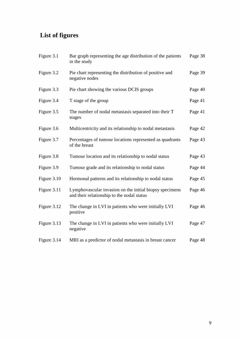
List of figures
Figure 3.1 Bar graph representing the age distribution of the patients in the study
Page 38
Figure 3.2 Pie chart representing the distribution of positive and negative nodes
Page 39
Figure 3.3 Pie chart showing the various DCIS groups
Page 40
Figure 3.4 T stage of the group
Page 41
Figure 3.5 The number of nodal metastasis separated into their T stages
Page 41
Figure 3.6 Multicentricity and its relationship to nodal metastasis
Page 42
Figure 3.7 Percentages of tumour locations represented as quadrants of the breast
Page 43
Figure 3.8 Tumour location and its relationship to nodal status
Page 43
Figure 3.9 Tumour grade and its relationship to nodal status
Page 44
Figure 3.10 Hormonal patterns and its relationship to nodal status
Page 45
Figure 3.11 Lymphovascular invasion on the initial biopsy specimens and their relationship to the nodal status
Page 46
Figure 3.12 The change in LVI in patients who were initially LVI positive
Page 46
Figure 3.13 The change in LVI in patients who were initially LVI negative
Page 47
Figure 3.14 MRI as a predictor of nodal metastasis in breast cancer Page 48

10
List of tables
Table 1.1 Features of malignant lymph nodes
Table 2.1 Flow Diagram showing the inclusion and exclusion
criteria
Page 26
Page 33
Table 4.1A comparison of ALND, SLNB and axillary ultrasound Page 54
11
Chapter 1
1.1 Literature Review
Breast cancer is a major public health care burden. Itis the most prevalent malignancy in
women worldwide and has surpassed cervical canceras the most prevalent female
malignancy in South Africa (1). Advances in awareness, screening and management
have led to a dramatic decrease in mortality from breast cancer(2). The transition to a
biological model with the understanding that breast cancer is a systemic disease has led
to more directed treatment. Anthracycline based chemotherapy is purported to have
reduced the mortality by 38%(3).
The predominant model for breast cancer in the 19th century proposed that breast
cancerwas a local phenomenon that spread contiguously along the chest wall then into
the lymphatic system and throughout the body, known as the mechanistic model. The
Halstedian radical mastectomy had this model in mind, affecting a cure by extensive
surgery - removing the breast along with its underlying muscles and including the
axillary structures(4). The associated severe morbidity was not unexpected and patients
carried the burden of their ‘cure’ for the rest of their lives(5). In the late 1960’s Fischer
suggested a biological model in which the disease outcome was based on the presence
of metastatic and micrometastatic disease leading to systemic spread(6). This ushered in
the modern management of breast cancer – local control of the breast and axilla with an
emphasis on systemic therapies to treat metastatic and micrometastatic disease
elsewhere in the body.
The Halstedianradical mastectomy was modified to a total mastectomy with an axillary
lymph node dissection. Surprisingly at the time, there was no change in mortality(6),
but a significant decrease in morbidity was apparent. The National Surgical Bowel and
Breast Project –B04 (NSABP –B04) was the first of the randomized breast cancer trials
that looked at these endpoints, supporting the biological theory of Fischer (7). The
NSABP –B04, confirmed that axillary nodal status was the basis for prognosis and that
systemic chemotherapy plays a significant role in the management of the disease(7).
12
The next evolutionary step in the management of breast cancer was to be more
surgically conservative with the breast and breast conservation techniques were applied.
Two large randomized control studies investigated breast conservation surgery
(lumpectomy) with irradiation, compared to a mastectomy. The results showed that
there was no difference in mortality, except the need for irradiation if a breast
conserving technique was employed(8,9).
As breast surgeries became less extensive, it was apparent that the axillary nodal status
was a key prognosticator of survival in early breast cancer. However, at the same time
advances in imaging techniques and screening programs leadto less advanced and
insitu(DCIS)tumours being detected. Consequently, the positive yield from axillary
nodal dissection was decreasing, with the majority of patients having a negative axillary
nodal dissection(10). Current statistics suggest that 60 – 70% of newly diagnosed
patients with breast cancer have negative axillarylymph nodes(10,11). An axillary
lymph node dissection(ALND)has associated complications such as lymphedema,
neuropathy, seroma and scarring (12,13). Therefore, decreasing the number of axillary
dissections would be beneficial to patients. This advantage provided the impetus for the
concept of a sentinel lymph node biopsy (SLNB).
A sentinel lymph node biopsy (SLNB) is a minimally invasive technique of axillary
lymph node sampling. It is based on the premise that breast malignancies spread in a
predictable manner to the axillary nodal basin, and the initial spread is to a node termed
the sentinel node(12). When compared to an axillary nodal dissection, a SLNB has a
95% sensitivity and an almost 100% specificity for nodal metastasis(13).
The rate of negative SLNBsin women with early breast cancer is 70 – 80%(14). In the
last decade, ultrasound of the axilla has been used to further stratify patients in order to
decrease the rate of positiveSLNBs by preselecting those patients that may proceed
directly to axillary nodal dissection. The detection of positive nodes has been further
enhanced by the use of a fine needle aspirate (FNA) ora core biopsy
technique(10,15).Due to the application of minimally invasive biopsy techniques to
exclude positive axillas, 70% of sentinel lymph nodes will be negative in patients with
early breast cancer (16).
13
In summary, there has been a revolution in the management of breast cancer within the
last century, from aggressive and debilitating surgeries to less invasive methods.
While substantial progress has been made to date, questions remain with regard to the
optimal management of the axilla in the modern management of breast cancer. The aim
of this study is to investigate the possibility of decreasing the rate of negative sentinel
node biopsies. Using a combination ofaxillary ultrasound findings, tumour
characteristics and additional imaging techniquesthe aim is to identify a cohort in whom
a ‘negative’ axilla can be safely predicted in a non-invasive manner. This would negate
the value of performing a SLNBin the identified cohort, thus taking a stride in the
advancement of treating breast cancer.
The breast cancer literature has not focused specifically on this question. In order to
better understand the subject, the literature review focused on the following subsections.
1. SLNBs: A comprehensive review on the subject was undertaken.
2. Imaging techniques of the axilla,and its value in predicting malignant lymph
node involvement.
3. Tumourcharacteristics and their value in predicting lymph node metastasis.
14
1.2 Sentinel lymph node biopsies
Sentinel lymph node biopsy has become the standard of care for patients with early
breast carcinomaand a clinically benign axilla. The first reported cases stem as far back
as 1994. It is a well recognized technique that has been validated by multiple
prospective randomized controlled studies. However, although it has been widely
accepted, there are many controversies that still exist. Within the aim of this study, if
aSLNB is intended to be replacedwith a non-invasive method, a thorough understanding
of the nuances of the technique is required. What follows is a detailed review of SLNBs.
1.2.1 Concept of sentinel lymph node biopsyand technique
The SLNB concept is an appealing one, and this in part is fuelled by its simplicity. The
‘sentinel nodes’ are the nodes that the tumour is thought to first spread to. The
procedure is facilitated by two techniques: a blue dye injection or by
lymphyscintigraphy aided by the use of a radioactive isotope and a gamma probe.
The term ‘sentinel node’ was coined by Gould et al in 1962 in describing a metastatic
lymph node of the parotid gland(19). His rationale for using the nodal histology as a
precursor to radical neck dissection was similar to the current concepts used in breast
cancer. The first critical use of a SLNB was by Cabanas in 1977. He observed that in
patients with penile cancer, if their ‘sentinel node’ was negative, their 5 year survival
was in excess of 90%. This is in comparison to patients with involved lymph nodes who
have a50%5-year survival (20). This led him to the conclusion that those patients with
a negative node did not require any further surgery. His study was elegant, however he
was erroneous in his presumption that the node was always in a fixed place(20).At the
same time, interest was mounting in the potential to avoid elective lymph node
dissection in intermediate thickness cutaneous melanoma. The pioneers of the technique
investigated patterns of nodal spread in melanomas of the back and around the
umbilicus, as the spread of melanomas in these regions wereknown to be variable. In
1977 cutaneous lymphoscintigraphy using vital blue dyes was used to map at risk nodal
basins in order to perform a more selective lymph node dissection. The technique was
then extrapolated to inguinal nodal dissection, with the deep system only requiring an
15
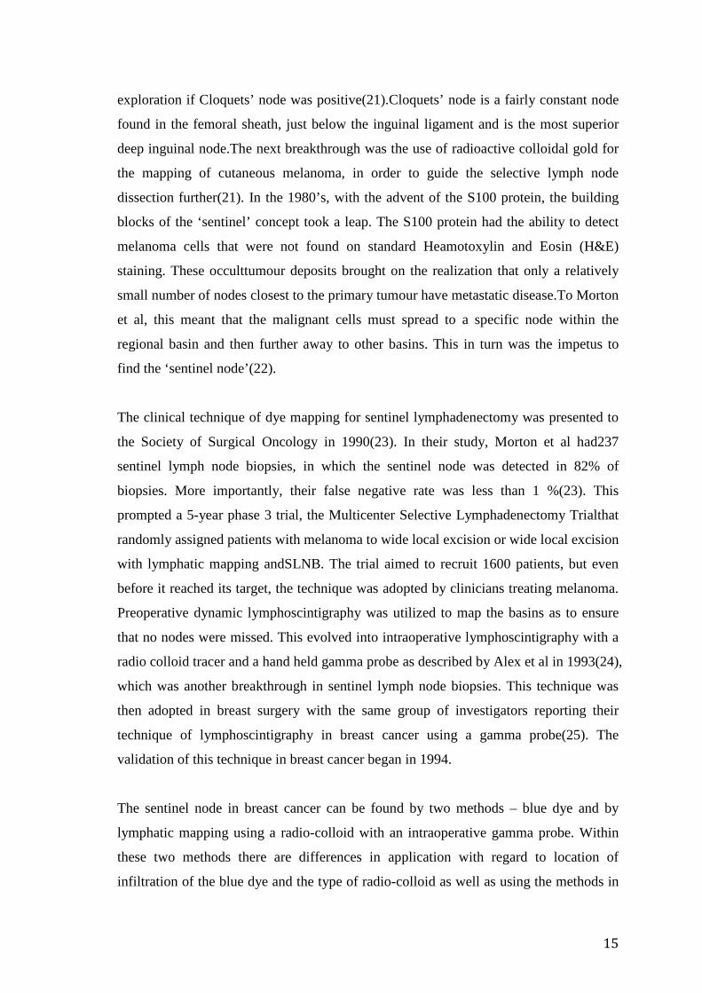
exploration if Cloquets’ node was positive(21).Cloquets’ node is a fairly constant node
found in the femoral sheath, just below the inguinal ligament and is the most superior
deep inguinal node.The next breakthrough was the use of radioactive colloidal gold for
the mapping of cutaneous melanoma, in order to guide the selective lymph node
dissection further(21). In the 1980’s, with the advent of the S100 protein, the building
blocks of the ‘sentinel’ concept took a leap. The S100 protein had the ability to detect
melanoma cells that were not found on standard Heamotoxylin and Eosin (H&E)
staining. These occulttumour deposits brought on the realization that only a relatively
small number of nodes closest to the primary tumour have metastatic disease.To Morton
et al, this meant that the malignant cells must spread to a specific node within the
regional basin and then further away to other basins. This in turn was the impetus to
find the ‘sentinel node’(22).
The clinical technique of dye mapping for sentinel lymphadenectomy was presented to
the Society of Surgical Oncology in 1990(23). In their study, Morton et al had237
sentinel lymph node biopsies, in which the sentinel node was detected in 82% of
biopsies. More importantly, their false negative rate was less than 1 %(23). This
prompted a 5-year phase 3 trial, the Multicenter Selective Lymphadenectomy Trialthat
randomly assigned patients with melanoma to wide local excision or wide local excision
with lymphatic mapping andSLNB. The trial aimed to recruit 1600 patients, but even
before it reached its target, the technique was adopted by clinicians treating melanoma.
Preoperative dynamic lymphoscintigraphy was utilized to map the basins as to ensure
that no nodes were missed. This evolved into intraoperative lymphoscintigraphy with a
radio colloid tracer and a hand held gamma probe as described by Alex et al in 1993(24),
which was another breakthrough in sentinel lymph node biopsies. This technique was
then adopted in breast surgery with the same group of investigators reporting their
technique of lymphoscintigraphy in breast cancer using a gamma probe(25). The
validation of this technique in breast cancer began in 1994.
The sentinel node in breast cancer can be found by two methods – blue dye and by
lymphatic mapping using a radio-colloid with an intraoperative gamma probe. Within
these two methods there are differences in application with regard to location of
infiltration of the blue dye and the type of radio-colloid as well as using the methods in

16
combination or alone. The underlying theme in these varying methods is the
understanding of the drainage patterns of the breast. Sappey et al described the
communication of the underlying lymphatics of the nipple areolar complex to the breast
parenchyma over 100 years ago – Sappey’ssubareolar lymphatic plexus. The
understanding was later extended to include the ipsilateral axillary nodes as the drainage
area of the breast parenchyma(26). In summary, lymphatic spread from the subareolar
plexus drains into the axillary lymph nodes.
The methods of dye/tracer infiltration are subareolarly, peritumorally with
intraparenchymal, intradermal or even subdermal injections. The gold standard was
thought to be intraparenchymalperitumoralinjection as it would reflect the drainage of
the individual tumour. However, when using radio-colloid in the upper outer quadrant
of the breast, the technique becomes more challenging as there is considerable ‘shine
through’ whilst using the gamma probe(27). Beitsch et al compared the drainage,as
described bySappey’ssubareolar plexus, using blue dye, withtheperitumoral injection of
radio-colloid. Their results showed a 94% detection rate of the sentinel node, and in this
there was a 99% concordance (blue and radioactive) between the dye and radio-
colloid(28). This concordance of subareolar blue dye and sulfur-colloid injected
peritumorally has been validated in other prospective studies as well(29). Subareolar
injection of dye is also an option in patients with non-palpable tumours. The combined
technique of peri-tumoralradio-colloid and subareolar blue dye or vise versa is a
technique commonly used whilst providing good results(27).
The literature is replete with conflicting studieswith regard to radio-colloid versus blue
dye or the combination of the two techniques. Kim et al published a meta-analysis on
the subject. They included 69 trials with 8059 patients. The results of this meta-analysis
favoured the combination of dye and colloid over either procedure alone(30). When the
combination is used, the false negative rate decreases to seven percent, as opposed to
10.9% with dye alone or 8.8% for radio-colloid alone (p = 0.0047)(30). This is the best
evidence available regarding the technique of dye or tracer infiltration and it favours a
combined technique. The learning curve of the procedure was highlighted in a
multicenter study using the combination technique, showing that the false negative rate
17
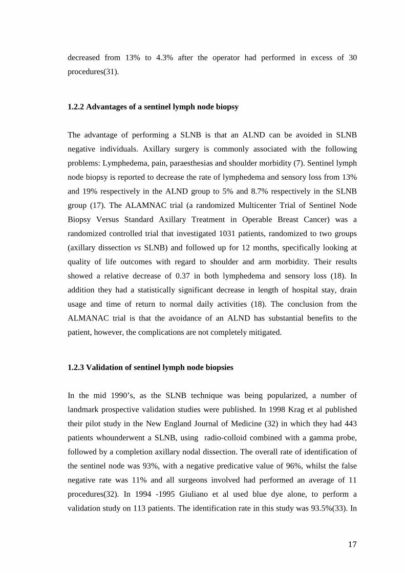
decreased from 13% to 4.3% after the operator had performed in excess of 30
procedures(31).
1.2.2 Advantages of a sentinel lymph node biopsy
The advantage of performing a SLNB is that an ALND can be avoided in SLNB
negative individuals. Axillary surgery is commonly associated with the following
problems: Lymphedema, pain, paraesthesias and shoulder morbidity (7). Sentinel lymph
node biopsy is reported to decrease the rate of lymphedema and sensory loss from 13%
and 19% respectively in the ALND group to 5% and 8.7% respectively in the SLNB
group (17). The ALAMNAC trial (a randomized Multicenter Trial of Sentinel Node
Biopsy Versus Standard Axillary Treatment in Operable Breast Cancer) was a
randomized controlled trial that investigated 1031 patients, randomized to two groups
(axillary dissection vs SLNB) and followed up for 12 months, specifically looking at
quality of life outcomes with regard to shoulder and arm morbidity. Their results
showed a relative decrease of 0.37 in both lymphedema and sensory loss (18). In
addition they had a statistically significant decrease in length of hospital stay, drain
usage and time of return to normal daily activities (18). The conclusion from the
ALMANAC trial is that the avoidance of an ALND has substantial benefits to the
patient, however, the complications are not completely mitigated.
1.2.3 Validation of sentinel lymph node biopsies
In the mid 1990’s, as the SLNB technique was being popularized, a number of
landmark prospective validation studies were published. In 1998 Krag et al published
their pilot study in the New England Journal of Medicine (32) in which they had 443
patients whounderwent a SLNB, using radio-colloid combined with a gamma probe,
followed by a completion axillary nodal dissection. The overall rate of identification of
the sentinel node was 93%, with a negative predicative value of 96%, whilst the false
negative rate was 11% and all surgeons involved had performed an average of 11
procedures(32). In 1994 -1995 Giuliano et al used blue dye alone, to perform a
validation study on 113 patients. The identification rate in this study was 93.5%(33). In
18
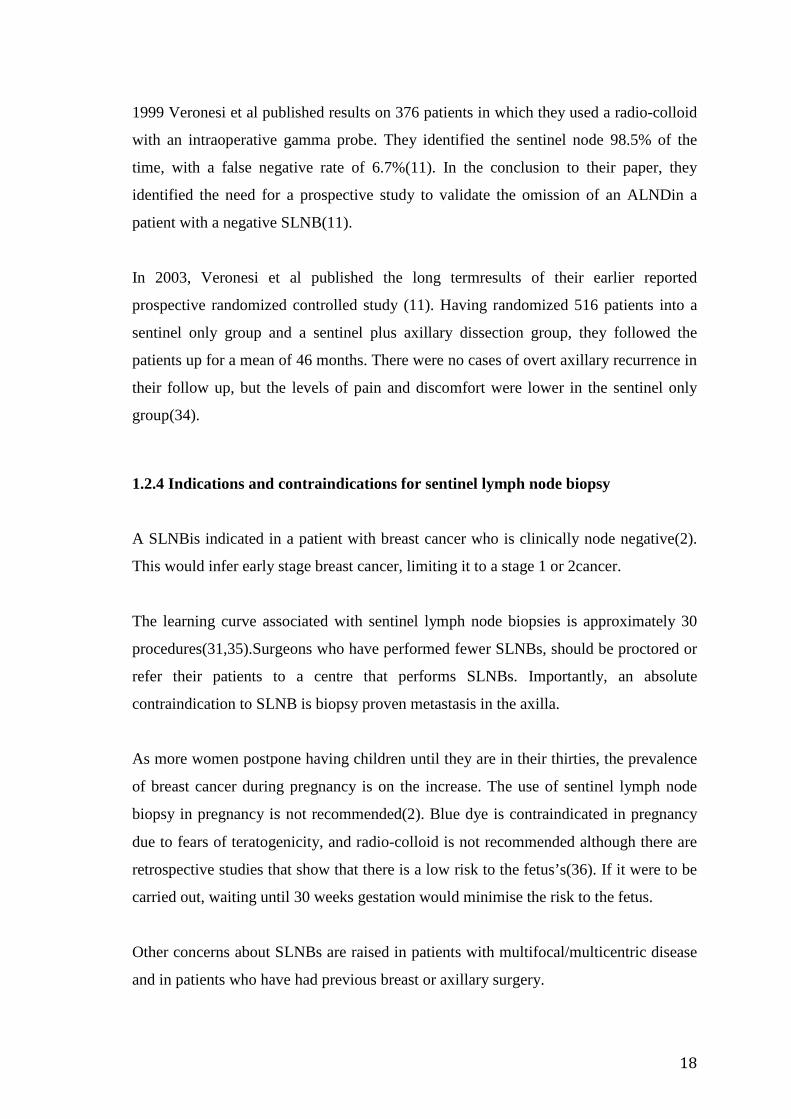
1999 Veronesi et al published results on 376 patients in which they used a radio-colloid
with an intraoperative gamma probe. They identified the sentinel node 98.5% of the
time, with a false negative rate of 6.7%(11). In the conclusion to their paper, they
identified the need for a prospective study to validate the omission of an ALNDin a
patient with a negative SLNB(11).
In 2003, Veronesi et al published the long termresults of their earlier reported
prospective randomized controlled study (11). Having randomized 516 patients into a
sentinel only group and a sentinel plus axillary dissection group, they followed the
patients up for a mean of 46 months. There were no cases of overt axillary recurrence in
their follow up, but the levels of pain and discomfort were lower in the sentinel only
group(34).
1.2.4 Indications and contraindications for sentinel lymph node biopsy
A SLNBis indicated in a patient with breast cancer who is clinically node negative(2).
This would infer early stage breast cancer, limiting it to a stage 1 or 2cancer.
The learning curve associated with sentinel lymph node biopsies is approximately 30
procedures(31,35).Surgeons who have performed fewer SLNBs, should be proctored or
refer their patients to a centre that performs SLNBs. Importantly, an absolute
contraindication to SLNB is biopsy proven metastasis in the axilla.
As more women postpone having children until they are in their thirties, the prevalence
of breast cancer during pregnancy is on the increase. The use of sentinel lymph node
biopsy in pregnancy is not recommended(2). Blue dye is contraindicated in pregnancy
due to fears of teratogenicity, and radio-colloid is not recommended although there are
retrospective studies that show that there is a low risk to the fetus’s(36). If it were to be
carried out, waiting until 30 weeks gestation would minimise the risk to the fetus.
Other concerns about SLNBs are raised in patients with multifocal/multicentric disease
and in patients who have had previous breast or axillary surgery.
19
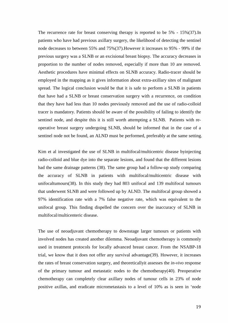
The recurrence rate for breast conserving therapy is reported to be 5% - 15%(37).In
patients who have had previous axillary surgery, the likelihood of detecting the sentinel
node decreases to between 55% and 75%(37).However it increases to 95% - 99% if the
previous surgery was a SLNB or an excisional breast biopsy. The accuracy decreases in
proportion to the number of nodes removed, especially if more than 10 are removed.
Aesthetic procedures have minimal effects on SLNB accuracy. Radio-tracer should be
employed in the mapping as it gives information about extra-axillary sites of malignant
spread. The logical conclusion would be that it is safe to perform a SLNB in patients
that have had a SLNB or breast conservation surgery with a recurrence, on condition
that they have had less than 10 nodes previously removed and the use of radio-colloid
tracer is mandatory. Patients should be aware of the possibility of failing to identify the
sentinel node, and despite this it is still worth attempting a SLNB. Patients with re-
operative breast surgery undergoing SLNB, should be informed that in the case of a
sentinel node not be found, an ALND must be performed, preferably at the same setting.
Kim et al investigated the use of SLNB in multifocal/multicentric disease byinjecting
radio-colloid and blue dye into the separate lesions, and found that the different lesions
had the same drainage patterns (38). The same group had a follow-up study comparing
the accuracy of SLNB in patients with multifocal/multicentric disease with
unifocaltumours(38). In this study they had 803 unifocal and 139 multifocal tumours
that underwent SLNB and were followed up by ALND. The multifocal group showed a
97% identification rate with a 7% false negative rate, which was equivalent to the
unifocal group. This finding dispelled the concern over the inaccuracy of SLNB in
multifocal/multicenteric disease.
The use of neoadjuvant chemotherapy to downstage larger tumours or patients with
involved nodes has created another dilemma. Neoadjuvant chemotherapy is commonly
used in treatment protocols for locally advanced breast cancer. From the NSABP-18
trial, we know that it does not offer any survival advantage(39). However, it increases
the rates of breast conservation surgery, and theoreticallyit assesses the in-vivo response
of the primary tumour and metastatic nodes to the chemotherapy(40). Preoperative
chemotherapy can completely clear axillary nodes of tumour cells in 23% of node
positive axillas, and eradicate micrometastasis to a level of 10% as is seen in ‘node
20
negative disease’ (41). However, does preoperative chemotherapy have any bearing on
the sentinel lymph node status? Some of the reasons given as to why pre-operative
chemotherapy might be a problem in assessing the true nodal status of patients, is
excessive fibrosis of the lymphatics caused by chemotherapy, a blockage of the
lymphatic channels by malignant debris, and the fact that patients given neoadjuvant
chemotherapy are more likely to have involved nodes(42). A prospective multicenter
trial is currently underway in an attempt to answer this question. Indeed, the ACOSOG
Z1071 trial will evaluate the effect of chemotherapy on biopsy proven axillary nodal
metastasis. The primary objective is to determine the false negative rate post
preoperative chemotherapy. There are reports of smaller published trials which also
investigated this problem to find an answer to this question. For example, Classe et al
enrolled 192 patients over 4 years in 12 centres, evaluating preoperative chemotherapy
on rates of SLNB detection. The conclusion of this study was that the results obtained
were similar to those inearly breast cancer. The detection rate was 90% and the false
negative rate was 11.5%(43). Furthermore, Kuerer et al published a review article
involving all available studies that assessed SLNB with neoadjuvant therapy. The
resultant identification rate was 83 – 100% in the 12 studies assessed and the conclusion
was that these results did not differ from SLNB without preoperative chemotherapy(42).
The German SENTINA (SENTInelNeoAdjuvant) trial of the AGO-B
(ArbeitsgemeinschaftgynäkologischeOnkologie) is a prospective multicenter study that
examines the role of SLNB in the neodjuvant setting. More than 1500 patients will be
included. This trial will also help answer this controversy.
In summary, the only contraindications to SLNB are biopsy proven metastasis or a
clinically positive axilla. The relative contraindications are pregnancy and previous
breast (not aesthetic) or axillary surgery. Importantly, multicentric/multifocal disease is
not a contraindication and in patients who have had neoadjuvant chemotherapy SLNB
appears safe, however we await the results of the ACOSOG Z1071 trial and the German
SENTINA study.
21
1.2.5 Ductal carcinomainsitu and sentinel lymph node biopsies
Controversy remains with regard to the indications for SLNB in patients with ductal
carcinoma insitu (DCIS). Ductal carcinoma insituis defined as a neoplasm that has no
histological features of invasion. It is commonly diagnosed on a core biopsy sample of
the breast, and a treatment plan is formulated on these results.Alarmingly, population
based screening programs have resulted in an exponential increase in the number of
patients with DCIS. The incidence in the USA has increased from 5.8/100000 in 1975to
32/100000 in 2005(44). Pure DCIS has a reported rate of axillary metastasis of between
2% - 13%(45,46). However, core needle biopsies which report DCIS, have invasive
cancer in the specimens in 8 – 38% of cases(47-49). Core needle biopsy has obvious
advantages over an excisional biopsy but due to the small area sampled, an invasive
malignancy may be missed. Ansari et al included 22 studies in their meta-analysis and
they found that 7.6% of patients with DCIS on biopsy specimens harboured axillary
metastasis. Their conclusion was that it was reasonable to offer a SLNB to patients with
DCIS (50).
Moreover, Goyal et al performed a retrospective review of 587 patients with DCIS.
Their invasive malignancy rate result on final histology was 38% (51). Multivariate
analysis revealed that a mass, on clinical examination or imaging, was the best predictor
of invasive disease(51). Of the patients that had an axillary examination, 13.6% had
metastasis – all of these patients had invasive disease. In another retrospective study of
110 patients, invasive disease was associated with high grade DCIS on core biopsy in
93.6% of the patients(52). This however is not a consistent finding, and invasion is
often associated with intermediate grade DCIS. Predictors of both a positive and
negative axilla are inconsistent in DCIS. The general consensus is that a SLNB is
required in DCIS if the clinical suspicion for invasive malignancy is high, the patient
has a mass or high grade DCIS or if a mastectomy is being performed.If a SLNB is not
performed during a mastectomy or lumpectomy for DCIS, and on final histology the
specimenshows invasive cancer, axillary staging will subsequently be required.
22
1.2.6 Controversies in sentinel lymph node biopsy
1.2.6.1 Micrometastasis and isolatedtumour cells
The significance of micrometastatic disease in breast cancer remains a controversy as its
clinical relevance is not fully elucidated. The current classification defines axillary
metastasis into three categories: as macrometastasis (> 2.0mm), micrometastasis
(0.2mm - 2.0 mm) and isolated tumour cells (ITC) (0.01 – 0.2mm) (2). Isolated tumour
cells are stained by immunohistochemistry only and are not visible on light microscopy.
The average harvest from a SLNB is 1.5 nodes as opposed to more than 10 nodes in an
ALND. As there are fewer nodes to examine, thishas increased the scrutinyofthe lymph
nodes and hence, thedetection of micrometastasishas increased. Immunohistochemistry
has aided in detecting metastasis which are less than 2mm in size,and these small
volume metastasis have led to intense debate.
In an effort to quantify the false negative rate of SLNB using standard light microscopy
with 0.2mm specimen slices, Weaver et al undertook a study where, they re-examined
the negative specimens from the NSABP-32 study(53). Specimens were sliced, from the
original 0.2mm slices, into 0.1mm and 0.05mm slices and were examined under light
microscopy. They wereaided by a computer assisted cell counter to examine the
specimens and found that 64 (27%) of the 236 samples showed metastasis, thirty were
detected by light microscopy alone and 34 were detected by the cell counter.
Twentyfiveof themetastasis were less than 0.03mm, and 49 were less than 0.1mm.
In their follow-up study the significance of micrometastases, and its impact on overall
survival was evaluated. Weaver et al in 2011published a study analysing 3887 patients,
with negative sentinel nodes. Women with breast cancer were randomly assigned to a
SLNB and ALND, or SLNB alone. The negative specimens were subjectedto further
sectioning and immunohistochemistry, resulting in a 15.9% rate of occult metastasis.
The results were separated into two groups - a completion ALND or no ALND (SLNB
23
only). The 5-year overall survival (OS) difference was small at 1.2%. The survival was
94.6%(ALND) and 95.8%(SLNB).However, this was statistically significant (p= 0.03).
There were also significant differences between disease free survival and distant disease
free survival, but again the absolute numbers were small. The recommendation was that
although there are differences in overall survival, further analysis of all negative nodes
was not recommended (54).
In contrast, the ACOSOG Z0010 trial, the MIRROR trial and in a review by Billamoria
et al, showed that there was no statically significant difference in overall survival
between a completion ALND and SLNB alone in patients with
micrometastsis(35,81,82).However it was noted that the majority of patients received
adjuvant systemic therapy and that it seemed to improve their 5-year disease free
survival. One would make the assumption that micrometastasis are smaller metastasis
and will grow and spread, however the literature does not support this finding. Indeed,
The NCCN guideline on breast cancer recommends basing treatment decisions on H&E
staining only and routine immunohistochemistry is cautioned against(2).
In summary, although micrometastatic disease is easily missed on routine H&E staining,
the presence of micrometastatic disease does not seem to have an impact on the overall
survival especially when adjuvant systemic adjuvant therapy is administered. With
reference to this study, it is important, as a clear limitation of ultrasonography would be
the detection of micrometastatic disease, however if it were not clinically relevant then,
this limitation would be minimised.
24
1.2.1.6 Isaxillary dissection necessary after a positive SLNB?
This is currently the most controversial topic in breast cancer and is also the most
relevant to our study. The ACOSOG Z0011 is a recently published prospective non-
inferiority trial(56) wherethe aim was to assess if SLNB alone was non-inferior to
completion ALND in T1 (< 2cm) and T2 (2 – 5cm) patients with positive SLNBs. All
patients had breast conservation therapy combined with whole breast irradiation (not
axillary radiation). The trial recruited patients from 1999 – 2004 and was terminated
prematurely due to poor accrual.
The results were surprising.No difference was found in local or regional recurrence
rates and the 5-year overall survival was 91.8% in the ALND group and 92.5% in the
SLNB alone group. The conclusion was that it was safe to omit an ALND in patients
with less than three nodes positive on SLNB, in T1 and T2 tumours receiving whole
breast irradiation. However, the criticism of this trial is that it was underpowered to
answer the question, and we await further evidence to support its findings.
Of note was that 97 of 355 (27.3%) patients in the ALND group had further metastasis
on ALND. Extrapolating this incidence to the SLNB group, an assumption can be made
on the number of metastatic nodes not operated on in the SLNB group. Importantly,
96% of patients in the ALND and 97% in the SLNB only group received anthracycline-
based chemotherapy(57). The chemotherapy decisions were based on the initial tumour
biology and characteristics and not on the SLNB findings. This confirms the assumption
that irradiation combined with systemic therapy, controlsthe axillary metastasis left
behind after a SLNB. So in summary, there is thus evidence for not proceeding to an
ALND in well selected patients with a positive SLNB and who are to receive
anthracycline based chemotherapy as well as whole breast irradiation.
25
1.2.1.6What is the role of intraoperative frozen section analysis of sentinel lymph
node biopsy?
There are two strategies for dealing with the staging of the axilla. The first is
performing an upfront SLNB, and the second is combining it with the definitive surgery.
The advantage of combining the procedures is the prevention of a second general
anaesthetic with its incumbent morbidity to the patient, but the disadvantage of frozen
section is the potential damage to the tissue during sectioning, rendering it inadequate
for H&E staining. Combining the procedures makes practical sense in a patient who
does not requirea complicated onco-plastic reconstruction or in a patient undergoing a
definitive mastectomy.However, the underlying question that remains is: - are the
results reliable and does the risk of a second procedure justify the false negative rate of
intraoperative frozen section?
The reported accuracy of frozen section is 90% - 93.3%, with a false negative rate of
28.4%(58,59). The converse is that the sensitivity is 66% - 71.6%, and these patients are
spared a second operation. Although the false negative rate might appear to be high,
only 26% of the false negative patients had macro-metastasis. The overwhelming
majority hadmicrometastatic disease – decreasing the false negative rate to an
acceptable 7% (59). This highlights the importance of micrometastatic disease and its
treatment.
In conclusion, frozen section is a valid technique for decreasing the morbidity of a
second anaesthetic, and is accurate in 90% of cases. The associated high false negative
rate represents small volume metastasis in the majority of patients and a SLNB alone, is
the current treatment for these micrometastasis.
1.3 Ultrasound and its use in the axilla
The use of ultrasound in the evaluation of a patient with breast cancer has become
commonplace. The reported sensitivity of clinical examination for detecting a negative
axilla is reported to be 38%(66) and patients with a clinically negative axilla are
candidates for a SLNB. In an effort to streamline the management algorithm, ultrasound
26
of the axilla with a FNA is being used to identify positive nodes, and patients with FNA
proven metastasis can proceed directly to an ALND or neo-adjuvant chemotherapy.
A high frequency probe is required to perform an axillary ultrasound. Close attention
should be paid to inferior aspect of the axilla below the insertion of pectoralis minor, as
this is a common place for the sentinel node to be found(60).
Lymph nodes have ultrasonographic features that suggest malignant infiltration.In a
normal node, the hilum is hyper-echoic and the cortex is hypo-echoic. Anatomically,
lymphatic channels enter lymph nodes at the peripheral cortex and the blood supply is
derived from the hilum of the node. Normal lymph nodes have the following features:
1. Oval in shape
2. Fatty hilum
3. Thin peripheral cortex(61)
As the metastatic cells infiltrate the node from the lymphatics into the cortex, the cortex
gets thicker and the hilum proportionately smaller, until it is entirely replaced by
malignancy. Ultrasonographically, the initial features are a thickened cortex with a
normal hilum. The cortex thickens, the node loses its oval shape as the hilum is
displaced, and the entire node is replaced by metastatic deposits(62). The hypo-echoic
cortex gradually fills the entire node(63).
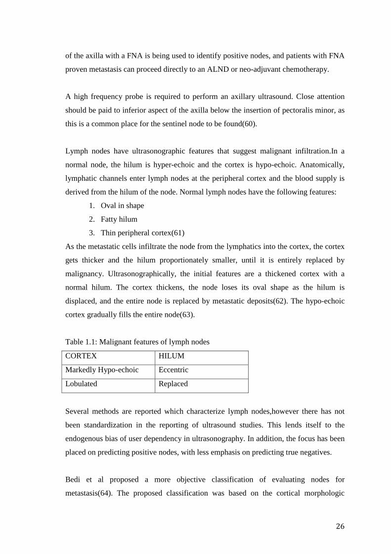
Table 1.1: Malignant features of lymph nodes
CORTEX HILUM
Markedly Hypo-echoic Eccentric
Lobulated Replaced
Several methods are reported which characterize lymph nodes,however there has not
been standardization in the reporting of ultrasound studies. This lends itself to the
endogenous bias of user dependency in ultrasonography. In addition, the focus has been
placed on predicting positive nodes, with less emphasis on predicting true negatives.
Bedi et al proposed a more objective classification of evaluating nodes for
metastasis(64). The proposed classification was based on the cortical morphologic
27
features of lymph nodes on ultrasound. In their study, inter-observer correlation of
positive or negative nodes was 88%. Their classification consisted of Types 1 – 6.
Types 1 – 4 were classified as benign, ranging from hypo-echoic with no visible cortex,
to thickened hypo-echoic generalized lobulation. Types 5 and 6 were malignant.
Combining a FNA with ultrasound can reduce the SLNB rate by 14%(60). Of interest
to my current study, the negative predictive value was 96%, and this increased if Type 4
was excluded. Maximum cortical thickness and ‘appearance of cortex’ are the best
predictors of nodal metastasis (60).
The accuracy of ultrasound of the axilla is difficult to ascertain due to the non-
standardized manner in which reporting takes place. Alvarez et al conducted a
systematic review on the subject, and their findings were very varied and
heterogeneous(65). Within the prediction of positivity using the sonographically
measured size of the lymph node, the sensitivity ranged between 48.8% and 87.1% and
the specificity was between 55.6% and 97.3%. Using morphological criteria for
positivity, the sensitivity was 26.4% to 75.9% and specificity was 88.4% to 98.1%.
Unfortunately, they did not investigate the accuracy of sonography in predicting a
negative axilla.
Nori et al published results of 147 women with breast cancer, in whom ultrasound was
combined with core biopsy if the node was suspicious for malignancy. The sensitivity
and positive predictive value in this study was 45.2% and 63.1% respectively. The
negative predictive value was 77.2% (78/101). Out of these 78 patients, 23 were
positive on histology (false negative). Twenty out of the 23 had micrometastases only,
and only 1/23 had more than 3 nodes involved(66). We can thus say that ultrasound
cannot accurately detect micrometastasis, however a negative ultrasound appears to
accurately exclude metastasis involving less than 3 nodes. With respect to the inability
to predict micrometastasis, this is probably insignificant, as micrometastasis do not
appear to have a clinical significance.These are important results, as they highlight the
question of the need for a SLNB in the face of a well performed negative ultrasound.
In the largest study published on this subject, 105 out of 398 patients with a negative
axillary ultrasound had positive nodes on SLNB(67). This is a negative predictive value
28
of 79%. Of the patients who had a positive SLNB and subsequent ALND, 59% had only
the sentinel involved and none had more than 3 nodes involved(67). Even though the
criteria for negative nodes were not standardized, the negative predictive value of 79%is
acceptable. Furthermore, in the ultrasound negative axilla, it is unlikely that more than 2
nodes will be positive. In addition, in another published series,Ragupathy et al showed a
sensitivity of 86% for predicting negative nodes(68).
To summarise the role of ultrasonography in detecting nodal metastasis from the above,
we can conclude that:
1. A comparison of the literature is problematic as there is no standard method of
reporting findings
2. The sensitivity of detecting positive nodes ranges only between 45% - 85%, and
when combined with FNA/Core biopsy, it improves the specificity of a positive
cytology to 100%.
3. The negative predictive value is underutilised. It predicts a negative axilla on
average 75% of the time, and when it is falsely negative the tumour deposits are
invariably micrometastasis, or involve less than three nodes.
1.4 Predictors of lymph node metastasis in breast cancer
The management of breast cancer has evolved.Individual characteristics of a tumour
prognosticate their behavior, aiding in the determination ofthe need for adjuvant therapy
fortumours with borderline indications, bringing us closer to individualizing cancer
treatment. The ability to predict lymph node metastasis on the basis of the primary
tumour characteristics would allow us to tailor treatment systemically and locally to the
axilla.
Patani et al conducted a systematic review on the topic and classified possible
predictors into the following categories: - clinical, radiological, pathological and
molecular(5). Ultimately the conclusion was that there was insufficient evidence to
recommend any specific marker to be used for the prediction of axillary lymph node
metastasis(5). Clinical parameters assessed in thestudy were age and palpability of the
primary tumour. The hypothesis was that younger age and palpability of tumour were
29
associated with lymph node metastasis. These lacked any sensitivity across the
spectrum of age categories(5). Of interest to this study is that the combination of an
ultrasound negative axilla together with other predictors has not specifically been
looked at.
The main pathological characteristics are tumour size, tumour grade,
multifocality/multicentricity, hormone receptor status and lymphovascular invasion.
With regard to these parameters, a size of less than 10mm correlates with a 10% chance
of having axillary metastasis, and a tumour of less than 25mm has a 15% chance of
carrying axillary metastasis(69), however,used in isolation this isa clinically unreliable
parameter as even small tumours can have extensive metastasis.
In breast cancer the tumour grade is commonly reported by using the modified Bloom
and Richardson scale (78). It includes a measure of nuclear pleomorphism, cellular
proliferation and tissue differentiation, and grades it out of a total score of nine. There is
a well recognized correlation between tumour grade and lymph node metastasis,
however, it does not have the sensitivity to be accurateenough(70). The Ki67 index
which is a measure of the mitotic activity, is an independent predictor of lymph node
metastasis(71). Histologically the following sub-types of ductal carcinoma have been
associated with fewer lymph node metastasis – Tubular, Mucinous, Cribriform,
Medullary(72).
The presence of a multifocal/multicentrictumour has been shown to be predictive of
axillary nodal metastasis(73). Lymphovascular invasion has also been associated with
lymph node involvement and in their study, Viale et al showed a 5.1 fold increase in
lymph node metastasis when lymphovascular invasion (LVI) was present(72). D2-40
(Podoplanin) is a pathological stain and a marker of lymphangiogenesis. When positive
it is widely used as a surrogate for lymphovascular invasion (78).
Hormone receptor status (Er/Pr) has not been identified as a specific marker of lymph
node involvement. Her-2 (Human epidermal growth factor 2) over expression has been
associated with a more aggressive type of breast cancer, however, over expression has
30
also not consistently being found to be associated with increased lymph node metastasis
(80).
The location of the tumour in relation to the breast has a bearing on lymph node
metastasis. In a prospective study of 135 patients by Susini et al specifically included
axillary ultrasound into their algorithm. A Ki67 > 10%, a positive ultrasound and a
tumour in the right upper quadrant (RUQ) were consistently associated with nodal
metastasis. Conversely, inner quadrant tumours with a Ki67 < 10% and a negative
ultrasound predicted negative nodes(74).
The breast research group at Memorial-Sloan Kettering recognized the factors alluded
to above as risk factors for metastasis, but also understood the cumbersome nature of
applying them within a clinical context(75). They included 3786 patients in their
database and developed a nomogram to predict nodal metastasis. Multivariate analysis
showed a statistical significance for the following variables: histology, size,
multifocality/multicentricity, location, and ER/PR status. Thedeveloped nomogramwas
adapted for use on smartphones and personal computers making it clinically applicable
(www.mskcc.org/nomograms).Although admitting that their nomogram is not perfect, it
can be used as a tool to educate patients on their risk of nodal metastasis (75).
31
1.5 Summary of the literature review
Significant strides have been taken in the management of breast cancer over the last
century. A level twoALND remains the gold standard for staging an axilla. However,
there is a clear benefit to a SLNBand there has been a shift towards performing SLNBs
in early breast cancer. In this literature review we have taken a journey through the
history, have examined the validation studies as well as evaluated the evidence for the
indications, the controversies and thecomplexities of this technique. A SLNB has been
proven to be equivalent to an ALND in the appropriately selected patient.
However, as shown, the majority of SLNBs (70%) are negative (14 – 16). This is the
motivation for this study. Can we safely predict which axilla will be free of metastasis
using non-invasive means?
Included in the literature search is a review on the utility of ultrasonography in staging
an axilla. Much of the focus in the literature has been on predicting positive nodes, and
this has been met with mixed success. However, the sensitivity for predicting a negative
axilla approaches 75% (66). This is not accurate enough to replace a SLNB. So, the data
was scrutinised, focusing on the false negatives within the cohort of Nori et al and it
was discovered that the majority of false negatives were patients with micrometastatic
disease. An ultrasound will never be able to detect micrometastatic disease, but
examining the literature led me to question the significance of micrometastatic disease.
I discovered that an ALND is not mandated after finding micrometastatic diseasein an
axilla(35, 81, 82), and this is counter-intuitive. Purported reasons for this are the use of
chemotherapy and irradiation to the breast. Also, from the literature it became apparent
that there was no widely adopted consensus on what constituted a malignant or benign
node, highlighting the user dependency and thus fallibility of ultrasound as a screening
tool.
Other predictors of axillary lymph node spread that are readily available were reviewed,
so as to determine if they could be combined with ultrasonography, to better predict a
negative axilla. Again, the results were mixed, with most studies concentrating on
predicting positive nodes, and not negative ones.
32
Frommy review of the literature, I conclude that a SLNB is currently the best method of
staging an axilla, however, there are a significant number of negative examinations. An
ultrasound in combination with other markers would be the most appropriate method of
staging an axilla non-invasively, but in order to achieve this we would need to
understand and overcome the shortcomings of an ultrasound study, notably its inability
to detect micrometatstatic disease and the lack of standardization in the reporting of
studies. In an effort to decrease these negative examinations, this is an important
preliminary study to conduct.
33
Chapter 2
2.1 Methods and measures
This is a retrospective review of patients who underwent a SLNB between December
2008 and November 2009.The University of Witwatersrand ethics committee granted
ethics approval for the study (M090435/2009). All SLNBs performed between
December 2008 and November 2009 were screened for eligibility.
The inclusion criteria were all patientsthat: -
- Had a documented negativeaxillary ultrasound (no suspicious nodes) or
- A negative FNA/core biopsy if one was carried out, and
- Fulfilled the criteria for a SLNB as per the indications proposed in the
NCCN breast cancer guidelines(2)
o Early breast cancer and
o Clinically negative nodes or
o A negative FNA/core biopsy of suspicious nodes
The exclusion criteria were: -
- Advanced breast cancer
- Patients without an axillary ultrasound report
- Inadequate documentation of findings on ultrasound
- Patients with an ultrasound report suspecting a metastatic node
- Recurrent breast cancer
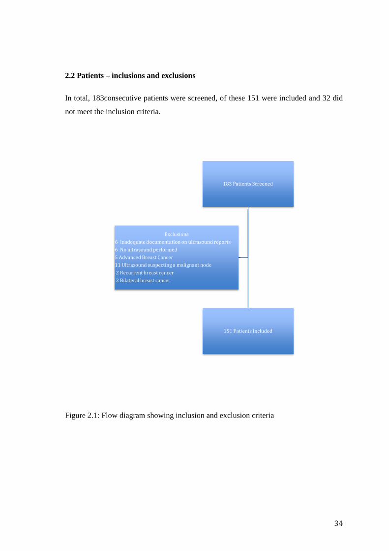
2.2 Patients – inclusions and exclusions
In total, 183consecutive
not meet the inclusion criteria
Figure 2.1: Flow diagram showing inclusion and exclusion criteria
6 Inadequate documentation on ultrasound reports
6 No ultrasound performed
5 Advanced Breast Cancer
11 Ultrasound suspecting a malignant node
2 Recurrent breast cancer
2 Bilateral breast cancer
nclusions and exclusions
consecutive patients were screened, of these 151 were included and 32
not meet the inclusion criteria.
Figure 2.1: Flow diagram showing inclusion and exclusion criteria
183 Patients Screened
151 Patients Included
Exclusions
6 Inadequate documentation on ultrasound reports
6 No ultrasound performed
5 Advanced Breast Cancer
11 Ultrasound suspecting a malignant node
2 Recurrent breast cancer
2 Bilateral breast cancer
34
f these 151 were included and 32 did
35
2.3Tumourcharacteristics
The following characteristics of the patients and their tumours were documented on a
Microsoft Excel (2011) spreadsheet.
The demographics of the patients, their age and race were captured.
I used the initial tumour biopsy specimen results to document the following: -
histological type (ductal, lobular, DCIS), tumour grade, hormonal receptor
status(Er/Pr/Her2-neu), lymphovascular invasion. The initial biopsy was used, as it
represents the information available before a SLNB is preformed, and thus is more
relevant than the final tumour histology when aiming to predict axillary metastasis. For
the purposes of comparison I grouped the receptor status of patients into Luminal A,
Luminal B, Triple negative and Her2-neu. A similar classification was initially
proposed by Perou et al in 2000 (76). Triple negative was used as the broader term to
encompass basal-like tumours as we could not specifically test for the basal phenotype.
To calculate the size, location and T-stage, the patient’sultrasound reports were used.
The ultrasound reports were recorded as being normal or reactive. A reactive node is a
histological change in a benign node in response to anon-malignant stimulus, however it
is a histological diagnosis and cannot be determined on ultrasound.Interpreting the
ultrasound reports with this in mind, the assumption made was that reactive nodes were
benign. As ultrasonography is user dependent, I thought it important to document the
ultrasonographers identity.
The multicentricity/multifocality of a tumour was determined primarily on the
mammogram findings, however a subset of patients had an MRI performed as well, and
some had multicentricity/multifocality diagnosed on MRI.
The number of sentinel nodes harvested and the presence of metastasis were
documented. This was further subdivided into macro and micrometastasis. The patients
with micrometastasis were further separated into those that had isolated tumour cells
only.
36
When available, the final histology specimens were used to record the following
variables: the size, histology, hormonal characteristics, lymphovascular invasion and
axillary lymph node histology and nodal yield. The aim was to compare the final
histology characteristics to the initial biopsy specimen, to gain an indication of the
reliability of initial specimens.
Statistica version 9 was used to analyse the collected data. Univariate analysis was used
to detect the significance of prognostic factors in predicting lymph node metastasis. P-
values were calculated using both Pearson and M-L Chi squared tests for each set of
recorded variables.
2.4Surgical practice
The study was conducted at the Netcare-Milpark Breast Centre wheremanagement
decisions are made by a multidisciplinary team that is lead by a specialist surgeon
whose area of interest is the breast. She runsa referral based hospital practice in
Johannesburg that deals exclusively with breast health care.
2.5 Multidisciplinary involvement
Although there is a radiologist within the multidisciplinary team at the Netcare-Milpark
hospital breast care centre, the majority of referrals are from other radiology practices.
Thus, the reporting of ultrasounds is not standardised. The pathologist in the multi-
disciplinary team is responsible for reporting on all SLNB specimens, but outside
referraltumour biopsiesare reported by various pathology laboratories around the greater
Johannesburg area. It is not economically feasible to repeat all imaging and biopsy
reporting.

37
2.6 Method and technique of sentinel lymph node biopsy
A single surgeon with the relevant expertise performed all the SLNBs at the Netcare-
Milpark breast care centre. The unit’s policy is to perform all SLNBs as a separate
procedure using H&E staining as well as immunohistochemistry, in order to identify
micrometastatic disease and isolated tumour cells. Patients with positive nodes had a
completion axillary lymph node dissection as a separate procedure, combined with their
definitive oncological breast procedure.
The SLNBs are performed in accordance with standard recommendations. A combined
dye and radio-colloid technique is employed. The 99mTc-labelled nano-colloid is
injected via the peritumoral route in the nuclear medicine department within the hospital.
Whenever possible, the patients are injected with 99mTc-labelled nano-colloid on the
morning of the operation, and their procedures are carried out in the afternoon.
In theatre, prior to the surgeon scrubbing, Patent V blue dye is infiltrated subareolarly.
Intraoperatively an axillary crease approach is used, and a gamma camera is used to
detect the 99mTc-labelled nano-colloid. All blue nodes, as well as all nodes with a
reading greater than 10% of the maximal nodal reading on the gamma camera are
removed.
Chapeter3.
Results
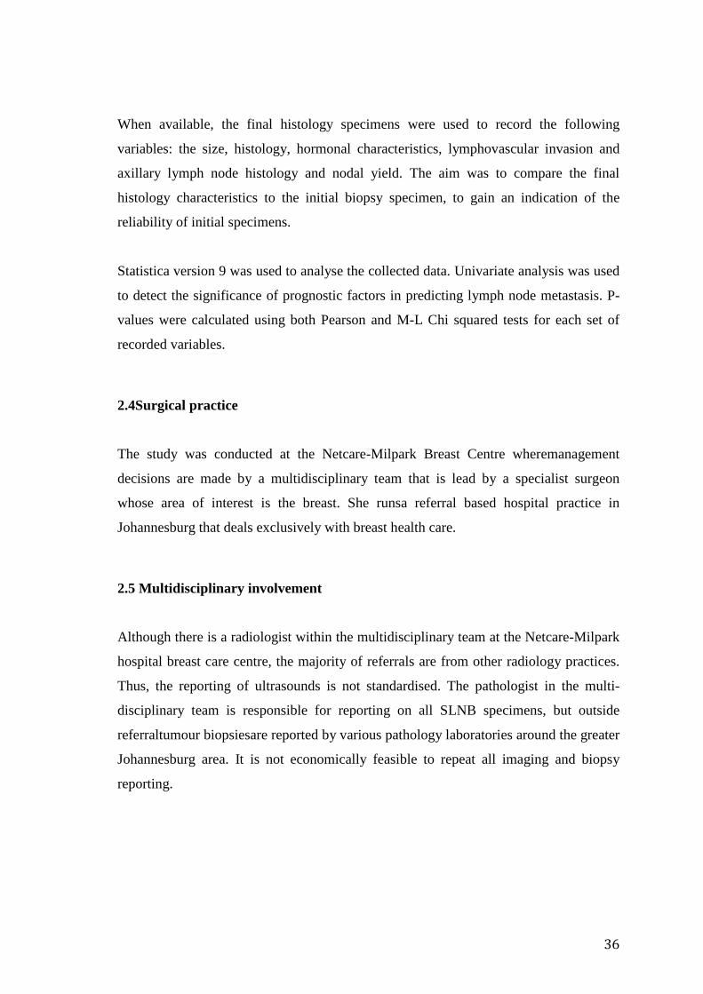
3.1 Demographics
The average age of presentation of the group was 53.3 years (27
being between 40 and 70 years old. The predominant ethnicity
Caucasian (87.6%).
Figure 3.1: Bar graph representing the age distribution of the patients in the
study(N=151)
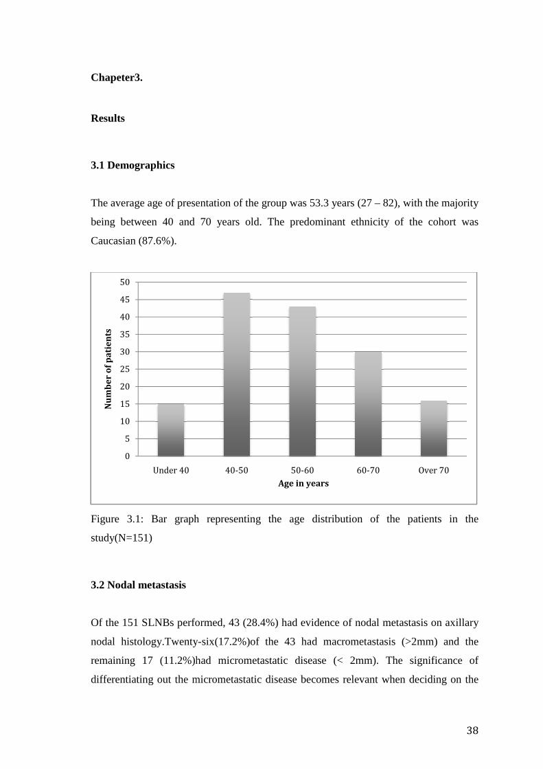
3.2 Nodal metastasis
Of the 151 SLNBs performed
nodal histology.Twenty
remaining 17 (11.2%)had
differentiating out the micrometastatic disease becomes relevant when deciding on the
0
5
10
15
20
25
30
35
40
45
50
Under 40
Nu
mb
er
of
pa
tie
nts
The average age of presentation of the group was 53.3 years (27 – 82), with the majority
being between 40 and 70 years old. The predominant ethnicity
Figure 3.1: Bar graph representing the age distribution of the patients in the
performed, 43 (28.4%) had evidence of nodal metastasis
Twenty-six(17.2%)of the 43 had macrometastasis (>2mm) and the
remaining 17 (11.2%)had micrometastatic disease (< 2mm). The
differentiating out the micrometastatic disease becomes relevant when deciding on the
40-50 50-60 60-70
Age in years
38
82), with the majority
being between 40 and 70 years old. The predominant ethnicity of the cohort was
Figure 3.1: Bar graph representing the age distribution of the patients in the
d evidence of nodal metastasis on axillary
asis (>2mm) and the
The significance of
differentiating out the micrometastatic disease becomes relevant when deciding on the
Over 70
management strategy. If we trea
brings the negative predictive value of an ultrasound to
Figure 3.2: Pie chart representing the distribution of positive and negative nodes as
percentages (n = 151)
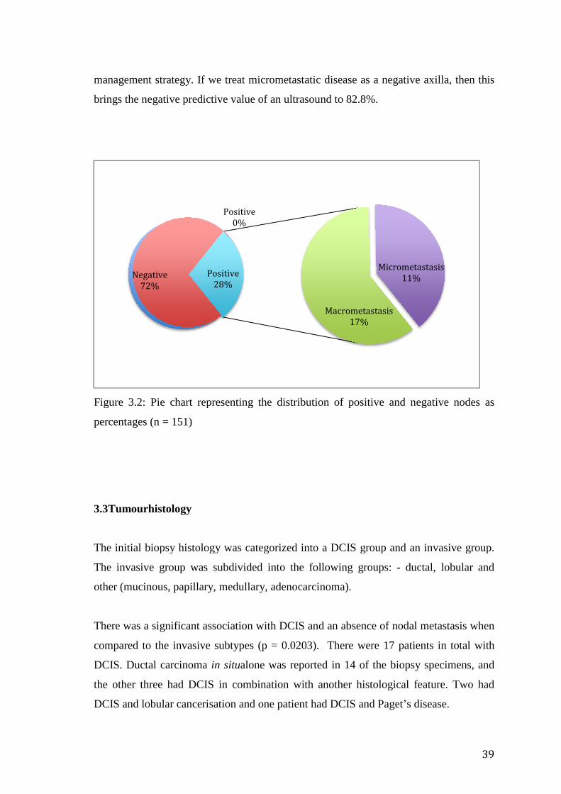
3.3Tumourhistology
The initial biopsy histology was categorized into
The invasive group was
other (mucinous, papillary, medullary, a
There was a significant association with DCIS and an absence of nodal metastasis when
compared to the invasive subtypes (p = 0.0203).
DCIS. Ductal carcinoma
the other three had DCIS in combination with another histological feature
DCIS and lobular canceris
Negative
72%
If we treat micrometastatic disease as a negative axilla, then this
predictive value of an ultrasound to 82.8%.
Figure 3.2: Pie chart representing the distribution of positive and negative nodes as
The initial biopsy histology was categorized into a DCIS group and an invasive group.
nvasive group was subdivided into the following groups: - ductal, l
other (mucinous, papillary, medullary, adenocarcinoma).
There was a significant association with DCIS and an absence of nodal metastasis when
compared to the invasive subtypes (p = 0.0203). There were 17 patients in total with
Ductal carcinoma in situalone was reported in 14 of the biopsy specimens
the other three had DCIS in combination with another histological feature
and lobular cancerisation and one patient had DCIS and Paget’s disease.
Positive
0%
Macrometastasis
17%
Micrometastasis
11%Positive
28%
39
as a negative axilla, then this
Figure 3.2: Pie chart representing the distribution of positive and negative nodes as
a DCIS group and an invasive group.
ductal, lobular and
There was a significant association with DCIS and an absence of nodal metastasis when
There were 17 patients in total with
in 14 of the biopsy specimens, and
the other three had DCIS in combination with another histological feature. Two had
ation and one patient had DCIS and Paget’s disease.
Micrometastasis
11%
Figure 3.3: Pie chart showing
Of the17 patients with
malignancy on their definitive histology.
limit for invasion on core biopsy specimens that is reported in the literature
In the invasive group, there was no correlation between the
specimen and the nodal status.
3.4 Size and T stage
The average diameter of the tumours
the T stage of the tumour
The T1 group had 74 patients in total,
had microcalcifications detected on imaging only
typical group of early breast cancer patients (
showing the various DCIS groups (N = 17)
an initial diagnosis of DCIS, six ultimately had an invasive
definitive histology.This is a rate of 35.2%, and is on the upper
limit for invasion on core biopsy specimens that is reported in the literature
there was no correlation between the histology type
nodal status.
of the tumourswas 16.7 mm ± 9.2mm (range: 0.9
the T stage of the tumour was calculated from this using the ultrasound measurements.
group had 74 patients in total, and they make up the majority
had microcalcifications detected on imaging only (T1mi). The study
ly breast cancer patients (Figure 3.4).
85%
6%
9%
DCIS
Pure DCIS
DCIS + Pagets
DCIS + Lobular
cancerisation
40
ultimately had an invasive
This is a rate of 35.2%, and is on the upper
limit for invasion on core biopsy specimens that is reported in the literature (47).
histology type of the biopsy
9.2mm (range: 0.9 – 48 mm) and
was calculated from this using the ultrasound measurements.
majority. Thirteen patients
study cohort represents a
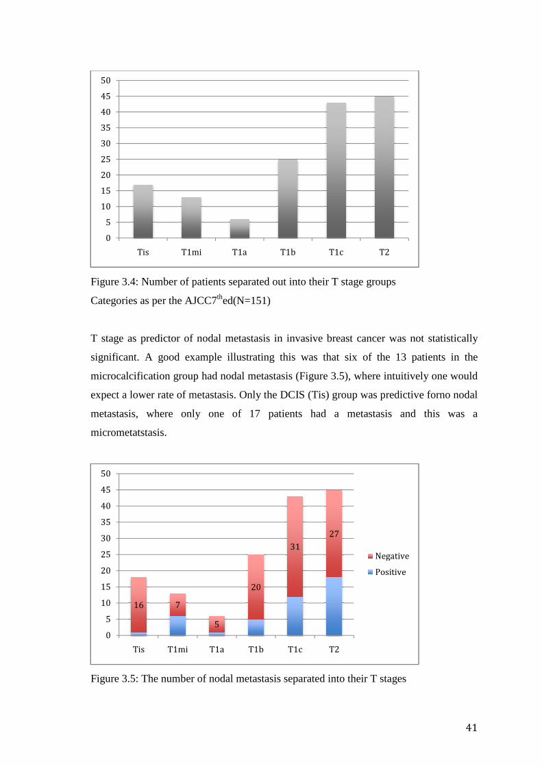
Figure 3.4: Number of patients separated out into their
Categories as per the AJCC
T stage as predictor of nodal metastasis in invasive breast cancer
significant. A good example illustrating this was that s
microcalcification group
expect a lower rate of metastasis
metastasis, where only one of 17 patients had a metastasis and this was a
micrometatstasis.
Figure 3.5: The number of n
0
5
10
15
20
25
30
35
40
45
50
Tis T1mi
16 7
0
5
10
15
20
25
30
35
40
45
50
Tis T1mi
Number of patients separated out into their T stage groups
Categories as per the AJCC7thed(N=151)
T stage as predictor of nodal metastasis in invasive breast cancer was not
A good example illustrating this was that six of the 13 patients in the
microcalcification group had nodal metastasis (Figure 3.5), where intuitively one would
expect a lower rate of metastasis. Only the DCIS (Tis) group was pre
, where only one of 17 patients had a metastasis and this was a
The number of nodal metastasis separated into their T stages
T1mi T1a T1b T1c T2
5
20
31
27
T1a T1b T1c T2
Negative
Positive
41
groups
was not statistically
of the 13 patients in the
, where intuitively one would
Tis) group was predictive forno nodal
, where only one of 17 patients had a metastasis and this was a
their T stages
Negative
Positive
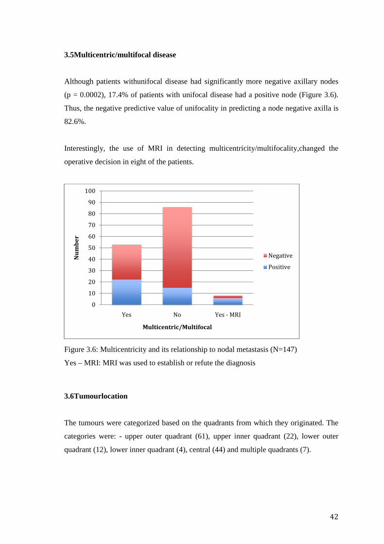
3.5Multicentric/multifocal
Although patients withunifocal
(p = 0.0002), 17.4% of patients with unifocal disease had a positive node
Thus, the negative predictive value of
82.6%.
Interestingly, the use of MRI
operative decision in eight
Figure 3.6: Multicentricity and
Yes – MRI: MRI was used to establish or refute the diagnosis
3.6Tumourlocation
The tumours were categorize
categories were: - upper oute
quadrant (12), lower inner quadrant (4), c
0
10
20
30
40
50
60
70
80
90
100
Yes
Nu
mb
er
ultifocal disease
unifocal disease had significantly more negative axillary node
17.4% of patients with unifocal disease had a positive node
the negative predictive value of unifocality in predicting a node negative axilla is
he use of MRI in detecting multicentricity/multifocality,
in eight of the patients.
: Multicentricity and its relationship to nodal metastasis (N=147)
MRI: MRI was used to establish or refute the diagnosis
categorized based on the quadrants from which they
pper outer quadrant (61), upper inner quadrant (2
quadrant (12), lower inner quadrant (4), central (44) and multiple quadrants (7
No Yes - MRI
Multicentric/Multifocal
Negative
Positive
42
negative axillary nodes
17.4% of patients with unifocal disease had a positive node (Figure 3.6).
in predicting a node negative axilla is
in detecting multicentricity/multifocality,changed the
(N=147)
they originated. The
pper inner quadrant (22), lower outer
quadrants (7).
Negative
Positive
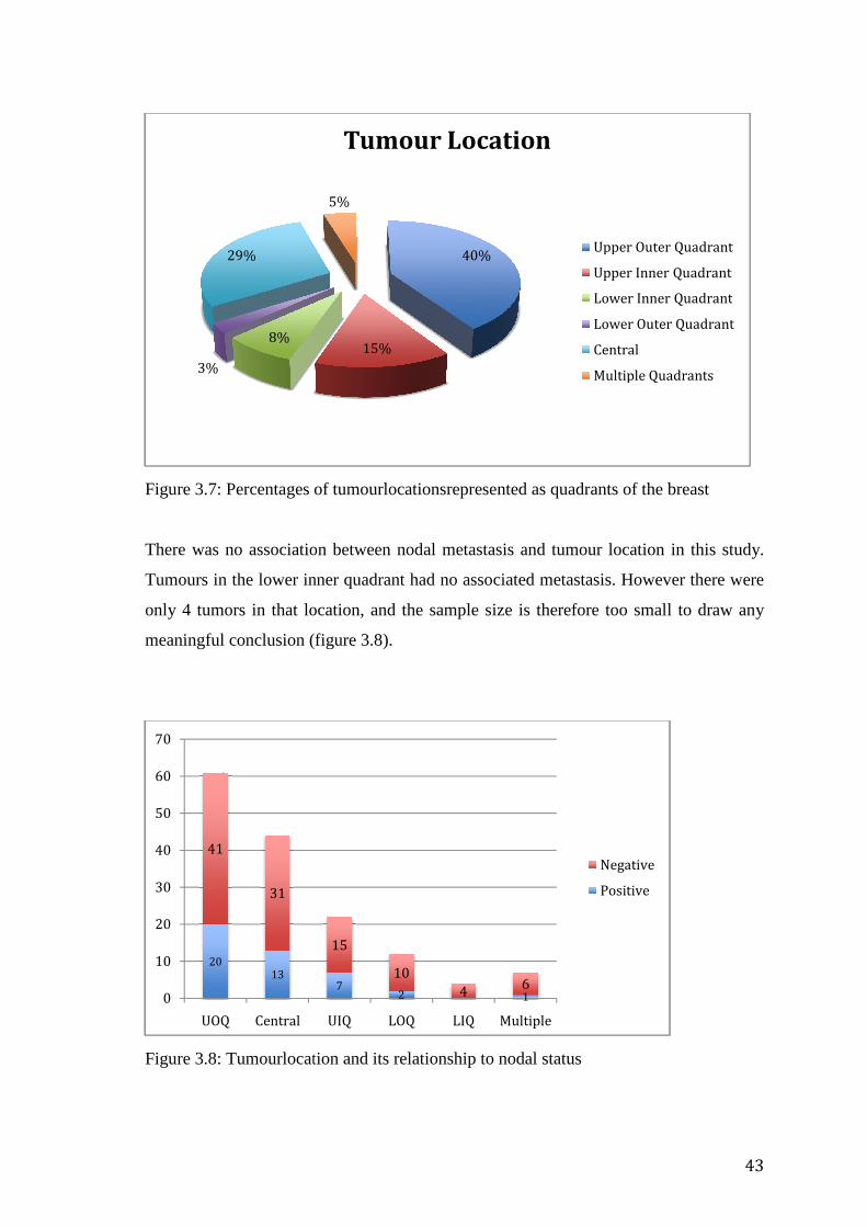
Figure 3.7: Percentages of t
There was no association
Tumours in the lower inner
only 4 tumors in that location, and the sample size is
meaningful conclusion (figure 3.8)
Figure 3.8: Tumourlocation
8%
3%
29%
2013
41
31
0
10
20
30
40
50
60
70
UOQ Central
j
Percentages of tumourlocationsrepresented as quadrants of the breast
association between nodal metastasis and tumour location
inner quadrant had no associated metastasis. H
only 4 tumors in that location, and the sample size is therefore too small to
(figure 3.8).
ocation and its relationship to nodal status
40%
15%
5%
Tumour Location
Upper Outer Quadrant
Upper Inner Quadrant
Lower Inner Quadrant
Lower Outer Quadrant
Central
Multiple Quadrants
72 1
15
10
46
UIQ LOQ LIQ Multiple
Negative
Positive
43
represented as quadrants of the breast
between nodal metastasis and tumour location in this study.
nt had no associated metastasis. However there were
too small to draw any
Upper Outer Quadrant
Upper Inner Quadrant
Lower Inner Quadrant
Lower Outer Quadrant
Central
Multiple Quadrants
Negative
Positive
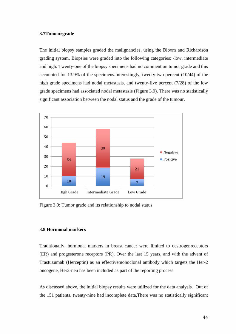
3.7Tumourgrade
The initial biopsy samples graded the malignancies, using the Bloom and Richardson
grading system. Biopsies were graded into the following cate
and high. Twenty-one of the b
accounted for 13.9% of the specimens.Interestingly, twenty
high grade specimens had nodal metastasis, and t
grade specimens had associated nodal
significant association between the nodal status and the grade of the tumour.
Figure 3.9: Tumor grade
3.8 Hormonal markers
Traditionally, hormonal markers in
(ER) and progesterone
Trastuzumab (Herceptin)
oncogene, Her2-neu has been included as
As discussed above, the
the 151 patients, twenty
10
34
0
10
20
30
40
50
60
70
High Grade
The initial biopsy samples graded the malignancies, using the Bloom and Richardson
grading system. Biopsies were graded into the following categories:
of the biopsy specimens had no comment on tumor grade
accounted for 13.9% of the specimens.Interestingly, twenty-two percent (10/44) of the
high grade specimens had nodal metastasis, and twenty-five percent
had associated nodal metastasis (Figure 3.9). There was no statistically
between the nodal status and the grade of the tumour.
rade and its relationship to nodal status
arkers
Traditionally, hormonal markers in breast cancer were limited to oestrogen
receptors (PR). Over the last 15 years, and with the advent of
(Herceptin) as an effectivemonoclonal antibody which targets the Her
has been included as part of the reporting process.
As discussed above, the initial biopsy results were utilized for the data analysis
twenty-nine had incomplete data.There was no statistically significant
19
7
39
21
Intermediate Grade Low Grade
Negative
Positive
44
The initial biopsy samples graded the malignancies, using the Bloom and Richardson
: -low, intermediate
comment on tumor grade and this
two percent (10/44) of the
five percent (7/28) of the low
. There was no statistically
between the nodal status and the grade of the tumour.
ted to oestrogenreceptors
Over the last 15 years, and with the advent of
monoclonal antibody which targets the Her-2
part of the reporting process.
for the data analysis. Out of
There was no statistically significant
Negative
Positive
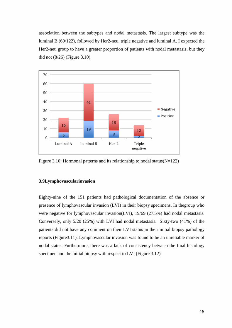
association between the subtypes and nodal metastasis.
luminal B (60/122), followed by Her2
Her2-neu group to have a greater proportion of patients with nodal metastasis, but they
did not (8/26) (Figure 3.10)
Figure 3.10: Hormonal patterns and its relationship to nodal status
3.9Lymphovascularinvasion
Eighty-nine of the 151 patients had pathological documentation of the absence or
presence of lymphovascular
were negative for lymphovascular invasion(
Conversely, only 5/20 (25
patients did not have any comm
reports (Figure3.11). Lymphovascular invasion was found to be an unreliable marker of
nodal status. Furthermore
specimen and the initial biopsy with respect to
6
16
0
10
20
30
40
50
60
70
Luminal A Luminal B
between the subtypes and nodal metastasis. The largest subtype was the
luminal B (60/122), followed by Her2-neu, triple negative and luminal A. I ex
neu group to have a greater proportion of patients with nodal metastasis, but they
(Figure 3.10).
: Hormonal patterns and its relationship to nodal status(N=122)
nvasion
nine of the 151 patients had pathological documentation of the absence or
presence of lymphovascular invasion (LVI) in their biopsy specimens.
lymphovascular invasion(LVI), 19/69 (27.5%) had nodal m
only 5/20 (25%) with LVI had nodal metastasis. Sixty
id not have any comment on their LVI status in their initial biopsy
Lymphovascular invasion was found to be an unreliable marker of
urthermore, there was a lack of consistency between the final histology
specimen and the initial biopsy with respect to LVI (Figure 3.12).
19
82
41
18
12
Luminal B Her-2 Triple
negative
Negative
Positive
45
The largest subtype was the
neu, triple negative and luminal A. I expected the
neu group to have a greater proportion of patients with nodal metastasis, but they
(N=122)
nine of the 151 patients had pathological documentation of the absence or
invasion (LVI) in their biopsy specimens. In thegroup who
had nodal metastasis.
Sixty-two (41%) of the
initial biopsy pathology
Lymphovascular invasion was found to be an unreliable marker of
, there was a lack of consistency between the final histology
Negative
Positive

Figure 3.11: Lymphovascular i
itsrelationship to the nodal status
Of the 17 specimens that were LVI positive, only seven
histology specimens (41%)
negative for LVI, eleven
3.12).This inconsistency brings the reliance of LVI as a marker of nodal disease into
question.
Figure 3.12: The change in LVI in patients
5
15
0
10
20
30
40
50
60
70
80
Yes
Remain positive in final histology
: Lymphovascular invasion on the initial biopsy specimens and
nodal status (N = 151)
s that were LVI positive, only seven remained positive in the final
histology specimens (41%) (Figure 3.12), and of the 55 patients that wer
negative for LVI, eleven were positive for LVI on final histology (20%) (Figure
is inconsistency brings the reliance of LVI as a marker of nodal disease into
: The change in LVI in patients who were initially LVI positive
19 19
5043
No No Comment
Negative
Positive
41%
59%
Positive LVI
Remain positive in final histology Changed to negative
46
initial biopsy specimens and
remained positive in the final
55 patients that were initially
were positive for LVI on final histology (20%) (Figure
is inconsistency brings the reliance of LVI as a marker of nodal disease into
positive.
Negative
Positive
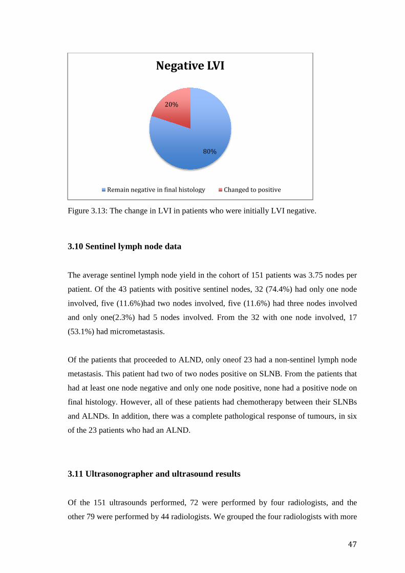
Figure 3.13: The change in LVI in patients who
3.10 Sentinel lymph node data
The average sentinel lymph node yield in the
patient. Of the 43 patients
involved, five (11.6%)had two nodes involved, five
and only one(2.3%) had 5 n
(53.1%) had micrometastasis.
Of the patients that proceeded to ALND, only oneof 23
metastasis. This patient had two
had at least one node negative and
final histology. However
and ALNDs. In addition, there was a complete pathological response of tumours, in six
of the 23 patients who had an ALND.
3.11 Ultrasonographer
Of the 151 ultrasounds performed,
other 79 were performed by 44 radiologists.
Remain negative in final histology
: The change in LVI in patients who were initially LVI negative
Sentinel lymph node data
mph node yield in the cohort of 151 patients
patients with positive sentinel nodes, 32 (74.4%) had only one
had two nodes involved, five (11.6%) had three
had 5 nodes involved. From the 32 with one
had micrometastasis.
s that proceeded to ALND, only oneof 23 had a non-sentinel lymph node
This patient had two of two nodes positive on SLNB. From the patients that
at least one node negative and only one node positive, none had a po
final histology. However, all of these patients had chemotherapy between their SLNBs
In addition, there was a complete pathological response of tumours, in six
of the 23 patients who had an ALND.
.11 Ultrasonographer and ultrasound results
the 151 ultrasounds performed, 72 were performed by four rad
performed by 44 radiologists. We grouped the four radiologists with more
80%
20%
Negative LVI
Remain negative in final histology Changed to positive
47
negative.
of 151 patients was 3.75 nodes per
%) had only one node
had three nodes involved
node involved, 17
sentinel lymph node
nodes positive on SLNB. From the patients that
only one node positive, none had a positive node on
all of these patients had chemotherapy between their SLNBs
In addition, there was a complete pathological response of tumours, in six
radiologists, and the
We grouped the four radiologists with more

than 10 procedures each together, and the remaining 44 were grouped together.
expected that the more experienced radiologists would be more accurate in their
assessment, however th
ultrasonographer had no bearing on the results
3.12 Magnetic Resonance Imaging
Onehundred and eight patients
Theirnodes were categorized as positive on MRI, negative on MRI or nodes visualized
but probably benign (Figure
predictor of nodal metastasis i
negative predictive value, but of the 72 negative MRI’s, 16 had positive nodes (22%)
and of the MRI’s that suggested positive nodes, eighteen of 32 were negative (56%).
Figure 3.14: MRI as a predictor of nodal metastasis
14
18
0
5
10
15
20
25
30
35
40
MRI +ve
than 10 procedures each together, and the remaining 44 were grouped together.
expected that the more experienced radiologists would be more accurate in their
assessment, however this did not hold true. Our data showed that
ultrasonographer had no bearing on the results when analysed in this manner
12 Magnetic Resonance Imaging
Onehundred and eight patients, in the cohort of 151 patients, had an MRI of the breast.
were categorized as positive on MRI, negative on MRI or nodes visualized
Figure3.13). Magnetic Resonance Imaging was an inconsistent
predictor of nodal metastasis in breast cancer. An MRI was anticipated
negative predictive value, but of the 72 negative MRI’s, 16 had positive nodes (22%)
and of the MRI’s that suggested positive nodes, eighteen of 32 were negative (56%).
as a predictor of nodal metastasis in breast cancer
7 9
29 27
MRI -ve Nodes but benign
Negative
Positive
48
than 10 procedures each together, and the remaining 44 were grouped together. We
expected that the more experienced radiologists would be more accurate in their
is did not hold true. Our data showed that the choice of
when analysed in this manner.
had an MRI of the breast.
were categorized as positive on MRI, negative on MRI or nodes visualized
was an inconsistent
cipated to have a good
negative predictive value, but of the 72 negative MRI’s, 16 had positive nodes (22%)
and of the MRI’s that suggested positive nodes, eighteen of 32 were negative (56%).
(N=108)
Negative
Positive

49
Chapter 4.
Discussion
Progress within the last 50 years has revolutionized the way breast cancer is treated.
Improved imaging and screening programs have changed the face of the
disease.Cancers that present at an early stageare treated more effectively and have better
outcomes. However, new treatments create new challenges, and the rate of negative
SLNBs is one such challenge. The rate of negative SLNBs is 70% - 80% (14 - 16)and
the possibility of decreasing this, needs to be investigated.
In order to accomplish this, the question that arises is, can we safely and non-invasively
predict which axillas will be free of metastasis and thus decrease the amount of negative
sentinel lymph node biopsies performed?
In order to answer the question, the problem needs to be contextualised. Seventy to
eighty percent(14 – 16)of SLNBs are negative, and if we extend this to
includemicrometastatic disease, my results show a negative (non significant) predictive
value of 82.8%.This impliesthat as a test for negative nodes, a SLNB is not specific
enough. A negative SLNBadds to the morbidityof a patient. When performed as a
separate procedure, it exposes patients to the risks of a general anaesthetic. Theyalso
have associated shoulder discomfort, paraesthesias, lymphedema(17) andthen there is
the psychological burden of an added procedure which is often forgotten. However,
omitting a SLNB may result in residual disease, which may impact on the mortality of
patients.
Therefore the risk of not performing a SLNB may be summarised as follows:
preventing morbidity in the majority at the expense of mortality in the minority,while,
even onedeath is unjustifiableas it is entirely preventable.
50
4.1 Aims and methods
The aim of my study was to improve prediction patterns of nodal spread with a view to
decrease negative SLNBs. I retrospectively studied the records of patients who had
undergone a SLNB over a ten-month period. Only those who had a documented
negative ultrasound or a negative FNA (if it was done) were included in my study,
resulting in a total of 151 patients.
My results showed that based on the information from the initial biopsy
(ER/PR/LVI/grade/histology) and radiology (size, location) I was unable to predict
nodal involvement. Patterns of spread appeared to be random, for example, even small
tumours with ‘favourable’ characteristics had nodal metastasis.
However, my study showed that a non-standardized ultrasound of the axilla can predict
that if axillary metastasis are present, three nodes or less will be affected in 97% of the
cases. This is an important finding considering the findings of the Z0011 trial (Axillary
dissection versus no axillary dissection in women with invasive breast cancer and
sentinel lymph node metastasis), which showed that a SLNB is equivalent to a
completion ALND in patients with one or two sentinel lymph nodes positive (56).
4.2 Radiological results
Importantly, I showed that ultrasound predicted a negative axilla in 71.6% of the
patients included in the study. This concurs with international literature(14). Following
the results of the NSABP-32(77) and the follow up study by Weaver et al(54) on occult
metastasis and its management, the recommendation by the NCCN was to not proceed
with a completion ALND in patients with occult metastasis(2). In this study, when I
exclude the patients with occult metastasis (17/43) the predictive value for an axilla
with non-significant metastases improved to 82.8%.
51
In the recent past, the major body of work done on ultrasound of the axilla and its use in
breast cancer has tried to improve the positive predictive value. In order to achieve this,
biopsy techniques have been employed with a view to avoid a SLNB and proceed
directly to an ALND in patients with a positive FNA or core biopsy of their axillary
nodes. This has reduced the rate of SLNBs by 14%(60).
Yet, there has been no coordinated effort by the breast care community to predict
negative lymph nodes and reduce the rate of negative SLNBs. In my study, there was no
standardization of ultrasound reporting and the reports were from multiple
ultrasonographers, with varying levels of experience. A lack of standardization in
reporting is the major impediment to progress in this arena.
However, Bedi et al(64) proposed a classification for characterizing lymph nodes.
Types 1-4 were benign, Type 5 and 6 suggested malignancy. Their negative predictive
value for Types 1-4 was 96% and this improved if Type 4 was removed. The
incorporation of this validated classification in reporting would allow us to objectively
stratify grades of negative axillas, and in that way identify a true negative axilla more
accurately.
If, for example, there are 100 patients with early breast cancer, we can assume that 70 of
these patients will not have axillary metastasis. If we were able to classify 30 of these
into Types 1-3, we could accurately rule out metastasis in these nodes and thus sparing
patients a SLNB. I would like to see us clinically validate this classification within a
trial setting.
4.3 Tumour characteristics
In the literature, there are conflicting results as to which markers accurately predict
nodal metastasis. However, the majority of publications find significance in one of their
studied parameters(69,72,74,75) and this may represent a publication bias.
52
I studiedtumour size, T stage, multicentricity/multifocality, grade, hormonal markers,
LVI, tumor location and histology and found a significant association with nodal
metastasis in only two of the studied parameters.
I found that multicentricity/multifocality predicted for nodal metastasis. However, 17%
of the patients with unifocaltumours had a positive node. Therefore, as a marker, it
lacks the accuracy required to make a firm recommendation.
4.4 Ductal carcinomainsitu
I found tumour histology to have a significant associationwith nodal metastasis,in
particular DCIS predicted a negative axilla. Initial biopsy specimens were utilized
instead of the final histology, as the aim was to recognise factors that influence nodal
metastasis, with a view to change surgical strategy. With this in mind, of the 17 patients
that had an initial diagnosis of DCIS on biopsy, six had an invasive malignancy on final
histology. This is an invasive malignancy rate of 35.2%, which is on the upper limit for
invasion of DCIS core biopsy specimens. The reported rate in the literature is between
8-38%(47).
Furthermore, oneof the patients that had a positive SLNB had a 0.75 mm
micrometastatic deposit in her node. Her initial tumour on preoperative imaging was
thought to be 13mm by 8mm (T1). The final primary tumour size was 8mm and
completion ALND yielded no additional nodal metastasis. Interestingly, this was an
intermediate grade tumour, not a high grade or a palpable DCIS lesion.
Although I was limited by an extremely small sample size, my data suggests negligible
benefit in performing a SLNB in DCIS, however our rate of invasion on final biopsy
specimens is high and thus the suspicion of invasive malignancy remains high. Thus, in
thisparticularbreast care centre, the practice of SLNBs in DCIS should continue.
However, in patients with DCIS,I found no evidence for performing a separate staging
SLNB as the risk of metastasis is small, and a combined breast procedure with frozen
section analysis would have been more appropriate. These conclusions were not part of
the aims of this study, and need to be further investigated.

53
4.5 Metastatic axillary nodes
In the group of patients with positive nodes, only one patient had more than three nodes
involved (1/43). Another, very interesting finding was that of the patients that had an
ALND, only one had an additional node involved (1/23). Twenty-two of the twenty-
three had a non-therapeutic ALND. All these patients had preoperative chemotherapy,
and this may explain the results. In addition, six of the twenty-three patients had a
complete pathological response to their primary tumour (CpR).
In the ACOSOG Z0011 (56) trial, patients with a positive SLNB were randomized to
ALND or observation. Their patients had less than three nodes involved on H&E
staining and had breast conservation therapy with whole breast irradiation (not axillary
radiation). Interestingly, twenty-seven percent of the ALND group (120/446) in the
Z0011 trial had additional metastasis and if we extrapolate this to the group that had a
SLNB alone, they would have had a similar rate of extra-sentinel lymph node metastasis.
Why is it that the two groups in the Z0011 trial had the same outcomes, when the SLNB
group had metastasis that was not surgically removed? Could it be that 96% percent of
the patients received adjuvant systemic therapy and all the patients had a lumpectomy
and breast irradiation? The inference is that the radiation and chemotherapy must confer
protection over smaller volume axillary lymph node metastasis and thus render them
inert. Furthermore, if this assumption is correct, then the question is, why the need for
an axillary staging procedure at all?
The question that this data raises, in conjunction with the Z0011 data, is whether
aSLNB is mandatory in a patient that is ultrasound negative, and is receiving the
combination of breast conservation therapy, radiotherapy and adjuvant chemotherapy.
My results show that a negative axillary ultrasound screened out axillas with threeor
less positive axillary nodes in 96% of cases, and tying this in with the Z0011 trial,
where the inclusion criteria for randomization was the presence of less than three nodes
that were positive, this would infer that patients with a negative ultrasound will be cured
by a SLNB.
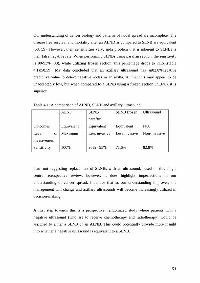
54
Our understanding of cancer biology and patterns of nodal spread are incomplete. The
disease free survival and mortality after an ALND as compared to SLNB are equivalent
(58, 59). However, their sensitivities vary, anda problem that is inherent to SLNBs is
their false negative rate. When performing SLNBs using paraffin section, the sensitivity
is 90-93% (30), while utilizing frozen section, this percentage drops to 71.6%(table
4.1)(58,59). My data concluded that an axillary ultrasound has an82.8%negative
predictive value to detect negative nodes in an axilla. At first this may appear to be
unacceptably low, but when compared to a SLNB using a frozen section (71.6%), it is
superior.
Table 4.1: A comparison of ALND, SLNB and axillary ultrasound
ALND SLNB
paraffin
SLNB frozen Ultrasound
Outcomes Equivalent Equivalent Equivalent N/A
Level of
invasiveness
Maximum Less invasive Less Invasive Non-Invasive
Sensitivity 100% 90% - 95% 71.6% 82.8%
I am not suggesting replacement of SLNBs with an ultrasound, based on this single
centre retrospective review, however, it does highlight imperfections in our
understanding of cancer spread. I believe that as our understanding improves, the
management will change and axillary ultrasounds will become increasingly utilized in
decision-making.
A first step towards this is a prospective, randomized study where patients with a
negative ultrasound (who are to receive chemotherapy and radiotherapy) would be
assigned to either a SLNB or an ALND. This could potentially provide more insight
into whether a negative ultrasound is equivalent to a SLNB.
55
4.6 Lymphovascular invasion and the reliance on the initial biopsy
Lymphovascular invasion (LVI) is reported in the literature to be a marker of nodal
metastasis(72). In this study initial biopsy resultswere used to understand the
association betweenlymphovascular invasion and nodal metastasis. The results from my
study do no not support the link between the two. However, a weakness of my study is
that a significant proportion (61/151) of the biopsy results did not test for it.
Unfortunately, the sample set is too small generalise, however, there is no association
between LVI and nodal status in this data. This is possibly because I used the biopsy
results and not the final histology results. Final pathology results are under greater
scrutiny than initial biopsy specimens, and therefore, probably have a higher yield of
positive LVI.
Ialso assessed the accuracy of initial biopsy results when compared to the final
histology specimens, in particular lymphovascular invasion. The correlation was only
20%, with a significant proportion of results changing when reviewed in the final
histology. The proportion of patients in the DCIS group that had invasive malignancies,
in conjunction with the poor LVI correlation, brings into question the reliance that is
placed on core biopsy results.
4.7 Limitations of this study
The limitations of this study are that this is a retrospective review and we were reliant
on information stored in files that were not designed to measure our outcomes. There
was a significant amount of missing data for most of the studied parameters, and this
weakened the robustness of the data calculations. Moreover, as this is a private practice,
and there were various pathology laboratories and radiology practices involved, each
had their own reporting standards, and this led to inconsistencies in documentation. In
retrospect, our sample size was also inadequate to answer this question, especially the
DCIS subset.
56
Chapter 5.
Conclusion and recommendations
My research concluded that in early breast cancer, neither an ultrasound nor various
tumour characteristics were able to accurately predict metastatic nodal spread to an
axilla. Furthermore, it questioned the reliance on initial biopsy specimens, as the
association with final tumour specimens was poor. This was particularly marked for
DCIS and LVI.
Our understanding of cancer biology and patterns of spread have progressed
substantially in the last century, yet our knowledge is incomplete. Breast cancer
research has provideda template for research in the surgical domain and the NSABP
trials have been aninvaluable repository of knowledge to turn to in order to make
evidence based management decisions. SLNBs have been validated through this body
of research and are an integral part of current practice.
A SLNB is a minimally invasive method of staging an axilla, and is the procedure of
choice in patients with early breast cancer and a clinically benign axilla. The issue is
that the majority (70%) of these SLNBs are negative.It would be incorrect to equate a
negative SLNB to an unnecessary one, as it remains a vital staging procedure, however,
it remainsan invasive procedure. Thus, in asking the question“Can we decrease the rate
of negative SLNBs”, we are exploring whether we can replace a SLNBwith a non-
invasive method to accurately stage an axilla in a select group of patients.
With regards to an ultrasound of the axilla, my data accurately predicted a negative
axilla in 71.6% of patients, and this improved to 82.8% when micrometastasis were
excluded. This is not accurate enough to replace a SLNB. However, when we compare
these results to the sensitivity of a frozen section SLNB, the accuracyis similar. In this
study a negative ultrasound could predict that an axilla, when positive would have three
or less lymph nodes involved.
57
A frozen section SLNB has equivalent outcomes to an ALND in terms of disease free
survival and 5 year mortality (58,59). This highlights the imperfections in our
understanding of disease patterns, as an ALND and a SLNB have varying sensitivities,
yet their outcome measures are the same. I suspect that if we compared a negative
ultrasound without subsequent axillary surgeryagainst these outcome measures, the
results will be similar.
Another key finding was that ultrasounds were not reported in a standardized manner or
classified according to an objective and validated method. The emphasis thus far has
been on predicting positive lymph nodes, and not negative ones. Scoring systems for
lymph nodes do exist. For example, an excellent scoring system has been proposed by
Bedi et al (64). This classification groups nodes into types 1-6, where, Type 1 is
definitely benign and Type 6 is definitely malignant. Using a classification similar to
this will screen the negative axillas more objectively, and provides a starting point for
future research on this topic. I believe this classification would allow us to identify a
sub-group within the group of negative ultrasounds that will definitely be benign. This
would be a significant step forwards in the treatment of breast cancer.
Insights from this study suggest that these issues could be further examined as follows: -
- A prospective study evaluating a classification system for reporting axillary
nodes with particular emphasis on predicting negative nodes is required.
- If the classification system is validated, it should be used to evaluate
ultrasound negative axillas in two randomized groups. One group to have a
SLNB, and the other to be observed. (Similar protocol to Z0011)
This will add to the body of knowledge and clarify the role of an ultrasound in early
breast cancer.
In addition, DICS patients have a low rate of positive nodes, and I recommend that their
SLNBsare combined with their definitive surgery with the use of intraoperative frozen
section.
58
References
1. Vorobiof DA, Sitas F and Vorobiof G. Breast Cancer incidence in South Africa. J
ClinOncolSept 2001, 19(18s):125s-127s.
2.Carlson R, Alfred C, Anderson B, Burnstein H, Stephen E, Farrar W, et al. NCCN
Breast Cancer Guideline. NCCN. 1st ed. 2012 Jan. 1;1–164.
3. Early Breast Cancer Trialists' Collaborative Group (EBCTCG). Effects of
chemotherapy and hormonal therapy for early breast cancer on recurrence and 15-year
survival: an overview of the randomised trials. Lancet. 2005 Apr.365(9472):1687–717.
4. Fisher B. From Halsted to prevention and beyond: advances in the management of
breast cancer during the twentieth century. Eur J Cancer. 1999 Dec. 35(14):1963–73.
5. Patani NR, Dwek MV, Douek M. Predictors of axillary lymph node metastasis in
breast cancer: A systematic review. European Journal of Surgical Oncology (EJSO).
2007 May. 33(4):409–19.
6. Fisher BB, Slack NN, Katrych DD, Wolmark NN. Ten year follow-up results of
patients with carcinoma of the breast in a co-operative clinical trial evaluating surgical
adjuvant chemotherapy. SurgGynecol Obstet. 1975 Mar. 31;140(4):528–34.
7. Fisher B, Redmond C, Fisher ER, Bauer M, Wolmark N, Wickerham DL, et al. Ten-
year results of a randomized clinical trial comparing radical mastectomy and total
mastectomy with or without radiation. N Engl J Med. 1985 Mar. 13;312(11):674–81.
8. Veronesi U, Cascinelli N, Mariani L, Greco M, Saccozzi R, Luini A, et al. Twenty-
year follow-up of a randomized study comparing breast-conserving surgery with radical
mastectomy for early breast cancer. N Engl J Med. 2002 Oct. 16;347(16):1227–32.
9. Fisher B, Anderson S, Bryant J, Margolese RG, Deutsch M, Fisher ER, et al. Twenty-
year follow-up of a randomized trial comparing total mastectomy, lumpectomy, and
lumpectomy plus irradiation for the treatment of invasive breast cancer. N Engl J Med.
2002 Oct. 16;347(16):1233–41.
59
10. Cools-Lartigue J, Meterissian S. Accuracy of Axillary Ultrasound in the Diagnosis
of Nodal Metastasis in Invasive Breast Cancer: A Review. World J Surg. 2011 Oct.
27;36(1):46–54.
11. Veronesi U, Paganelli G, Viale G, Galimberti V, Luini A, Zurrida S, et al. Sentinel
Lymph Node Biopsy and Axillary Dissection in Breast Cancer: Results in a Large
Series. JNCI Journal of the National Cancer Institute. 1999 Feb. 17;91(4):368–73.
12. Kell MR, Kerin MJ. Sentinel lymph node biopsy. BMJ. 2004 Jun.
5;328(7452):1330–1.
13. Fraile MM, Rull MM, Julián FJF, Fusté FF, Barnadas AA, Llatjós MM, et al.
Sentinel node biopsy as a practical alternative to ALND in breast cancer patients: an
approach to its validity. Annals of Oncology. 2000 May 31;11(6):701–5.
14. Rattay T, Muttalib M, Khalifa E, Duncan A, Parker SJ. Clinical utility of routine
pre-operative axillary ultrasound and fine needle aspiration cytology in patient selection
for sentinel lymph node biopsy. Breast. 2012 Apr.;21(2):210–4.
15. Abe H, Schmidt RA, Sennett CA, Shimauchi A, Newstead GM. US-guided Core
Needle Biopsy of Axillary Lymph Nodes in Patients with Breast Cancer: Why and How
to Do It. RadioGraphics. 2007 Oct. 1;27(Supplement 1):S91–9.
16. Jakub JW, Pendas S, Reintgen DS. Current Status of Sentinel Lymph Node
Mapping and Biopsy: Facts and Controversies. The Oncologist. 2003 Feb. 1;8(1):59–68.
17. Fleissig A, Fallowfield LJ, Langridge CI, Johnson L, Newcombe RG, Dixon JM, et
al. Post-operative arm morbidity and quality of life. Results of the ALMANAC
randomised trial comparing sentinel node biopsy with standard axillary treatment in the
management of patients with early breast cancer. Breast Cancer Res Treat. 2006 Jan.
31;95(3):279–93.
18. Mansel RE, Fallowfield L, Kissin M, Goyal A, Newcombe RG, Dixon JM, et al.
Randomized Multicenter Trial of Sentinel Node Biopsy Versus Standard Axillary
Treatment in Operable Breast Cancer: The ALMANAC Trial. JNCI Journal of the
National Cancer Institute. 2006 May 2;98(9):599–609.
60
19. Gould EA, Winship T, Philbin PH, Kerr HH. Observations on a “sentinel node” in
cancer of the parotid. Cancer. Wiley Subscription Services, Inc., A Wiley Company;
1960;13(1):77–8.
20. Cabanas RMR. An approach for the treatment of penile carcinoma. Cancer. 1977
Jan. 31;39(2):456–66.
21. Holmes ECE, Moseley HSH, Morton DLD, Clark WW, Robinson DD, Urist MMM.
A rational approach to the surgical management of melanoma. Ann Surg. 1977 Sep.
30;186(4):481–90.
22. Morton DL, Chan AD. The concept of sentinel node localization: How it started.
Seminars in Nuclear Medicine. 2000 Jan.;30(1):4–10.
23. Morton DL. Technical Details of Intraoperative Lymphatic Mapping for Early Stage
Melanoma.Archives of Surgery. American Medical Association; 1992 Apr
1;127(4):392–9.
24. Alex JCJ, Weaver DLD, Fairbank JTJ, Rankin BSB, Krag DND. Gamma-probe-
guided lymph node localization in malignant melanoma. SurgOncol. 1993 Sep.
30;2(5):303–8.
25. Krag DND, Weaver DLD, Alex JCJ, Fairbank JTJ. Surgical resection and
radiolocalization of the sentinel lymph node in breast cancer using a gamma probe.
SurgOncol. 1993 Nov. 30;2(6):335–40.
26. Turner-Warwick RT. The lymphatics of the breast.British Journal of Surgery. 1959
May;46(200):574–82.
27. Beitsch PDP, Clifford EE, Whitworth PP, Abarca AA. Improved lymphatic
mapping technique for breast cancer. Breast J. 2000 Dec. 31;7(4):219–23.
28. Beitsch PD, Clifford E, Whitworth P, Abarca A. Improved Lymphatic Mapping
Technique for Breast Cancer. The Breast Journal. Blackwell Science Inc;
2001;7(4):219–23.
29. Kargozaran H, Shah M, Li Y, Beckett L, Gandour-Edwards R, Schneider PD, et al.
Concordance of Peritumoral Technetium 99M Colloid and Subareolar Blue Dye

61
Injection in Breast Cancer Sentinel Lymph Node Biopsy. Journal of Surgical Research.
2007 Nov.;143(1):126–9.
30. Kim T, Giuliano AE, Lyman GH. Lymphatic mapping and sentinel lymph node
biopsy in early-stage breast carcinoma: a metaanalysis. Cancer. 2005 Dec. 31;106(1):4–
16.
31. Tafra L, Lannin DR, Swanson MS et al. Multicenter Trial of Sentinel Node Biopsy
for Breast Cancer Using Both Technetium Sulfur Colloid and Isosulfan Blue Dye. Ann
Surg. Lippincott, Williams, and Wilkins; 2001 Jan. 1;233(1):51.
32. Krag DD, Weaver DD, Ashikaga TT, Moffat FF, Klimberg VSV, Shriver CC, et al.
The sentinel node in breast cancer--a multicenter validation study. N Engl J Med. 1998
Sep. 30;339(14):941–6.
33. Giuliano AEA, Jones RCR, Brennan MM, Statman RR. Sentinel lymphadenectomy
in breast cancer. Journal of Clinical Oncology. 1997 May 31;15(6):2345–50.
34.Veronesi U, Paganelli G, Viale G, Luini A, Zurrida S, Galimberti V, et al. A
randomized comparison of sentinel-node biopsy with routine axillary dissection in
breast cancer. N Engl J Med. 2003 Aug 7;349(6):546–53.
35. Bilimoria KY, Bentrem DJ, Hansen NM, et al. Comparison of sentinel lymph node
biopsy alone and completion axillary lymph node dissection for node-positive
breast cancer. J ClinOncol 2009;27(June (18)):2946–53.
36. Khera SY, Kiluk JV, Hasson DM, Meade TL, Meyers MP, Dupont EL, et al.
Pregnancy-associated breast cancer patients can safely undergo lymphatic mapping.
Breast J. 2007 Dec. 31;14(3):250–4.
37. Port ER, Fey J, Gemignani ML, Heerdt AS, Montgomery LL, Petrek JA, et al.
Reoperative sentinel lymph node biopsy: a new option for patients with primary or
locally recurrent breast carcinoma. J Am Coll Surg. 2002 Jul. 31;195(2):167–172
38. Kim HJH, Lee JSJ, Park EHE, Choi SLS, Lim WSW, Chang MAM, et al. Sentinel
node biopsy in patients with multiple breast cancer. Breast Cancer Res Treat. 2007 Jul.
28;109(3):503–6.
62
39. Fisher BB, Brown AA, Mamounas EE, Wieand SS, Robidoux AA, Margolese RGR,
et al. Effect of preoperative chemotherapy on local-regional disease in women with
operable breast cancer: findings from National Surgical Adjuvant Breast and Bowel
Project B-18. Journal of Clinical Oncology. 1997 Jun. 30;15(7):2483–93.
40. Rastogi P, Anderson SJ, Bear HD, Geyer CE, Kahlenberg MS, Robidoux A, et al.
Preoperative Chemotherapy: Updates of National Surgical Adjuvant Breast and Bowel
Project Protocols B-18 and B-27. Journal of Clinical Oncology. 2008 Feb.
10;26(5):778–85.
41. Kuerer HM, Sahin AA, Hunt KK, Newman LA, Breslin TM, Ames FC, et al.
Incidence and impact of documented eradication of breast cancer axillary lymph node
metastases before surgery in patients treated with neoadjuvant chemotherapy. Ann Surg.
1999 Jul.;230(1):72–8.
42. Kuerer HMH, Newman LAL. Lymphatic mapping and sentinel lymph node biopsy
for breast cancer: developments and resolving controversies. Journal of Clinical
Oncology. 2005 Mar. 9;23(8):1698–705.
43. Classe J-M, Bordes V, Campion L, Mignotte H, Dravet F, Leveque J, et al. Sentinel
Lymph Node Biopsy After Neoadjuvant Chemotherapy for Advanced Breast Cancer:
Results of Ganglion Sentinelle et ChimiotherapieNeoadjuvante, a French Prospective
Multicentric Study. Journal of Clinical Oncology. 2009 Feb. 5;27(5):726–32.
44. Kohler BA, Ward E, McCarthy BJ, Schymura MJ, Ries LAG, Eheman C, et al.
Annual report to the nation on the status of cancer, 1975-2007, featuring tumors of the
brain and other nervous system. JNCI Journal of the National Cancer Institute. 2011
May 4;103(9):714–36.
45. Kelly TAT, Kim JAJ, Patrick RR, Grundfest SS, Crowe JPJ. Axillary lymph node
metastases in patients with a final diagnosis of ductal carcinoma in situ. Am J Surg
[Internet]. 2003 Oct.;186(4):368–70. Available from:
http://www.sciencedirect.com/science/article/pii/S0002961003002769

63
46. Klauber-DeMore NN, Tan LKL, Liberman LL, Kaptain SS, Fey JJ, Borgen PP, et al.
Sentinel lymph node biopsy: is it indicated in patients with high-risk ductal carcinoma-
insitu and ductal carcinoma-insitu with microinvasion? Ann SurgOncol. 2000 Sep.
30;7(9):636–42.
47. Mittendorf MEA, Arciero CA, Gutchell V, Hooke J, Shriver CD. Core biopsy
diagnosis of ductal carcinoma in situ: An indication for sentinel lymph node biopsy.
Curr Surg. 2004 Dec. 31;62(2):253–7.
48. Crowe JPJ, Rim AA, Patrick RJR, Rybicki LAL, Grundfest-Broniatowski SFS, Kim
JAJ, et al. Does core needle breast biopsy accurately reflect breast pathology? Surgery.
2003 Sep. 30;134(4):523–8.
49. Ciatto SS, Houssami NN, Ambrogetti DD, Bianchi SS, Bonardi RR, Brancato BB,
et al. Accuracy and underestimation of malignancy of breast core needle biopsy: the
Florence experience of over 4000 consecutive biopsies. Breast Cancer Res Treat. 2007
Feb. 28;101(3):291–7.
50. Ansari B, Ogston SA, Purdie CA, Adamson DJ, Brown DC, Thompson AM. Meta
‐analysis of sentinel node biopsy in ductal carcinoma in situ of the breast. British
Journal of Surgery. John Wiley & Sons, Ltd; 2008;95(5):547–54.
51. Goyal AA, Douglas-Jones AA, Monypenny II, Sweetland HH, Stevens GG, Mansel
RER. Is there a role of sentinel lymph node biopsy in ductal carcinoma in situ?: analysis
of 587 cases. Breast Cancer Res Treat. 2006 Jul. 31;98(3):311–4.
52. Schneider CC, Trocha SS, McKinley BB, Shaw JJ, Bielby SS, Blackhurst DD, et al.
The use of sentinel lymph node biopsy in ductal carcinoma in situ. Am Surg. 2010 Aug.
31;76(9):943–6.
53. Weaver DLD, Krag DND, Manna EAE, Ashikaga TT, Waters BLB, Harlow SPS, et
al. Detection of occult sentinel lymph node micrometastases by immunohistochemistry
in breast cancer. An NSABP protocol B-32 quality assurance study. Cancer.
2006;107(4):661–7.
54. Weaver. Effect of Occult Metastases on Survival in Node-Negative Breast Cancer.
N Engl J Med. 2011 Feb. 2;364(5):412–21.
64
55. Wilson JP, Mattson D, Edge SB. Is there a need for axillary dissection in breast
cancer? J NatlComprCancNetw 2011;9:225-230
56. Giuliano AE, Hunt KK, Ballman KV, Beitsch PD, Whitworth PW, Blumencranz
PW, et al. Axillary Dissection vs No Axillary Dissection in Women With Invasive
Breast Cancer and Sentinel Node Metastasis. JAMA. 2011 Feb. 8;305(6):569–75.
57. Güth U, Myrick ME, Viehl CT, Schmid SM, Obermann EC, Weber WP. The post
ACOSOG Z0011 era: Does our new understanding of breast cancer really change
clinical practice? Eur J SurgOncol. 2012 Aug;38(8):645-50.
58. Van de Vrande S, Meijer J, Rijnders A, Klinkenbijl JHG. The value of
intraoperative frozen section examination of sentinel lymph nodes in breast cancer.
European Journal of Surgical Oncology. 2009 Mar. 1;35(3):276–80.
59. Jensen AJ, Naik AM, Pommier RF, Vetto JT, Troxell ML. Factors influencing
accuracy of axillary sentinel lymph node frozen section for breast cancer. AJS. 2010
May 1;199(5):629–35.
60. Deurloo EEE, Tanis PJP, Gilhuijs KGAK, Muller SHS, Kröger RR, Peterse JLJ, et
al. Reduction in the number of sentinel lymph node procedures by preoperative
ultrasonography of the axilla in breast cancer. Eur J Cancer. 2003 Apr. 30;39(8):1068–
1073.
61. Abe H, Schmidt RA, Kulkarni K, Sennett CA, Mueller JS, Newstead GM. Axillary
Lymph Nodes Suspicious for Breast Cancer Metastasis: Sampling with US-guided 14-
Gauge Core-Needle Biopsy--Clinical Experience in 100 Patients1. Radiology. 2009 Jan.
1;250(1):41–9.
62. Mainiero MB. Regional Lymph Node Staging in Breast Cancer: The Increasing
Role of Imaging and Ultrasound-Guided Axillary Lymph Node Fine Needle Aspiration.
Radiologic Clinics of NA. Elsevier Ltd; 2010 Sep. 1;48(5):989–97.
63. Koelliker SL, Chung MA, Mainiero MB, Steinhoff MM, Cady B. Axillary lymph
nodes: US-guided fine-needle aspiration for initial staging of breast cancer--correlation
with primary tumor size. Radiology. 2007 Dec. 31;246(1):81–9.
65
64. Bedi DGD, Krishnamurthy RR, Krishnamurthy SS, Edeiken BSB, Le-Petross HH,
Fornage BDB, et al. Cortical morphologic features of axillary lymph nodes as a
predictor of metastasis in breast cancer: in vitro sonographic study. CORD Conference
Proceedings. 2008 Aug. 31;191(3):646–52.
65. Alvarez S, Orbe EA, Alcorta P, López F, Alonso I, s JC. Role of sonography in the
diagnosis of axillary lymph node metastases in breast cancer: a systematic review.
American Journal of Roentgenology. 2006 Apr. 30;186(5):1342–8.
66. Nori J, Vanzi E, Bazzocchi M, Bufalini FN, Distante V, Branconi F, et al. Role of
axillary ultrasound examination in the selection of breast cancer patients for sentinel
node biopsy. Am J Surg. 2007 Jan.;193(1):16–20.
67. Mills P, Sever A, Weeks J, Fish D, Jones S, Jones P. Axillary ultrasound assessment
in primary breast cancer: an audit of 653 cases. The Breast Journal. 2010
Sep.;16(5):460–3.
68. Ragupathy S, Flynn S, Duncan KA. 16. Axillary ultrasound accuracy in assessing
nodal metastases in a breast screening service. Cancer Imaging. 2010 Dec. 31;11 Spec
No A:S184–4.
69. Westman G, Stål O, Arnesson L. Prediction of axillary lymph node metastases in a
screened breast cancer population. ActaOncologica. 1994. Apr; 33(6) 603-608
70. Elston CWC, Ellis IOI. Pathological prognostic factors in breast cancer. I. The value
of histological grade in breast cancer: experience from a large study with long-term
follow-up. Histopathology. 1991 Oct. 31;19(5):403–10.
71. Orucevic AA, Reddy VBV, Bloom KJK, Bitterman PP, Magi-Galluzzi CC, Oleske
DMD, et al. Predictors of lymph node metastasis in T1 breast carcinoma, stratified by
patient age. Breast J. 2001 Dec. 31;8(6):349–55.
72. Viale G, Zurrida S, Maiorano E, Mazzarol G, Pruneri G, Paganelli G, et al.
Predicting the status of axillary sentinel lymph nodes in 4351 patients with invasive
breast carcinoma treated in a single institution. Cancer 2005;103(3):492–500.
66
73. Chua B, Ung O, Taylor R, Boyages J. Frequency and predictors of axillary lymph
node metastases in invasive breast cancer. ANZ J Surg. 2001 Nov. 30;71(12):723–8.
74. Susini T, Nori J, Olivieri S, Molino C, Marini G, Bianchi S, et al. Predicting the
status of axillary lymph nodes in breast cancer: A multiparameter approach including
axillary ultrasound scanning. The Breast.; 2009 Apr. 1;18(2):103–8.
75. Bevilacqua JLB, Kattan MW, Fey JV, Cody HS, Borgen PI, Van Zee KJ. Doctor,
What Are My Chances of Having a Positive Sentinel Node? A Validated Nomogram for
Risk Estimation.Journal of Clinical Oncology. 2007 May 7;25(24):3670–9.
76. Perou C, Sørlie T, Eisen M, van de Rijn M. Molecular portraits of human breast
tumours. Nature. 2000.
77. Krag DN, Anderson SJ, Julian TB, Brown A, Harlow SP, Costantino JP, et al.
Primary outcome results of NSABP B-32, a randomized phase III clinical trial to
compare sentinel node resection (SNR) to conventional axillary dissection (AD) in
clinically node-negative breast cancer patients. ASCO Meeting Abstracts. 2010 Jun.
20;28(18_suppl):505.
78. Bloom H, Richardson WW. Histological grading and prognosis in breast cancer: a
study of 1409 cases of which 359 have been followed for 15 years. British Journal of
Cancer. 1957.
79. Braun MM, Flucke UU, Debald MM, Walgenbach-Bruenagel GG, Walgenbach K-
JK, Höller TT, et al. Detection of lymphovascular invasion in early breast cancer by
D2-40 (podoplanin): a clinically useful predictor for axillary lymph node metastases.
Breast Cancer Res Treat. 2008 Nov 30;112(3):503–11.
80. Olayioye MA. Update on HER-2 as a target for cancer therapy: intracellular
signaling pathways of ErbB2/HER-2 and family members. Breast Cancer Res.
2001;3(6):385–9.
81. Cote R, Giuliano AE, Hawes D, Ballman KV. ACOSOG Z0010: a multicenter
prognostic study of sentinel node (SN) and bone marrow (BM) micrometastases in
67
women with clinical T1/T2 N0 M0 breast cancer. Journal of Clinical Oncology
2010.(Meeting Abstracts)June 2010vol. 28 no. 18_suppl CRA504
82.de Boer M, van Deurzen CH, van Dijck JA, Borm GF, van Diest PJ, Adang EM, et
al. Micrometastases or isolated tumor cells and the outcome of breast cancer. N Engl J
Med. 2009 Aug 12;361(7):653–63.