camouflage treatment in adult skeletal class iii cases … … · tients with “borderline”...
TRANSCRIPT
ORIGINAL ARTICLE
349
aGraduate Student,
bProfessor, Department of Orthodontics,
School of Stomatology, The Fourth Military Medical University,
China.
Corresponding author: Yinzhong Duan.
Department of Orthodontics, School of Stomatology, The Fourth
Military Medical University, No.145 West Changle road, Xi’an, P.
R.China, 710032.
+86 29 84776137; e-mail, [email protected].
Received February 3, 2010; Last Revision March 19, 2010;
Accepted March 23, 2010.
DOI:10.4041/kjod.2010.40.5.349
Camouflage treatment in adult skeletal Class III cases by
extraction of two lower premolars
Fang Ning, PhD,a Yinzhong Duan, PhDb
Objective: The purpose of this study was to evaluate the dentoskeletal and soft tissue profile changes after extraction of two lower first or second premolars in “borderline” adult skeletal Class III cases. Methods: Twenty-eight patients with “borderline” skeletal Class III malocclusion were studied. All of them were treat-ed by extraction of two lower first or second premolars. Lateral cephalometric radiographs taken at the start and end of treatment were analysed. Twenty-five cephalometric variables were calculated and paired t-tests were performed. Results: After treatment, no significant changes were noted in the skeletal parameters (p ≥ 0.05). Regarding the dental parameters, the L1-MP angle decreased by 8.1o, the U1-L1 angle increased by 7.7o (p < 0.01), the overjet distance increased by 5.7 mm (p < 0.01), the L1-NB angle decreased by 7.3o and the L1-NB distance decreased by 4.8 mm (p < 0.01). The soft tissue parameters of Li-E, Li-H and Li-RL2 distance decreased by 3.2 mm, 3.4 mm and 4.1 mm respectively (p < 0.01). Conclusions: Orthodontic camouflage treatment by extraction of two lower first or second premolars provides a viable treatment alternative for “borderline” skeletal Class III cases to achieve a good occlusal relationship. (Korean J Orthod 2010;40(5):349-357)
Key words: Class III treatment, Diagnosis and treatment planning, Adult treatment, Tooth movement
INTRODUCTION
Skeletal Class III malocclusion is a common maloc-
clusion in orthodontic clinics in China.1 It often pres-
ents the clinician with extreme difficulties for success-
ful treatment. Studies have reported that skeletal Class
III discrepancy worsens with age.2,3 Early intervention
of skeletal Class III deformities in the mixed dentition
or even in the deciduous dentition has received in-
creasing attention in the orthodontic field. The alter-
native approaches include the use of reverse headgears,
chin cups and functional appliances.4-8 However, not
all patients can achieve good results using these
methods. Furthermore, the patient seeking treatment at
the clinic may be past their adolescent growth spurt
and present with a severe skeletal Class III deformity.
In some of these patients, treatment with either surgi-
cal-orthodontic therapy or orthodontic camouflage
treatment is possible. We refer to these cases as the
“borderline case”. For adult borderline skeletal Class
III patients, orthognathic surgery is often the recom-
mended choice of therapy as this can achieve a good
result and the outcomes tend to be stable. However, in
China, some patients do not readily accept surgery be-
cause of economical and psychological reasons as well
as the potential surgical risks involved. Can ortho-
dontic camouflage treatment achieve a similar result as
successful as orthognathic surgery?
Fang Ning, Yinzhong Duan 대치교정지 40권 5호, 2010년
350
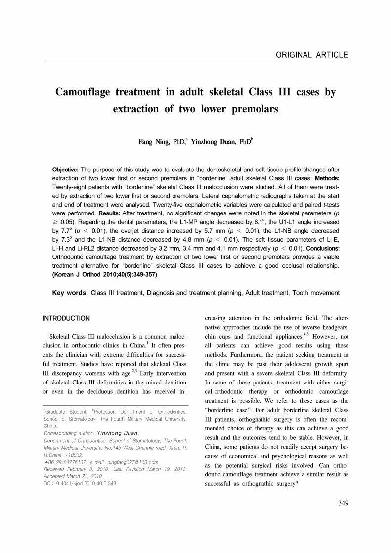
Fig 1. Skeletal measurements used in the study. SN in-dicates Sella-nasion plane; RL1, horizontal reference line; RL2, vertical reference line; 1, SNA; 2, SNB; 3, ANB; 4, SN-MP; 5, Pg-NB; 6, A-RL1; 7, A-RL2; 8, B-RL2; 9, Ar-Pg.
Lin and Gu9 reported that they had successfully
treated 13 severe skeletal Class III cases using the
method of extraction of two lower second molars.
After treatment, the soft tissue change was remarkable
and concave facial profiles were improved to straight
profiles. Sato10 introduced the MEAW (multiloop edge-
wise archwire) therapy with extraction of two lower
third molars for skeletal Class III patients. In our clin-
ic, we conducted a search for the available treatment
methods for adult borderline skeletal Class III cases
using orthodontic camouflage treatment. For example,
extraction of two lower first or second premolars was
used to treat skeletal Class III malocclusion and a
Class III molar relationship and proper overjet and
overbite were achieved at the end of the treatment.
The aim of this study was to evaluate dentoskeletal
and soft tissue profile changes after extraction of two
lower first or second premolars in adult borderline
skeletal Class III cases.
MATERIAL AND METHODS
Cases selection
Fifteen males and thirteen female consecutive pa-
tients with “borderline” skeletal Class III malocclusion
were included in this study. The ages of the patients
ranged from 17.0 to 22.4 years with a mean age of
18.8 years. All the patients were treated with extraction
of two lower first or second premolars in the Depart-
ment of Orthodontics at the Fourth Military Medical
University. The patients' first visit dates were all from
2002 to 2007. The study protocol was approved by the
Ethics Committee of the Fourth Military Medical Uni-
versity and informed consent was obtained from
patients. The selection criteria were as follows:
(1) Anterior crossbite;
(2) Mesial or superior mesial Class III molar rela-
tionship, with maxillary second premolar occluding in
the buccal groove of the mandibular first molar;
(3) No mandibular shift due to occlusal interference
or premature contact of teeth;
(4) Concave facial profile;
(5) −4.0o ≤ ANB < 0o;
(6) Over the adolescent growth spurt;
(7) Originally classified as surgical cases by other
orthodontists but the patients and their families rejected
surgical treatment.
Treatment approach
All patients were treated with the standard edgewise
technique. In these cases, two lower first or second
premolars were extracted. The basis for making ex-
traction decisions of first vs. second premolars was as
follows: Commonly, the two first premolars were ex-
tracted to correct the marginal Class III cases.
However, if the second premolars were carious, then
the decayed teeth were extracted. Also, the reversed
overjet and molar relationship should be considered
when extraction. If the reversed overjet distance was
short and the molar relationship was not a superior
mesial Class III, then the second premolars were con-
sidered to be extracted to correct the molar
relationship. Class III elastics were used in some cases
when required. Light and continuous force was recom-
mended (about 50 g on each side) and the “torque”
force in the lower anterior teeth was used to avoid the
lingual inclination. TAD were not used for anchorage

Vol. 40, No. 5, 2010. Korean J Orthod Class III camouflage treatment
351
Fig 2. Dental measurements used in the study. SN in-dicates Sella-nasion plane; RL1, horizontal reference line; RL2, vertical reference line; 1, U1-SN; 2, L1-MP; 3, U1-L1; 4, U1-NA (degree); 5, U1-NA (mm); 6, L1-NB (degree); 7, L1-NB (mm); 8, OP-FH; 9, overjet.
Fig 3. Soft tissue measurements used in the study. SN indicates Sella-nasion plane; RL1, horizontal ref-erence line; RL2, vertical reference line; 1, upper lip to E plane; 2, lower lip to E plane; 3, lower lip to Hline; 4, Cm-Sn-Ls; 5, Ls-RL2; 6, Li-RL2; 7, A-Ls.
control in this study. The mean duration of treatment
was 2.0 ± 0.6 years.
Cephalometric analysis
Standardized lateral cephalometric radiographs of
each patient were obtained at the start and end of
treatment. Each radiograph used in the present study
were taken in the same cephalostat and traced on ace-
tate paper. Twenty-five cephalometric landmarks (Figs
1 - 3) were identified.11-14 All the tracings and meas-
urements were manually carried out twice with a
2-week interval by one examiner with a sharp pencil
under optimal conditions.
Statistical analysis
The statistical analysis was processed with SPSS
10.0 for Windows. The arithmetic mean and standard
deviation were calculated for each variable. Paired
t-tests were performed to assess the statistical sig-
nificance of any dental and skeletal change. The levels
of significance were: p ≥ 0.05 (NS), *p < 0.05; †p
< 0.01.
The method error in locating, superimposing and
measuring the changes of different landmarks was cal-
culated by Dahlberg’s formula
╱ ,
where d represents the difference between two registra-
tions and n is the number of duplicate registrations.
The method error determined was 0.3 mm for linear
measurement and 0.4o for angular measurement, which
were both statistically insignificant (p ≥ 0.05).
RESULTS
After using the standard edgewise technique and ex-
traction of two lower first or second premolars, most
of the patients achieved efficient treatment. At the end
of the treatment, the facial profile was improved from
a concave to a straight tendency. The anterior crossbite
was corrected and a Class III molar relationship and
Class I canine relationship were achieved in all sub-
jects. There was no sign of active periodontal disease
or gingival inflammation after treatment. The occlusal
relationship in the upper and lower arches was stable,
tight and concordant after the treatment.

Fang Ning, Yinzhong Duan 대치교정지 40권 5호, 2010년
352
Pre-treatment Post-treatment Difference‐‐‐‐‐‐‐‐‐‐‐‐‐‐‐‐‐‐‐‐‐‐‐‐‐‐‐‐‐‐‐‐‐‐‐‐‐‐ ‐‐‐‐‐‐‐‐‐‐‐‐‐‐‐‐‐‐‐‐‐‐‐‐‐‐‐‐‐‐‐‐‐‐‐‐‐‐‐‐‐‐‐ ‐‐‐‐‐‐‐‐‐‐‐‐‐‐‐‐‐‐‐‐‐‐‐‐‐‐‐‐‐‐‐‐‐‐‐‐‐‐ p valueMean SD Mean SD Mean SD
SNA(o) 80.5 3.5 80.6 3.7 0.1 2.3 NS
SNB (o) 82.9 4.0 82.6 3.8 -0.3 2.5 NS
ANB (o) -2.4 2.5 -2.0 2.1 0.4 1.2 NS
SN-MP (o) 36.3 3.3 36.9 3.5 0.6 2.6 NS
Pg-NB (mm) 1.0 1.2 0.8 1.1 -0.2 1.1 NS
A-RL1 (mm) 55.2 7.3 55.5 7.5 0.3 5.7 NS
A-RL2 (mm) 63.1 5.9 63.2 6.1 0.1 6.4 NS
B-RL2 (mm) 67.8 6.1 67.5 5.7 -0.3 4.5 NS
Ar-Pg (mm) 117.3 8.2 117.5 8.6 0.2 7.4 NS
U1-SN (o) 105.1 3.4 108.7 4.7 3.6 3.1 *
L1-MP (o) 85.3 5.2 77.2 6.6 -8.1 4.3 †
U1-L1 (o) 128.5 5.7 136.2 5.5 7.7 3.5 †
U1-NA (o) 33.2 6.7 34.5 4.8 1.3 5.9 NS
U1-NA (mm) 7.5 4.7 7.9 4.3 0.4 2.3 NS
L1-NB (o) 21.9 6.3 14.6 4.1 -7.3 5.5 †
L1-NB (mm) 6.3 2.2 1.5 0.9 -4.8 1.6 †
OP-FH (o) 13.1 2.7 12.2 3.1 -0.9 2.5 NS
Overjet (mm) -2.7 1.4 3.0 1.2 5.7 1.5 †
Ls-E (mm) -1.9 2.7 -1.0 1.9 0.9 1.7 *
Li-E (mm) 3.6 2.9 0.4 2.0 -3.2 2.5 †
Li-H (mm) 4.5 1.1 1.1 0.8 -3.4 1.4 †
Cm-Sn-Ls (o) 92.1 5.6 89.0 5.9 -3.1 4.2 *
Ls-RL2 (mm) 87.4 7.8 88.4 7.9 1.0 6.4 NS
Li-RL2 (mm) 88.6 8.6 84.5 7.8 -4.1 6.9 †
A-Ls (mm) 30.6 4.8 30.9 2.5 0.3 3.7 NS
NS, Not significant; SD, standard deviation; *p < 0.05; †p < 0.01.
Table 1. Comparison of cephalometric values before and after orthodontic treatment (n = 28)
Skeletal and vertical changes
No significant anteroposterior or vertical skeletal
changes were identified during treatment (p ≥ 0.05).
The ANB angle increased by 0.4° and the mandibular
plane angle increased by 0.6° but were significantly
different (p ≥ 0.05). The occlusal plane angle (OP-
FH) rotated counterclockwise with a mean value of
0.9o (p ≥ 0.05).
Dental changes
The upper incisors to the SN plane were proclined
a mean of 3.6o (p < 0.05). L1-MP angle decreased by
8.1o, U1-L1 angle increased by 7.7o, L1-NB angle de-
creased by 7.3o and L1-NB distance decreased by 4.8
mm (p < 0.01). The overjet was increased by 5.7 mm
(p < 0.01) (Table 1).
Soft tissue changes
After treatment, the results were statistically sig-
nificant with Cm-Sn-Ls decreased by 3.1o and Ls-E
distance increased by 0.9 mm (p < 0.05). Also Li-E
distance decreased by 3.2 mm, Li-H distance decreased
by 3.4 mm and Li-RL2 distance decreased by 4.1 mm

Vol. 40, No. 5, 2010. Korean J Orthod Class III camouflage treatment
353
Fig 6. Pretreatment intraoral photographs. A, Lateral view on the right side; B, frontal view; C, lateral view on theleft side; D, occlusal view of maxillary dentition; E, occlusal view of mandibular dentition; F, lateral view of anteriorteeth.
Fig 5. Posttreatment facial photographs. A, Frontal view; B, lateral view.
Fig 4. Pretreatment facial photographs. A, Frontal view;B, lateral view
(p < 0.01) (Table 1).
Case report
A 17-year-old male presented with anterior crossbite
and mandibular protrusion (Figs 4-10). The intraoral
examination showed a Class III molar relationship on
both sides and slight crowding in the upper and lower
arches. A crossbite from 14 to 22 was noted. The
overjet was −2 mm and ANB angle was −4o. The
mandibular dental midline was 2 mm to the left of the
facial midline. Therefore, a combined surgical-ortho-
dontic treatment was recommended, but the patient re-
fused this treatment. Treatment started with extraction
of two lower first premolars and standard edgewise
technique was applied. In order to avoid the occlusal
interference during buccal movement of upper teeth, a
lower bite-plate was fitted. The bite-plate was then
transferred to the upper arch after extraction of two
lower first premolars. After eight months of retraction
Fang Ning, Yinzhong Duan 대치교정지 40권 5호, 2010년
354
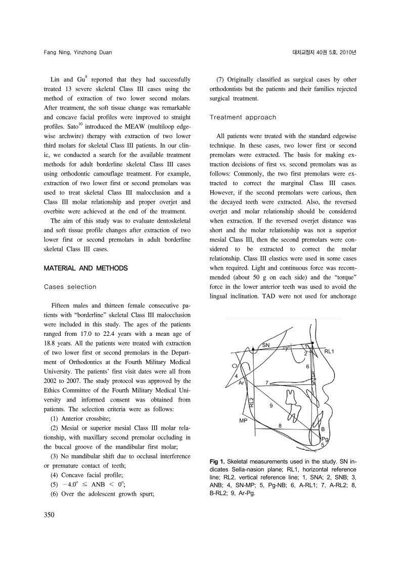
Fig 7. Posttreatment intraoral photographs. A, Lateral view on the right side; B, frontal view; C, lateral view on theleft side; D, occlusal view of maxillary dentition; E, occlusal view of mandibular dentition; F, lateral view of anteriorteeth.
Fig 8. Panoramic radiographs. A, Pretreatment; B, posttreatment.
of the mandiblular anterior teeth and Class III elastics,
the anterior crossbite was corrected. Sixteen months
later, a Class III molar relationship and Class I canine
relationship was established with a corrected midline.
At the end of treatment, the concave facial profile of
the patient was changed to a straight profile. The over-
jet and overbite were normal without deleterious ef-
fects to the periodontium and the lower incisors were
stable. The active treatment time for this patient was
twenty three months. The skeletal Class III tendency
remained after treatment with an ANB angle of −3.5o,
but the facial profile showed significant improvement.
DISCUSSION
Select identifications for treatment in “bor-derline skeletal Class III malocclusion”
“Borderline” surgical/orthodontic cases refer to pa-
tients with mild to moderate skeletal problems that can
be treated by either orthodontic or surgical means.15
Cassidy et al.16
defined “borderline cases” as those pa-
tients who were similar with respect to the character-
istics on which the orthodontic/surgical decision ap-
peared to have been based. As for the adult skeletal
Class III cases, however, how should clinicians de-
termine if patients are suitable for surgery? It still
lacks a clear consensus.9,17,18
Kerr et al.17
tried to es-
tablish some cephalometric yardsticks in adult patients
with class III malocclusion to find objective criteria for

Vol. 40, No. 5, 2010. Korean J Orthod Class III camouflage treatment
355
Fig 9. Lateral cephalometric radiographs. A, Pretreatment; B, posttreatment.
Fig 10. Superimposition of pretreatment and posttreat-ment cephalometric tracings.
treatment options. These researchers indicated that sur-
gery should be performed for patients with an ANB
angle of less than −4o, a maxillary/mandibular (M/M)
ratio of 0.84, an inclination of the lower incisors to the
mandibular of 83o, and a Holdaway angle of 3.5
o. In
2002, a formula was developed to determine whether
patients with class III malocclusion underwent either
orthodontic treatment or orthognathic surgery, on the
basis of the four variables: Wits appraisal, length of
the anterior cranial base, M/M ratio, and lower gonial
angle.19
Zeng et al.18
reported that orthodontic doctors
should consider orthognathic surgery when the ANB
angle was under −4o and L1-MP angle was under 82o.
Ning et al.14
suggested that orthodontic camouflage
treatment could achieve good results when the ANB
angle was from −3o to 0o. However, these studies did
not provide methods to specifically distinguish between
patients with “borderline” surgical- orthodontic class
III malocclusion. Furthermore, the treatment choices
largely depended on the clinicians' personal pre-
ferences. In our research for all patients, the ANB an-
gle was under 0o and the lowest value was −4.0o.
Using the orthodontic camouflage treatment, all pa-
tients achieved a proper overjet and overbite.
Influence of extraction of two lower first or second premolars on skeletal, dental and soft-tissue profiles
Extraction of two lower first or second premolars
has little effect on skeletal parameters and there were
no statistical significance observed (p ≥ 0.05). The
change in ANB angle was not obvious with a value of
−2.0o after treatment. This proved that orthodontic
treatment of skeletal Class III cases with the extraction
method had camouflage effects and surgical-ortho-
dontic treatment should be suggested for severe skel-
etal Class III cases to achieve a good effect. In our
study the change seen in the lower incisors was sig-
nificant with several items changing significantly (p <
0.01) due to the extraction of premolars in the
mandible. After treatment, the overjet and overbite was
proper. Pellegrino G20 and Fukui and Tsuruta et al.21
both reported a skeletal crossbite case with severe
crowding in the upper and lower arches. Both of the
patients were treated by extraction of four premolars.
The overjet and overbite became normal after treat-
ment and good occlusal relationship was achieved with
Fang Ning, Yinzhong Duan 대치교정지 40권 5호, 2010년
356
straight profiles attained. Our results with cephalo-
metric radiographs were consistent with their study.
When closing the extraction space in the mandible, we
should pay close attention to the thickness of the buc-
cal plate and avoid excessive lingual inclination during
retraction. The improvement in soft tissue profile was
obvious with concave profiles before treatment chang-
ing to straight profiles after treatment. The imbalance
of soft tissue change and skeletal change after ortho-
dontic treatment is valuable as a nonsurgical treatment
of severe skeletal Class III malocclusion.1,9 Also, the
compensatory mechanism is worthy of further study.
The tongue position of Class III patients is quite
large and flat. The forceful tongue could cause relapse,
and also occlusal trauma to upper anterior teeth could
occur if the lower teeth are uprighted due to force
from the tongue. For this, we instruct the patients to
do tongue exercises and ask them to wear retainers
carefully for at least two years.
Comparison of the extraction of two lower first or second premolars with other ex-traction modes for camouflage treatment of skeletal Class III malocclusion
According to the study by Lin and Gu,9 the in-
dications to extract two lower second molars include
severe skeletal Class III malocclusion, a mesial rela-
tionship of first molars and a well arranged arch or on-
ly minor crowding of the lower arch. Compared with
extraction of two lower second molars, whether the
third molar erupted into the second molar’s place was
not a concern when using our method. Also, after the
extraction of two lower second molars, all of the teeth
including the first molars in the mandible need to be
retracted back one at a time. The distance moved is
relatively long and the number of teeth moved is more
than that required during extraction of the two lower
first or second premolars. In our study, the extraction
space was closed by moving the anterior teeth back-
wards and posterior teeth forwards. The distance
moved and the treatment time were relatively short. In
the study by Lin JX, the mean treatment time was
comparatively longer, about 2.6 ± 0.6 years, but in our
study the mean treatment time was 2.0 ± 0.6 years.
Another extraction method in the treatment of skel-
etal Class III malocclusions was extraction of one low-
er incisor. This method was adopted for mild Class III
malocclusions and was noted for a shorter treatment
time. However, a midline deviation existed and stable
interlocking occlusal relationship was not easily ach-
ieved after treatment.
For camouflage treatment in skeletal Class III cases
combined with severe crowding, the extraction of four
premolars in the upper and lower arches was adopted
at times. Ning et al.14 indicated that the upper arch
length discrepancy should be carefully analyzed before
extraction of two upper premolars. Otherwise the facial
profile may be poor due to over-retraction of the ante-
rior teeth. The mean treatment time was 2.1 ± 0.4
years.
In orthodontic treatment, we have objectives to be
met which are function, esthetics and stability.
However, because long-term records are not yet avail-
able for this treatment group, caution is advised in as-
suming the permanency of acceptable outcomes at this
time. Proper diagnosis and the establishment of real-
istic treatment objectives by the clinician and the pa-
tient are necessary to prevent undesirable sequelae in
Class III camouflage treatment.
CONCLUSION
Extraction of two lower first or second premolars
provides a viable treatment alternative for skeletal
Class III cases to achieve good results.
After treatment, the overjet and overbite were nor-
mal and Class III molar relationship and Class I canine
relationship were achieved in all subjects. The occlu-
sion relationship was stable, tight and concordant.
ACKNOWLEDGEMENT
The authors thank Prof. Hee-Moon Kyung at the
Kyungpook national university in Korea and Prof.
Somchai Satravaha in Thailand, for amending the
English of the manuscript and helpful comments for
the discussion.

Vol. 40, No. 5, 2010. Korean J Orthod Class III camouflage treatment
357
-국문 록 -
성인 골격성 III 부정교합환자의 하악 소구치
발치를 통한 보상치료
Fang Ning, Yinzhong Duan
이번 연구의 목 은 성인 골격성 III 부정교합환자의 하악 제1소구치 는 제2소구치 발치 후에 치조골격성 변화와 연조직 변화를 평가하는 데 있다. 28명의 경계선상에 있는 골격성 III 부정교합환자들이 연구되었다. 모든 환자는 하악 제1소구치 는 제2소구치를 발치한 후 치료되었으며 치료 과 치료 후 측모두부방사선사진을 촬 하여 paired t-test를 통해 분석하 다. 치료 후에 골격 으로 유의성 있
는 변화는 찰되지 않았다. 치성변화에 해 하악 치각도가 8.1도 정도 감소하 으며 interincisal angle은 7.7도 정도 증가하 다 (p < 0.01). 수평 피개는 5.7 mm로 증가하 으
며 (p < 0.01), L1-NB 각은 7.3도 감소하 고 L1-NB 거리는 4.8 mm 감소하 다 (p < 0.01). 연조직 변화에 있어 Li-E, Li-H과 LiRL2 거리는 각각 3.2 mm, 3.4 mm, 4.1 mm 감소하 다 (p < 0.01). 심하지 않은 골격성 III 부정교합환자에 한 하악 소구치 발치를 통한 교정 인 보상치료는
우수한 교합 계를 달성할 수 있으며 측이 가능한 치료
안이 될 수 있다.
주요 단어: III 치료, 진단과 치료계획, 성인교정, 치아이동
REFERENCES
1. Lin J, Gu Y. Preliminary investigation of nonsurgical treatment
of severe skeletal Class III malocclusion in the permanent
dentition. Angle Orthod 2003;73:401-10.
2. Guyer EC, Ellis EE 3rd, McNamara JA Jr, Behrents RG.
Components of Class III malocclusion in juveniles and
adolescents. Angle Orthod 1986;56:7-30.
3. Dietrich UC. Morphological variability of skeletal Class 3 rela-
tionships as revealed by cephalometric analysis. Rep Congr
Eur Orthod Soc 1970;131-43.
4. Duan YZ, Yang ZH, Leng J. Effects of face mask/Class III
elastic therapy on severe skeletal crossbite. J Modern Stomatol
2003;17:251-2.
5. Gu Y, Rabie AB, Hägg U. Treatment effects of simple fixed
appliance and reverse headgear in correction of anterior
crossbites. Am J Orthod Dentofacial Orthop 2000;117:691-9.
6. Uner O, Yüksel S, Uçüncü N. Long-term evaluation after
chincap treatment. Eur J Orthod 1995;17:135-41.
7. Cha KS. Skeletal changes of maxillary protraction in patients
exhibiting skeletal class III malocclusion: a comparison of
three skeletal maturation groups. Angle Orthod 2003;73:26-35.
8. Tollaro I, Baccetti T, Franchi L. Craniofacial changes induced
by early functional treatment of Class III malocclusion. Am J
Orthod Dentofacial Orthop 1996;109:310-8.
9. Lin J, Gu Y. Lower second molar extraction in correction of
severe skeletal Class III malocclusion. Angle Orthod 2006;76:
217-25.
10. Sato S. Case report: developmental characterization of skeletal
Class III malocclusion. Angle Orthod 1994;64:105-11.
11. Lew KK. Soft tissue profile changes following orthodontic
treatment of Chinese adults with Class III malocclusion. Int J
Adult Orthodon Orthognath Surg 1990;5:59-65.
12. Demir A, Uysal T, Sari Z, Basciftci FA. Effects of camouflage
treatment on dentofacial structures in Class II division 1 man-
dibular retrognathic patients. Eur J Orthod 2005;27:524-31.
13. Nalbantgil D, Arun T, Sayinsu K, Fulya I. Skeletal, dental and
soft-tissue changes induced by the Jasper Jumper appliance in
late adolescence. Angle Orthod 2005;75:426-36.
14. Ning F, Duan Y, Huo N. Camouflage treatment in skeletal
Class III cases combined with severe crowding by extraction
of four premolars. Orthod Waves 2009;68:80-7.
15. Rabie AB, Wong RW, Min GU. Treatment in borderline Class
III malocclusion: orthodontic camouflage (extraction) versus
orthognathic surgery. Open Dent J 2008;2:38-48.
16. Cassidy DW Jr, Herbosa EG, Rotskoff KS, Johnston LE Jr. A
comparison of surgery and orthodontics in "borderline" adults
with Class II, division 1 malocclusions. Am J Orthod Dentofa-
cial Orthop 1993;104:455-70.
17. Kerr WJ, Miller S, Dawber JE. Class III malocclusion: surgery
or orthodontics? Br J Orthod 1992;19:21-4.
18. Zeng XL, Lin JX, Huang JF. Skeletal crossbite: surgery or or-
thodontics? West China J Stomatol 1985;3:233-7.
19. Stellzig-Eisenhauer A, Lux CJ, Schuster G. Treatment decision
in adult patients with Class III malocclusion: orthodontic ther-
apy or orthognathic surgery? Am J Orthod Dentofacial Orthop
2002;122:27-37.
20. Pellegrino G. Italian board of orthodontics: case N. 2 adult
malocclusion. Prog Orthod 2005;6:102-12.
21. Fukui T, Tsuruta M. Invisible treatment of a Class III female
adult patient with severe crowding and cross-bite. J Orthod
2002;29:267-75.