calpains: markers of tumor aggressiveness?
TRANSCRIPT
E X P E R I M E N T A L C E L L R E S E A R C H 3 1 6 ( 2 0 1 0 ) 1 5 8 7 – 1 5 9 9
ava i l ab l e a t www.sc i enced i r ec t . com
www.e l sev i e r . com/ loca te /yexc r
Research Article
Calpains: Markers of tumor aggressiveness?
Hélène Roumesa, Ludovic Leloupb,1, Elise Dargelosa, Jean-Jacques Brustisa,Laetitia Daurya,⁎, Patrick Cottina
aUniversité Bordeaux 1, Unité Protéolyse, Croissance et Développement Musculaire, INRA USC-2009, Avenue des Facultés,33405 Talence Cedex, FrancebDepartment of Pathology, S713 Scaife Hall, University of Pittsburgh, Pittsburgh, PA 15261, USA
A R T I C L E I N F O R M A T I O N
⁎ Corresponding author. Tel.: +33 (0)5 40 00 2E-mail address: [email protected] (L. Daury)
1 Tel.: (412) 624 9366; fax: (412) 624 8946.
0014-4827/$ – see front matter © 2010 Elseviedoi:10.1016/j.yexcr.2010.02.017
A B S T R A C T
Article Chronology:
Received 16 November 2009Revised version received5 February 2010Accepted 16 February 2010Available online 1 March 2010
Rhabdomyosarcoma (RMS) are soft-tissue sarcoma commonly encountered in childhood. RMScells can acquire invasive behavior and form metastases. The metastatic dissemination implicatesmany proteases among which are μ-calpain and m-calpain.Study of calpain expression and activity underline the deregulation of calpain activity in RMS.Analysis of kinetic characteristics of RMS cells, compared to human myoblasts LHCN-M2 cells,shows an important migration velocity in RMS cells. One of the major results of this study is thepositive linear correlation between calpain activity and migration velocity presenting calpains as amarker of tumor aggressiveness. The RMS cytoskeleton is disorganized. Specifying the role of μ-and m-calpain using antisense oligonucleotides led to show that both calpains up-regulate α- andβ-actin in ARMS cells. Moreover, the invasive behavior of these cells is higher than that of LHCN-M2 cells. However, it is similar to that of non-treated LHCN-M2 cells, when calpains are inhibited.In summary, calpains may be involved in the anarchic adhesion, migration and invasion of RMS.The direct relationship between calpain activity and migration velocities or invasive behaviorindicates that calpains could be considered as markers of tumor aggressiveness and as potentialtargets for limiting development of RMS tumor as well as their metastatic behavior.
© 2010 Elsevier Inc. All rights reserved.
Keywords:
CalpainsRhabdomyosarcoma cells
MigrationInvasionCytoskeleton
Alveolar and embryonal rhabdomyosarcoma (ARMS and ERMSrespectively) are soft-tissue sarcoma commonly encountered inchildhood and adolescence. These rhabdomyosarcoma (RMS) arisefrom immature cells which fate is to form striated skeletal muscle.These cells express, similarly to muscle cells, some muscle specificproteins such as myogenic factors (MRF), desmin, and myogenin[1,2]. ARMS are characterized by two chromosomal translocations:t(2;13)(q35;q14) or t(1;13)(p36;q14) [3]. ERMS histological typeis associated with a loss of heterozygosis at the 11p15 locus [4].RMS can acquire invasive behavior and can form metastases inlung, bone, marrow, and lymphatic nodes. The development of
1 94; fax: +33 (0)5 40 00.
r Inc. All rights reserved.
such metastases is associated with poor prognosis [5]. Thecomprehension of mechanisms that regulate cancer cells migra-tion and invasion may be a key for the development of newtherapies for limiting metastases. Among the many proteasesimplicated in cell motility, the calpains play an essential role inregulating migration and invasion phenomena, both involved inmetastases development [6–8]. Targeting calpains may present anovel approach toward restraining metastasis and development ofRMS cancers. Calpains are calcium-dependent cysteine proteaseswhich best-characterized members are μ-calpain and m-calpain(i.e. CAPN1 and CAPN2 respectively), known as ubiquitous
84 96.

1588 E X P E R I M E N T A L C E L L R E S E A R C H 3 1 6 ( 2 0 1 0 ) 1 5 8 7 – 1 5 9 9
heterodimeric enzymes consisting of a large 80-kDa catalyticsubunit and a smaller regulatory subunit (about 28 kDa, css1 orcss2). Css1 is ubiquitous while css2 is tissue-specific [9].
Calpain activity is Ca2+-dependent. Among the different factorsregulating calpain activity, the critical factor is its specificendogenous inhibitor, calpastatin [10]. Calpastatin is a ubiquitousprotein regulated by several factors [11] that requires Ca2+ to bindand inhibits calpains [12].
The present study focuses on the role of both μ- and m-calpainin the metastatic characteristics of the two cancer cell lines, ARMSand ERMS, assuming the role of calpains in cell adhesion andmigration (that are limited steps of invasion and metastasis).
To reach that goal, we first characterized the calpain system inRMS before studying the role of the calpains in RMS cell adhesion,on RMS morphology and cytoskeleton organization, as well as inRMS migration characteristics and invasive behavior. Non-tumorhuman myoblasts LHCN-M2 [13] were chosen as non-metastaticcontrol cells.
The main result is that calpains play a crucial role in adhesion,high migration velocities and invasive behavior of RMS. Morespecifically, calpains may be considered as markers of tumoraggressiveness since their global activity is linked to migrationvelocities by a positive linear correlation. Moreover, inhibition ofcalpain activity in RMS led to migration velocities and invasivebehavior similar to those of non-treated LHCN-M2 cells. Becausecalpain activity is directly involved in migration and invasion ofRMS, the proteases would be a good target for the development ofnovel therapies to control metastasis.
Materials and methods
Cell culture
Cells were grown under 5% CO2 atmosphere at 37°C in DMEM(Dulbecco'smodified Eaglemedium)with pyruvate supplementedwith 10% FBS (fetal bovine serum) for RMS, and a culture mediumcontaining DMEMwithout pyruvate (60%), medium 199 (20%) andFBS (20%) for LHCN-M2 cells (humanmyoblasts cell lines) [13]. Allcell culture reagents were purchased from Gibco-BRL. Someexperiments have been done only with the tumor cell linepresenting the most aggressive phenotype, ARMS.
Calpain inhibitor treatments
The chemical calpain inhibitor calpeptin (Z-Leu-Nle-CHO) waspurchased from Calbiochem and used at 40 and 60 μM for adhesionassay, 50 μM for migration and invasion assay for immunolocaliza-tion and for cytoskeleton organization and 80 μM forWestern blot.A second calpain inhibitor, calpain inhibitor III (Z-Val-Phe-CHO,Calbiochem) was used at 50 μM. Antisense oligodeoxynucleotidetransient transfections has been realized as previously described[14].
Quantification of calpain activity
All cell lines were cultured in DMEM supplemented with 0.1%FBS during 48 h. The global calpain activity was observed andquantified on living cells using t-BOC-LM-CMAC (7-amino-4-chloromethylcoumarin, t-BOC-L-leucyl-L-methionine amide,
Molecular Probes) [7,15]. This molecule enters passively in cellsand becomes fluorescent after calpain cleavage. Culture mediumwas replaced by DMEMwithout serum containing 50 μM of t-BOC-LM-CMAC. Cells were incubated during 30 min in darkness. The μ-calpain activity was observed and quantified on fixed andpermeabilized cells using Calpain 1 substrate (Chemicon) whichbecome fluorescent after μ-calpain cleavage. After PAF fixation(15 min) and permeabilization with 1% Triton X-100 (3 min), cellswere incubated with Calpain 1 substrate 50 μM during 40 min indarkness. The global calpain or the μ-calpain activities were thenobserved using an inverted epifluorescence microscope (Leica AFDMI6000). The results were quantified using the Metamorphsoftware (Molecular Device).
Immunolocalization of calpains and calpastatin
All cell lines were cultured in DMEM supplemented with 0.1% FBSfor 48 h. Next, cells were fixed and permeabilized. A specificity wasdisrupted by incubation (1 h) with PBS/BSA (3%). Then, m-calpain,μ-calpain and calpastatin were detected using correspondingantibodies (N-19 sc-7533 raised in goat (1/50), Santa CruzBiotechnology; MAB3104, raised in mouse (1/500), Chemicon;H-300 sc-20779, raised in rabbit (1/50), Santa Cruz Biotechnologyrespectively, for 3 h). Then, cells were incubated with anappropriate secondary Alexa fluor antibody (anti-goat-Alexa594-conjugated (1/1000), anti-mouse-Alexa594-conjugated (1/3000),and anti-rabbit-Alexa488-conjugated (1/1000) antibodies respec-tively for 90 min). Next cells were observed using an epifluores-cence microscope (Leica AF DMI6000).
Adhesion assay
Adhesion assays were performed as described previously [14].These assays were carried out with (40 μM or 60 μM) or withoutcalpeptin treatment.
Migration assay
All cell lineswere seeded in DMEM containing 0.1% FBS at a densityof 4.103 cells/cm2 in 35 mm diameter glass-bottom plate. Eighthours after seeding, time-lapse microscopy experiments wereperformed on an inverted Leica AF DMI6000 microscope equippedwith an environmental chamber with phase-contrast optic(images taken every 15 min). Cell velocities of migration, definedas the average of 73 subsequent cell centroid displacements/onetime interval between two successive images (15 min), wereevaluated using the tracking object of the Metamorph software(Molecular Device). The cell trajectories were recorded for 18 h.These assays have been made in presence vs. absence of calpeptin(50 μM) added at the beginning of the time-lapse.
Invasion assay
The invasive capacity of RMS and LHCN-M2 cells was measuredusing BD Biocoat Growth Factor Reduced Matrigel InvasionChambers (BD Biosciences). Cells were seeded at a density of 105
cells/cm2 on the membrane in DMEM with 0.1% FBS. Quiescentmedium was also added under the membrane. After 48 h ofincubation, cells staying on the membrane were scraped off. Cellslying under the membrane were fixed with PAF (4%), and

1589E X P E R I M E N T A L C E L L R E S E A R C H 3 1 6 ( 2 0 1 0 ) 1 5 8 7 – 1 5 9 9
dehydrated overnight using ethanol. Cells were then stained withHansen's hemalun and evaluated. The same experiment wasrealized with culture insert whose PET membrane was recoveredby BD Matrigel matrix. Cells crossing the membrane were stainedand evaluated. The invasive capacity of all cell lines was expressedby the percentage of cells going through the membrane withMatrigel matrix as compared to the percentage of cells goingthrough the membrane without any Matrigel matrix. The implica-tion of calpains in this phenomenon was evaluated using the sameprocedures after calpeptin treatment (50 μM).
Cytoskeleton analysis and vinculin localization
Cell actin fibers were stained as previously described [16], with vs.without calpeptin or inhibitor III, and observed using anepifluorescence microscope (Leica AF DMI6000). Vinculin wasdetected after fixation and permeabilization, using primaryantibody (V-9131, Sigma-Aldrich; 1/50) (3 h). Next, cells wereincubated with a secondary Alexa fluor antibody from MolecularProbes (anti-mouse-Alexa594-conjugated 1/1000) (90 min).
Western blots
Western blots were performed as described previously [16]. Thebicinchoninic acid protein assay kit was purchased from Pierce,polyvinylidene fluoride membrane from Millipore and nitrobluetetrazolium/5-bromo-4chloro-3-indoyl-1-phosphate from Pro-mega. The membranes were incubated with specific antibodiesagainst m-calpain (raised in sheep as previously described([17]; 1/1000), β-tubulin (Santa Cruz Biotechnology (H-235 sc-9104); 1/500), μ-calpain (Sigma-Aldrich (C-0355); 1/2500) andcalpastatin (Santa Cruz Biotechnology (H-300 sc-20779);1/250). The secondary antibody conjugated to alkaline phos-phatase (Sigma-Aldrich; 1/10,000) was used for detection. Theapparent density of the band on the membranes was quantifiedwith Totalab (Progenesis) and corrected according to β-tubulinexpression.
Extraction of total RNA, reverse transcription andquantitative PCR
Extraction of total RNA, reverse transcription and quantitative PCRwere performed as previously described [18]. Forward and reverseprimer sequences for μ- and m-calpain amplification were agtcg-tgccgcagcatggtga, acttgtccaggtcaaacttccg and gccatcaagtacctcaac-cagg, gctccttggcagatgtctgtg, respectively. Transcript concentrationof both isoforms was determined from the standard curveand compared to the control LHCN-M2. GAPDH (glyceraldehydephosphate dehydrogenase) was used as control; its forward andreverse primer sequences were aaggtcatcccagagctgaacgc andacaacctggtcctcagtgtagcc.
Statistical analysis
The statistical significance of the differences between multiplegroupswas determined using ANOVA.When Fwas associatedwitha probability p<0.05, intergroup comparisons were conductedusing Student's t-test. Unless indicated, all data were expressed asmeans±SEM and were representative of an average of at leastthree independent experiments.
Results
Characterization of the calpain system inrhabdomyosarcoma cells
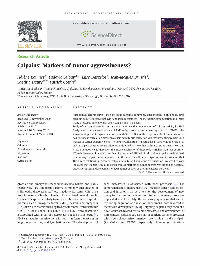
In RMS cells, the expression of μ-calpain and m-calpain wassignificantly reduced compared to that of LHCN-M2 cells(Fig. 1A1–2). Moreover, the expression of calpastatin was reducedby 85±1% in ARMS cells and 74±3% in ERMS cells, compared tothat of LHCN-M2 cells (Fig. 1A3).
Specific mRNA levels for μ- and m-calpains were quantified byreal time RT-PCR and compared to that of LHCN-M2 control cells(Fig. 1B). The mRNA level of μ-calpain was similar in all cell lines(Fig. 1B1) whereas the expression level of m-calpain mRNA wasreduced by more than 40% and 50% for ARMS and ERMS cells,respectively (Fig. 1B2).
Using t-BOC-LM-CMAC, the global calpain activity of RMS cellswasmeasured and compared to that of LHCN-M2 cells (Fig. 1C1–3).Calpain activity was significantly higher in both RMS cell lines thanin LHCN-M2 cells (2.6±0.6 and 2.1±0.1 fold for ARMS and ERMScells respectively) (Fig. 1C4). Furthermore, μ-calpain activity(calpain 1 substrate) was higher in RMS than in LHCN-M2 cells(2.3±0.4 and 2.7±0.4 fold for ARMS and ERMS cells respectively)(Fig. 1C5).
In LHCN-M2myoblasts, μ-calpain was concentrated around thenucleus with a weak decreasing gradient towards the cellperiphery presenting a linear structure parallel to the main axisof the cell (Fig. 1D1). In these cells, m-calpain was moreparticularly localized around the nucleus in a spot-shaped fashion(Fig. 1D2) and calpastatin presented a higher concentrationaround the nucleus, decreasing towards the cell periphery (Fig.1D3). In RMS cells, μ-calpain presented a higher concentrationaround the nucleus, decreasing towards the cell periphery. m-Calpain and calpastatin localizations were not different betweenall cell lines (Fig. 1D4–6 for ARMS, data not shown for ERMS cells).
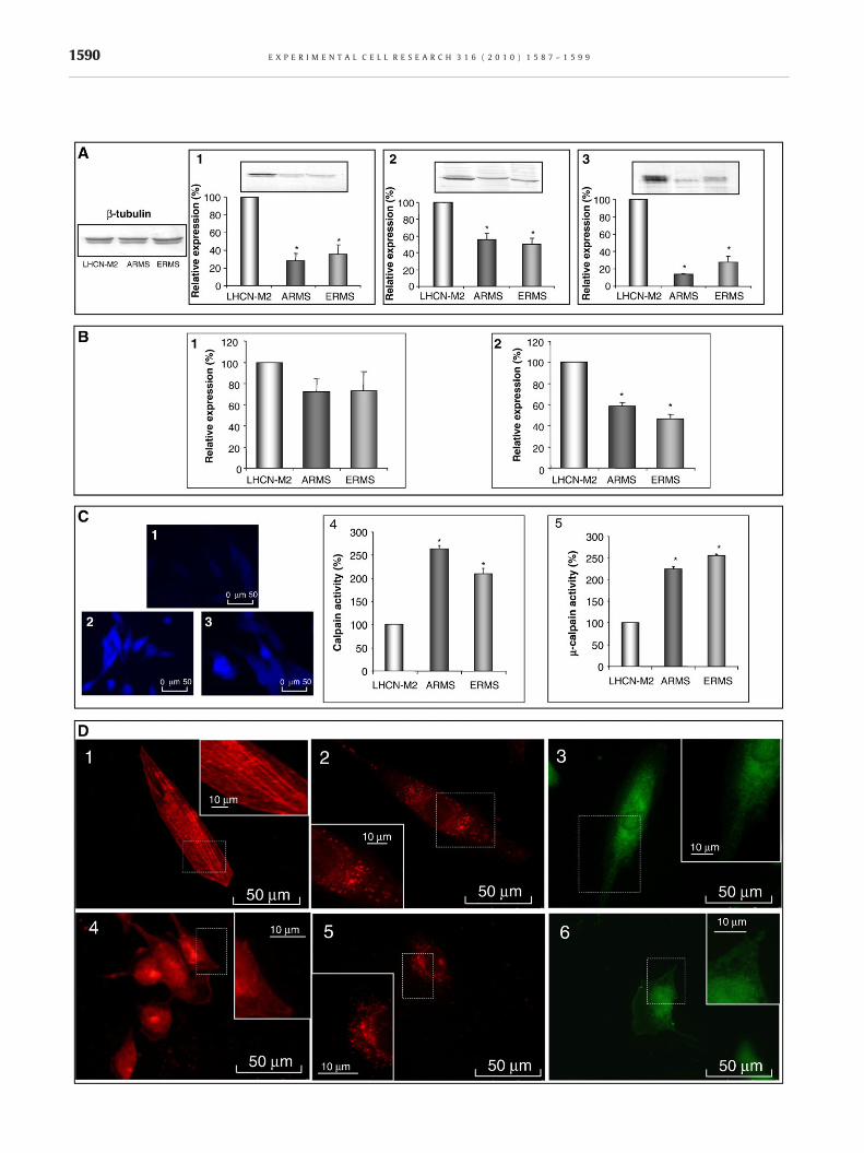
Implication of calpains in adhesion
As shown in Fig. 2A, RMS cells presented a delay and a decrease intheir adhesion rates. Eight hours after seeding, the adhesion ratesof ARMS and ERMS were 37±3 % and 35±4% respectively,whereas LHCN-M2 myoblasts adhered completely (p<0.05).
Calpain inhibition drastically decreased the adhesion rate ofLHCN-M2 (Fig. 2B1). The presence of calpeptin led to a decrease inthe adhesion rate of ARMS. However, this decrease was significantonly 2 and 4 h after seeding at 40 μM of calpeptin and after 2 h at60 μMof calpeptin (Fig. 2B2). The adhesion rates of ERMSwere notaffected by the presence of calpeptin neither at 40 μM of calpeptinnor at 60 μM (data not shown).
In order to understand the difference of adhesiveness betweenRMS and LHCN-M2 cells, vinculin (one of the main focal adhesioncomponents) localization was investigated in all cell lines (Fig. 2).In LHCN-M2 cells, vinculin was regularly localized at the ventralsurface of the cell (Fig. 2C1). On the contrary, in both RMS celllines, vinculin was principally localized at the periphery of the cell(Fig. 2C2 for ARMS, data not shown for ERMS). The inhibition ofcalpain activity led to a reorganization of vinculin. In all cell linespresenting a rounded shape, vinculin was localized predominantlyat the periphery of the ventral surface of the cell (Fig. 2C3 and C4
1590 E X P E R I M E N T A L C E L L R E S E A R C H 3 1 6 ( 2 0 1 0 ) 1 5 8 7 – 1 5 9 9

1591E X P E R I M E N T A L C E L L R E S E A R C H 3 1 6 ( 2 0 1 0 ) 1 5 8 7 – 1 5 9 9
for LHCN-M2 and ARMS cells, respectively, data not shown forERMS). Contrary to LHCN-M2 myoblasts, in which the presence ofcalpeptin led to a 107±14% increase in vinculin expression, thepresence of calpeptin in ARMS led to a 14±3% decrease in vinculinexpression (Fig. 2C5). Antisense oligodeoxynucleotide treatmentsagainst μ-calpain led to an increase in LHCN-M2 vinculinexpression (14±4%) but did not affect the vinculin expressionlevels of ARMS (p=0.30) (Fig. 2C6–7). Antisense oligodeoxynu-cleotide treatments againstm-calpain led to an increase in vinculinexpression in LHCN-M2 cells (Fig. 2C6). Conversely, this treatmentin ARMS led to a decrease in vinculin expression (Fig. 2C7).
Morphological characteristics and actin cytoskeletonorganization of migrating rhabdomyosarcoma cells
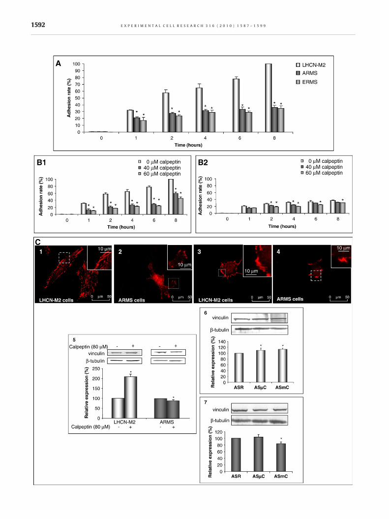
Eight hours after seeding, control cells exhibited an elongatedshape and membrane protrusions. Migrating RMS cell linesexhibited an elongated shape with a reduced cytoplasm andlarge nuclei. In addition, membrane protrusions were smaller andthinner than those of LHCN-M2 myoblasts (Fig. 3A1 and 3A2 forLHCN-M2 and ARMS cells respectively; data not shown for ERMS).The inhibition of calpain activity impaired cell morphology, all celllines presenting a rounded shape with very short membraneprotrusions (Fig. 3B1 and B2 for LHCN-M2 and ARMS cells,respectively; data not shown for ERMS).
Since the actin cytoskeleton is responsible for cell morphologyand considered to be the motor of cell migration [19], we studiedits organization in the different cell lines. In elongated LHCN-M2cells, the actin cytoskeleton formed stress fibers parallel to themain axis of the cell (Fig. 3C1). On the contrary, in both RMS celllines, actin cytoskeleton failed to form parallel stress fibers (Fig.3C2 for ARMS; data not shown for ERMS). The actin cytoskeletonwas disorganized and mainly concentrated at the periphery of thecell.
In presence of calpeptin, the LHCN-M2 F-actin was no longer atthe center of the cell. However it still remained organized at theperiphery of the cell where it was concentrated (Fig. 3C3). In bothRMS cell lines, the inhibition of calpains induced an increase in theconcentration of F-actin at the periphery of the cell (Fig. 3C4, datanot shown for ERMS).
In all cell lines, the presence of calpeptin led to an increase inthe quantity of F-actin (Fig. 3D1). This result was confirmed by theuse of calpain inhibitor III leading to an increase in the level of F-actin by 18±3% in ARMS (Fig. 3D2).
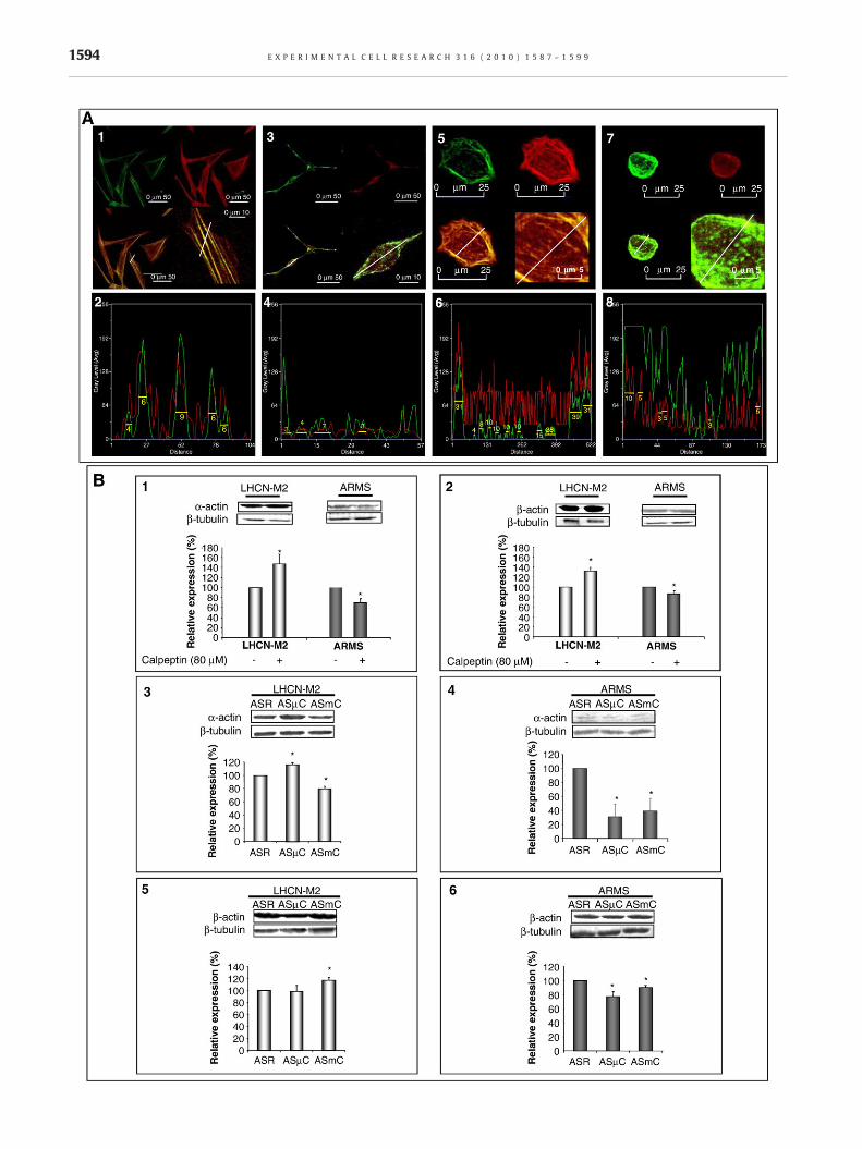
Implication of calpain in the arrangement of actincytoskeleton
In LHCN-M2 cells, F-actin and μ-calpain colocalized by 93% (F-actinoverlapping μ-calpain: 93.2±0.5% and μ-calpain overlapping F-actin: 93.2±1.0%) (Fig. 4A1). In these cells, stress fibers were
Fig. 1 – Characterization of calpain system components in rhabdomcalpastatin in all cell lines (A1, A2, and A3, respectively). Quantificat(B1 and B2, respectively). Global calpain activity in LHCN-M2, ARMof global calpain activity and μ-calpain activity (C4 and C5, respectLHCN-M2 cells (D1, D2, and D3, respectively) and in ARMS cells (D4, Dleast 3 independent experiments. ⁎Significantly different from therepresentative of at least three separated sets of culture.
associated with a peak of μ-calpain concentration (Fig. 4A2). Inaddition, F-actin in ARMS cells colocalized with μ-calpain at alower extent than in LHCN-M2 myoblasts (F-actin overlapping μ-calpain: 66.5±3.5% and μ-calpain overlapping F-actin: 46.7±2.9)(Fig. 4A3–4). In both cell lines, the presence of calpeptin led to arelocalization of F-actin as well as μ-calpain at the periphery of thecell (Fig. 4A5–8).
In order to precise the role of each calpain on α-actin and β-actin expression (components of F-actin), the expression of eachisoform of actin monomer was determined in the presence vs.absence of calpeptin. In LHCN-M2 cells, the inhibition of calpainsinduced an increase of bothα-actin and β-actin (α-actin increasedby 51±14% and β-actin increased by 33±7) (Fig. 4B1–2). InARMS conversely, this inhibition induced a decrease of both α-actin and β-actin (α-actin decreased by 38±5% and β-actindecreased by 15±3%) (Fig. 4B1–2).
Moreover, in LHCN-M2 cells, the inhibition of μ-calpain (byantisense oligodeoxynucleotide treatments) up-regulated theexpression of α-actin but did not affect the expression level ofβ-actin (Fig. 4B3 and 5) while the inhibition of m-calpain down-regulated the expression of α-actin and up-regulated the expres-sion of β-actin (Fig. 4B4 and 6). It appeared that the inhibition of μ-calpain as well as m-calpain in ARMS down-regulated theexpression of both α-actin and β-actin (Fig. 4B4 and 6).
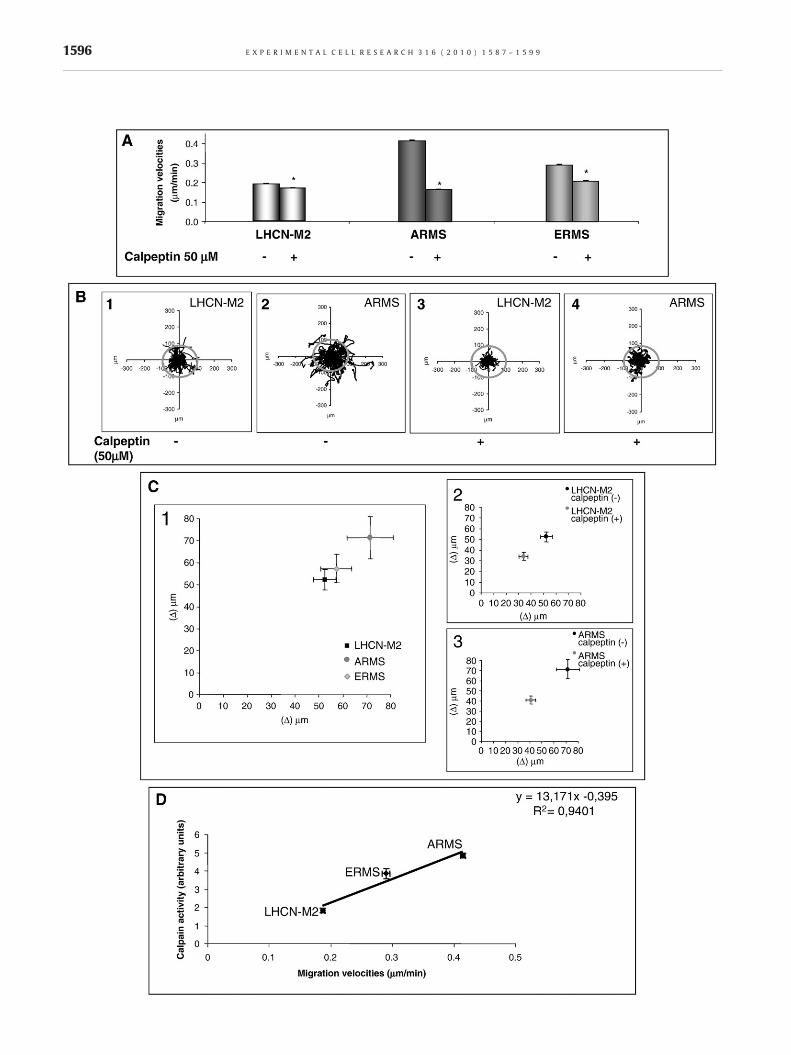
Kinetic characteristics of rhabdomyosarcoma cell lines
As presented in Fig. 5A, the migration velocities of cancer cellswere significantly higher (about 2.3 and 1.6 fold for ARMS andERMS respectively) than those measured in controls. The inhibi-tion of calpain activity drastically impaired migration velocities ofall cell lines (Fig. 5A). The velocities decreased by 11% for LHCN-M2, 60% for ARMS and 28% for ERMS cells. Similar results wereobtained using the calpain inhibitor III (data not shown).
To complete this study, the different cell trajectories wereanalyzed and are presented in Fig. 5B. The ARMS dispersion areawas higher than that of LHCN-M2 cells (Fig. 5B2 vs. B1, ERMSdispersion area was intermediate, data not shown). As presentedin Fig. 5C1, Δ (vector between the initial point of each trajectoryand the final localization of the cell) confirmed that the dispersivecapacity was higher for the ARMS cell line. The inhibition of calpainactivity significantly impaired the dispersion area of all cell lines(Fig. 5B3 vs. B1 and B4 vs. B2) as well as the dispersive behavior(Fig. 5C2–3; data not shown for ERMS). The dispersive capacitydecreased by 35%, 44% and 49% for LHCN-M2, ARMS and ERMScells respectively. Moreover, the dispersive capacity of treatedARMS cells was similar to the dispersive capacity of LHCN-M2 instandard conditions (p=0.07).
These results highlight the role of calpains in migration anddispersive behavior of all cell lines. Moreover, the global activityof calpains was directly correlated with the velocity of each cell
yosarcoma cells. Expression of μ-calpain, m-calpain andion of μ-calpain andm-calpainmRNA expression, in all cell linesS and ERMS cells (C1, C2, and C3, respectively). Quantificationively). Localization of μ-calpain, m-calpain and calpastatin in5, and D6, respectively). Results are mean values±SEM from atcontrol LHCN-M2 cells (p<0.05). Pictures shown are
1592 E X P E R I M E N T A L C E L L R E S E A R C H 3 1 6 ( 2 0 1 0 ) 1 5 8 7 – 1 5 9 9
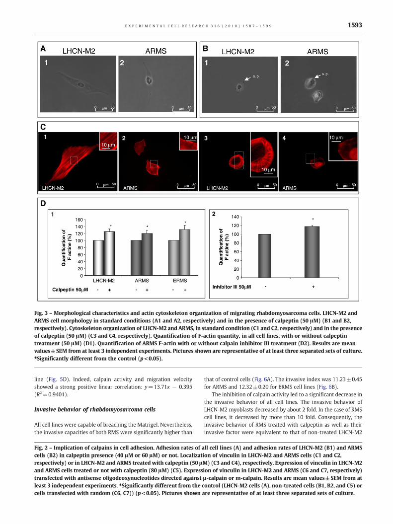
Fig. 3 – Morphological characteristics and actin cytoskeleton organization of migrating rhabdomyosarcoma cells. LHCN-M2 andARMS cell morphology in standard conditions (A1 and A2, respectively) and in the presence of calpeptin (50 μM) (B1 and B2,respectively). Cytoskeleton organization of LHCN-M2 and ARMS, in standard condition (C1 and C2, respectively) and in the presenceof calpeptin (50 μM) (C3 and C4, respectively). Quantification of F-actin quantity, in all cell lines, with or without calpeptintreatment (50 μM) (D1). Quantification of ARMS F-actin with or without calpain inhibitor III treatment (D2). Results are meanvalues±SEM from at least 3 independent experiments. Pictures shown are representative of at least three separated sets of culture.⁎Significantly different from the control (p<0.05).
1593E X P E R I M E N T A L C E L L R E S E A R C H 3 1 6 ( 2 0 1 0 ) 1 5 8 7 – 1 5 9 9
line (Fig. 5D). Indeed, calpain activity and migration velocityshowed a strong positive linear correlation: y=13.71x − 0.395(R2=0.9401).
Invasive behavior of rhabdomyosarcoma cells
All cell lines were capable of breaching the Matrigel. Nevertheless,the invasive capacities of both RMS were significantly higher than
Fig. 2 – Implication of calpains in cell adhesion. Adhesion rates of acells (B2) in calpeptin presence (40 μM or 60 μM) or not. Localizatrespectively) or in LHCN-M2 and ARMS treated with calpeptin (50 μand ARMS cells treated or not with calpeptin (80 μM) (C5). Expresstransfected with antisense oligodeoxynucleotides directed againstleast 3 independent experiments. ⁎Significantly different from the ccells transfected with random (C6, C7)) (p<0.05). Pictures shown a
that of control cells (Fig. 6A). The invasive index was 11.23±0.45for ARMS and 12.32±0.20 for ERMS cell lines (Fig. 6B).
The inhibition of calpain activity led to a significant decrease inthe invasive behavior of all cell lines. The invasive behavior ofLHCN-M2 myoblasts decreased by about 2 fold. In the case of RMScell lines, it decreased by more than 10 fold. Consequently, theinvasive behavior of RMS treated with calpeptin as well as theirinvasive factor were equivalent to that of non-treated LHCN-M2
ll cell lines (A) and adhesion rates of LHCN-M2 (B1) and ARMSion of vinculin in LHCN-M2 and ARMS cells (C1 and C2,M) (C3 and C4), respectively. Expression of vinculin in LHCN-M2ion of vinculin in LHCN-M2 and ARMS (C6 and C7, respectively)μ-calpain or m-calpain. Results are mean values±SEM from atontrol (LHCN-M2 cells (A), non-treated cells (B1, B2, and C5) orre representative of at least three separated sets of culture.
1594 E X P E R I M E N T A L C E L L R E S E A R C H 3 1 6 ( 2 0 1 0 ) 1 5 8 7 – 1 5 9 9
1595E X P E R I M E N T A L C E L L R E S E A R C H 3 1 6 ( 2 0 1 0 ) 1 5 8 7 – 1 5 9 9
cells (Fig. 6A–B). Similar results were obtained using the calpaininhibitor III (data not shown).
Discussion
RMS cells can acquire invasive behavior and can form metastasesin lung, bone, marrow, and lymphatic nodes decreasing thechances of patients' healing to less than 20% [5]. The comprehen-sion of the mechanisms that regulate cancer cells migration andinvasion may be a key for the development of new therapies forlimiting metastases.
The determination of the mRNA expression level of μ-calpainand m-calpain, in all cell lines showed that it was similar for μ-calpain and it was reduced for m-calpain, in RMS cells, comparedto control. However, Western blots showed that μ-calpain as wellas m-calpain were reduced in RMS when compared to control.These results could be explained by a potential post-transcrip-tional regulation for RMS μ-calpain and by an absence of suchregulation for m-calpain. In RMS cell lines, even though theexpression of the calpain system components was decreasedcompared to LHCN-M2 myoblasts, both global and μ-calpainactivities were higher than in LHCN-M2 cells. This deregulationof RMS calpain system is in agreement with the high activity orexpression of calpains detected in human renal cell carcinoma[20], in squamous cell carcinoma [21], and in human colorectaladenocarcinoma [22]. In RMS cells, calpastatin expression is highlydown-regulated. Thereby, the increase in calpain activity could bedue to an accumulation of the non-calpastatin-inhibited proteo-lyzed active forms of the proteases. The immunolocalization ofcalpain system components was different between the cell lines. InRMS cells, μ-calpain presented a gradient of concentrationdecreasing toward the periphery of the cell. Conversely, inLHCN-M2 cells, μ-calpain presented linear structures parallel tothe main axis of the cell. This difference of μ-calpain organizationbetween LHCN-M2 and RMS cells presumes a different role for thisprotease as a function of the cell type. The localization of m-calpainwas identical in all cell types and this observation confirms theresults obtained by Samanta et al. showing the localization of m-calpain at the endoplasmic reticulum level [23].
In RMS cells, the weak adhesion rate compared to LHCN-M2 ischaracteristic of the metastatic phenotype of these cells. In bothRMS cell lines, the quantity of focal adhesions, marked by vinculin,was weaker than that of LHCN-M2 cells. Moreover, in RMS,vinculin was concentrated essentially at the periphery of the cell.
Fig. 4 – Implication of calpain in the actin cytoskeleton arrangemenLHCN-M2 cells, in standard conditions (A1). Representation of colothe line drawn on A1 (A2). A width at themiddle of the peak overlapF-actin and μ-calpain (white line). Numbers indicate the amount oImmunolocalization and representation of colocalization of F-actinrespectively). Immunolocalization and representation of colocalizapresence of calpeptin (50 μM) (A5 and A6, respectively, for LHCN-MQuantification of α-actin and β-actin expression in LHCN-M2 and Arespectively). Quantification of α-actin expression in LHCN-M2 andB4, respectively) and quantification of β-actin expression in LHCN-are mean values±SEM from at least 3 independent experiments. ⁎Sitransfected with random) (p<0.05). Pictures shown are representa
This vinculin distribution may be responsible for the weakadhesion rate of RMS. These results are in agreement with thedecreased adhesiveness observed in circulating tumor cells inprostate cancer [24]. A reduction in the adhesiveness may increasethe motility of RMS cells which is a key step of invasion [25].Inhibition of calpain activity impaired the adhesion of LHCN-M2and ARMS cells involving calpains in this process. Moreover, theinhibition of μ-calpain as well as m-calpain led to an over-expression of vinculin in LHCN-M2 cells. These results indicate thatvinculin is a calpain substrate in human myoblasts, in agreementwith the results obtained by Taylor et al. in Z-disc [26]. In ARMS,the expression of vinculin was regulated only by m-calpain thatstimulated its expression. Together, these results show the role ofcalpain in adhesion of LHCN-M2 and ARMS. They also support adifferential role for μ-calpain and m-calpain in the regulation ofvinculin depending on the cell type. These differences at theadhesive capacity level may be responsible for some differences atthe migration level.
Indeed, in all cell lines, the determination of migrationvelocities showed that RMS migrated faster and further awaythan LHCN-M2. The highmigration velocities of RMS cells linked tothe high invasive behavior of these cell lines are both character-istics of tumor and metastatic cells. In LHCN-M2 as well as in RMScell lines, the inhibition of calpain activity led to a decrease in cellmigration velocities. This decrease is in agreement with thedecrease of migration observed in fibroblasts capn 4−/− [27].Moreover, in presence of calpeptin, the migration velocities ofARMS were similar to the migration velocities of LHCN-M2 cells instandard conditions (p=0.52, NS). Similarly, the dispersionbehavior of all cell lines was reduced and in presence of calpeptin,the dispersive capacity of ARMS was similar to the dispersivecapacity of LHCN-M2 cells in standard conditions. Together, theseresults indicate that calpains are involved in migration velocitiesand dispersive capacity of migrating cells.
To summarize, RMS showed a high calpain activity and highmigration velocities. One of the main results was the determina-tion of a positive linear correlation between migration velocitiesand calpain activity. Indeed, calpain activity was directly linked tomigration velocities pointing these proteases as markers of tumoraggressiveness.
The reorganization of the actin cytoskeleton is a fundamentalstep in cell adhesion, migration and invasion [19]. In LHCN-M2cells, the actin cytoskeleton was organized in stress fibers thatwere parallel to the main axis of the cell. This organization confersa strong adhesiveness to these cells. Moreover, the F-actin
t. Immunolocalization of F-actin (green) and μ-calpain (red) incalization of F-actin (green) and μ-calpain (red), at the level ofping zone higher than 2 pixels illustrated colocalization of bothf pixel between the limit of the overlapping zone (A2).and μ-calpain in ARMS cells, in standard conditions (A3 and A4,tion of F-actin and μ-calpain in LHCN-M2 and in ARMS, in the2 cells and A7 and A8, respectively, for ARMS cells).RMS cells with or without calpeptin (80 μM) (B1 and B2,ARMS cells after antisense oligonucleotides treatment (B3 andM2 and ARMS cells, in the same conditions (B5 and B6). Resultsgnificantly different from the control (non-treated cells or cellstive of at least three separated sets of culture.
1596 E X P E R I M E N T A L C E L L R E S E A R C H 3 1 6 ( 2 0 1 0 ) 1 5 8 7 – 1 5 9 9

Fig. 6 – Invasive behavior of rhabdomyosarcoma cells. Invasive capacity of LHCN-M2, ARMS and ERMS cells with orwithout calpeptin(50 μM) (A). Invasive index of LHCN-M2, ARMS and ERMS cells with or without calpeptin (50 μM) (B). Results aremean values±SEMfrom at least 3 independent experiments. ⁎Significantly different from the control LHCN-M2 cells; ⁎⁎significantly different fromdata without calpeptin (p<0.05).
1597E X P E R I M E N T A L C E L L R E S E A R C H 3 1 6 ( 2 0 1 0 ) 1 5 8 7 – 1 5 9 9
constituting the stress fibers colocalized with μ-calpain. The samecolocalization was previously reported in dendritic cells [28] andmay implicate μ-calpain in the organization and rearrangement ofthe actin cytoskeleton. Even if μ-calpain seems to play a role at thetime of cell adhesion [29], its association with the stress fiberspresumes of a singular role in migration. On the contrary, the actincytoskeleton of RMS was disorganized and concentrated at theperiphery of the cell. This lack of stress fibers has already beendescribed in v-Src transformed cells in which calpain activitysimulates the loss of actin stress fibers and induces somemorphological modifications [30]. This disorganization may beresponsible for the weak adhesion rate and the high migrationvelocities of RMS cells [31]. In all cell lines, the inhibition of calpainactivity induced morphological modifications: cells becamerounded, with small protrusions. These changes contrast withthe modifications observed in CHO cells treated with calpaininhibitor 1 which present longer shape than controls [32]. Indeed,
Fig. 5 – Kinetic characteristics of rhabdomyosarcoma cell lines. Avecalpeptin (A). Individual trajectories of cell displayed in diagrams drof the plot (B). LHCN-M2 and ARMS cells trajectories in standard co(B3 and B4, respectively). Distance Δ between the initial point of etime-lapse (C). Δ for LHCN-M2, ARMS, and ERMS cells, in standard ccalpeptin (C2 for LHCN-M2 and C3 for ARMS cells). Positive linear cResults are mean values±SEM from at least 3 independent experim(p<0.05).
fibroblasts over-expressing calpastatin show a rounded morphol-ogy as observed in treated LHCN-M2 and RMS cells [33]. In LHCN-M2 cells, calpain inhibition induced a disorganization of the centralactin cytoskeleton and the F-actin was concentrated at theperiphery of the cell. These results are in agreement with thoseobtained in C2C12 cells over-expressing calpastatin [34]. Thisrearrangement of actin cytoskeleton was coupled with therelocalization of μ-calpain. This result is in agreement with thechanges in μ-calpain localization coordinated with the actin-related cytoskeleton rearrangements observed during early em-bryonic development of Drosophila [35]. In LHCN-M2 cells, thepersistence of peripheral actin stress fibers, coupled with thedisorganization of central fibers could be explained by studiesshowing that two different pathways are involved in stress fiberregulation: one regulates peripheral stress fibers, depending oncalcium and involving calmodulin while the other implies Rho andRho-kinase [36]. The same concentration of F-actin at the
rage of migration velocities of all cell lines, with or withoutownwith the initial point of each trajectory placed at the originnditions (B1 and B2, respectively) and in calpeptin presenceach trajectory and the final localization of cell, after 18 h ofonditions (C1). Δ for LHCN-M2 and ARMS cells with or withoutorrelation between calpain activity and migration velocity (D).ents. ⁎Significantly different from the control LHCN-M2 cells

1598 E X P E R I M E N T A L C E L L R E S E A R C H 3 1 6 ( 2 0 1 0 ) 1 5 8 7 – 1 5 9 9
periphery of the cell was observed in RMS cell lines. These resultsmay involve calpains in the Rho and Rho-kinase-dependant stressfiber regulation.
Although the F-actin was disorganized when calpain activitywas inhibited, its quantity was higher in all treated cell lines thanin controls. Such increase has already been observed in dendriticcells [28]. In these cells, the accumulation of vinculin, that bindsthe Arp 2/3 complex, may stimulate the actin polymerization andthen increase F-actin quantity. In LHCN-M2 cells, same interac-tions may be responsible for F-actin increase since inhibition ofcalpain induced an increase in vinculin expression. On thecontrary, in ARMS cells, the increase in F-actin quantity observedwhen calpain activity was inhibited may result from anotherprocess given the fact that calpain inhibition induced a decrease invinculin expression.
F-actin is formed by α-actin and β-actin. As a consequence, theincrease in F-actin caused by the inhibition of calpain may be dueto either a stimulation of the polymerization or to an inhibition ofthe depolymerization. In LHCN-M2 cells, the inhibition of calpainsinduced an increase of both actin isoforms. Indeed, the increase inF-actin may be due to a stimulation of actin polymerization. Theseresults are in agreement with those of Calle et al. proposing avinculin-Arp2/3 stimulation of actin polymerization [28]. On thecontrary, in the same conditions in ARMS, both actin isoformsdecreased. The increase in F-actin observed in ARMS may resultfrom an inhibition of F-actin depolymerization. Moreover, inLHCN-M2 cells, μ-calpain seemed to play a preponderant role inthe down-regulation ofα-actin expressionwhile m-calpain down-regulated β-actin expression. In ARMS cells, both μ-calpain and m-calpain had the same effect on α-actin and β-actin. Both calpainsstimulated the expression of the two actin isoforms.
To summarize, regulation of actin cytoskeleton organizationand arrangement was dependent on calpain activity and on thespecific cell type.
The invasive tests realized with all cell lines showed the higherinvasive capacities of RMS compared to that of LHCN-M2 cells. Thisresult confirms the invasive phenotype of RMS.
The inhibition of calpain activity induced a decrease in theinvasive capacity of all cell lines. These results showed thatcalpains play a significant role in the invasive process and are inagreement with those obtained in other different cell types suchas cancer prostate cells [6], lung cancer cells [8] or osteoclastomacells [37]. More specifically, in the presence of calpeptin, theinvasive capacities of both RMS cell lines were similar to that ofLHCN-M2 cells in standard conditions. This result is the secondmajor point of this study directly involving calpain activity intumor aggressiveness.
The different implications of calpains in adhesion, migrationand invasion which are all key steps of metastatic disseminationindicate that μ-calpain and m-calpain are potential therapeutictargets in the aim of stemming tumor invasion and development ofmetastases. Moreover, the direct relationship between calpainactivity and migration velocities or invasive behavior indicatesthat calpains can serve as useful markers of tumor aggressiveness.
Acknowledgments
We thank very much Dr V. Mouly and Dr G. S. Butler-Browne forthe gift of LHCN-M2 myoblasts and Dr A. Bonnieu for the gift of
RMS. We also thank Dr M. Godfroy for reading the manuscript andA. Pires-Alves for helping with additional experiments.
The authors appreciate the assistance provided by Dr J.-L. Morelfor confocal imagery on confocal microscope Leica SP5.
This work was supported by grants from the AssociationFrançaise contre les Myopathies (AFM), the Ligue National Contrele Cancer, Comités Aquitaine Charente, and from the InstitutNational de Recherche Agronomique (INRA-France, PHASEDepartment).
R E F E R E N C E S
[1] P. Dias, D.M. Parham, D.N. Shapiro, B.L. Webber, P.J. Houghton,Myogenic regulatory protein (MyoD1) expression in childhoodsolid tumors: diagnostic utility in rhabdomyosarcoma, Am. J.Pathol. 137 (1990) 1283–1291.
[2] D.M. Parham, B. Webber, H. Holt, W.K. Williams, H. Maurer,Immunohistochemical study of childhood rhabdomyosarcomasand related neoplasms. Results of an intergrouprhabdomyosarcoma study project, Cancer 67 (1991) 3072–3080.
[3] W.J. Fredericks, N. Galili, S. Mukhopadhyay, et al., The PAX3-FKHRfusion protein created by the t(2;13) translocation in alveolarrhabdomyosarcoma is a more potent transcriptional activatorthan PAX3, Mol. Cell. Biol. 15 (1995) 1522–1535.
[4] H.J. Scrable, D.P. Witte, B.C. Lampkin, et al., Chromosomallocalization of the human rhabdomyosarcoma locus by mitoticrecombination mapping, Nature 329 (1987) 645–647.
[5] R. Dagher, L. Helman, Rhabdomyosarcoma: an overview,Oncologist 4 (1999) 34–44.
[6] A. Mamoune, J.H. Luo, D.A. Lauffenburger, A. Wells, Calpain-2 as atarget for limiting prostate cancer invasion, Cancer Res. 63 (2003)4632–4640.
[7] L. Xu, X. Deng, Tobacco-specific nitrosamine4-(methylnitrosamino)-1-(3-pyridyl)-1-butanone inducesphosphorylation of mu- and m-calpain in association withincreased secretion, cell migration, and invasion, J. Biol. Chem.279 (2004) 53683–53690.
[8] L. Xu, X. Deng, Suppression of cancer cell migration and invasionby protein phosphatase 2A through dephosphorylation ofmu- and m-calpains, J. Biol. Chem. 281 (2006) 35567–35575.
[9] E. Schád, A. Farcas, G. Jékely, P. Tompa, P. Friedrich, A novelhuman small subunit of calpains, Biochem. J. 362 (2002)383–388.
[10] A.Wendt, V.F. Thompson, D.E. Goll, Interaction of calpastatin withcalpain: a review, Biol. Chem. 385 (2004) 465–472.
[11] P. Raynaud, C. Jayat-Vignoles, M.P. Laforêt, H. Levéziel, V.Amarger, Four promoters direct expression of the calpastatingene, Arch. Biochem. Biophys. 437 (2005) 69–77.
[12] Y. Otsuka, D.E. Goll, Purification of the Ca2+-dependentproteinase inhibitor from bovine cardiac muscle and itsinteraction with the millimolar Ca2+-dependent proteinase,J. Biol. Chem. 26 (1987) 5839–5851.
[13] C.H. Zhu, V. Mouly, R.N. Cooper, et al., Cellular senescence inhuman myoblasts is overcome by human telomerase reversetranscriptase and cyclin-dependent kinase 4: consequences inagingmuscle and therapeutic strategies for muscular dystrophies,Aging Cell. 6 (2007) 515–523.
[14] G. Mazères, L. Leloup, L. Daury, P. Cottin, J.J. Brustis, Myoblastattachment and spreading are regulated by different patterns byubiquitous calpains, Cell. Motil. Cytoskeleton 63 (2006) 193–207.
[15] N.O. Carragher, B.D. Fonseca, M.C. Frame, Calpain activity isgenerally elevated during transformation but hasoncogene-specific biological functions, Neoplasia 6 (2004) 53–73.
[16] L. Leloup, G. Mazères, L. Daury, P. Cottin, J.J. Brustis, Involvementof calpains in growth factor-mediated migration, Int. J. Biochem.Cell. Biol. 38 (2006) 2049–2063.

1599E X P E R I M E N T A L C E L L R E S E A R C H 3 1 6 ( 2 0 1 0 ) 1 5 8 7 – 1 5 9 9
[17] J.J. Brustis, N. Elamrani, D. Balcerzak, et al., Rat myoblast fusionrequires exteriorized m-calpain activity, Eur. J. Cell. Biol. 64(1994) 320–327.
[18] L. Leloup, L. Daury, G. Mazères, P. Cottin, J.J. Brustis, Involvementof the ERK/MAP kinase signaling pathway in milli-calpainactivation and myogenic cell migration, Int. J. Biochem. Cell. Biol.39 (2007) 1177–1189.
[19] A. Lambrechts, M. Van Troys, C. Ampe, The actin cytoskeleton innormal and pathological cell motility, Int. J. Biochem. Cell. Biol. 36(2004) 1890–1909.
[20] C. Braun, M. Engel, M. Seifert, et al., Expression of calpain Imessenger RNA in human renal cell carcinoma: correlation withlymph node metastasis and histological type, Int. J. Cancer 84(1999) 6–9.
[21] J. Reichrath, C. Welter, T. Mitschele, U. Classen, V. Meineke, W.Tilgen, M. Seifert, Different expression patterns of calpainisozymes 1 and 2 (CAPN1 and 2) in squamous cell carcinomas(SCC) and basal cell carcinomas (BCC) of human skin, J. Pathol.199 (2003) 509–516.
[22] A. Lakshmikuttyamma, P. Selvakumar, R. Kanthan, S.C. Kanthan,R.K. Sharma, Overexpression of m-calpain in human colorectaladenocarcinomas, Cancer Epidemiol. Biomarkers Prev. 13 (2004)1604–1609.
[23] K. Samanta, P. Kar, B. Ghosh, T. Chakraborti, S. Chakraborti,Localization of m-calpain and calpastatin and studies of theirassociation in pulmonary smooth muscle endoplasmic reticulum,Biochim. Biophys. Acta 1770 (2007) 1297–2307.
[24] E.W. Howard, S.C. Leung, H.F. Yuen, et al., Decreasedadhesiveness, resistance to anoikis and suppression of GRP94 areintegral to the survival of circulating tumor cells in prostatecancer, Clin. Exp. Metastasis 25 (2008) 497–508.
[25] J. Kassis, R. Radinsky, A. Wells, Motility is rate-limiting forinvasion of bladder carcinoma cell lines, Int. J. Biochem. Cell. Biol.34 (2002) 762–775.
[26] R.G. Taylor, G.H. Geesink, V.F. Thompson, M. Koohmaraie, D.E.Goll, Is Z-disk degradation responsible for postmortemtenderization? J. Anim. Sci. 73 (1995) 1351–1367.
[27] N. Dourdin, A.K. Bhatt, P. Dutt, et al., Reduced cell migration anddisruption of the actin cytoskeleton in calpain-deficientembryonic fibroblasts, J. Biol. Chem. 276 (2001) 48382–48388.
[28] Y. Calle, N.O. Carragher, A.J. Thrasher, G.E. Jones, Inhibition ofcalpain stabilises podosomes and impairs dendritic cell motility,J. Cell. Sci. 119 (2006) 2375–2385.
[29] A. Glading, D.A. Lauffenburger, A. Wells, Cutting to the chase:calpain proteases in cell motility, Trends Cell. Biol. 12 (2002)46–54.
[30] N.O. Carragher, V.J. Fincham, D. Riley, M.C. Frame, Cleavage offocal adhesion kinase by different proteases during SRC-regulatedtransformation and apoptosis. Distinct roles for calpain andcaspases, J. Biol. Chem. 276 (2001) 4270–4275.
[31] B.J. Perrin, K.J. Amann, A. Huttenlocher, Proteolysis of cortactin bycalpain regulates membrane protrusion during cell migration,Mol. Biol. Cell. 17 (2006) 239–250.
[32] A. Huttenlocher, S.P. Palecek, Q. Lu, et al., Regulation of cellmigration by the calcium-dependent protease calpain, J. Biol.Chem. 272 (1997) 32719–32722.
[33] D.A. Potter, J.S. Tirnauer, R. Janssen, et al., Calpain regulates actinremodelling during cell spreading, J. Cell. Biol. 141 (1998)647–662.
[34] S. Dedieu, S. Poussard, G. Mazères, F. Grise, E. Dargelos, P. Cottin, J.Brustis, Myoblasts migration is regulated by calpain through itsinvolvement in cell attachment and cytoskeletal organization,Exp. Cell. Res. 292 (2004) 187–200.
[35] Y. Emori, K. Saigo, Calpain localization changes in coordinationwith actin-related cytoskeletal changes during early embryonicdevelopment of Drosophila, J. Biol. Chem. 269 (1994)25137–25142.
[36] K. Katoh, Y. Kano, S. Ookawara, Rho-kinase dependentorganization of stress fibers and focal adhesions in culturedfibroblasts, Genes to Cells 12 (2007) 623–638.
[37] D.G. Fan, J.Y. Dai, J. Tang, M.M. Wu, S.G. Sun, J.L. Jiang, Q.Y. Fan,Silencing of calpain expression reduces the metastatic potentialof human osteosarcoma cells, Cell. Biol. Int. 33 (2009)1263–1267.