ca n an revue journal of canadienne botany botanique · bucciarelli et al. 549 intreduction sequent...
TRANSCRIPT

VMPR 4502Prob 4I..712
l_ [] National Research ConseUnational availBI Council Canada de recherches Canada
-CN3Reprinted from Reimpression de la
:' n •Ca adnan RevueJournal of canadienne
_ Botany de botanique
and suscewith Entoleuca mammata (=--Hypoxyion
R,G. Fulcher, N.A. Anderson,
Volume 77 • Number 4 • 1999 "_
&U1i.n
I

548
Histochemical and microspectrophotometricanalyses of early wound responses of resistantand susceptible Populus tremuloides inoculatedwith Entoleuca mammata (=-Hypoxylonmammatum)B. BucciareUi, M.E. Ostry, R.G. Fulcher, N.A. Anderson, and C.P. Vance
Abstract: Stem tissue of resistant and susceptible genotypes of Poyulus tremuloides Michx. wounded or wound-inoculated with Entoleuca mammata (Wahlenberg: Fr.) J.D. Rogers & Y.-M. Ju was prepared for histochemical andmicrospectrophotometric analysis. Samples were collected over a 96-h period. Parenchyma cell walls associated withthe response zone of infected resistant and susceptible genotypes accumulated phenolic substances having lignin-likeproperties. Features of the lignified zone distinguished resistant from susceptible genotypes, This zone in the resistantgenotype was uniformly lignified, while in the susceptible genotype, it was discontinuous. Wound ca]lus developed inthe infected resistant but not in the susceptible genotype, in the former, callus developed internal to the lignified zone,
contained phenolic substances, and was visible 48 h after inoculation. In the susceptible, callus failed to develop.Wounded tissue of both genotypes displayed no distinguishing response characteristics. Both produced equivalentamounts of callus, accumulated similar levels of lignin-like substances, and deposited it in identical locations. It isconcluded that resistant P. tremuloldes limits infection by E. mammat, by two distinct mechanisms: (i) by thedevelopment of an intact and localized lignified harrier zone and (ii) by the development of wound callus rich inphenolic substances. The susceptible is ineffective at developing either of these barriers.
Key words: aspen, Hypoxylon canker, disease resistance.
1R_sum6 : Les auteurs ont pr6par6 les tissus caulinaires de g6notypes r6sistant et susceptible du Populus trernuloidesMichx. bless6s ou bless6s et inocul6s avec l'Entoleuca mammata (Wahlenberg: Ft.) J.D, Rogers & Y.-M. Ju, aria d'en
loire ranalyse histochimique et microspectrophotomgtrique. Les 6chantillons ont gt6 rdcoltgs pendant une pdriode de96 h. Les parois des cellules de parenchyme associ6es avec la zone de rdaction des g6notypes inocul6s, r6sistants etsusceptibles, sccumulent des substances ph6noliques ayant des propri6t6s ressemblant h la lignine. Les caractdristiquesde la zone lignifi6e permettent de distinguer les g6notypes r6sistants de ceux qui sont susceptibles. Chez le g6notyper6sistant, cette zone est uniform6ment lignifi6e alors qu'elle est discontinue chez le gdnotype susceptible, Un callustraumatique se d6veloppe chez le g6notype r6sistant infect_ mais est absent chez le g6notype susceptible. Chez lepremier, le col se d6veloppe _t l'int6rieur de la zone lignifi6e, ¢ontient des substances ph6notiques et est visible 48 hapr_s l'inoculation. Chez le g6notype susceptible, le col ne se d_veloppe pas. Les tissus blessds des deux g6notypes nemontrent pas de r6actions caractdristiques distinctives. Les deux produisent des quantitgs 6quivalentes de cal,accumulent les m_mes quantit& de susbstance ressemblant h la lignine et les d6posent dans les m_mes endroits. Les
auteurs concluent que le/_ tremuloides limite l'infection par I'E. mammata 5. l'aide de deux m6canismes distincts :(i) par le d6veloppement d'un zone de barrage ligaifide intacte et localisde et (ii) en d6veloppant un callus traumatiquefiche en susbtances phdnoliques. Le phdnotype susceptible n'arrive pas _ ddvelopper ces deux harri6res.
Mots clds : peuplier lhux-tremble, chancre hypoxylon, r6sistance _ la tnaladie.
[Traduit par la Redaction]
Received August 6, 1998.
B. Bucciareni and N.A. Anderson. Department of Plant Pathology, 495 Borlaug Hall. University of Minnesota. St. Paul.MN 55108, U.S.A.M.E. Ostry. t Department of Forest Resources, 115 Green Hall. University of Minnesota. and North Central Research Station.1992 Folwell Avenue, St. Paul, MN 55108, U.S.A.R.G. Fulcher. Department of Food Science and Nutrition, 225 Food Science Building, Universit'y of Minnesota. St. Paul.MN 55108, U.S.A.C.P. Vance. Agronomy and Plant Genetics Department and Plant Science Research Unit. Agricultural Research Service. U.SDepartment of Agriculture, 411 Borlang Hall, 1991 Upper Buford Circle. Universit_ of Minnesota. St. Paul. MN 55108. U.S.A.
IAuthor to whom all correspondence should be addressed. Present address: North Central Research Station. 1992 Folwell Avenue.St. Paul, MN 55108. U.S.A. e-mail: [email protected]
Can. J. Bin. 77:548-555 (1999) © 1999 NRC Canada

Bucciarelli et al. 549
Intreduction sequent lateral root cuttings excised from tile above stock plants.These plants were maintained as hedges in the greeenhouse under
PolndUS tremuloides Michx. (trembling aspen), widely a 16 h light : 8 h dark photoperiod for 1 year prior to experi-
used in the paper industry, is the predominant forest tree in mentation.the Great Lakes States. Emoleuca mammata (Wahlenberg: A pathogenic culture of E. mammata was isolated from a can-
FrO J.D. Rogers & Y.-M. Ju (=--Hypoxylon mammatum kered aspen grown at the poplar research plantation at the Uni-(Wahlenberg: Fr.) P. Karst.) (Rogers and Ju 1996), the causal versity of Minnesota Agricultural Research Station (Rosemount,Minn.). The isolate was cultured on one layer of dialysis tubingagent of Hypoxylon canker, is the most important pathogen (mwco = 6000-8000) placed over malt agar (DifcoT_). All inocu-of P. tremuloides in this area. lations were performed using a 3-week-old mycelial culture grown
Infection by E. mammata occurs through aspen stem in the dark at 20°C.wounds that penetrate the periderm. Established hyphae
cause extensive sapwood decay resulting in branch and stem Wounding and inoculationcanker formation. Resistant aspen genotypes are capable of Wounds were made on green internodal stem tissue, specifically,walling off the cankered area and limiting penetration internode six and seven (3_- mm stem diameter) proximal to the(Baranyay 1967; Ostry and Anderson 1983). Conversely, first unrolled leaf of the shoot apex. Wounds were made with asusceptible genotypes fail to effectively isolate and wall off scalpel and extended the length of the internode. Wounds werethe canker. Unrestricted cankers are capable of girdling the approximately 2 mm wide and 0.5 mm deep, penetrating the cortexinfected stem, resulting in tissue death distal to the infection and phloem and exposing newly formed xylem tissue. In thesite. This can be fatal to the tree if the pathogen spreads to wound-inoculated treatment, strips of mycelium were placedthe main trunk, mycelium-side-down along the entire length of the wound.
Considerable evidence indicates that lignification of cell Nonwounded, wounded, and wound-inoculated internodes werewalls in some plants is crucial to disease resistance (see re- wrapped with one layer of Parafilm TM to prevent desiccation. Tis-sue was collected 12, 24, 48, 72, and 96 h after treatment.views by Bostock and Stermer 1989; Biggs 1992a, 1992b; Wounding and inoculations were staggered over a 96-h period al-Vance et al. 1980; Ride 1983; Hahlbrock and Scheel 1989). lowing tissue to be collected on the same day for all time points.Histochemical analysis of woody species show that rapid Nonwounded internodes were also collected at this time. Individ-development of an intact lignified response zone near the ual trees, with four to six treated branches, were used exclusivelywound site is highly correlated to resistance (Biggs 1992a, for each specified treatment and time point. Three internedes from1992b). The development of this lignified barrier is also es- each treatment were evaluated.sential to subsequent defense responses such as suberin de-position and restoration of periderm integrity (Mullick 1975; Histoehemical analysis
Biggs 1992a). Delayed development or the development of Samples were collected and immediately placed in formalin -discontinuous barriers result in a response zone easily pene- acetic acid - alcohol (FAA) fixative, dehydrated in a tertiary butyltrated by pathogens (Rioux and Ouellette 199l; Biggs alcohol series (Johansen 1940), and embedded in TissuePrep ®1992a, 1992b). (Fisher Scientific Co., Fairlawn, N.J.). Embedded material was
Limited work has been done on the aspen - E. mammata sectioned at 10 lain using a rotary microtome. Tissue was mountedinteraction in relation to barrier zone formation and com- on glass slides using Haupt's adhesive (Johansen 1940) and de-
paraffinized with xylene. Histochemical tests to detect lignin,partmentalization of infected tissue. Past research has shown subetin, and general phenolics were conducted as follows: phloro-that rapid development of a physical barrier, such as wound glucinol-HC1 (PG-HCI) and the Maule tests (Johansen 1940;callus, in various poplar species is associated with resistance Faulkner and Kimmins 1975) were used to detect lignin; Sudan IV,to infection (Berbee and Rogers 1964). However, in some Sudan Black B (Jensen 1962), and the fluorescence technique de-
resistant genotypes wound callus formation was not ob- veleped by Biggs (1984b) were used to localize suberin; generalserved, suggesting that an alternate host response is involved phenolics were detected by exposing tissue for 15 rain in a solutionin resistance to infection (Berbee and Rogers 1964). Our of 0.02 M ferric chloride - 0.02 M potassium ferricyanide
study evaluated barrier zone formation and compartmentaliza- (Sherwood and Vance 1976); hyphae were observed with fiuor-tion in E. mammata resistant and susceptible aspen genotypes, escein isothiocyanate coupled to wheat germ agglutinin (FITC-
The goals of this research were to (i) evaluate differences in WGA) (Vector Laboratories Inc., Burliagame, Calif.) by followingthe procedure of McManus et al. (1989) and Morrell et al. (1985).early response zone development in resistant and susceptible Tissue autofluorescence was examined under UV illuminationaspen genotypes and (ii) determine resistance mechanisms (Osram HBO 100W/2 bulb, with a filter cube having the followinginvolved in preventing infection of aspen by E. mammata, wavelength parameters: BP365 FT395 LP397
Materials and methods UV microspectrophotometryStems were sectioned as above and mounted in glycerine under
Plant and fungal material a quartz coverslip on quartz slides. A Zeiss UMSP-80 scanningResistant (Pike Bay®) and susceptible (clone 422) genotypes of microspectrophotometer equipped with an Osram high-pressure xe-
E tremuloides were propagated from lateral root cuttings (Start non lamp (XBO 75Wl. an ultrafluar quartz condenser, and a 100×1971) and maintained in the greenhouse as stock plants. Pike ultrafluar quartz ol_iective was used to analyze the tissue. A inca-Bay ®, a mature superior aspen genotype, was located near Cass suring diaphragm of 0.08 mm was positioned over fluorescentLake, Minnesota. Clone 422 is a 30-year-old aspen originating areas of cell wails. An absorpuon spectrum, ranging from 200-from a cross between two cankered parents. This clone was located 400 nm, of the fluorescent area was obtained using the spectralin the poplar research plantation at the University of Minnesota analysis program Lambda-Scan _Carl Zeiss. Inc.. Thornwood,Agricultural Research Station in Rosemount, Minn. (Anderson et N.I.). The procedure followed was similar to that described byal. 1979). All plants used in this study were propagated from sub- Hartley et al. (1990). Statistical analysis was performed on the
© 1999 NRC Canada

550 Can. J. Bot. VoL 77, 1999
spectral data using a principal component analysis. Analysis of .E x" "_ '_variance was subsequently performed on the first principal compo- x: o _ _ x_ = _nent since it captured 92% of the variability within the data. _ i_ = _ _: o _ .2 E
Specimen scanning at a specified wavelength was done using _ _ ._ _ _" ,= _ Z o _ .- _ =the MAPS (Microscopic Analysis for Photometric Scanning) soft- _ . _ _ ._ _=_ = = = <
ware program (Carl Zeiss, Inc., Thornwood, N.J.). This program _ _ _ _ _ o= _ _ _ "_ _ odcontrolled a scanning stage and permitted measurement of a field _ _ ._ _ _ .= <,_ _ = _ =consisting of up to 65 000 data points. Prior to scanning, adjust- _ = '_ '_ _ E _ _ 0Jments were made at 270 nm on an area devoid of plant tissue. This r_ _ o o - _ "_analysis was similar to that described by Ames et ah (1992). _ ._ ._ _ __' _ _ .._ _ _ _o-=;_'~
• , °Results _ _ _ . _ _'__ _ _ "
Histochemical response zone development _ ,_ o _.¢ _= _-= _ _ _ =_ _._ o >'_
Genotypic differences in E. mammata infected resistant _ < _ = _ _ _ _ ._ -_and susceptible aspen stem tissue was evaluated by follow- _ -'2 = % ._ _ _ .= =ing the PG-HCI staining pattern of phloem and cortical pa- _ _g _ _ rm ¢,9
renchyma cell walls near the infection site. Three separate _ ._ _ ._ -_ _ _ N ._internodes from each treatment were evaluated. The staining _ R = = _ ._pattern observed was consistent among all three individual _ _ _,_=_ o _ _ _ ,'_= _internodes for each specific time point. Results showed that _ _ x: oby 24 h after inoculation the response zone in the resistant ._ =o _o_ _ _ -=.-= _=genotype developed directly adjacent to the wound margin _._v ,_"g _ _ _._ ,_ _,in both phloem and cortical parenchyma, whereas in the sus- _= == _ ._ _ ,_,._ _,)._= _°ceptible genotype, it developed approximately 0.1 mm inter- .= _: _ _ _ = > = _ _ _nal to the wound margin and predominately in phloem par- "_=o o_ _ ._ ._,_o _ _ -,,_=_,_enchyma. _ - -_
By 48 h after inoculation the response zone of the resis- _ -o -_ o _ "= ,_ 2 = otant genotype extended approximately 0.2 mm internal from _ _ _ _ = -_ _ ,. _ _ _the wound margin. In the susceptible genotype, the response ._ = _ _ -_ -_ _ -_ _ _ _
zone exteuded 0.45-0.5 mminternally, more than twice that "_=_ o-_.= o __°_ _, = _ _._ _.of the resistant genotype. Both phloem and cortical paren- o _ ._ _ _ .~ o =chynm cell walls within the response zone of the resistant _ _ _ _= _ o _ _ _ _ o - _
genotype were uniformly stained withPG-HCl. In contrast, _ o _: _ o o _m-__e = - o _= =the PG-HCI positive-staining cell walls in the susceptible _ _ _""_ o s _ ._ ogenotype continued to be predominately visible only in the _ o _ = " "_ " o
phloem parenchyma, with the cell corners of these respond- "-<_ _, _ _._,_ _ _ _'_= _e_'__ _ing cell walls being the mostPG-HC1 reactive. Pithparen- _ =_ _ __,_,_'., o " _e- = _ =_ = -chyma cell walls closest to the wound margin in the resistant _ _- _ = e .= ,_ ."2 _ _., .genotype stained positive for PG-HCI. Contrarily, in the sus- _ ,- =_' --,__ .5 "= _ - "_ _ _ ..°ceptible genotype, pith parenchyma failed to react with PG- x_ =__ . o= _: _ _ _ _'_='_ _HCI. ._ =_ _ _-_ _'_ _ ,o _'
Seventy-two hours after inoculation the cell walls in the _ _ _ r, _ _ , = _, _ _response zone of the resistant genotype displayed an intense _, ._ ._ _ - 8 _ _z _ N '= _ _
and localized PG-HCI positive-staining zone that extended "_ o _U_ _ _._. _ _"_ _ _ o _ _-u _ _. __-__ _ ,
0.15-0.3 mm internal from the wound margin, In the suscep- =_ _ o _ _ _ _ _._.=o-o_"..........tible genotype, the PG-HC1 positive-staining zone appeared _ _: _ ._ _ _ _ "_ _ _ ._ _ ._ ._discontinuous and extended up to 0.5 mm internally. '_'_._ __'"_ = _ _,,o, _ _, __-=t--.= _ _ _ o
Ninety-six hours after inoculation the PG-HC1 response ._ _ ,) _ ._ _ _ _ _ 2 "_._ •zone of the resistant genotype (Fig. 1A) had not changed in _ -= _ .= _ >. _ "_ .-2.-= - o
intensity. However, the area occupied by this zone decreased _ o _ _ _, _ _=_ _: _ _ _ _because of the growth of wound callus tissue developing ira- _- _ = _ -5 _ _ _ o = _ _ _ =mediately internal to the PG-HC1 response zone. In the corti- _ _ _ _ _ _cal cell walls of the susceptible genotype (Fig. IB). PG-HC1 "_ _ _ _ Z _ _ _ o_ _ _staining appeared to be distributed uniformly; however, in = " = 2 o - ,. _ _ "_phloem parenchyma cell walls, PG-HC1 staining continued _ < ,-__ _=_ _ '_ _ -_ _ _: _c-- __ _"to be distributed in a discontinuous pattern. _ _ • _ .= -_ = _ _ - =_ =
In wounded stem tissue (Figs. IC and 1D), the area occu- _ _ '._ .._ ._ o ,_ _ = _ _ _pied by the PG-HC1 positive-staining zone was insignificant ,,z ,_ _ o ._ - _ _ o _ _ _ _relative to that observed in E. mammata infected tissue. The - ,- _ _ .- _ _ _ _ _ _ =
positive-staining cell walls developed as a single cell layer _ _ _ o _ _ = _ "_ _ _ =along the wound margin, and by 96 h these cells appeared _ '7 ._ _ _ _ _ u_ 7 ._ ._ _ _
© 1999NRC Canada

551Bucciarelli et al.
© 1999NRC Canada

552 Can. J. Bot. Vol. 77, 1999
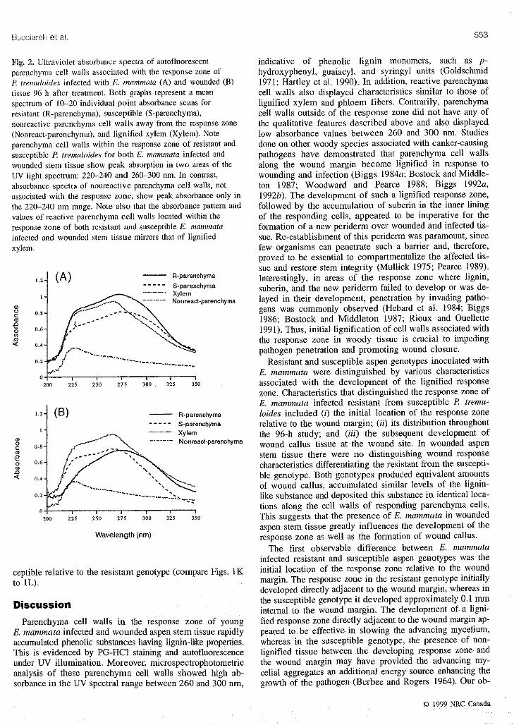
compressed by the rapidly forming wound callus tissue Principal component analysis of the lambda-scan spectral(Figs. IG and IH). The development of the PG-HCI posi- data cmlfirm that absor'oance values for wavelengths be-tire-staining zone in wounded tissue occurred 24 h later than tween 260 and 300 nm provide the greatest variability in thisobserved in E. mammata infected tissue, data set. This indicates that differences between reactive
Another feature distinguishing resistant and susceptible parenchyma cell walls within the response zone and non-E. mammata infected aspen genotypes was the development reactive parenchyma cell walls outside of the response zoneof wound callus. Wound callus developed internal to the PG- was due predominately to the accumulation of UV absorbingHC1 response zone in the E. mammata infected resistant substances having maximum absorbance values in the 260-genotype but not in the susceptible genotype (compare to 300-nm range. Hartley et al. (1990) and GoldschmidtFigs. 1A and IB). Wound callus, in the resistant genotype, (1971) have demonstrated that phenylpropanoid tigninwas initiated near the vascular cambium and was microscop- monomers such as p-coumaryl, guaiacyl, and syringyl units,ically visible by 48 h after inoculation. By 72 h a callus display absorbance maxima between 270 and 300 nm.ridge was visible around the wound perimeter, and by 96 h Mean absorbance values of reactive parenchyma cellafter inoculation, the growth of wound callus lead to the ap- walls of resistant and susceptible E. mammata infected as-pearance of crushed cells in the PG-HCI positive response pen stem tissue displayed distinct genotypic differences 96 hzone (Fig. IA). The cell walls and cytoplasm of wound cal- after inoculation even though their spectral pattern of ab-lus cells were PG-HCI negative but stained positive for both sorption was similar (Fig. 2A). Higher overall absorptionferric chloride - potassium ferricyanide (Fig. lG) and the values were measured in the resistant relative to the suscep-Manle reaction (data not shown). In contrast, the susceptible tible genotype (P = 0.03). In contrast, mean absorbance val-genotype showed no visible sign of wound callus formation ues of reactive parenchyma cell walls of wounded resistantor reaction of the response zone to either the Maule reaction and susceptible aspen stem tissue (Fig. 2B) displayed no dis-(data not shown) and ferric chloride - potassium ferricyan- tinct genotypic differences 96 h after wounding (P > 0.5).ide staining (Fig. IH) above background levels. The absorbance pattern and values of reactive parenchyma
The development of wound callus in wounded stem tissue cell walls located within the response zone of both resistantwas similar for both resistant and susceptible genotypes and susceptible E. mammata infected and wounded stem tis-(compare Figs. 1C and 1D) and exceeded that observed sue mirrors that of liguified xylem (Figs. 2A and 2B). Thisin the E. mammata infected resistant genotype (compare strong spectral relationship is suggestive of a similar pheno-Figs. 1A and 1C). Callus tissue in wounded stem tissue was lic composition between developmentally lignified xylemproduced in such large amounts that, on occasion, the entire cell walls and reactive parenchyma cell walls associatedwound site became enclosed (data not shown), with the response zone of aspen stem wounds.
Suberin was not detected in cell walls associated with the Distribution of the deposited phenolic substances withinPG-HC1 response zone or in callus tissue using either Sudan the response zone of E. mammata infected and woundedIV, Sudan black B, or the PG-HC1 quenching technique de- resistant and susceptible aspen stem tissue was examinedveloped by Biggs (1984b). However, epidermal cells reacted using a UV absorption scanning microspectrophotometerpositive using all three methods, controlled by the MAPS software program. This technique
Phloroglucinol-HC1 positive-staining parenchyma cell resulted in a visual image of the distribution pattern of phe-walls within the response zone autofluoresced yellow-blue nolic substances having maximum absorbanee at the speci-when viewed with epifluorescent UV illumination (Figs. IE fled wavelength used in the scan. Figures 11and IJ representand 1F). Lignified xylem, phloem fibers, and suberized epi- the distribution patterns of substances absorbing at 270 nmdermal cell walls also emitted autofluorescent characteristics in E. mammata infected resistant and susceptible aspen stemsimilar to those of the response zone. Parenchyma cell walls tissue 96 h after inoculation. The 270-nm wavelength wasnot associated with the response zone did not autofluoresce, chosen to scan the response zone because 270 nm was aCongruent with the PG-HC1 data, the autofluorescent cell common peak absorbance wavelength identified in the spec-walls in the resistant genotype displayed an intact boundary tra of reactive parenchyma cell walls. Spectral analyseszone extending the length of the wound margin from cortex show that areas of peak absorbance at 270 mn closely cot-to xylem (compare Figs. 1A and 1E). In the susceptible go- respond to celt walls that are PG-HC1 positive (comparenotype, the autofiuorescent cell walls of the boundary zone Figs. 1A and 1B with Figs. 1I and 1J) and autofluorescentdisplayed a fragmented and discontinuous appearance (corn- under UV illumination (compare Figs. 1E and 1F withpare Figs. tB and 1F). Figs. lI and IJ).
UV mierospeetrophotometry Fungal penetration
Reactive parenchyma cell walls within the response zone Tissue penetration and degradation were caused by largeof both E. mammata infected and wounded stem tissue show mycelial aggregates. In the susceptible genotype (Fig. 1L),peak absorption in two areas of the UV light spectrum: 220- fungal penetration occurred equally in phloem and cortical240 and 260-300 nm (Figs. 2A and 2B). Absorbance values tissue that stained poorly with PG-HC1 and showed a discon-in both of these spectral zones increased progressively from tinuous autofluorescent pattern. In contrast, if penetration24 to 96 h after treatment (data not shown). In contrast, occurred in the resistant genotype it did so predominantlyabsorbance spectra of nonreactive parenchyma cell walls, through cells near the vascular cambium (Fig. IK). A1-not associated with a response zone, show peak absorbance though the rate of fungal penetration was difficult to accu-only in the 220-240 nm range, the values remained rela- rately assess, degradation, as evidenced by phloem fiberstively low and did not change over time (Figs. 2A and 2B). encircled by the mycelial aggregate, was greater in the sus-
© 1999NRC Canada
Buccia:oiliet al. 553
Fig. 2. Ultraviolet absorbance spectra of autofluorescent indicative of phenolic lignin monomers, such as p-parenchyma cell walls associated with the response zone of hydroxyphenyl, guaiacyl, and syringyl units (GoldschmidP. tremuloides infected with E. mammata (A) and wounded(B) 1971; Hartley etal. 1990). In addition, reactive parenchymatissue 96 h after treatment. Both graphs represent a mean cell walls also displayed characteristics similar to those ofspectrum of 10-20 individual point absorbance scans for lignified xylem and phloem fibers. Contrarily, parenchymaresistant (R-parenchyma), susceptible (S-parenchyma), cell walls outside of the response zone did not have any ofnonreactive parenchyma cell wails away from the response zone the qualitative features described above and also displayed(Nonreact-parenchyma), and ligrtified xylem (Xylem). Note low absorbance values between 260 and 300 nm. Studiesparenchyma cell walls within the response zone of resistant and done on other woody species associated with canker-causingsusceptible R tremuloides for both E. mammata infected and pathogens have demonstrated that parenchyma cell wallswounded stem tissue show peak absorption in two areas of the along the wound margin become lignified in response toUV light spectrum: 220-240 and 260-300 am. In contrast, wounding and infection (Biggs 1984a; Bostock and Middle-absorbance spectra of nonreactive parenchyma cell walls, not ton t987; Woodward and Pearce 1988; Biggs 1992a,associated with the response zone, show peak absorbance only in 1992b). The development of such a lignified response zone,the 220-240 nm range. Note also that the absorbance pattern and followed by tile accumulation of suberin in the inner liningvalues of reactive parenchyma cell walls located within the of the responding cells, appeared to be imperative for theresponse zone of both resistant and susceptible E. mammata formation of a new periderm over wounded and infected tis-infected and wounded stem tissue mirrors that of lignified sue. Re-establishment of this periderm was paramount, sincexylem, few organisms can penetrate such a barrier and, therefore,
proved to be essential to compartmentalize the affected tis-sue and restore stem integrity (Mullick 1975; Pearce 1989).
1.2-_ (A) -- R-parenehyma Interestingly, in areas of the response zone where lignin,
/ ..... S-parenchyma suberin, and the new periderm failed to develop or was de-|. "............. Xylem,oo,o o....... t-parenohyma layed in their development, penetration by invading patho-
• ._-_" -_ _ xN gens was commonly observed (Hebard et al. 1984; Biggso 0.8'
,_ 1986; Bostock and Middleton 1987; Rioux and Ouellettes_ o.6. 1991). Thus, initial lignification of cell walls associated with
the response zone in woody tissue is crucial to impeding0.4. pathogen penetration and promoting wound closure.o.2. Resistant and susceptible aspen genotypes inoculated with
/" E. mammata were distinguished by various characteristics2oo 2z5 2_o 27_ 3_0 3_5 3_0 associated with the development of the lignified response
zone. Characteristics that distinguished the response zone ofE. mammata infected resistant from susceptible P. tremu-
1,2. (a) -- R-parenehyma loides included (i) the initial location of the response zone..... S-pareneh'cma relative to the wound margin; (it) its distribution throughout
x- :./_... •............ Xylem the 96-h study; and (iii) the subsequent development of_o 0.8. .,,_.:,_ \ ....... Nonreact-parenehymawound callus tissue at the wound site. In wounded aspen2, .---" stem tissue there were no distinguishing wound response
_ 0'6" ___ characteristics differentiating the resistant from the suscepti-a_
< 04. --" /- --. ""-,._ ble genotype. Both genotypes produced equivalent amounts
_-J --.- -- _-f _-'"--.._.__ ",_ of wound callus, accumulated similar levels of the lignin-o._.",_,,.4,-'" ............. 2.,..... like substance and deposited this substance in identical loca-
,-" tions along the cell walls of responding parenchyma cells.200 2_5 _0 275 3_o 3_5 3'5o This suggests that the presence of E. mammata in wounded
aspen stem tissue greatly influences file development of theWavelength(nm) response zone as well as the formation of wound callus.S
The first observable difference between E. mammatainfected resistant and susceptible aspen genotypes was the
ceptible relative to the resistant genotype (compare Figs. 1K initial location of the response zone relative to the woundto 1L). margin. The response zone in the resistant genotype initially
developed directly adjacent to the wound margin, whereas in
DiscussiOn the susceptible genotype it developed approximately 0.1 mminternal to the wound margin. The development of a ligni-
Parenchyma cell walls in the response zone of young fled response zone directly adjacent to the wound margin ap-E. mammata infected and wounded aspen stem tissue rapidly peared to he effective in slowing the advancing mycelium,accumulated phenolic substances having lignin-like properties, whereas in the susceptible genotype, the presence of non-This is evidenced by PG-HC1 staining and autofluorescence lignified tissue between the developing response zone andUnder UV illumination. Moreover, microspectrophotometric the wound margin may have provided the advancing my-analysis of these parenchyma cell walls showed high ab- celial aggregates an additional energy source enhancing thesorbance in the UV spectral range between 260 and 300 am, growth of the pathogen (Berbee and Rogers 1964). Our ob-
© 1999NRCCanada

554 Can. J. Bot. VoL T7, 1999
servations coutrasl with those of Biggs (1984a) who lotmd euces in the anloant of callus produced was observed (Pinnabarrier zones iu peach (Pnmus persica (L.) Bartsch) infected 1986; Berbee and Rogers 1964). In addition, our study alsowith Cytosporu leucostoma (Sacc,) to initially develop up showed that callus cells developing in resistant E. mammatato 1 tour internal to the wound margin. However, Biggs infected stem tissue displayed a uuique hislochemical re-
(1984a) studied the wound response in mature stem tissue, sponse in contrast to cells associated with the liguified re-whereas our study focused on juvenile stem tissue, spouse zone. The cytoplasm and cell walls of callas cells
The second distinguishing characteristic between resistant were PG-HC1 negative but stained positive with ferricand susceptible aspen genotypes in response to E. mamlt_aa chloride - potassium ferrous sulfate and the Maule reaction.
infection was the distribution of PG-HC1 positive-staining Such a histochemical response of wound callus tissue sug-cell walls of the response zone. In the resistant genotype the gests that the phenolic composition of this tissue is differentcell walls associated with the response zone displayed a lig- than that observed in the lignilied response zone aud, there-nification pattern that was intact and localized as evidenced fore, may provide an additional barrier that ptx_vents or ira-by PG-HCI staining and autofluorescense by UV illumina- pedes infection.tion. In the susceptible genotype, these cell walls were dis- in conclusion, these results indicate that, in the presencetributed in a discontinuous pattern along the response zone, of the pathogen, the resistant genotype is capable of devel-In various woody species previously studied, the distribution oping two types of barriers that restrict infl_ction (an intactof the newly deposited lignin along the response zone ap- lignified barrier and a wound callus bancier rich in phenolicpeared to he important in controlling pathogen penetration substances), whereas the susceptible genotype is iueft?ctiveinto noninfected tissue. Biggs (1984a) found that C. leuco* at developing either one of these barriers. This implicatesstoma penetrated boundary zones in peach through areas two distinct mechanisms by which /_ ttvemuloides limits in-where cell walls were poorly lignified and suberized such as fection by E, mammat_t. The extent to which these mecha-
through discontinuities along the developing boundary zone nisms occur ttttx)ughout the population of/:: tremnloides isor through newly initiated cells along the vascular cambium, not known. However, the ability to identify these nmcha-In elm (Ulmus americana L.), a discontinuous response zone aisms plus other defense responses would be of benefit todeveloping in wounded stem tissue has been associated with screen aspen genotypes resistant or tolerant to E. mammatasusceptibility to Ophiostoma ulmi (Buism.) Nannf., whereas infection.intact response zones were effective in resisting ingress ofthis pathogen (Rioux and Ouellette 1991). Berbee and Rog-ers (1964) reported that E. mammatu selectively penetrates Aelcnowledgementfl
and degrades aspen stem tissue composed of nonlignified The authors thank Mr. Thomas Medin for assistance in the
cell walls as opposed to stem tissue composed of lignified use of the microspectrophometric equipment and Mr. Da-walls. In our study, E. mammata penetration in the sus- vid D. Rugg for expertise in the statistical analysis andceptible genotype occurred indiscriminately and extensively interpretation of the data. The authors are grateful to thethrough cortical and phloem tissue where the response zone United States Forest Service, North Central Research Sta-appeared discontinuous and cell walls were nonlignified. In
lion; Department of Plant Pathnlogy, University of Minne-the resistant genotype, if fungal penetration through the re- sota; and the United States Department of Agriculture, Agri-sponse zone did occur, it started and progressed along the culture Research Service, for providing financial support forvascular cambium where cells were newly initiated and this project.poorly lignified. E. mammata penetration was not observedin cortical or phloem tissue where the response zone was in-
tact and uniformly lignified. Therefore, congruent with stud- I_e'feleen¢efiies on other woody species, in addition to earlier studies inthe E. mammata - aspen interaction, the continuity of the re- Ames, N.E, Hartley, R.D., and Akin, D.E. 1992. Distribution ofsponse zone in E. mammata infected aspen stem tissue up- aromatic compounds in coastal bermudagrass cell walls usingpears to be crucial in restricting penetration of the pathogen ultraviolet absorptioJ_ scanning mJcrospeclrophotometry. Foodand therefore vital to resistance. Struct. ll: 25-32,
The development of wound callus is a third feature distin- Anderson, N.A., Ostry, M.E., and Anderson, G.W. 1979. Insectwomlds as infection sites for tIypoxylon mamtnanor_ on trem-
guishing resistant from susceptible aspen genotypes in re- bling aspen. Phytopathology, 69: 476-479.spouse to E. mammata infection. In the resistant genotype, Baranyay, J.A. 1967. Notes on Hypoxylon cauker of aspen in AI-wound callus developed immediately internal to the lignified berta. For. Chron. 43: 372-380.
response zone and was first visible 48 h after iuoeulation. Berbee, J.G., and Rogers, J.D. 1964. Life cycle and host range ofBy 96 h, callus ridges were macroscopically evident along Hypoxyh)n pruinatum and its pathogenesis on poplars. Phyto-the perimeter of the wound margin. The resistant E. mum- pathology, 54:257-261mata infected genotype consistently produced wound callus. Biggs, A.R. 1984a. Boundary-zone formanon in peach bark in re-although less than that observed in noninfected wounded sponse to wounds and C_;to_pora teucosmma infection. Can. J.tissue. In contrast, susceptible [2 mammata infected tissue Bot. 62:2814-2821failed to develop wound callus throughout the duration of Biggs, A.R 1984b. lntracellular suberin: occurrence and detectionthe study despite its ability to form it in the absence of the in tree bark. Int. Assoc. Wood Anat. Bull. 5: 243-248.pathogen. Berbee and Rogers (1964/ reported that Popuhts Blggs A.R. 1986. Comparative anatom2, and host response of twospp. resistant to E. mammatu infection rapidly developed peach eultivars inoculated with Leucosmma ctncta andlarge amounts of wound callus; however, genotypie differ- L. personii. Phytopathology, 76: 905-912.
© 1999 NRC Canada
Bucciare}li et al. 555
Biggs, A.R. 1992a. Anatomical and physiological responses of Morrell, J.J., Gibson, D.G., and Krahmer, R.L. 1985. Effect of flu-bark tissues to mechanical injury. In Defense mechanisms of orescent-labeled lectins on visualization of decay fungi in wood
woody plants against fungi. Edited by R.A. Blanchette and A.R. sections. Phytopathology, 75: 329-332.Biggs. Springer-Verlag, Berlin. pp. 13_.0. Mullick, D.B. 1975. A new tissue essential to necrophylactic peri-
Biggs, A.R. 1992b. Responses of angiosperm bark tissues to fungi derm formation in the bark of four conifers. Can. J. Bot. 53:causing cankers and canker rots. In Defense mechanisms of 2443-2457.woody plants against fungi. Edited by R.A. Blanchette and A,R. Ostry, M,E., and Anderson, N.A. 1983. Infection of tremblingBiggs. Springer-Verlag, Berlin. pp,41-60, aspen by Hypoxylon mammatum through cicada oviposition
Bostock, R.M., and Middleton, G.E. 1987. Relationship of wound wounds. Phytopathology 73: 1092-1096.periderm formation to resistance to Ceratocystisfimbriata in al- Pearce, R.B, 1989. Cell wall alterations and antimicrobial defensemend bark. Phytopathology, 77:1174-ll 80. ill perennial plants. In Plant Cell Wall Polymers, Biogenesis and
Bostock, R.M., and Stermer, B.A. 1989. Perspectives on wound Biodegradation, Proceedings of the Symposium of the 3rdhealing in resistance to pathogens. Annu. Rev. Phytopathol. 27: Chemical Congress of North America, 5-11 June 1988, Toronto,343-371. Ont. Edited by N.G. Lewis and M.G. Paice. American Chemical
Faulkner, G., and Kimmins, W.C. 1975. Staining reactions of the Society, Washington, D.C. pp, 346-360.tissue bordering lesions induced by wounding, tobacco mosaic Pinon, J. 1986. Test of inhibition of cambial activity of poplarvirus, and tobacco necrosis virus in bean. Phytopathology, 65: by Hypoxylon mammatum: development and application. Eur. J.1396-t400. For. Pathol, 16: 230-238.
Goldschmid, O. 1971. Ultraviolet spectra. In Lignin: occurrence, Ride, J.P, 1983. Cell walls and other structural barriers in defence.formation, structure and reactions. Edited by K.V. Sarkanen, and In Biochemical plant pathology. Edited by J.A. Callow. JohnC,H. Ludwig. Wiley-lnterscience, New York. pp. 241-267. Wiley & Sons Ltd., New York. pp. 215-236.
Hahlbrock, K., and Scheel, D. 1989. Physiology and molecular Rioux, D., and Ouellette, G.B. 1991. Barrier zone formation in
biology of phenylpropanoid metabolism. Annu. Rev. Plant host and nonhost trees inoculated with Ophiostoma ulmi. I.Physiol. Plant Mol. Biol. 40: 347-369. Anatomy and histochemistry. Can. J. Bot. 69: 2055-2073.
Hartley, R.D., Atkin, D.E., Himmelsbach, D.S., and Beach, D.C. Rogers, J.D., and Ju, Y.-M. 1996, Entoleuca mammata comb. nov.1990. Microspectrophotometry of bermudagrass (Cynodon for Hypoxylon mammatum and the genus Entoleuca.dactylon) cell wails in relation to lignification and wall bio- Mycotaxon, 59: 441448.degradability. J. Sci. Food Agric. 50: 179-189. Sherwood, R.T, and Vance, C.P. 1976. Histochemistry of papillae
Hebard, EV., Griffin, G.J., and Elkins, J.R. 1984. Developmental formed in reed canarygrass leaves in response to noninfectinghistopathology of cankers incited by hypovirulent and virulent pathogenic fungi, Phytopathology, 66: 503-510.isolates of Endothia parasitica on susceptible and resistant Starr, G.H. 1971. Propagation of aspen trees from lateral roots. J.chestnut trees. Phytopathology, 74: 140-149. For. 69: 866-867.
Jensen, W.A. 1962. Botanical histochemistry. W.H. Freeman & Vance, C.P., Kirk, T.K., and Sherwood, R.T. 1980, Lignification asCo., San Francisco. a mechanism of disease resistance. Annu. Rev. Phytopathoh 18:
Johansen, D.A. 1940. Plant microtechnique. McGraw-Hill Book 259-288.Co., New York. Woodward, S., and Pearce, R.B. 1988. Wound-associated responses
McManus, P.S., Ewers, F.W., and Fulbright, D.W. 1989. Character- in Sitka spruce root bark challenged with Phaeolus schweinitzii.ization of the chestnut blight canker and the localization and Physiol. Mol. Plant Pathol. 33: 151-162.isolation of the pathogen Cryphonectria parasitica. Can. J. Bot.67: 3600-3607.
1999NRC Canada