c•00(ow7o606 stin gu< - defense technical information center · a stand or rack to hold the...
TRANSCRIPT
LOXI,.AHAZARD .DIVI SIO0NChief, Tuxiz Hazandz Division
AMRL-TR-65-202 ' iskA
ELECTROPHORETIC SEPARATIONS ON ACRYLAMIDE GELS:DISC ELECTROPHORESIS
LEANDRO RENDON
DECEMBER 1965
Distribution of this documentis unlfimited
c•00(oW7o606 STIN O Gu< '
AEROSPACE MEDICAL RESEARCH LABORATORIESAEROSPACE MEDICAL DIVISIONAIR FORCE SYSTEMS COMMAND
WRIGHT-PATTERSON AIR FORCE BASE, OHIO
Notices
When US Government drawings, specifications, or other data are used for anypurpose other than a definitely related Government procurement operation, theGovernment thereby incurs no responsibility nor any obligation whatsoever, andthe fact that the Government may have formulated, furnished, or in any way sup-plied the said drawings, specifications, or other data, is not to be regarded byimplication or otherwise, as in any manner licensing the holder or any other personor corporation, or conveying any rights or permission to manufacture, use, or sellany patented invention that may in any way be related thereto.
Requests for copies of this report should be directed to either of the addresseeslisted below, as applicable:
Federal Government agencies and their contractors registered withDefense Documentation Center (DDC):
DDCCameron StationAlexandria, Virginia 22314
Non-DDC users (stock quantities are available for sale from):
Chief, Input SectionClearinghouse for Federal Scientific & Technical Information (CFSTI)Sills Building5285 Port Royal RoadSpringfield, Virginia 22151
Change of Address.
Organizations and individuals receiving reports via the Aerospace MedicalResearch Laboratories' automatic mailing lists should submit the addressograpbplate stamp on the report envelope or refer to the code number when correspond-ing about change of address or cancellation.
Do not return this copy. Retain or destroy.

ELECTROPHORETIC SEPARATIONS ON ACRYLAMIDE GELS:DISC ELECTROPHORESIS
LEANDRO RENDON
Distribution of this documentis unlimited

Foreword
This work was performed under Project No. 6302, "Toxic Hazards of Pro-pellants and Materials," Task No. 630202, "Pharmacology - Biochemistry," fromAugust 1963 to June 1965 in the Toxic Hazards Branch, Physiology Division,Biomedical Laboratory, Aerospace Medical Research Laboratories, Wright-Pat-terson Air Force Base, Ohio. The assistance and suggestions of 1/Lt Duncan E.McVean, and the assistance of TSgts. W. D. Leonard, G. W. Craig, and W. F.Hunt, Jr., who evaluated the procedure, are gratefully acknowledged.
This technical report has been reviewed and is approved.
WAYNE H. McCANDLESSTechnical DirectorBehavioral Sciences LaboratoryAerospace Medical Research Laboratories
ii

Abstract
Disc Electrophoresis, a new method for fractionating serum proteins andenzymes developed by Ornstein and Davis (Mt. Sinai Hospital, New York, N. Y.),possesses great sensitivity, speed and reproducibility while requiring a sampleas little as 3 microliters in routine separations. The technique as adapted andstandardized for use in the Toxic Hazards Branch, Aerospace Medical ResearchLaboratories, for serum protein and Lactic Dehydrogenase (LDH) isozyme sepa-rations is presented along with suggestions for making the apparatus needed toperform disc electrophoresis.
iii

Table of ContentsSection Page
I INTRODUCTION ------------------------------------------------------------------------------- 1
II EQUIPMENT---------------------------------------------------------------------------- 3
III M T RILI---MA TERIA LS - - ---------------------- ------------- ----------- --------------- -------- 5
Reagents----------------------------------------- ---------------------- --- ---------------------- ----- 5
Stock Solutions ---------------------------------------------------------------------------------- 5
Working Solutions ----------------------------- ----- -------------- -------- -------------- 6
IV PROCEDURE ----------------------------------------------------- ------------------------- 7
Polymerizing the Gels----------------------------------------------------------------------- --- 7
Electrophoresis--------------------------------------------------------------------------------- -11
Staining and Destaining Gels - Locating Protein Fractions ---------------------- 15
Staining Gels for LDH Isozyme Fractions - - ------- ----------------- 17
APPENDIX: Helpful Hints in Disc Electrophoresis Methodology-------------------------------- 21
REFERENCES ----------------------- --------- - ---------------- -2
iv

List of Illustrations
Figure No. Page
1 Parts of Equipment for Disc Electrophoresis ------------------------- 3
2 Gel Containers in Place on Polymerization Stand ---------------------------------------------------------------- 7
3 Refractile Line at Boundary of Lower Gel and the Water Overlay --------------- 8
4 The "Spacer" Gel becomes Opaque. The water overlay can be seen above theopaque section of the column of gel -..........................................----------------------------------- 9
5 Mixture of Large-Pore Solution and Sample being Transferred to Tubes. Thethree tubes on the right show complete photopolymerization of the acrylamide gels .... 10
6 Removing Gel Container from Polymerization Stand ------------------------ 11
7 Inserting Tubes into Grommets of Upper Reservoir ---------------------------------------------------------.12
8 Arrangement of Equipment for Disc Electrophoresis ---------------------------------------------------------- 13
9 First Step in Removal of Gel from Glass Tube following Electrophoretic Run ------- 14
10 Second Step in Removal of Gel from Glass Tube following Electrophoretic Run ---------- 16
11 Acrylamide Gels in Storage Tubes. Typical serum protein separation patternsfor (I to r): Rat, Dog, Monkey, and Human ------------------------------------------------------------------------ 18
12 Normal Serum LDH Isozyme Separation Patterns in (1 to r): Human, Monkey,and Dog (Beagle). The isozymes visible are LDH-1, LDH-2, and LDH-3 --------- 19
13 Sample Data Sheet ----------.-.-------.-.----.-.-.-...............................------------------------------------------ ------ 23
14 Sample Densitometric Scan of Human Serum Protein Fractions Separated onAcrylamide Gel -----------------------------------------...............................-------------------------------------------------- 24
15 Sample Densitometric Scan of Human Serum LDH Isozyme Fractions Sepa-rated on Acrylamide Gel ---------------------------------------------------------------------------------------------- ------------- 25
V

[t Up per Rese rvoirSample Gel
PowerS Spacer Gel
Small-PoreLower Gel
LowerReservoir
(frontispiece) Disc Electrophoresis
vi
SECTION I
Introduction
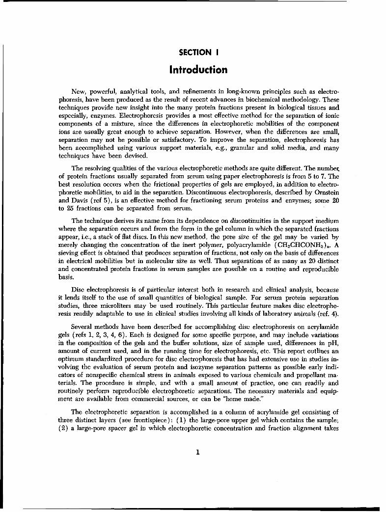
New, powerful, analytical tools, and refinements in long-known principles such as electro-phoresis, have been produced as the result of recent advances in biochemical methodology. Thesetechniques provide new insight into the many protein fractions present in biological tissues and.especially, enzymes. Electrophoresis provides a most effective method for the separation of ioniccomponents of a mixture, since the differences in electrophoretic mobilities of the componentions are usually great enough to achieve separation. However, when the differences are small,separation may not be possible or satisfactory. To improve the separation, electrophoresis hasbeen accomplished using various support materials, e.g., granular and solid media, and manytechniques have been devised.
The resolving qualities of the various electrophoretic methods are quite different. The numberof protein fractions usually separated from serum using paper electrophoresis is from 5 to 7. Thebest resolution occurs when the frictional properties of gels are employed, in addition to electro-phoretic mobilities, to aid in the separation. Discontinuous electrophoresis, described by Ornsteinand Davis (ref 5), is an effective method for fractioning serum proteins and enzymes; some 20to 25 fractions can be separated from serum.
The technique derives its name from its dependence on discontinuities in the support mediumwhere the separation occurs and from the form in the gel column in which the separated fractionsappear, i.e., a stack of fiat discs. In this new method, the pore size of the gel may be varied bymerely changing the concentration of the inert polymer, polyacrylamide (CH 2CHCONH 2 ),. Asieving effect is obtained that produces separation of fractions, not only on the basis of differencesin electrical mobilities but in molecular size as well. Thus separations of as many as 20 distinctand concentrated protein fractions in serum samples are possible on a routine and reproduciblebasis.
Disc electrophoresis is of particular interest both in research and clinical analysis, becauseit lends itself to the use of small quantities of biological sample. For serum protein separationstudies, three microliters may be used routinely. This particular feature makes disc electropho-resis readily adaptable to use in clinical studies involving all kinds of laboratory animals (ref. 4).
Several methods have been described for accomplishing disc electrophoresis on acrylamidegels (refs 1, 2, 3, 4, 6). Each is designed for some specific purpose, and may include variationsin the composition of the gels and the buffer solutions, size of sample used, differences in pH,amount of current used, and in the running time for electrophoresis, etc. This report outlines anoptimum standardized procedure for disc electrophoresis that has had extensive use in studies in-volving the evaluation of serum protein and isozyme separation patterns as possible early indi-cators of nonspecific chemical stress in animals exposed to various chemicals and propellant ma-terials. The procedure is simple, and with a small amount of practice, one can readily androutinely perform reproducible electrophoretic separations. The necessary materials and equip-ment are available from commercial sources, or can be "home made."
The electrophoretic separation is accomplished in a column of acrylamide gel consisting ofthree distinct layers (see frontispiece): (1) the large-pore upper gel which contains the sample;(2) a large-pore spacer gel in which electrophoretic concentration and fraction alignment takes
1

place; and (3) the small-pore lower gel in which the actual separation occurs. The rest of thearrangement includes an upper and lower buffer reservoir, electrodes for each reservoir, and aconstant power supply source. Electrophoresis is performed in a vertical position, with the gelcontainers attached to the upper reservoir and the lower ends immersed in the buffer solution ofthe lower reservoir. The electrodes are arranged so that the upper one is the cathode and thelower one the anode. In this manner, the migration of sample ions is toward the small-pore gelin the lower section of the gel column. Current is applied for a specific period of time, afterwhich the gels are rimmed out of the containers and placed in special staining solutions forlocation of the separated fractions.
2

SECTION II
Equipment
(See fig. 1) The buffer reservoirs can be purchased from commercial sources or can bemade from inert, nonconductive materials. Round plastic (polystyrene) dishes, 4 to 6 inches indiameter and 2 to 2% inches deep, or pie-plate covers of similar type plastic material, can beused. These are generally available at local housewares stores.
Holes, approximately ¾A-inch in diameter, are made in the bottom of one of the plastic dishesby heating the end of a brass cork borer of the proper size and touching it to the dish. The holesare made at equal intervals along a circumference and equi-distant from the electrode located inthe center of the dish. Rubber electrical grommets, lY4-inch-inside diameter, are then inserted tofit tightly into the holes. The grommets should have holes that fit snugly around the tubular con-
Figure IParts of Equipment for Disc Electrophoresis. Power Supply and support for the upper reservoir not shown.
3

tainers for the gels. In use, the upper buffer reservoir, fitted with electrode and grommets, issupported above the lower reservoir by means of a tripod or support ring.
The electrodes are made from thoroughly cleaned cylindrical graphite poles removed fromflashlight batteries, and are attached at the center of each reservoir by inserting into a flat-toprubber test tube cap (B-D Vacutainer cap) cemented to the bottom of the dish. The leads tothe electrodes should be made of insulated wire soldered to the metal caps of the electrodes.Platinum wire electrodes can also be used. A simple way to make a suitable electrode is to windthe platinum wire around a 1¾-inch length of rigid polyethylene tubing with a ¼/4-inch-outside.diameter. After soldering the electrode leads to the platinum wire, the electrode can be insertedinto the flat-top rubber test tube cap cemented to the bottom of the dish.
The tubular containers for the gel columns are made from glass tubing of a 5-mm-insidediameter and cut to 63-mm lengths. The ends of the tubes are square cut but not fire polished,since this may reduce the size of the opening. Sharp edges are removed with an emery cloth orfine-grade sandpaper. While the diameter of the tubes is not critical, all tubes of one set shouldbe cut from the same piece of tubing to eliminate differences in electrical resistance from cylin-der to cylinder.
A stand or rack to hold the tubes during filling and polymerization of the gels is made bycementing fiat-top rubber test tube caps to a fiat piece of wood or plastic. The open end of thecap fits snugly around a tube to prevent leakage of the gel solution.
The power supply source should provide a constant current to a maximum of 200 to 250milliamperes. Current must be maintained with little or no change when an electrophoretic runis being conducted, to avoid introducing variations in the electrical mobility of the fractions beingseparated.
4

SECTION III
Materials
REAGENTS
The reagents in the form of pre-mixed, standardized solutions are available from CanalIndustries Co., ("Canalco"), 4935 Cordell Ave., Bethesda, Md. They can also be obtained fromDistillation Products Industries, Division of Eastman Kodak Co., Rochester 3, N. Y., and MathesonColeman and Bell (MCB), 2909 Highland Ave., Norwood (Cincinnati) 12, Ohio, as reagentgrade compounds.
1. Acrylamide (Eastman #5521; MCB #AX330) (CH 2CHCONH 2). This reagent should bekept in a cool, dark place to slow down polymerization. Its shelf life in the crystal form lasts foryears.
2. N,N'-Methylenebisacrylamide (Eastman #8383) ("BIS") (CH 2(NHCOCHCH 2)2 ). "BIS"is a crystalline solid that should be stored in a cool, dry, dark place.
3. 2-Amino-2-(hydroxymethyl)-l, 3-propanediol (Eastman #4833) ("TRIS") (Tris Buffer -Sigma Chemical Co., 3500 DeKalb St., St. Louis 18, Mo.).
4. N,N,N',N'-Tetramethylethylenediamine (Eastman #8178) ("TEMED") (MCB #TX405).
5. Riboflavin (Eastman #5181; MCB #VX 165).
6. HCl, reagent grade, 1 N.
7. Ammonium persulfate, reagent grade (MCB #AX 1340).
8. Glycine (ammonia-free) (Eastman #445; MCB #GX 205).
9. Glacial Acetic Acid (Eastman #763).
10. Amido Schwarz; also known as Naphthol Blue Black, Aniline Blue Black, C. 1. No. 20470(MCB #AX 1485).
11. Bromphenol Blue (Eastman #752; MCB #BX 1415).
STOCK SOLUTIONS
(The following solutions are identified in a manner similar to that used by Ornstein andDavis.)
(A) 1 N HCl - 48.00 ml (B)* 1 M H3PO4 - 25.60 mlTRIS - 38.30 grams TRIS - 5.70 gramsTEMED - 0.23 ml TEMED - 6.46 mlH20 to - 100.00 ml H_,O to - 100.00 ml
(pH 8.9) (pH 6.7)
*To make 1 M H3P0 4, dilute 68.3 ml of 85%, or
67.0 ml of 86% acid to make 1 liter.
5

(C) Acrylamide - 30.0 grams (D) Acrylamide - 10.0 gramsBIS - 0.8 grams BIS - 2.5 gramsH,20 to - 100.0 ml H2 0 to - 100.0 ml
(E) Riboflavin - 4.0 gramsH20 to - 100.0 ml
The stock solutions are kept in amber glass or plastic bottles at temperatures around 10°Cand have a shelf life of several weeks.
WORKING SOLUTIONSThese are also stored in plastic or amber glass bottles. The shelf life is up to several weeks
with the exception of Small-Pore Solution #2 which must be prepared weekly.
Small-Pore #1 (15% Gel) Small-Pore #21 Part Stock Solution A Ammonium Persulfate 0.14 gram2 Parts " . . C H20 to 100.00 ml1 Part H20 (Prepare weekly)
(pH 8.9-.1)
Large-Pore Solution (21/2% Gel) Buffer Solution for Reservoirs1 Part Stock Solution B TRIS 6.0 grams2 Parts " D Glycerine 28.8 grams1 Part ". . E H20 to 1 liter4 Parts H2 0 (pH 8.3)
(pH 6.7)
Dilute the buffer solution 1 part plus 9 of distilled water for use in the reservoirs. The solu-tion can be used for 24 samples before being discarded. However, do not mix up the solutionsonce they have been used. If they are to be re-used, they should be kept in separate containers.
Fixative - Stain SolutionAmido Schwarz - 1.0 gram7% Acetic Acid - 100.0 ml
(by volume)
After filtering, store in a reagent bottle. This solution can be re-used several times.
Bromphenol Blue Solution Wash Solution for Destaining and Storing GelsBromphenol Blue 0.01 gram Glacial Acetic Acid 70.0 mlDist. H20 to 1 liter Dist. H20 to. 1 liter
*Polyacrylamide Solution for Destaining
Acrylamide - 6.00 gramsRiboflavin - 0.50 mgmTEMED - 0.05 mlH20 to - 100.00 ml
*After the ingredients have been completely dissolved, the solution is placed in a 100-ml
cylinder and exposed to a fluorescent light with the bulb at a distance of about 3 inches fromthe cylinder and parallel to it. The riboflavin is generally bleached in about 1½/2 hours. When thishas been accomplished, the solution is mixed with an equal volume of distilled water and storedin a brown glass or plastic bottle.
6

SECTION IV
Procedure
POLYMERIZING THE GELS1. Insert the glass tube in which the gels are to be polymerized into the rubber caps on the
polymerization stand, carefully setting them in a vertical position. The tubes should be pushedinto the holders so that they rest firmly against the base (see fig. 2).
2. Since polymerization rate is affected by temperature, the gel Working Solutions shouldbe permitted to warm to room temperature before using. For each set of 8 gels, mix 5 ml eachof Small-Pore Solution No. 1 and No. 2 in a small Erlenmeyer flask or test tube. Blend thoroughlybut avoid excessive aeration. Using a pipette, fill the polymerization tubes with this mixturewithout delay to within /2-inch of their tops.
Figure 2
Gel Containers in Place on Polymerization Stand.
7

3. Very carefully overlay the solution in each tube with a ¼4-inch column of distilledwater. A 25-gauge needle, %-inch long, attached to a 2-ml syringe and bent about 200 can beused for this purpose. Fill the barrel with enough distilled water so that surface tension preventsformation of a drop at the end of the needle. When the needle is laid against the inside wall ofthe tube, water will ooze out evenly. Disposable pipettes with rubber bulbs are most practicalfor this step because the long pipette tips permit reaching to the level of the gel solution to over-lay with water. The water should drip down the sides of the tube but should not "bomb" thelower gel. This water overlay should produce a sharp refractile boundary at the interface (seefig. 3). Discard any tube in which swirling or mixing is apparent.
Figure 3
Refractile Line at Boundary of Lower Gel and the Water Overlay.
8

4. Allow gels to polymerize for about 40 minutes at room temperature. The first refractileline will disappear due to diffusion and a second will appear, about 3 mm below the locationof the first, in about 25 minutes. This will mark the junction of the clear gel polymer. At the endof the 40 minutes, remove the water and unreacted polymer solution by inverting and shakingthe tubes gently and draining. A disposable pipette with rubber bulb can also be used to aspiratethe water and unreacted polymer from the tube.
5. Then, using a pipette, rinse the inside of the tubes gently with the Large-Pore Solutionand discard the rinse. Add about %-inch of Large-Pore Solution to each tube and overlay withabout ¼/-inch column distilled water.
6. Place the base with the tubes about 3 inches away from a 15-watt flourescent lamp. (Thisprocedure may also be used in step 4.) Care should be taken not to disturb the water-gel inter-faces when moving the base to the lamp. The clear solution becomes increasingly opaque afterabout 5 minutes of exposure (see fig. 4) and total polymerization occurs in about 15 minutes.The polymerized Large-Pore gel forms the "spacer" section of the gel column.
Figure 4The "Spacer" Gel becomes Opaque. The water overlay can be seen
above the opaque section of the column of gel.
9

7. Decant and shake off the water layer over the "spacer gel." Next, add to each tube athoroughly but gently mixed solution containing 0.15 ml of Large-Pore Solution and 3 microlitersof sample for serum protein separation. A disposable plastic tray of the type used for spot testsis ideal to use for mixing the gel solution and the sample. Hamilton syringes, 10 microliter size,can be used to measure volumes of the sample. After thorough mixing, the resultant solution canbe transferred to the tubes by means of a disposable pipette (see fig. 5). No water overlay isrequired for this step. For lactic dehydrogenase (LDH) isozyme separations, more readily identi-
fied fractions are obtained when 6 microliters of sample are used. In the case of rat serum LDHisozyme separations, 15 microliters of serum should be used. Set support stand with tubes in front
of the fluorescent lamp for 20 minutes for photopolymerization of the upper gel.
Figure 5
Mixture of Large-Pore Solution and Sample being Transferred to Tubes. The three tubeson the right show complete photopolymerization of the acrylamide gels.
10

8. When polymerization has been completed (upper gel becomes opaque), remove thetubes from the base by tilting sharply to break any seal that might have been formed (see fig. 6).
Figure 6
Removing Gel Container from Polymerization Stand.
ELECTROPHORESIS
1. The electrophoresis is started within an hour after the "spacer" gel has been polymerized.Delays beyond this period result in the gel becoming more tightly packed, and variations in themovement of the fractions in the gel column will occur. Fill the lower reservoir with buffer toabout ½-inch from the top of the dish.
2. Insert the tubes, upper gel at the top, into the grommets of the upper reservoir (see fig. 7).
3. Next, place a hanging drop of buffer at the bottom end of each tube by means of apipette to prevent trapping of bubbles.
4. Lower the reservoir so that the bottoms of the tubes are immersed at least '4-inch inthe buffer of the bottom reservoir.
11

5. Fill the remaining empty spaces above the gel tubes with buffer by means of a pipetteand then fill the upper reservoir with enough buffer to reach a level of about one inch from thebottom of the dish.
6. Add a few drops of Bromphenol Blue Solution to the upper reservoir and stir to mix thedye thoroughly. This dye is added to act as an indicator of electrophoretic movement. The dyemolecules move faster than the protein fractions which form behind the dye as it passes throughthe gels.
7. Connect the power supply, cathode to the upper and anode to the lower reservoir (seefig. 8).
a. For serum protein separations, a current of 2% ma per tube for 45 minutes (or until thedye has moved at least 25 mm into the lower gel) should be used. An exception to this ishuman sera in which best results are obtained after 25 minutes.
Figure 7
Inserting Tubes into Grommets of Upper Reservoir.
12

o,44•.
Figure 8
Arrangement of Equipment for Disc Electrophoresis.
b. For lactic dehydrogenase (LDH) isozyme separations, a current of 2% ma per tube for60 minutes should be used except in the case of rat serum in which instance 21/2 ma pertube for 21/2 hours should be used.
Currents higher than 5 ma per tube should be avoided since heating may produce artifacts.When electrophoresis is conducted at low temperatures (10 to 15'C) to reduce enzyme inactiva-tion, the current is reduced to 1 ma per tube and the time is extended to about 2 hours.
8. Within 5 minutes after the current is applied, a thin ring of Bromphenol Blue will beseen migrating into the gels. If it does not appear, the current should be disconnected and theassembly thoroughly checked. The following possible sources of this difficulty should be investi-gated:
a. Electrodes not properly connected as specified in step 7;
b. Power supply not functioning properly;
13

Figure 9
First Step In Removal of Gel from Glass Tube following Electrophoretic Run.
14

c. "Spacer" or sample gel too tightly packed due to being too concentrated or too old; roomtemperatures above the average range of 22* to 25WC may also accelerate rate and extentof polymerization;
d. Tubes not extending into the lower buffer solution.
Concentration of the separated proteins is completed in the lower end of the spacer gel. Theproteins are visible at this point as a thin refractile band just behind the dye band. Then, as theyenter the lower (small-pore) gel, the proteins will begin to separate from one another and fromthe dye. As they separate, the fractions can often be seen as thin refractile bands. The free dyeappears bluish-red and will be easily visible. Since the dye is bound by albumin, the deep blueband represents the location of the albumin fraction.
The electrophoresis is performed for the time indicated depending upon the purpose of theseparation or until the albumin band has moved about 25 mm into the lower gel. By that time,the free dye will have moved some 6 mm ahead of the albumin band.
9. When the electrophoretic run is completed, turn off the power supply and decant thebuffer solution. Remove the gel tubes from the upper reservoir.
10. Next, release the gels from the tubes by rimming under water. A simple method ofdoing this consists of introducing a teasing needle or wire into the top end of the tube betweenthe gel and the wall of the tube for a distance of several millimeters while rotating the tube (seefig. 9). The needle or wire is pulled out and introduced into the bottom end of the tube in thesame manner as just described. As the needle or wire is withdrawn, a slight pressure appliedagainst the gel will bring the gel out about 5 mm beyond the end of the tube (see fig. 10). Now,attach a medicine dropper rubber bulb filled with water on the top end of the tube. As the bulbis squeezed, the hydrostatic pressure is uniformly applied against the gel which is easily forcedout without sustaining damage.
STAINING AND DESTAINING GELS - LOCATING PROTEIN FRACTIONS
1. After removal from the containers, place each gel in separate test tubes containing enoughFixative-Stain Solution to cover the gel completely. At the end of ½ hour, the solution is decantedand the gels rinsed for a few minutes in a wash solution of 7% Acetic Acid. The gels can be leftovernight in this wash solution if necessary.
2. The excess dye is removed by use of the same apparatus as for the electrophoresis. De-staining tubes are easily made from "disposable" pipettes cut to the appropriate length. The gels,upper gel section at the top, are placed in the destaining tubes which are about 1 mm in diameterlarger than the gels. Medicine dropper tubes are also suitable for this purpose. The gels slidedown in the tubes and wedge firmly against the constructed ends of the tubes.
3. Add enough 7% Acetic Acid solution to the lower reservoir so that a level about oneinch from the bottom of the dish is obtained.
4. Insert the destaining tubes into the grommets of the upper reservoir. To prevent backflow of the free dye up the tube, add Polyacrylamide Solution for Destaining to each tube bymeans of a pipette. No bubbles should remain in the tube.
5. Place a hanging drop of the 7% Acetic Acid solution at the bottom end of each tube.
6. Now, lower the reservoir so that the bottoms of the tubes are immersed at least ¼4-inchin the solution of the bottom reservoir.
15

Figure 10Second Step in Removal of Gel from Glass Tube following Electrophoretic, Run.
16

7. Fill the upper reservoir with enough 7% Acetic Acid solution to reach a level aboutone inch from the bottom of the dish.
8. Connect the electrodes, cathode to the upper reservoir. The unbound dye migrates downthe gels and into the lower reservoir. Generally, the destaining is accomplished in about 2 hoursusing current at the rate of 5 ma per tube.
9. When destaining is completed, turn off the power supply and discard the destainingsolution in both reservoirs. Transfer the gels to small test tubes containing 7% Acetic Acid. Thegels can be preserved for long periods (at least 2 years) in this solution with negligible loss ofdye intensity.
10. Sample Storage Sets (Model A) manufactured by R. P. Cargille Labs, Inc., CedarGrove, New Jersey, provide a simple and efficient method of storing gels (see fig. 11).
STAINING GELS FOR LDH ISOZYME FRACTIONS
1. After the gels are removed from the containers following the electrophoresis procedure,they should be rinsed in distilled water prior to immersing in the following LDH isozyme stain-ing solution modified in this laboratory from Allen's (U. of Michigan) procedure described in the"Canalco" Information Center Newsletter:
TRIS Buffer (0.05M) (pH 8.3) 11.00 ml
KCN (pH 8.3) 3.00 ml
Sodium Lactate 1.25 ml
Nitro-Blue Tetrazolium (NBT) 7.00 mg
b-Diphosphopyridine Nucleotide (b-DPN) 10.00 mg
Phenazine Methosulfate (2 mg/ml in dist. water) 0.15 ml
In the above solution, all of the ingredients except the Phenazine Methosulfate can be mixedwithin 15 to 20 minutes of use if kept in a cool, dark place. The Phenazine Methosulfate is lightsensitive and breaks down very rapidly. It should be added just before the gels are immersedin the solution.
2. A special tray can be designed to hold each gel in a separate compartment (see fig. 1,upper left corner) or the gels can be placed in separate test tubes. Each gel should be immersedin the solution and incubated at 37'C in the dark for a minimum of 2 hours but not more than2½ hours. This will assure uniform staining of the isozyme fractions. The fractions will appearas purple-colored bands, equi-distant from each other. The fastest moving fraction, LDH-1,appears in the region of the albumin and alpha, globulin bands, while the slowest movingfraction, LDH-5, remains near the top of the small-pore gel in the region of the gamma globulins.The other isozyme bands appear in the region between the LDH-1 and LDH-5.
3. LDH-1 is associated with cardiac muscle activity; LDH-2 and LDH-3 with kidney, pan-creas, erythrocytes, normal serum and lung; LDH-4 with skeletal muscle and LDH-5 with liverinjury or damage. Aberrations from the "normal" isozyme separation pattern can provide infor-
17

7" '6'
Figure I1I
Acryla mid. Gels in Storage Tubes. Typical serum protein separation patterns for (I to r):rat, dog, monkey, and human. Only the major fractions are visible in the photograph.
18

I? -
:ME 4 J
Figure 12Normal Serum LDH Isozyme Separation Patterns in (I to r): Human, Monkey, and Dog (Beagle).
The isozymes visible are LDH-1, LDH-2, and LDH-3.
19

mation pertaining to specific organ damage and as possible indication of nonspecific chemicalstress (see fig. 12).
4. To prepare above individual reagent solutions:
0.05 M TRIS Buffer (pH 8.3): 0.5 M Sodium Lactate (pH 8.3):TRIS 5.98 gins Sodium Lactate 5.60 gins1 N HCl 25.00 ml "Tris" buffer to 100.00 mlDist. water to 1 liter
0.06 M KCN (pH 8.3):KCN 0.39 gms"Tris" buffer to 100.00 ml
These stock solutions should be kept in amber glass or plastic bottles at temperatures around10'C. The Nitro-Blue Tetrazolium (NBT), d-Diphosphopyridine Nucleotide (b-DPN), also knownas Cozymase and Coenzyme-1, Sodium Lactate, and Phenazine Methosulfate are available fromthe following commercial sources:
Sigma Chemical Co., Nutritional Biochemicals Corp.,3500 DeKalb St., 21010 Miles AvenueSt. Louis 18, Mo. Cleveland 28, Ohio
The gels can be preserved in 7% Acetic Acid for periods of at least 2 years with little lossof dye intensity.
20

L
APPENDIX
Helpful Hints in Disc Electrophoresis Methodology
Cleaning glass tubes: Glassware must be cleaned very carefully and thoroughly. Cotton-tipped applicator sticks can be used to remove gel residue and clean the inner walls of the gelcontainers after immersion in a detergent solution. After any remaining gel material, especiallyupper gel, is removed from inside the tubes, the tubes are rinsed and immersed in dichromate-sulfuric acid cleaning solution overnight. This step should be followed by a thorough rinse indistilled water, and drying in an oven.
"Curved" bands: These can be produced by pulling the tubes off the base cap too abruptlyafter the gel has been polymerized and just before electrophoresis without first breaking the sealby permitting air to leak into the cap. Overheating as a result of using too much current mayalso produce "curved" bands. Overage gel or old and improperly stored serum can also produce"curved" bands.
Sample Concentration: The usual amount of sample (3 microliters in protein fraction sepa-rations and 6 microliters for LDH isozyme separations) mentioned in the general instructionssometimes may result in a faint pattern after staining. If this happens, the amount of samplerelative to upper gel should be increased until the desired intensity is obtained.
Protein staining reaction: If the protein bands appear blackish rather than blue, it is prob-ably time for fresh stain. The persulfate carries over from the lower gel and after the stain hasbeen used several times, the accumulated persulfate causes degradation of the Amido-Schwarz.
Number of serum bands: Ordinarily, it is possible to separate the proteins in serum intosome 20 or more bands. If only a few bands are obtained, the possible causes may be:
1. "Swirling" or mixing of the overlaid water with the lower gel, in other words, "bombing"of the lower gel;
2. Blood not fresh or allowed to clot too long. Serum should be removed from cells andused within 2 hours after the blood has been withdrawn.
Effect of storage and age of serum: In working with over 4000 serum samples, personnel inthis laboratory have observed that serum frozen within 2 hours after blood has been drawn andstored can be used for LDH isozyme separation. But once thawed and then re-frozen, the samplemay not successfully reproduce the same band intensity as before. Storage itself in the froienstate will result in a gradual loss in band intensity. Some protein bands disappear after 2 hourseven if the serum is frozen during the interval before electrophoresis is performed. In reportingelectrophoretic data, the length of time sample has been stored and the temperature at whichstored should be stated. If the samples are not frozen, the time interval and temperature betweentime blood is drawn and sample actually used should be noted. Rapid freezing to temperatures-20'C or below and rapid thawing have the least effect on serum samples.
Breaking gels: Sometimes gels break upon removal from the glass tube. This usually occursat points of heavy protein concentration. The protein-laden gel tends to swell and adhere to theglass walls. To eliminate this, less sample material should be used. It is also helpful to chill thetubes before removal of the gels and to make certain that water is permitted to enter the tubeduring the rimming operation to act as a lubricant.
21

Removing gels from glass columns: (Dr. R. Reisfeld, NIH) Attach a blunted 22 to 25gauge hypodermic needle to a water source by means of rubber tubing. Control the flow ofwater to a very slow trickle. Rim the gel with this needle from the front end of the gel, to adepth of about ½-inch. The water forces its way between the gel and the glass tubing, lubri-cating the gel and causing it to slide smoothly out. The needle works equally well on a water-filled syringe, but this is slightly less convenient to handle.
Gel removal: It has been reported in the literature that propylene glycol is an even better-lubricant than water for use in removing gels with a hypodermic needle as described above.
Sample data sheets: Figures 13 through 15 are sample data sheets for recording densito-metric results of scans of acrylamide gels.
22

Sample Data Sheet for Recording Densitometric Results of Scans of Acrylamide Gels
RECORD OF ELECTROPHORETIC SEPARATIONS
Animal Number, Sex: Male Female (Encircle one)
Termatrex Accession Number- Age: Young Adult Old ( " "
Animal Species-
Control - Experimental (Encircle one) Body Fluid:__
Age of Temperature at Contaminant:
Sample: which stored____ Duration of Exposure:
Percentage Distribution of Activity
Proteins LDH
0-~ -z z
aO - E - -0 a V
.- -- 0 o 0, o-.E 0
Date a _ .a a 0 a 0 a
Figure 13Sample Data Sheet
23

(a) (b) ()(d) (e) (f)
Figure 14
Sample Densitametric Scan of Human Serum Protein Fractions Separated on Acrylamide Gel.Main visible protein fractions are-. (a) pro-albumin; (b) albumin; (c) alpha,; (d) alpha2,(e) beta; (f) gamma globulins. Many bands separated on the gel are not detected by thedensitometer.
24

Figure 15
Sample Densitometric Scan of Human Serum LDH IsozymeFractions Separated on Acrylamide Gel. The bands observedare (I to r): LDH-1, LDH-2, and LDH-3. (Bands LDH-4 and LDII-5not present in this sample.)
25

References
1. Broome, J., "A Rapid Method of Disc Electrophoresis," Nature, Vol. 199, No. 4889, p. 179,1963.
2. Caldwell, Robert C. and Ward Pigman, "Disc Electrophoresis of Human Saliva in Poly-acrylamide Gel," Arch. Biochem & Biophysics, Vol. 110, p. 91-96, 1965.
3. "Gel Electrophoresis," Annals of the New York Academy of Sciences, Vol. 121, Art. 2, 1964.
4. Narayan, K. Anantb, Suhasini Narayan and Fred A. Kummerow, "A Modified Disc Electro-phoretic Method for Animal Blood Serum Proteins," J. Chromatog., 16, 187-193, 1964.
5. Ornstein, L. and B. J. Davis, "Gel Electrophoresis," Annals of the N. Y. Acad. of Sciences,Vol. 121, Art. 2, p. 321 and p. 404, 1964. Also, "Disc Electrophoresis," preprinted by Distilla-tion Products Industries, Division of Eastman Kodak Co., Rochester 3, N. Y.
6. Reisfeld, R. A., U. J. Lewis and D. E. Williams, "Disc Electrophoresis of Basic Proteins andPeptides on Polyacrylamide Gels," Nature, Vol. 195, No. 4838, pp. 281-283, 1962.
700 - July 1966 - 773-50-1142
26

Security Classification
DOCUMENT CONTROL DATA- R&D(Security classification of title, body of abstract and indexing annotation must be entered when the overall report ia classified)
I.- ORIGINATIN G. ACTIVITY (Corporate author) 12s.2. REPORT SECURITY C LASSIFICATION
Aerospace Medical Research Laboratories, Aerospace UNCLASSIFIEDMedical Division, Air Force Systems Command, -ROUP
Wright-Patterson Air Force Base, Ohio 45433 N/A3. REPORT TITLE
ELECTROPHORETIC SEPARATIONS ON ACRYLAMIDE GELS:DISC ELECTROPHORESIS
4. DESCRIPTIVE NOTES (Type of report and inciusivo dates)
Final reportS. AUTHOR(S) (Last name, first name, initial)
Rendon, Leandro
6. REPORT DATE 7*. TOTAL NO. OF PAGES NO. OF REFS
December 1965 34 6Sa. CONTRACT OR GRANT NO. 9a. ORIGINATOR'S REPORT NUMBER(S)
b. PROJECT NO. 6302AMRL-TR-65-202
c Task No. 630202 sb. OTHER RIPORT NO(S) (Any othernumbere thatmay be assign.d9b 0'his report)
d.
10. AV A IL ABILITY/LIMITATION NOTICES
Distribution of this document is unlimited.
II. SUPPLEMENTARY NOTES 12. SPONSORING MILITARY ACTIVITY
Aerospace Medical Research Laboratories,Aerospace Medical Div., Air Force SystemsCommand, Wright-Patterson AFB, Ohio
13. ABSTRACT
Disc Electrophoresis, a new method for fractionating serum proteins andenzymes developed by Ornstein and Davis (Mt. Sinai Hospital, New York,N. Y.), possesses great sensitivity, speed and reproducibility whilerequiring a sample as little as 3 microliters in routine separations. Thetechnique as adapted and standardized for use in the Toxic Hazards Branch,Aerospace Medical Research Laboratories, for serum protein and LacticDehydrogenase (LDH) isozyme separations is presented along with suggestionsfor making the apparatus needed to perform disc electrophoresis.
DD IJAN64 1473Security Classification

Security Classification14. LINK A LINK B LINK C
KEY WORDSROLE WT ROLE WT ROLE WT
ElectrophoresisIsozymesSerum proteinsBiochemistry
PharmacologyToxicology
INSTRUCTIONS
1. ORIGINATING ACTIVITY: Enter the name and address imposed by security classification, using standard statementsof the contractor, subcontractor, grantee, Department of De- such as:fense activity or other organization (corporate author) issuing (1) "Qualified requesters may obtain copies of thisthe report. report from DDC."
2a. REPORT SECUITY CLASSIFICATION: Enter the over- (2) "Foreign announcement and dissemination of thisall security classificition of the report. Indicate whether"Restricted Data" is included. Marking is to be in accord- report by DDC is not authorized."ance with appropriate security regulations. (3) "U. S. Government agencies may obtain copies of
this report directly from DDC. Other qualified DDC2b. GROUP: Automatic downgrading is specified in DoD Di- users shall request throughrective 5200. 10 and Armed Forces Industrial Manual. Enterthe group number. Alsb, when applicable, show that optionalmarkings have been used for Group 3 and Group 4 as author- (4) "U. S. military agencies may obtain copies of thisized. report directly from DDC. Other qualified users
3. REPORT TITLE: Enter the complete report title in all shall request throughcapital letters. Titles in all cases should be unclassified.If a meaningful title cannot be selected without classifica-tion, show title classification in all capitals in parenthesis (5) "All distribution of this report is controlled. Qual-immediately following the title, ified DDC users shall request through
4. DESCRIPTIVE NOTES: If appropriate, enter the type of ..... ,"report, e.g., interim, progress, summary, annual, or final. If the report has been furnished to the Office of TechnicalGive tOe inclusive dates when a specific reporting period is Services, Department of Commerce, for sale to the public, indi-covered. cate this fact and enter the price, if known.
S. AUTHOR(S): Enter the name(s) of author(s) as shown on IL SUPPLEMENTARY NOTES: Use for additionpl explana-or in the report. Entei last name, first name, middle initial, tory notes.If military, show rank end branch of service. The name ofthe principal atithor is an absolute minimum requirement. 12. SPONSORING MILITARY ACTIVITY: Enter the name of
the departmental project office or laboratory sponsoring (pay-6. REPORT DATZ. Enter the date of the report as day, ing for) the research and development. Include address.month, year; or month, year. If more than one date appearson the report, use date of publication. 13. ABSTRACT: Enter an abstract giving a brief and factual
summary of the document indicative of the report, ,even though7a. TOTAL NUMBER OF PAGES: The total page count it may also appear elsewhere in the body of the technical re-should follow normal pagination procedures, Le., enter the port. If additional space is required, a continuation sheet shallnumber of pages containing information. be attached.
7b. NUMBER OF REFERENCES: Enter the total number of It is highly desirable that the abstract of classified reportsreferences cited in the report. be unclassified. Each paragraph of the abstract shall end with8a. CONTRACT OR GRANT NUMBER: If appropriate, enter an indication of the military security classification of the in-the applicable number of the contract or grant Under which formation in the paragraph, represented as (TS), (S), (C), or (U).the report was written. There is no limitation on the length of the abstract. How-
8b, Sc, & Sd. PROJECT NUMBER: Enter the appropriate ever, the suggested length is from 150 to 225 words.military department identification, such as project number,subproject number, system numbers, task number, etc. 14. KEY WORDS: Key words are technically meaningful terms
or short phrases that characterize a report and may be used as9a. ORIGINATOR'S REPORT NUMBER(S): Enter the offi- index entries for cataloging the report. Key words must becial report number by which the document will be identified selected so that np security classification is required. Identi-and controlled by the originating activity. This number must fiers, such as equipment model designation, trade name, militarybe unique to this report. project code name, geographic location, may be used as key
9b. OTHER REPORT NUMBER(S): If the report has been words but will be followed by an indication of technical con-assigned any other report numbers (either by the originator text. The assignment of links, rules, and weights is optional.or by the sponsor), also enter this number(s).
10. A'VAILABILITY/LIMITATION NOTICES: Enter any lim-itations on further dissemination of the report, other than those
Security Classification