c h a p t e r 16dr-sanchez.net/b231/downloads/lecture/16_specialsenses.pdf · c h a p t e r the...
TRANSCRIPT
1
C H A P T E R
The Special Senses
16
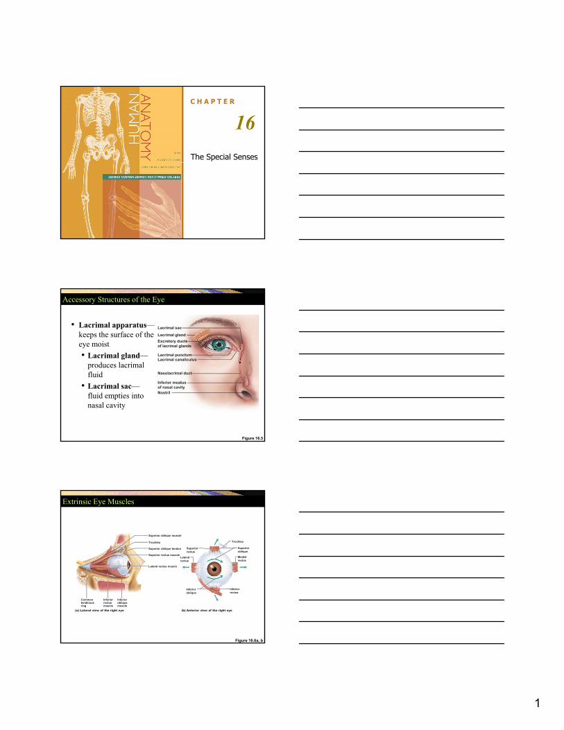
Accessory Structures of the Eye
• Lacrimal apparatus—keeps the surface of the eye moist• Lacrimal gland—
produces lacrimal
Lacrimal glandExcretory ducts of lacrimal glands
Lacrimal punctumLacrimal canaliculus
Lacrimal sac
produces lacrimal fluid
• Lacrimal sac—fluid empties into nasal cavity
Figure 16.5
Nasolacrimal duct
Inferior meatusof nasal cavityNostril
Extrinsic Eye Muscles
Superior oblique muscle
Trochlea
Superior oblique tendon
Superior rectus muscle
Lateral rectus muscle
Trochlea
Superioroblique
Lateralrectus
Superiorrectus
Medialrectus
Figure 16.6a, b
Inferiorrectusmuscle
Commontendinousring
Inferiorobliquemuscle
Lateral rectus muscle
(a) Lateral view of the right eye (b) Anterior view of the right eye
Inferiorrectus
Inferioroblique

2
Anatomy of the Eyeball
• External wall consists of three layers (tunics)• Fibrous layer• Vascular layer• Sensory layer
I l i i fl id (h )• Internal cavity contains fluids (humors)• Anterior segment- aqueous humor• Posterior segment- vitreous humor
Medial View of the Eye
Ora serrata
Ciliary body
Ciliary zonule(suspensoryligament)CorneaIrisP il Posterior pole
Fovea centralisMacula lutea
Retina
Choroid
Sclera
Figure 16.7a
(a) Diagrammatic view. The vitreous humor is illustrated only in the bottom part of the eyeball.
Anteriorpole
Pupil
Anteriorsegment(containsaqueous humor)LensScleral venoussinusPosterior segment (contains vitreous humor)
Optic nervePosterior pole
Central arteryand vein of the retinaOptic disc(blind spot)
Fibrous Layer
• Sclera- white of the eye (dura mater of brain); provides anchoring for extrinsic eye muscles
• Cornea- light enters here, transparentCornea light enters here, transparent• Junction of sclera and cornea- limbus

3
Vascular Layer• Choroid-forms posterior 5/6 of vascular tunic,
continuous with ciliary body; prevents light scatter
• Ciliary body- a thickened ring of tissue that encircles the lens- consists of smooth muscle (ciliary muscle) – focuses the lens.
• Ciliary processes-generate suspensory ligament• Accomodation• Makes aqueous humor
• Iris- visible, colored part of eye. Allows light to enter. Has smooth muscle fibers
• Pupil- opening of iris (pupillary light reflex)-opening for light
The Vascular Layer
Ora serrata
Ciliary body
Ciliary zonule(suspensoryligament)CorneaIrisP il Posterior pole
Fovea centralisMacula lutea
Retina
Choroid
Sclera
Figure 16.7a
(a) Diagrammatic view. The vitreous humor is illustrated only in the bottom part of the eyeball.
Anteriorpole
Pupil
Anteriorsegment(containsaqueous humor)LensScleral venoussinusPosterior segment (contains vitreous humor)
Optic nervePosterior pole
Central arteryand vein of the retinaOptic disc(blind spot)

4
Sensory Layer (Retina)
• Consists of two layers• Pigmented layer • Neural layer (external to internal)
• Photoreceptors (cones and rods)• Bipolar neurons• Ganglion cells
Posterior Aspect of the Eyeball
Neural layer of retina
Pigmentedlayer ofretina
ScleraChoroid
Optic disc
Pathway of light
Figure 16.8a
(a) Posterior aspect of the eyeball
Central arteryand vein of retina Optic
nerve

5
Microscopic Anatomy of the Retina
RodPhotoreceptors
ConeBipolarcellsGanglion
cells
ChoroidOuter segmentsof rods and conesNuclei of
ganglioncells
Figure 16.8b, c
Pigmentedlayer of retinaPathway of light
Pathway of signal output
(b) Cells of the neural layer of the retina
Amacrine cellHorizontal cell
Pigmentedlayer of retina
Axons ofganglion cells
Nuclei ofrods andcones
Nucleiof bipolarcells
(c) Photomicrograph of retina
PhotoreceptorsProcessof bipolarcell
Outerfiber
Innerfibers
Rodcellbody
Conecellbody
Synapticterminals
Rod cellbody
Nuclei
Mitochondria
Connectingcilia
ent
Figure 16.9
Apicalmicrovillus
Discscontainingvisual pigments
Melaningranules
Discs beingphagocytized
Pigmentcellnucleus
Basal lamina(border with choroid)
Inne
r se
gme
Out
er s
egm
ent
Pig
men
ted
laye
r
Blood Supply of the Retina
• Retina receives blood from two sources• Outer third of the
retina—supplied Maculalutea
Central arteryand veinemergingfrom theoptic disc
Optic disc
Figure 16.10
by capillaries in the choroid
• Inner two-thirds of the retina—supplied by central artery and vein of the retina
Retina

6
Internal Chambers, Fluids and the Lens
• Posterior cavity- vitreous humor• Anterior cavity- anterior chamber (aqueous humor) and
posterior chamber• Aqueous humor- is renewed continuously and is in constant
motion- formed as filtrate of the blood from capillaries in ciliary processes, flows through pupil into anterior chamber, drains into veins via canal of Schlemm and returns to blood.
• Lens- a thick transparent, biconvex disc that changes shape to allow focusing of the light on the retina.
Internal Chambers and Fluids
Anterior chamberAnteriorsegment
Cornea
Cornea
Corneal epithelium
Corneal endothelium
Aqueous humor
Iris
LensLens epithelium
Lens
Posterior segment(contains vitreoushumor)
Ciliary zonule(suspensory
2
Figure 16.11
Aqueous humor is formed by filtration from the capillaries in the ciliary processes.
Sclera
Bulbarconjunctiva
Scleral venous sinus
Posterior chambersegment(containsaqueoushumor)
Corneoscleraljunction
(suspensoryligament)
Ciliaryprocesses
Ciliarymuscle
Ciliary body
Aqueous humor flows from the posterior chamber through the pupil into the anterior chamber. Some also flows through the vitreous humor (not shown).
Aqueous humor is reabsorbed into the venous blood by the scleral venous sinus.
1
2
3
13
Visual Pathways
• Most visual information travels to the cerebral cortex
• Responsible for conscious “seeing”• Other pathways travel to nuclei in the midbrain
d di h land diencephalon
7
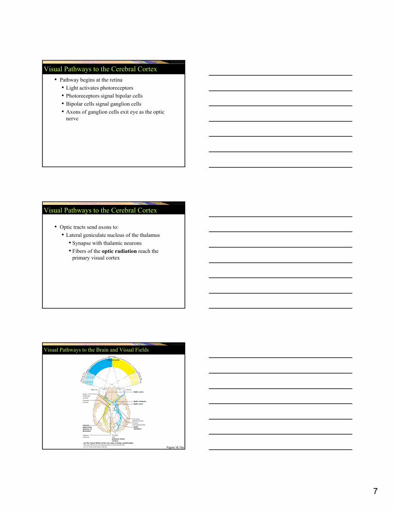
Visual Pathways to the Cerebral Cortex• Pathway begins at the retina
• Light activates photoreceptors • Photoreceptors signal bipolar cells• Bipolar cells signal ganglion cells • Axons of ganglion cells exit eye as the opticAxons of ganglion cells exit eye as the optic
nerve
Visual Pathways to the Cerebral Cortex
• Optic tracts send axons to:• Lateral geniculate nucleus of the thalamus
• Synapse with thalamic neurons • Fibers of the optic radiation reach the
primary visual cortex
Right eye Left eye
Fixation point
Optic nerveSupra-chiasmatic
Both eyes
Visual Pathways to the Brain and Visual Fields
Figure 16.15a
Pretectalnucleus
Opticradiation
Optic tractOptic chiasma
Uncrossed(ipsilateral) fiberCrossed(contralateral) fiber Lateral
geniculatenucleus ofthalamus
Superiorcolliculus
Occipitallobe(primary visualcortex)
(a) The visual fields of the two eyes overlap considerably.Note that fibers from the lateral portion of each retinal fielddo not cross at the optic chiasma.
chiasmaticnucleus

8
Visual Pathways to Other Parts of the Brain
• Some axons from the optic tracts• Branch to midbrain
• Superior colliculi• Pretectal nuclei
• Other branches from the optic tractsOther branches from the optic tracts• Branch to the suprachiasmatic nucleus
The Ear and Equilibrium
• The ear is divided into the outer, middle and inner ear.
• Outer and middle ear participate in hearing only• Inner ear functions for both hearing and
ilib iequilibrium
Structure of the Ear
Auricle(pinna)
External ear Middleear
Internal ear(labyrinth)
Figure 16.17a
Externalacousticmeatus
(a) The three regions of the ear
Helix
Lobule
Pharyngotympanic(auditory) tube
Tympanicmembrane

9
Tympanic Membrane
• Separates outer from middle ear.• Shaped like a flattened cone• Transmits air vibrations to auditory ossicles
Structures of the Middle Ear
Auditory
Entrance to mastoid antrum in the epitympanic recess
Semicircularcanals
Vestibularnerve
Oval window(deep to stapes)
Incus(anvil)
Malleus(hammer)
Vestibule
Figure 16.17b
Pharyngotympanic(auditory) tube
ossicles
Tympanicmembrane
Cochlea
Cochlearnerve
Round window
(anvil)Stapes(stirrup)
(b) Middle and internal ear
Middle Ear
• Tympanic cavity• Ossicles- malleus, incus, stapes (merge onto oval
window)- transmit sound from external ear to internal ear. Increase force but not the amplitude of vibrations transmitted by tympanic membrane
• eustachian tube/ auditory tube- equalizes air pressure
• Oval window (stapes)• Round window – dissipates left-over energy in
cochlea

10
Middle Ear
• Round window – dissipates left-over energy in cochlea
• Reflexive muscles that protect from loud sounds (tympanic reflex)
S di ( )• Stapedius (stapes)• Tensor tympani (malleus)
Malleus
View
Superior
Anterior
Lateral
Incus Epitympanic recess
The Middle Ear
• Ear ossicles—smallest bones in the body• Malleus—attaches to
the eardrum • Incus—between the
malleus and stapes• Stapes vibrates
Pharyngotym-panic tube
Tensortympanimuscle
Tympanicmembrane(medial view)
Stapes Stapediusmuscle
Figure 16.18
• Stapes—vibrates against the oval window
• Tensor tympani and stapedius• Two tiny skeletal
muscles in the middle ear cavity
Inner Ear
• Vestibule
• Semicircular canals
• Utricle and saccule- balance and position of head; movement
• Semicircular ducts-concerned with movement; ampullae are swellings near utricle
• Cochlea• Cochlear duct- contains spiral
organ of Corti, converts mechanical sound to nerve impulses

11
Structure of the Ear
Auricle(pinna)
External ear Middleear
Internal ear(labyrinth)
Figure 16.17a
Externalacousticmeatus
(a) The three regions of the ear
Helix
Lobule
Pharyngotympanic(auditory) tube
Tympanicmembrane
The Internal Ear
Anterior
Semicircular ducts insemicircular canals
PosteriorL t l
Temporalbone Facial nerve
Vestibular nerveSuperior vestibularganglionInferior vestibularganglion
Figure 16.19
Lateral
Cristae ampullaresin the membranousampullaeUtricle investibuleSaccule investibule
Stapes inoval window
ganglionCochlear nerveMaculaeSpiral organ (of Corti)Cochlear duct incochlea
Round window
Utricle and Saccule-vestibular apparatus
• Concerned with balance and position of head while stationary
• Acceleration (linear)• Endolymph • Has special sensory epithelium called Macula
• Receptor hair cells

12
The Maculae in the Internal Ear
Macula ofsaccule
Otoliths
Hair bundle
Kinocilium
Stereocilia
Otolithicmembrane
Macula ofutricle
Figure 16.22a
Hair bundle
Vestibularnerve fibers
Hair cellsSupportingcells
(a)
The Maculae in the Internal EarHaircell
OtolithsOtolithicmembrane
Force ofgravity
Figure 16.22b(b)
Head upright Head tilted
Semicircular Ducts &Vestibular apparatus
• Rotational movement• Anterior, posterior, and lateral semicircular duct.• Opens onto utricle• Ampulla
• Crista ampullaris- (crest with hair cells)• Cupula- jelly-like pointed cap

13
Structure and Function of the Crista Ampullaris
Hair bundle (kinociliumplus stereocilia)
Crista ampullaris
Endolymph
Cupula
Figure 16.23a, b
Fibers of vestibular nerve
plus stereocilia)
Hair cell
Supporting cell
Membranous labyrinth
Cristaampullaris
(a) Anatomy of a crista ampullaris in a semicircular canal
(b) Scanning electron micrographof a crista ampullaris (45X)
Structure and Function of the Crista Ampullaris
Figure 16.23c
Fibers ofvestibular
nerve
At rest, the cupula stands upright.
Section ofampulla,filled withendolymph
(c) Movement of the cupula during rotational acceleration and deceleration
Cupula Flow of endolymph
During rotational acceleration, endolymph moves inside the semicircular canals in the direction opposite the rotation (it lags behind because of inertia). Endolymph flow bends the cupula and excites the hair cells.
As rotational movement slows, endolymph keeps moving in the direction of the rotation, bending the cupula in the opposite direction from acceleration and inhibiting the hair cells.
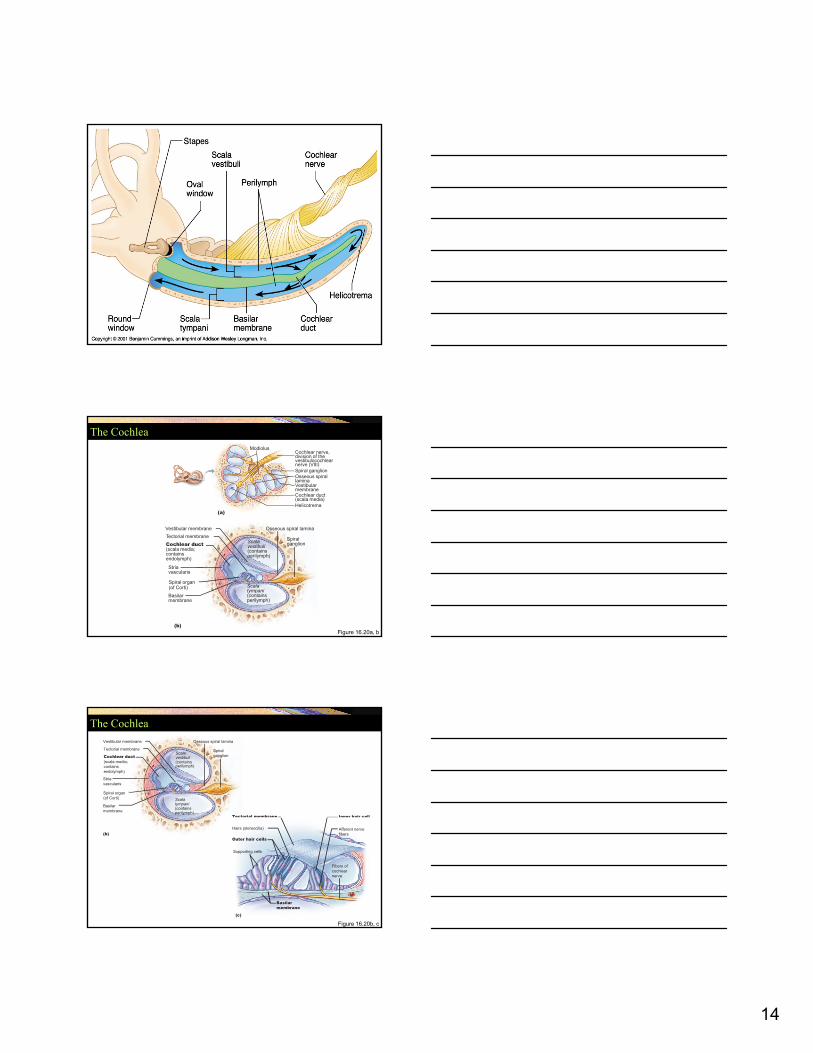
Cochlear Duct• A spiral blind tube • Perilymph-filled chambers- scala vestibuli (starts
from oval window) and scala tympani Merge onto round window• Vestibular membrane• Basilar membrane
• EndolymphEndolymph• Organ of Corti
• Hair cells- stereocilia• Supporting cells
• Tectorial membrane
14
The Cochlea
(a)Helicotrema
ModiolusCochlear nerve,division of thevestibulocochlearnerve (VIII)
Cochlear duct(scala media)
Spiral ganglionOsseous spirallaminaVestibularmembrane
Figure 16.20a, b(b)
Cochlear duct(scala media;containsendolymph)
Tectorial membraneVestibular membrane
Scala vestibuli(containsperilymph)
Scala tympani(containsperilymph)
Basilarmembrane
Spiral organ(of Corti)
Striavascularis
Spiralganglion
Osseous spiral lamina
The Cochlea
Cochlear duct(scala media;containsendolymph)
Tectorial membrane
Vestibular membrane
Scala vestibuli(containsperilymph)
Scala tympani(containsperilymph)
Basilarmembrane
Spiral organ(of Corti)
Striavascularis
Spiralganglion
Osseous spiral lamina
Tectorial membrane Inner hair cell
Figure 16.20b, c
(b)
(c)
Tectorial membrane Inner hair cell
Outer hair cells
Hairs (stereocilia) Afferent nervefibers
Basilarmembrane
Fibers ofcochlearnerve
Supporting cells

15
Hearing Pathway• Sound vibrations• Ossicles- stapes and oval window• Perilymph of scala vestibuli and tympani• Endolymph of scala media• Tectorial membrane stays still, hair cells on basilar
membrane (stereocilia) have mechanically gated ion h l (K+) l d tchannels (K+) leads to
• Hair cells stimulated (excess released via round window)
• AP in CNVIII to thalamus (via medulla and pons)• Thalamus• Primary auditory cortex in temporal lobe
The Role of the Cochlea in Hearing
Sound waves vibrate the tympanic membrane.
Malleus Incus
Auditory ossicles
Stapes
Ovalwindow
Scala vestibuli
Helicotrema
Cochlear nerve
Scala tympani
Cochlear duct
Basilarmembrane
Auditory ossicles vibrate. Pressure is amplified.
Pressure waves created by the stapes pushing on the oval window move through fluid in the scala vestibuli.
Sounds with frequencies below hearing travel through the
1
2
3
4a
2 3
4a
4b
Figure 16.21
Roundwindow
Tympanicmembrane
ea g t a e t oug t ehelicotrema and do not excite hair cells.
Sounds in the hearing range go through the cochlear duct, vibrating the basilar membrane and deflecting hairs on inner hair cells.
4b
1