by lucy suliman. sleep related breathing disorders defintions sleep related breathing disorders...
TRANSCRIPT
ByLucy suliman
obstructive sleep apneaobstructive sleep apnea


Sleep related Sleep related breathing disorders breathing disorders defintionsdefintions ApneaApnea:: Apnea is the cessation, or near Apnea is the cessation, or near
cessation, of airflow for 10 seconds. cessation, of airflow for 10 seconds. Oronasal thermal sensor (or Oronasal thermal sensor (or alternative) drops by ≥90% of alternative) drops by ≥90% of baseline.baseline.
Duration ≥ 10 sec. At least 90% of Duration ≥ 10 sec. At least 90% of events duration must meet apnea events duration must meet apnea amplitude reduction criteria .amplitude reduction criteria .
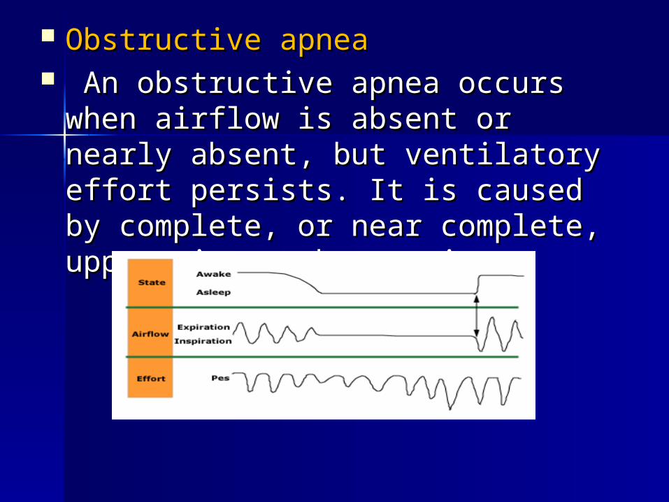
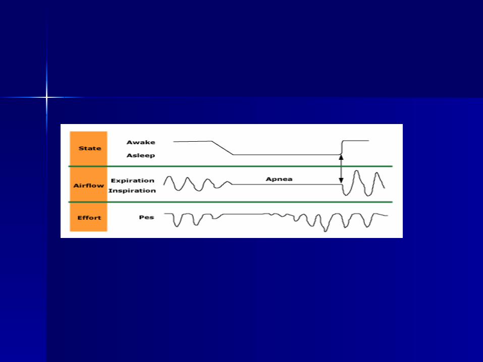
Obstructive apneaObstructive apnea An obstructive apnea occurs when An obstructive apnea occurs when
airflow is absent or nearly absent, airflow is absent or nearly absent, but ventilatory effort persists. It is but ventilatory effort persists. It is caused by complete, or near caused by complete, or near complete, upper airway obstruction.complete, upper airway obstruction.

Central apneaCentral apnea A central apnea occurs when both A central apnea occurs when both
airflow and ventilatory effort are airflow and ventilatory effort are absent Breathing cessation is proven absent Breathing cessation is proven by an absence of diaphragmatic by an absence of diaphragmatic activation, measured by activation, measured by electromyography (EMG).electromyography (EMG).

Mixed apneaMixed apnea During a mixed apnea, there is an During a mixed apnea, there is an
interval during which there is no interval during which there is no respiratory effort (ie, central apnea respiratory effort (ie, central apnea pattern) and an interval during which pattern) and an interval during which there are obstructed respiratory there are obstructed respiratory efforts .The central apnea pattern efforts .The central apnea pattern usually precedes the obstructive usually precedes the obstructive apnea pattern during mixed apnea apnea pattern during mixed apnea

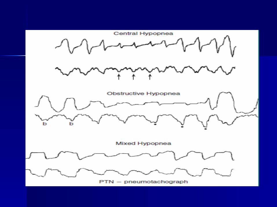
HypopneaHypopnea Hypopnea is a reduction of airflow to a degree Hypopnea is a reduction of airflow to a degree
that is insufficient to meet the criteria for an that is insufficient to meet the criteria for an apnea.apnea.
The most recent definition, endorsed by the The most recent definition, endorsed by the
American Academy of Sleep Medicine, American Academy of Sleep Medicine, recommends that hypopnea be scored when all of recommends that hypopnea be scored when all of the following four criteria are met: the following four criteria are met:
Airflow decreases at least 30 percent from Airflow decreases at least 30 percent from baseline baseline
There is diminished airflow lasting at least 10 There is diminished airflow lasting at least 10 secondsseconds
at least 4 percent oxyhemoglobin desaturation at least 4 percent oxyhemoglobin desaturation Associated with arousalAssociated with arousal

ororAirflow decreases at least 50 Airflow decreases at least 50
percent from baselinepercent from baseline There is diminished airflow There is diminished airflow
lasting at least 10 seconds lasting at least 10 seconds 3 percent oxyhemoglobin 3 percent oxyhemoglobin
desaturation or an arousal desaturation or an arousal

Respiratory effort related arousalsRespiratory effort related arousals — — Respiratory effort related arousals Respiratory effort related arousals
(RERAs) exist when there is a (RERAs) exist when there is a sequence of breaths that lasts at sequence of breaths that lasts at least 10 seconds, is characterized byleast 10 seconds, is characterized by
increasing respiratory effort orincreasing respiratory effort or flattening of the nasal pressure flattening of the nasal pressure
waveformwaveform leads to an arousal from sleepleads to an arousal from sleep does not meet the criteria of an does not meet the criteria of an
apnea or hypopnea apnea or hypopnea
HypoventilationHypoventilation Hypoventilation during sleep is defined Hypoventilation during sleep is defined
as an increase in the arterial carbon as an increase in the arterial carbon dioxide (PaCO2) of 10 mm Hg during dioxide (PaCO2) of 10 mm Hg during sleep (compared with an awake supine sleep (compared with an awake supine value) that lasts at least 25 percent of value) that lasts at least 25 percent of the sleep time.the sleep time.
Directly measuring the pCO2 in an Directly measuring the pCO2 in an arterial blood gas during a sleep study is arterial blood gas during a sleep study is optimal, but impractical. optimal, but impractical.
Transcutaneous CO2 measurements and Transcutaneous CO2 measurements and expired end tidal CO2 are alternatives .expired end tidal CO2 are alternatives .
Polysomnography Polysomnography indicesindices

Apnea indexApnea index The apnea index (AI) is the total The apnea index (AI) is the total
number of apneas per hour of sleep number of apneas per hour of sleep Apnea-hypopnea indexApnea-hypopnea index — — The apneaThe apnea––
hypopnea index (AHI) is the total hypopnea index (AHI) is the total number of apneas and hypopneas per number of apneas and hypopneas per hour of sleep. The AHI is most hour of sleep. The AHI is most commonly calculated per hour of total commonly calculated per hour of total sleep.sleep.
Respiratory disturbance indexRespiratory disturbance index The respiratory disturbance index (RDI) The respiratory disturbance index (RDI)
is the total number of events (eg, is the total number of events (eg, apneas, hypopneas, and RERAs) per apneas, hypopneas, and RERAs) per hour of sleep. hour of sleep.
DesaturationDesaturation — — Oxygen desaturation is Oxygen desaturation is a frequent consequence of apnea and a frequent consequence of apnea and hypopnea. Several measures are used hypopnea. Several measures are used to quantify the severity of desaturation to quantify the severity of desaturation and should be included in and should be included in polysomnography report .polysomnography report .
Oxygen desaturation index (ODI)Oxygen desaturation index (ODI) This is the number of times that the This is the number of times that the
oxygen saturation falls by more than 3 oxygen saturation falls by more than 3 or 4 percent per hour of sleep or 4 percent per hour of sleep Minimum levels Minimum levels

Arousal indexArousal index The arousal index (ArI) is the The arousal index (ArI) is the
total number of arousals per hour total number of arousals per hour of sleep. It is generally lower than of sleep. It is generally lower than the AHI or RDI because the AHI or RDI because approximately 20 percent of approximately 20 percent of apneas or hypopneas are not apneas or hypopneas are not accompanied by arousals accompanied by arousals

syndromes of sleep syndromes of sleep related breathing related breathing disorders disorders


Obstructive sleep apnea syndromeObstructive sleep apnea syndrome Obstructive sleep apnea syndrome Obstructive sleep apnea syndrome
encompasses obstructive sleep apnea (OSA) encompasses obstructive sleep apnea (OSA) in adults and OSA in children. OSA in adults in adults and OSA in children. OSA in adults is defined as either :is defined as either :
More than 15 apneas, hypopneas, or RERAs More than 15 apneas, hypopneas, or RERAs per hour of sleep (ie, an AHI or RDI >15 per hour of sleep (ie, an AHI or RDI >15 events/hr) in an asymptomatic patientevents/hr) in an asymptomatic patient
OR OR More than 5 apneas, hypopneas, or RERAs More than 5 apneas, hypopneas, or RERAs
per hour of sleep (ie, an AHI or RDI >5 events per hour of sleep (ie, an AHI or RDI >5 events per hour) in a patient with symptoms (eg, per hour) in a patient with symptoms (eg, sleepiness, fatigue and inattention) or signs sleepiness, fatigue and inattention) or signs of disturbed sleep (eg, snoring, restless of disturbed sleep (eg, snoring, restless sleep, and respiratory pauses). sleep, and respiratory pauses).
Central sleep apnea syndromeCentral sleep apnea syndrome Central sleep apnea syndrome Central sleep apnea syndrome
(CSAS) can be:(CSAS) can be: idiopathicidiopathic (eg, primary central sleep (eg, primary central sleep
apnea [CSA])apnea [CSA]) secondarysecondary. Examples of secondary . Examples of secondary
CSAS include Cheyne-Stokes CSAS include Cheyne-Stokes breathing, CSA due to high altitude breathing, CSA due to high altitude periodic breathing, CSA due to a periodic breathing, CSA due to a medical condition, and CSA due to a medical condition, and CSA due to a drug or substance. More than 75 drug or substance. More than 75 percent of events should be central percent of events should be central to qualify for this syndrome category. to qualify for this syndrome category.
Complex Sleep Apnea (CompSA)
CompSA consists of all or predominantly obstructive apneas which convert to all or predominantly central apneas when treated with a CPAP or bilevel devices
Cheyne-Stokes breathingCheyne-Stokes breathing — — Cheyne-Stokes Cheyne-Stokes breathing refers to a cyclic pattern of breathing refers to a cyclic pattern of crescendo-decrescendo tidal volumes and crescendo-decrescendo tidal volumes and central apneas, hypopneas, or both. It is central apneas, hypopneas, or both. It is commonly associated with heart failure or commonly associated with heart failure or stroke. stroke.
Hypoventilation syndromesHypoventilation syndromes — — Patients with a Patients with a hypoventilation syndrome generally have mild hypoventilation syndrome generally have mild hypercarbia or elevated serum bicarbonate hypercarbia or elevated serum bicarbonate levels when awake, which worsen during levels when awake, which worsen during sleep. sleep.
Two hypoventilation syndromes:Two hypoventilation syndromes: congenital central hypoventilation syndrome congenital central hypoventilation syndrome
(CCHS) (CCHS) obesity hypoventilation syndrome (OHS), obesity hypoventilation syndrome (OHS),

Cheyne-Stokes Cheyne-Stokes breathingbreathing

Obstructive sleep apnea :-Obstructive sleep apnea :- (OSA) is a disorder that is (OSA) is a disorder that is
characterized by obstructive apneas characterized by obstructive apneas and hypopneas caused by repetitive and hypopneas caused by repetitive collapse of the upper airway during collapse of the upper airway during sleep.sleep.
The diagnosis should be considered The diagnosis should be considered whenever a patient presents with whenever a patient presents with risk factors or clinical manifestations risk factors or clinical manifestations that are compatible with OSA. that are compatible with OSA.

RISK FACTORSRISK FACTORS :: obesity. obesity. craniofacial abnormalities.craniofacial abnormalities. upper airway soft tissue upper airway soft tissue
abnormalities.abnormalities. Potential risk factors include: Potential risk factors include:
heredity,smoking, nasal congestion, heredity,smoking, nasal congestion, and diabetes .and diabetes .
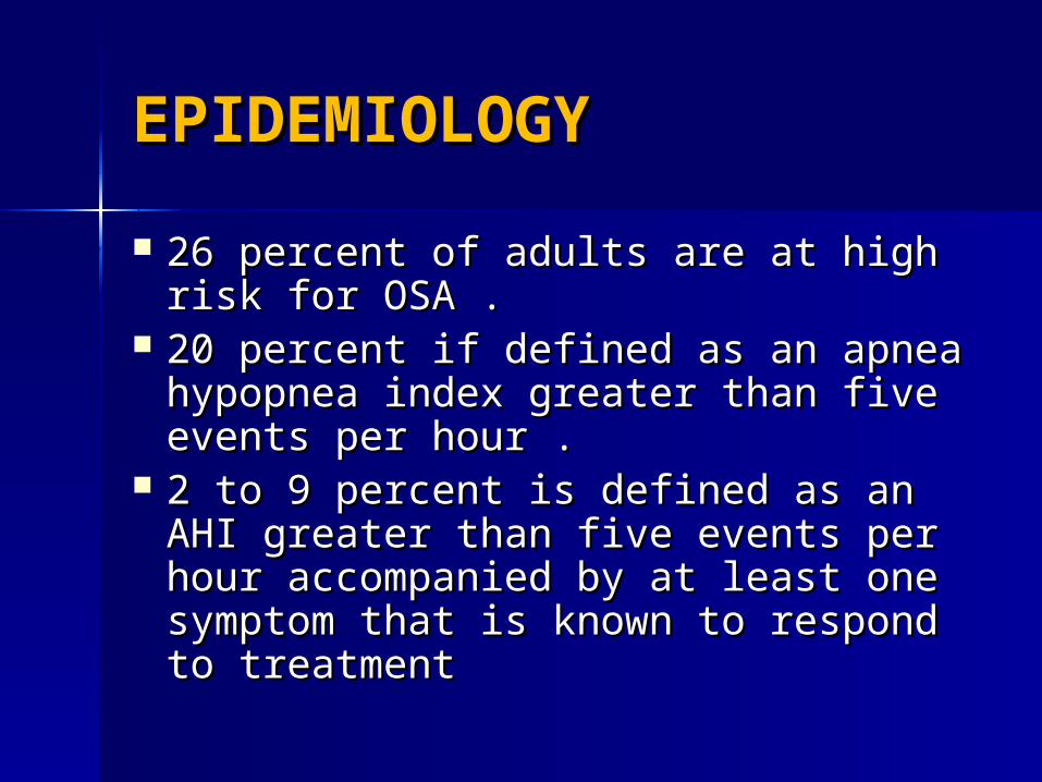
EPIDEMIOLOGY EPIDEMIOLOGY
26 percent of adults are at high risk 26 percent of adults are at high risk for OSA .for OSA .
20 percent if defined as an apnea 20 percent if defined as an apnea hypopnea index greater than five hypopnea index greater than five events per hour .events per hour .
2 to 9 percent is defined as an AHI 2 to 9 percent is defined as an AHI greater than five events per hour greater than five events per hour accompanied by at least one accompanied by at least one symptom that is known to respond to symptom that is known to respond to treatment treatment

AgeAge :- :- The prevalence of OSA increases The prevalence of OSA increases
from 18 to 45 years of age.from 18 to 45 years of age. There is a two- to three-fold higher There is a two- to three-fold higher
prevalence among individuals who prevalence among individuals who are 65 years and older, compared to are 65 years and older, compared to those who are 30 to 64 years old. those who are 30 to 64 years old.

Grades of OSAGrades of OSA
MildMild :-:-AHI between 5 and 15 events per AHI between 5 and 15 events per
hourhour Moderate:-Moderate:-
an AHI between 15 and 30 events an AHI between 15 and 30 events per hourper hour
Severe:-Severe:- AHI greater than 30 events AHI greater than 30 events
per hour, as well as an oxyhemoglobin per hour, as well as an oxyhemoglobin saturation below 90 percent for more saturation below 90 percent for more than 20 percent of the total sleep timethan 20 percent of the total sleep time

Diagnosis of OSADiagnosis of OSA
HistoryHistory Physical examinationsPhysical examinations laboratorylaboratory Subjective questionnaire:-Subjective questionnaire:-
Berline questionnairBerline questionnair
Epworth Sleepness Epworth Sleepness scalescale
Polysomnography.Polysomnography.


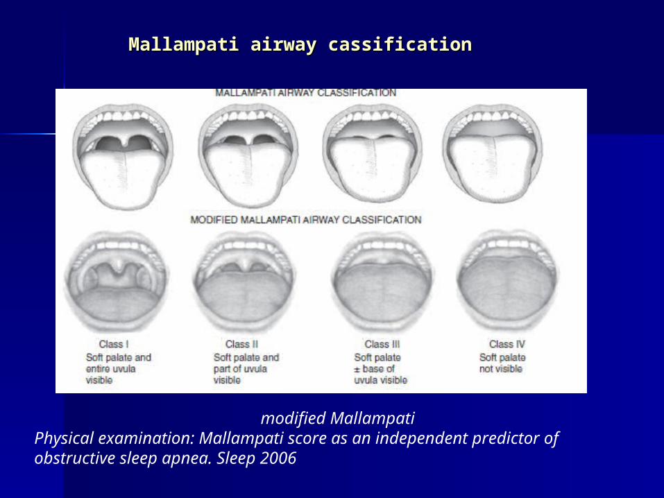
Mallampati airway cassificationMallampati airway cassification
modified MallampatiPhysical examination: Mallampati score as an independent predictor of obstructive sleep apnea. Sleep 2006

LaboratoryLaboratory 1)protinuria 1)protinuria —— Less than 10 percent of Less than 10 percent of
patients with OSA have proteinuriapatients with OSA have proteinuria
2) Hypercapnia 2) Hypercapnia —— Although uncommon Although uncommon in OSA alone, awake hypercapnia (and in OSA alone, awake hypercapnia (and hypoxemia) may be present if obesity hypoxemia) may be present if obesity hypoventilation syndrome coexistshypoventilation syndrome coexists
3)Hypothyroidism 3)Hypothyroidism —— OSA can be OSA can be caused or exacerbated by caused or exacerbated by hypothyroidismhypothyroidism

Imaging Studies
lateral cephalometry, endoscopy, fluoroscopy, CT scanning, MRI.


Epworth sleepiness scale:The chance to doze off or fall asleep in the
following situation: 1-sitting and reading 2- watching TV 3- sitting inactive in public place 4-as a passenger in a car for an hour without
a break 5-lying down to rest in the afternoon when
circumstances permit 6-sitting and talking to someone 7- sitting quietly after a lunch without alcohol. 8- in a car while stopped for few minutes in
traffic

Berline questionnairequestionnaire

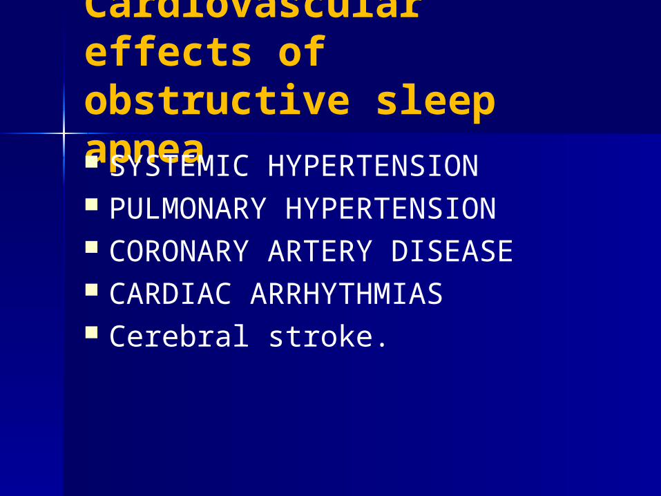
Cardiovascular effects of obstructive sleep apnea SYSTEMIC HYPERTENSION PULMONARY HYPERTENSION CORONARY ARTERY DISEASE CARDIAC ARRHYTHMIAS Cerebral stroke.
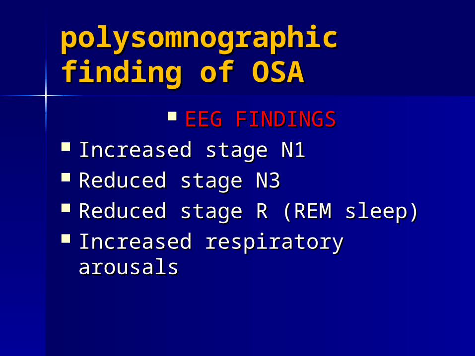
polysomnographic polysomnographic finding of OSAfinding of OSA
EEG FINDINGSEEG FINDINGS Increased stage N1Increased stage N1 Reduced stage N3Reduced stage N3 Reduced stage R (REM sleep)Reduced stage R (REM sleep) Increased respiratory arousalsIncreased respiratory arousals
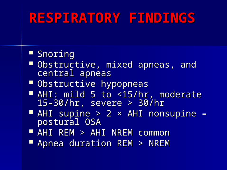
RESPIRATORY RESPIRATORY FINDINGSFINDINGS
SnoringSnoring Obstructive, mixed apneas, and Obstructive, mixed apneas, and
central apneascentral apneas Obstructive hypopneasObstructive hypopneas AHI: mild 5 to <15/hr, moderate 15AHI: mild 5 to <15/hr, moderate 15––
30/hr, severe > 30/hr30/hr, severe > 30/hr AHI supine > 2 × AHI nonsupine AHI supine > 2 × AHI nonsupine ––
postural OSApostural OSA AHI REM > AHI NREM commonAHI REM > AHI NREM common Apnea duration REM > NREMApnea duration REM > NREM

ARTERIAL OXYGEN ARTERIAL OXYGEN DESATURATIONDESATURATION
Lowest SaO2 during REM sleepLowest SaO2 during REM sleep Longest REM periods in the early Longest REM periods in the early
morning hours typically have the morning hours typically have the worst desaturationworst desaturation
CYCLIC VARIATION IN CYCLIC VARIATION IN HEART RATEHEART RATE
Slowing of heart rate at apnea Slowing of heart rate at apnea onset and speeding at event onset and speeding at event termination.termination.

diagnostic criteria of OSA diagnostic criteria of OSA 1) an obstructive respiratory disturbance 1) an obstructive respiratory disturbance
index (RDI) greater than 15 events per index (RDI) greater than 15 events per hour.hour.
2) an obstructive RDI between 5 and 14 2) an obstructive RDI between 5 and 14 events per hour that is accompanied by events per hour that is accompanied by daytime sleepiness, loud snoring, daytime sleepiness, loud snoring, witnessed breathing interruptions, or witnessed breathing interruptions, or awakenings due to gasping or choking. awakenings due to gasping or choking.
Academy of Sleep Medicine: The AASM Manual for ScoringAcademy of Sleep Medicine: The AASM Manual for Scoring
of Sleep and Associated Events: American Academy of Sleep Medicine, 2007.of Sleep and Associated Events: American Academy of Sleep Medicine, 2007.


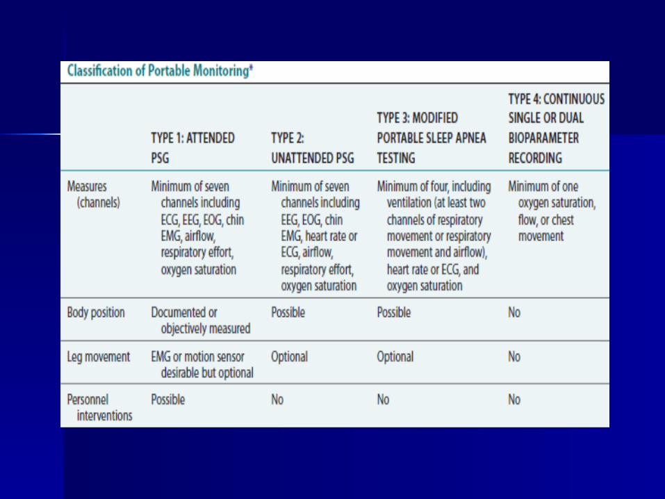
Types of PolysomnographyTypes of Polysomnography
Full-nightFull-night, attended, in-laboratory , attended, in-laboratory polysomnography (PSG) is considered the polysomnography (PSG) is considered the gold-standard diagnostic test for OSA. It gold-standard diagnostic test for OSA. It involves monitoring the patient during a involves monitoring the patient during a full night's sleepfull night's sleep
Patients who are diagnosed with OSA and Patients who are diagnosed with OSA and
choose positive airway pressure therapy choose positive airway pressure therapy are subsequently brought back for another are subsequently brought back for another study, during which their positive airway study, during which their positive airway pressure device is titrated pressure device is titrated
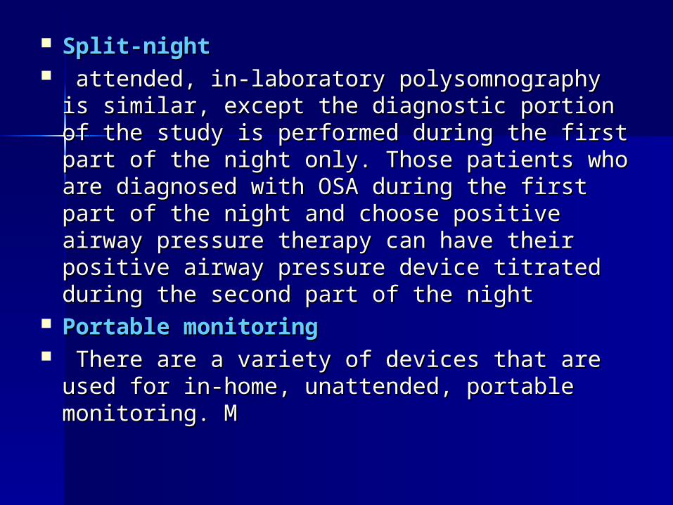
Split-nightSplit-night attended, in-laboratory polysomnography is attended, in-laboratory polysomnography is
similar, except the diagnostic portion of the similar, except the diagnostic portion of the study is performed during the first part of the study is performed during the first part of the night only. Those patients who are diagnosed night only. Those patients who are diagnosed with OSA during the first part of the night and with OSA during the first part of the night and choose positive airway pressure therapy can choose positive airway pressure therapy can have their positive airway pressure device have their positive airway pressure device titrated during the second part of the night titrated during the second part of the night
Portable monitoringPortable monitoring There are a variety of devices that are used There are a variety of devices that are used
for in-home, unattended, portable monitoring. for in-home, unattended, portable monitoring. M M


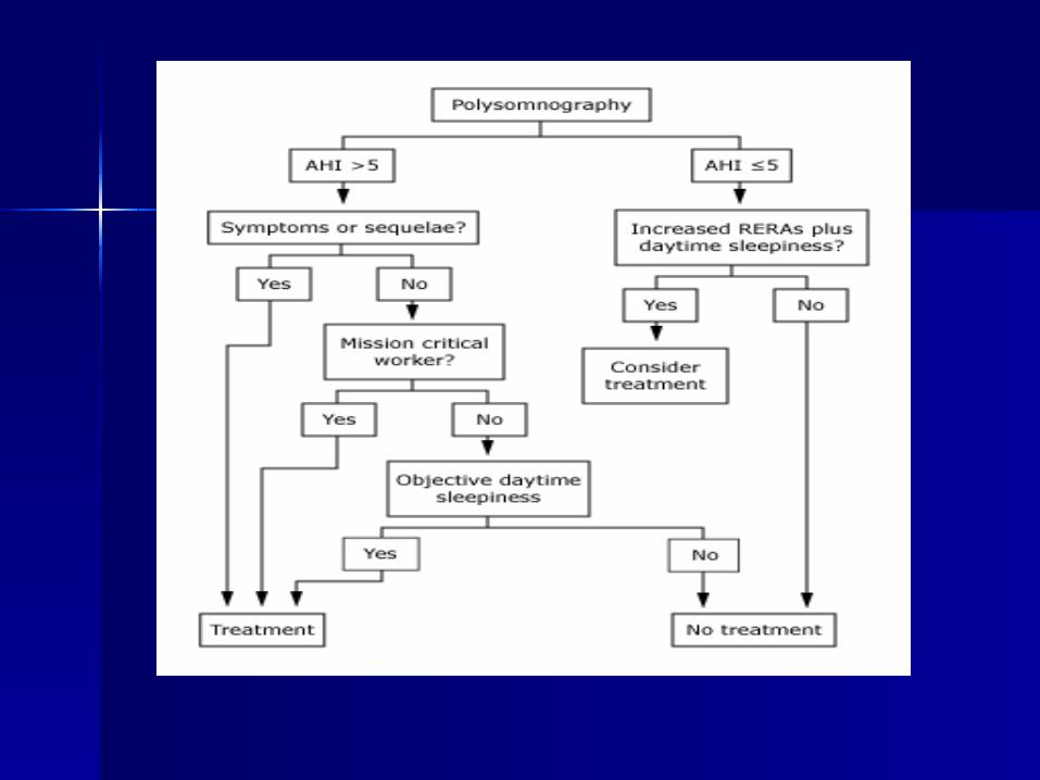
Indications for Indications for treatmenttreatment 1.1. respiratory disturbance index (RDI) respiratory disturbance index (RDI)
greater than 15 events per hour greater than 15 events per hour 2.2. an obstructive RDI between 5 and 14 an obstructive RDI between 5 and 14
events per hour that is accompanied by events per hour that is accompanied by daytime sleepiness, loud snoring, daytime sleepiness, loud snoring, witnessed breathing interruptions, or witnessed breathing interruptions, or awakenings due to gasping or choking. awakenings due to gasping or choking.
3.3. The AHI is greater than five events per The AHI is greater than five events per hour and the patient performs mission hour and the patient performs mission critical work (eg, airline pilot, bus critical work (eg, airline pilot, bus driver) driver)
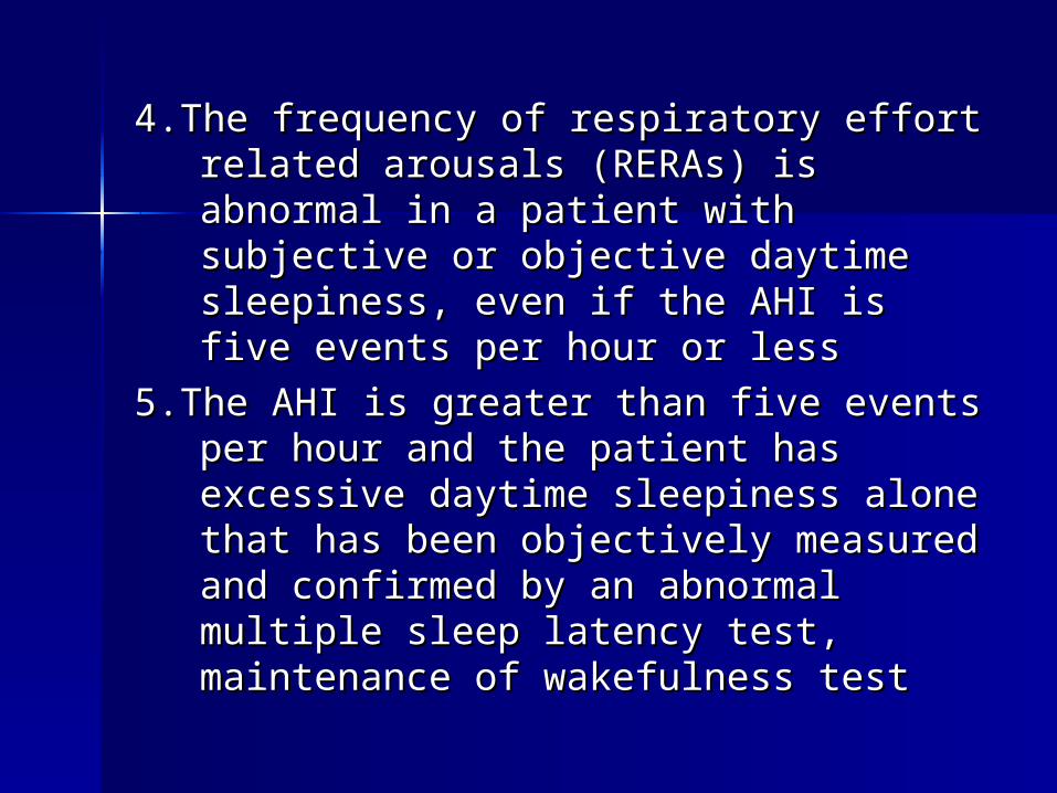
4.The frequency of respiratory effort 4.The frequency of respiratory effort related arousals (RERAs) is abnormal related arousals (RERAs) is abnormal in a patient with subjective or in a patient with subjective or objective daytime sleepiness, even if objective daytime sleepiness, even if the AHI is five events per hour or less the AHI is five events per hour or less
5.The AHI is greater than five events per 5.The AHI is greater than five events per hour and the patient has excessive hour and the patient has excessive daytime sleepiness alone that has daytime sleepiness alone that has been objectively measured and been objectively measured and confirmed by an abnormal multiple confirmed by an abnormal multiple sleep latency test, maintenance of sleep latency test, maintenance of wakefulness test wakefulness test
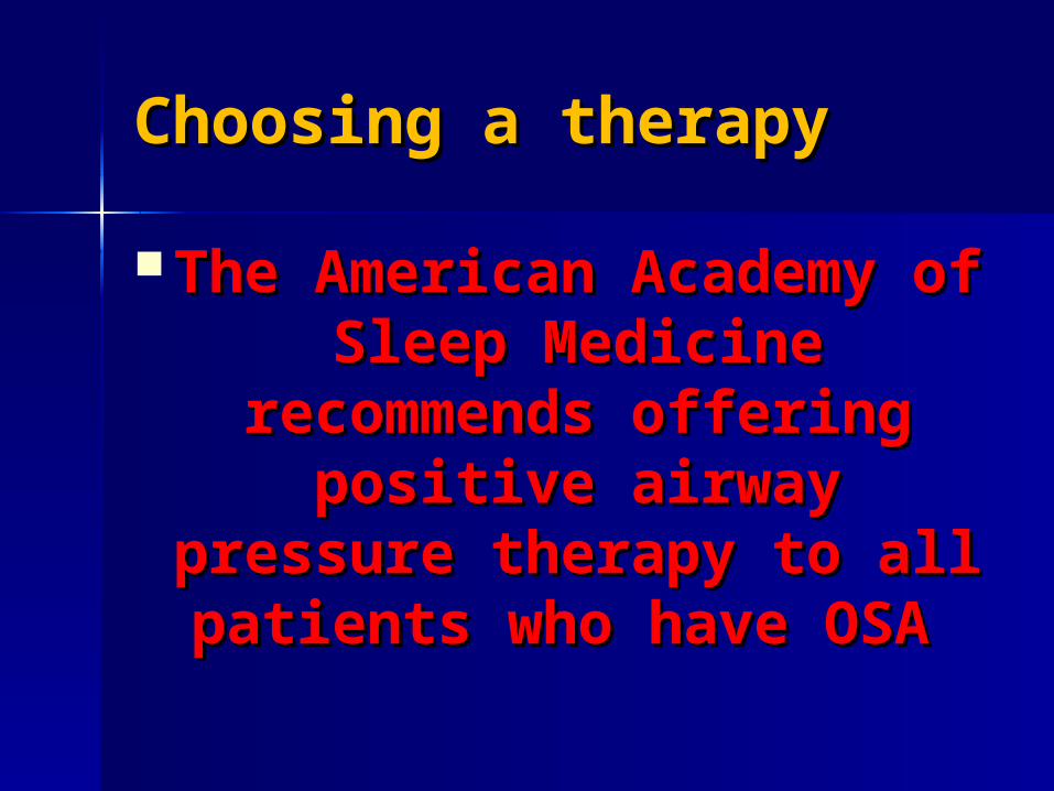
Choosing a therapyChoosing a therapy
The American Academy The American Academy of Sleep Medicine of Sleep Medicine
recommends offering recommends offering positive airway positive airway
pressure therapy to all pressure therapy to all patients who have OSA patients who have OSA

The choice of an OSA-specific The choice of an OSA-specific therapy:-therapy:-
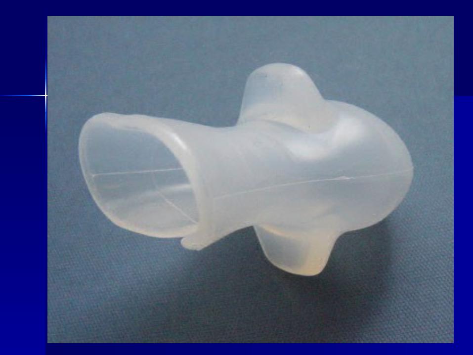
positive airway pressure, positive airway pressure, an oral appliance, an oral appliance, upper airway surgery.upper airway surgery. Medical treatment.Medical treatment.
depends on the severity of the OSA .depends on the severity of the OSA .
severe OSA (AHI >30 events severe OSA (AHI >30 events per hour and/or severe per hour and/or severe clinical sequelaeclinical sequelae
we use positive airway pressure we use positive airway pressure as first-line therapy. This is based as first-line therapy. This is based on the variable efficacy of oral on the variable efficacy of oral appliances in this patient appliances in this patient population. population.

For patients with mild or moderate For patients with mild or moderate OSA (AHI ≤ 30 events per hour with OSA (AHI ≤ 30 events per hour with severe clinical sequelaesevere clinical sequelae
we prefer positive airway pressure to an we prefer positive airway pressure to an oral appliance because it is superior at oral appliance because it is superior at reducing the frequency of obstructive reducing the frequency of obstructive events. events.
In contrast, for patients with mild or In contrast, for patients with mild or moderate OSA without severe clinical moderate OSA without severe clinical sequelaesequelae
we initiate an oral appliance rather than we initiate an oral appliance rather than positive airway pressure. This is based on positive airway pressure. This is based on our recognition that most patients prefer our recognition that most patients prefer an oral appliancean oral appliance
and both modalities have a similar effect and both modalities have a similar effect on symptoms on symptoms


surgical therapysurgical therapy when positive airway pressure or an when positive airway pressure or an
oral appliance is declined, oral appliance is declined, ineffective (after at least a three ineffective (after at least a three month trial of therapy), or not an month trial of therapy), or not an option option
For patients whose OSA is due to a For patients whose OSA is due to a surgically correctable obstructing surgically correctable obstructing lesion, surgical resection of the lesion, surgical resection of the obstructing lesion is first-line obstructing lesion is first-line therapy therapy


BEHAVIOR BEHAVIOR MODIFICATIONMODIFICATION

Weight lossWeight loss Sleep positionSleep position Alcohol avoidanceAlcohol avoidance Medication avoidanceMedication avoidance