by dr. salahuddin m. kamal ( prof. emeritus in eaea) associate professor medical radiation physics...
TRANSCRIPT

ByDr. Salahuddin M. Kamal
(Prof. Emeritus in EAEA)
Associate Professor Medical Radiation Physics
King Abdulaziz UniversityFaculty of Medical Applied Sciences
Diagnostic Radiology Department
Radiobiology and
Radiation Protection(Rad-222)
Radiobiology
When x-rays enter the body, they interact at the atomic level to cause ionization. Radiobiology is the response of living systems to ionizing radiation.

Ionization
Ionization is the process of removing an electron from an electrically neutral atom to produce an ion pair. An ion is an atom or subatomic particle with a positive or negative charge.
Ionization negative ion (electron)
positive ion: atom with 3 protons, 2 electrons
X-ray enters atom and strikes electron, knocking it out of its orbit and creating two ions (ion pair). The ejected electron is the negative ion and the atom with a net positive charge is the positive ion.
+++
Ionizing Radiation
The two types of ionizing radiation are electromagnetic and particulate.
Electromagnetic: Electromagnetic radiation has both electrical and magnetic properties. It includes x-rays and gamma rays. X-rays are produced by a machine and gamma rays are produced when radioactive materials decay. Neither one has any mass.
Particulate: Particulate radiation consists of particles that have mass and travel at high speeds. Included are alpha particles (helium nuclei), electrons (beta particles; also called beta rays), protons and neutrons.
Attenuation
Attenuation is the reduction of the x-ray beam intensity by interaction with the tissue that the x-rays pass through. The three types of interactions are:
1. Coherent scattering
2. Compton scattering
3. Photoelectric absorption
Approximately 9% of the x-rays pass through the tissues without any interactions.

Coherent Scattering
A low-energy x-ray interacts with an outer-shell electron and causes it to vibrate briefly. A scattered x-ray of the same energy as the primary x-ray is then emitted, going in a different direction than the primary x-ray. An electron is not ejected from the atom. (No ionization). This interaction occurs about 8% of the time.

primary x-ray strikes electron in outer shell
scattered x-ray emitted with same energy as primary x-ray, different direction
Coherent Scattering
electron vibrates

Compton Scattering
In Compton Scattering, an outer shell electron is ejected when struck by an x-ray, creating an ion pair (ionization). The primary x-ray loses some of its energy and continues in a different direction as a scattered x-ray. Compton Scattering accounts for about 62% of the interactions occurring within the tissues. Approximately 30% of the scattered x-rays exit the head.

ejected electron (negative ion)
Compton Scattering
primary x-ray
The primary x-ray strikes an outer-shell electron, knocking it out of its orbit (ionization). The primary x-ray loses some of its energy and continues in a different direction as a scattered x-ray.
atom with net positive charge is positive ion
scattered x-ray

Photoelectric Absorption
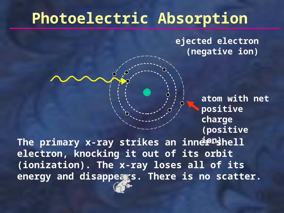
In Photoelectric Absorption, an inner-shell electron is ejected when struck by an x-ray, creating an ion pair (ionization). The primary x-ray loses all of its energy in knocking the electron out of its orbit; the x-ray ceases to exist (no scatter radiation). Photoelectric Absorption accounts for about 30% of the interactions occurring within the tissues.
Photoelectric Absorption
The primary x-ray strikes an inner-shell electron, knocking it out of its orbit (ionization). The x-ray loses all of its energy and disappears. There is no scatter.
ejected electron (negative ion)
atom with net positive charge (positive ion)

threshold
linear
non-linear
Dose-Response Curves
non-threshold
Re
sp
on
se
Dose
Dose-Response curves represent the relationship between the dose of radiation a person receives and the cellular response to that exposure. These responses may be linear or non-linear and may, or may not, have a threshold dose; the responses (effects) may be stochastic or deterministic. (See next two slides for definitions of these terms).

Linear: the response is directly related to the dose. As the dose increases, the response increases proportionately.
Non-linear: the response is not proportionate to the dose. An increase in dose may result in a larger or smaller increase in the response depending on the location on the dose-response curve.
Threshold: this represents the dose at which effects are produced; below this dose, there are no obvious effects.
Non-threshold: any dose, no matter how small, will produce a response.

Stochastic effect: occurs by chance, usually without a threshold level of dose. The probability of a stochastic effect is increased with increasing doses, but the severity of the response is not proportional to the dose (e.g., two people may get the same dose of radiation, but the response will not be the same in both people). Genetic mutations and cancer are the two main stochastic effects.
Deterministic effect: health effects that increase in severity with increasing dose above a threshold level. Usually associated with a relatively high dose delivered over a short period of time. Skin erythema (reddening) and cataract formation from radiation are two examples of deterministic effects.
DNA
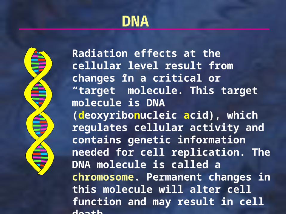
Radiation effects at the cellular level result from changes in a critical or “target” molecule. This target molecule is DNA (deoxyribonucleic acid), which regulates cellular activity and contains genetic information needed for cell replication. The DNA molecule is called a chromosome. Permanent changes in this molecule will alter cell function and may result in cell death.

Direct vs. Indirect Effect
DNA
If an x-ray or some type of particulate radiation interacts with the DNA molecule, this is considered a direct effect. Particulate radiation, because of its mass, is more apt to cause damage to the DNA by this direct effect. Other molecules that contribute to cell function, such as RNA, proteins, and enzymes, may also be affected by the direct effect.
x-ray or particulate radiation
Direct effect=

Direct vs. Indirect Effect
H2Oions and
free radicals
Most of the damage to DNA molecules from x-rays is accomplished through the indirect effect. When x-rays enter a cell, they are much more likely to hit a water molecule because there are a large number of water molecules in each cell. When the x-ray ionizes the water molecule, ions and free radicals are produced which in turn bond with a DNA molecule, changing its structure. Since the x-ray interacted with the water molecule before the DNA was involved, this is considered an indirect effect.
x-ray or particulate radiation
DNA =Indirect effect

A free radical is an atom or molecule that has an unpaired electron in the valence shell, making it highly reactive. These free radicals aggressively join with the DNA molecule to produce damage. In the presence of oxygen, the hydroperoxyl free radical is formed; this is one of the most damaging free radicals that can be produced. Free radicals are the primary mediator of the indirect effects on DNA.
Free Radical
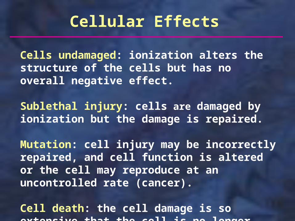
Cells undamaged: ionization alters the structure of the cells but has no overall negative effect.
Sublethal injury: cells are damaged by ionization but the damage is repaired.
Mutation: cell injury may be incorrectly repaired, and cell function is altered or the cell may reproduce at an uncontrolled rate (cancer).
Cell death: the cell damage is so extensive that the cell is no longer able to reproduce.
Cellular Effects
Sublethal Injury: Cellular Repair
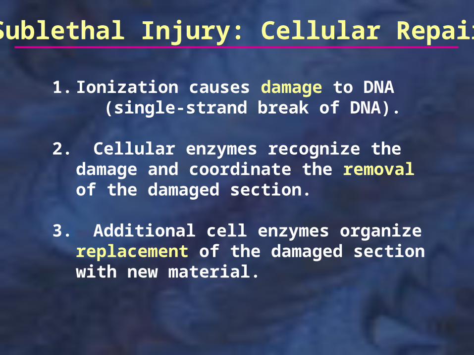
1. Ionization causes damage to DNA (single-strand break of DNA).
2. Cellular enzymes recognize the damage and coordinate the removal of the damaged section.
3. Additional cell enzymes organize replacement of the damaged section with new material.

When the DNA is damaged, cell function may be altered or reproductive capacity may be accelerated. Cancer is the most harmful result of cellular mutation.
Mutation
Normal
Mutation
Cell Death
If there is extensive damage to the cell following irradiation or if cell division (mitosis) is disrupted, the cell may die. This will depend on how sensitive the cells are to radiation. The loss of a few cells or small group of cells is usually of no consequence, since there are so many cells present in the body. In most cases, the dead cells will soon be replaced through normal reparative processes.

Cell CycleMore damage results when the cell is irradiated during the G1/early S portion of the cell cycle (before DNA synthesis); the damaged DNA (chromosome) will be duplicated during DNA synthesis and will result in a break in both arms of the chromosome at the next mitosis.
G1 = gap phase 1 in which nuclear components are replicatedS = synthesis phase; DNA is synthesized during the last 2/3 of this phase
G2 = gap phase 2, a preparatory stage to cell division
M = mitosis, during which cells divide
Cell most sensitive to radiation

1. High reproductive rate (many mitoses)
2. Undifferentiated (immature)
3. High metabolic rate
Radiosensitive CellsCells that are more easily damaged by radiation are radiosensitive. The characteristics of radiosensitive cells are:
Lymphocytes, germ cells, basal cells of skin and mucosa, and erythroblasts are examples of radiosensitive cells.

Radioresistant Cells
1. Low reproductive rate (few mitoses)
2. Well differentiated (mature)
3. Low metabolic rate
Cells that are not as susceptible to damage from radiation are radioresistant. The characteristics of radioresistant cells are:
Nerve and muscle cells are examples of radioresistant cells.

Radiation Effect Modifiers
The biological response to radiation is dependent on several different factors. These include:
• Total Dose: the higher the radiation dose, the greater the potential cellular damage.
• Dose Rate: A high dose given over a short period of time (or all at once) will produce more damage than the same dose received over a longer period of time.
• Total Area Covered: the more cells that are exposed to radiation, the greater the effects will be.

Radiation Effect Modifiers (continued)
• Type of tissue: As discussed earlier, radiosensitive cells are more likely to be damaged by radiation than are radioresistant cells.
• Age: Because the cells are dividing more frequently in a growing child, young people are affected more by radiation than are older people.
• Linear Energy Transfer: This measures the rate of the loss of energy as radiation moves through tissue. Particulate radiation (alpha particles, electrons, etc.) has a higher LET because it has mass and interacts with tissues much more readily than do x-rays.

• Oxygen Effect: Radiation effects are more pronounced in the presence of oxygen. Oxygen is required for the formation of the hydroperoxyl free radical, which is the most damaging free radical formed following ionization.
Radiation Effect Modifiers (continued)

The amount of exposure a patient receives from dental diagnostic radiography (effective dose) is relatively small. Most of the radiation damage will be repaired. The effects of the radiation damage that is not repaired may not show up for many years. The time between the exposure and the appearance of the effects of that exposure is called the latent period. In general, the higher the dose, the shorter the latent period.
Latent Period

Since repair of radiation injury is not 100%, radiation effects are accumulative. However, these effects will usually not be noticeable, since they are masked by the normal aging processes.
The effects from extreme levels of radiation exposure are potentially life threatening. Since these high levels will never be seen with diagnostic radiography, the effects will not be discussed. Check radiology texts or online sources for more information.

Somatic Cells vs. Germ Cells
There are two general types of cells in the body; these are somatic and genetic. Somatic cells are all the cells except for the germ (reproductive) cells. If somatic cells are irradiated, only the person exposed will be affected. Germ cells are the sperm and ova. If the germ cells are irradiated, the offspring of the individual may be affected.

Hormesis
Hormesis is a dose response phenomenon in which small doses of a toxin have the opposite effect of large doses. For example, exposing mice to small doses of radiation shortly before exposing them to very high levels of radiation actually decreases the likelihood of cancer. The initial low dose of radiation may activate certain repair mechanisms in the body and these mechanisms are efficient enough to not only neutralize the radiation effects but may even repair other defects not caused by the radiation. There is a lot of debate about hormesis, but the general opinion is that this is not something that can be relied on when discussing the effects of radiation exposure.

Dosimetry
Measuring the dose of radiation emitted by a radioactive source.
As mentioned previously, radiation effects are dependent on the total area covered. If the entire body is exposed, it would be considered whole-body radiation. If only a localized area is exposed, as in dental radiography, it would be called specific-area radiation. The effects from a given dose of radiation would be expected to be more severe if the whole body is exposed to that dose rather than a specific area.

Traditional Units SI* Units
Roentgen (R) Coulombs per kilogram
rad Gray
rem Sievert
Units of Radiation Measurement
* SI = International System of Units; used worldwide

Roentgen
The Roentgen (R) is the traditional unit of measuring radiation exposure. This measures the ionization of air. (The exact definition of Roentgen is complicated and not worth remembering). The Roentgen measures radiation quantity before the radiation enters the body. There is no exact SI unit comparable to the Roentgen, but in keeping with the metric system it is measured in coulombs per kilogram.

The rad (radiation absorbed dose) is the traditional unit used to measure the energy absorbed by the body. The SI unit is the Gray (Gy). 1 Gray = 100 rads; 1 cGy (centiGray) = .01 Gray = 1 rad.
rad/Gray

The rem (roentgen equivalent man) is the traditional unit used for comparing the effects of different types of ionizing radiation (electromagnetic and particulate).The dose (in rads) is multiplied by a quality (weighting) factor. The quality factor for x-rays is 1. Therefore the dose in rems (dose equivalent) is the same as the dose in rads. For alpha particles the quality factor is 20. Therefore the dose in rems (dose equivalent) would be 20 times the dose in rads for alpha particles. The higher the LET, the higher the qualifying factor.The SI unit is the Sievert (Sv). 1 Sievert = 100 rems;
1cSv (.01 Sievert) = 1 rem.
rem/Sievert

1 R = 1 rad = 1 rem
1 Gray = 1 Sievert = 100 rads = 100 rems
c (centi) = .01 m (milli) = .001 µ (micro) = .000001
1000 mrem = 1 rem = 1 cSv
100 mrem = 1 mSv = 1000 µSv
1 Sv = 100 cSv = 1,000 mSv = 1,000,000 µSv
* For x-rays, not particulate radiation
Conversions*

Each year, people are exposed to various types of ionizing radiation (listed below) and receive an average dose of 3.6 mSv (360 mrem ) per year. The actual dose depends on the degree of exposure to the ionizing radiation sources.
Radon 2 mSv(55%)
Cosmic 0.27 mSv (8%)
Rocks/soil 0.28 mSv (8%)
Food/water 0.4 mSv (11%)
Medical x-rays 0.39 mSv (11%)
Nuclear medicine 0.14 mSv (4%)
Consumer products 0.1 mSv (3%)
Other sources <0.01 mSv (<1%)
Annual Radiation Exposure

Natural (Background) RadiationEnvironmental radiation that we are exposed to daily is called natural or background radiation. It is composed of both external and internal sources. Background radiation averages 3.0 mSv (300 mrem) per year.
External Sources:
Cosmic (8%*): Ionizing radiation from space. Increased exposure at higher altitudes and during airline travel.
Terrestrial (8%*): Results from radioactive materials in soil and rocks. May be incorporated into some building materials. (See next slide).
* Percent of average annual radiation exposure

Certain black sand beaches in Brazil produce radiation levels as high as 5 mrem/hour. This would be equivalent to getting a full series of x-ray films every hour (photo left).
Some plants in another area of Brazil have absorbed so much radium that they will produce an autoradiograph when placed on photographic paper (photo right).

Natural (Background) Radiation(continued)
Internal Sources:
Radon (55%*): Radon and its decay products enter our homes via the atmosphere and water. Inhalation of these products contributes more than half of our average annual radiation exposure.
Food/Water (11%*): Some of the food and water we ingest contains radioactive materials.
* Percent of average annual radiation exposure, both natural and artificial
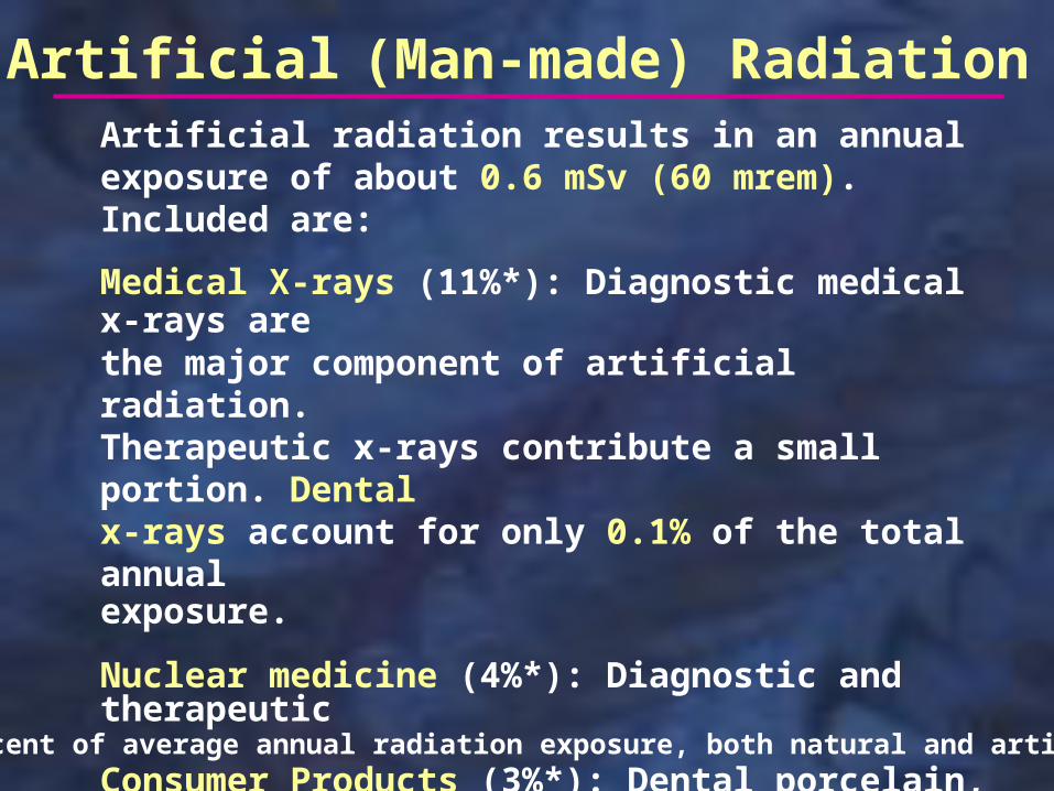
Artificial (Man-made) RadiationArtificial radiation results in an annual exposure of about 0.6 mSv (60 mrem). Included are:
Medical X-rays (11%*): Diagnostic medical x-rays are the major component of artificial radiation. Therapeutic x-rays contribute a small portion. Dental x-rays account for only 0.1% of the total annual exposure.
Nuclear medicine (4%*): Diagnostic and therapeutic
Consumer Products (3%*): Dental porcelain, smoke alarms, televisions, airport inspections, etc..
Other Sources (<1%*): Primarily nuclear fallout
* Percent of average annual radiation exposure, both natural and artificial

Effective Dose Equivalent
Exposure and dose are not related to the amount or type of tissue covered by the x-ray beam. A dose (or exposure) of 1 Sv could cover just the teeth or the entire body. Obviously, the overall effects would be different, even though the dose is the same. The effective dose equivalent takes into account the dose, the volume of tissue covered and the radiosensitivity of the cells. Using the effective dose equivalent, different types of x-ray examinations can be more realistically compared regarding the risk factor of each. The following slide lists the effective dose equivalents for some typical radiographic exams.

Effective Dose Equivalent
AFM* (round, F) 60 µSv
AFM* (rect., F) 27 µSvPanoramic 7 µSvCeph 220 µSvChest 80 µSvUpper GI 2400 µSvNatural Radiation 3000 µSv
Notice that the effective dose equivalent for natural (background) radiation (to which everyone is exposed) is 50 times as much as that for an AFM with round collimation and using F-speed film.
*AFM = adult full mouth series of x-ray films

Comparable risk from:
AFM (cancer)Smoking 1 cigarette (cancer)
Drinking 30 cans of diet soda (cancer)
Riding a bicycle 10 miles (accident)
Driving a car 300 miles (accident)
Flying 1000 miles (accident)
The preceding slide shows that taking a complete series of radiographs increases the average yearly exposure to the patient by a very small amount. The risk ( of cancer formation) is increased slightly. The following table compares this risk with the risk involved in common activities or habits.

The accepted cumulative dose of ionizing radiation during pregnancy is 5 rad (.05 Sv). According to the American Academy of Family Physicians, you would need 50,000 dental x-ray examinations to reach the 5-rad cumulative dose to the fetus.
An airline flight of 5 hours results in an exposure of 25 µSv. The exposure to the pelvic region from a full-mouth series of radiographs (done properly) is 1 µSv. (average natural (background) radiation is 8 µSv per day).
The decision to order films during pregnancy is a personal one. Because of the relatively low dose, it is not expected that there will be any harm to the fetus. However, my recommendation is to limit the films to those needed to treat the patient during the pregnancy (symptomatic teeth or very active caries).
Pregnancy
Radiation Protection forPatient and Operator
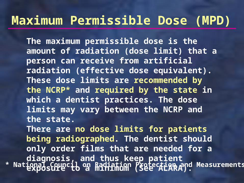
Maximum Permissible Dose (MPD)
The maximum permissible dose is the amount of radiation (dose limit) that a person can receive from artificial radiation (effective dose equivalent). These dose limits are recommended by the NCRP* and required by the state in which a dentist practices. The dose limits may vary between the NCRP and the state. There are no dose limits for patients being radiographed. The dentist should only order films that are needed for a diagnosis, and thus keep patient exposure to a minimum (See ALARA).
* National Council on Radiation Protection and Measurements

Dose limits (MPD’s) are set for occupationally exposed personnel (dentist, dental hygienist, and dental assistant) and for non-occupationally exposed individuals (front-office staff, people in waiting room, etc.). The dose limits are as follows:
Maximum Permissible Dose (MPD)
Occupationally exposed:
Adult 50 mSv (NCRP & Ohio) Minor 5 mSv (Ohio) Pregnant 5 mSv (Ohio)
Non-Occupationally exposed:
NCRP 5 mSv Ohio 1 mSv

Patient Protection
It is important to do everything we can to reduce the amount of exposure when a patient has dental radiographs taken. The following slides identify the ways in which we can do this.

ALARA
ALARA stands for “As Low As Reasonably Achievable”. If we assume that there is no threshold for stochastic effects (mutations and cancer) to occur, then it is important to keep the exposure to the minimum needed to provide an accurate diagnosis. In other words, take only those films needed to properly identify patient problems.

Professional Judgment/Selection Criteria
Deciding which films are needed for a particular patient is dependent on two things: Professional Judgment and Selection Criteria.
Professional Judgment: Through education and experience, each dentist develops an expertise in deciding which films will be needed to obtain an accurate diagnosis.
Selection Criteria: In 2005, the ADA, in conjunction with the Food and Drug Administration, released updated guidelines for prescribing dental radiographs. These guidelines identify which films should be taken, based on clinical findings. (For more detailed discussion, see “Self-study: Patient Management/Film Ordering”)

Equipment Reliability
X-ray equipment must be functioning properly to insure that the patient does not receive unnecessary radiation exposure. The settings for the exposure factors (exposure time, mA, kVp) must accurately reflect the output. Each state has requirements for the inspection of x-ray equipment to make sure that everything is working properly. The Ohio Department of Health requires that equipment be checked every five years (fee charged) and that the operator renew the registration of the equipment every two years (fee charged). If new x-ray equipment is purchased or if an x-ray unit is received from another dentist, the state must be notified. Disposal of old units must also be documented.

Direct Current (Constant Potential)
60-cycle Alternating Current
Many machines now convert the alternating current into a direct current (constant potential). Instead of cycles going from zero to the maximum, both positive and negative, the voltage stays at the maximum positive value, creating more effective x-ray production. This allows for shorter exposure times and a 20% reduction in patient exposure.
Constant Potential X-ray Machine

Filtration
Low-energy x-rays do not contribute to the formation of an x-ray image; all they do is expose the body to radiation. Therefore, we need to get rid of them. The process of removing these low-energy x-rays from the x-ray beam is known as filtration. Filtration increases the average energy (quality) of the x-ray beam. The x-ray beam becomes more penetrating, providing good image formation on the film with reduced patient exposure. (See “Self-study: X-ray Production” for more information on filtration).
Low-energy x-rays
high-energy x-ray

Collimation
Collimation is used to restrict the size of the x-ray beam, covering the entire film with the x-ray beam but not exposing unnecessary tissue. By reducing the amount of tissue exposed, the production of scatter radiation is also reduced. The shape of the opening (round or rectangular) in the collimator determines the shape of the x-ray beam. The size of the opening determines the size of the beam at the end of the PID. If you switch from a 7 cm diameter round PID to a 6 cm diameter round PID, the patient receives 25% less radiation. Rectangular collimation results in the patient receiving 55 % less radiation when compared to what they would receive with a 7 cm round PID. (See “Self-study: X-ray Production” for more on collimation).
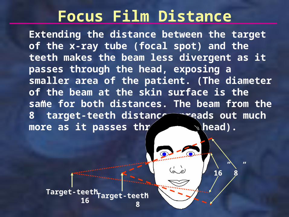
Focus Film DistanceExtending the distance between the target of the x-ray tube (focal spot) and the teeth makes the beam less divergent as it passes through the head, exposing a smaller area of the patient. (The diameter of the beam at the skin surface is the same for both distances. The beam from the 8” target-teeth distance spreads out much more as it passes through the head).
16” 8”
Target-teeth 16”
Target-teeth 8”

Using a faster film requires less radiation. Using F-speed film (Insight) instead of D-speed film reduces patient exposure by 60%. F-speed film has larger silver halide crystals, which more readily intercept the x-rays. (See “Self-study: Film and Screens” for more on films).
Intraoral Film Speed
Extraoral Screen Speed
Extraoral films are exposed by light from intensifying screens; this light is produced when x-rays contact phosphor crystals on the surface of the screens. The light is either blue or green, depending on the type of screen. Intensifying screens have different speeds, depending on the type of phosphor crystal (rare earth recommended) and the thickness of the phosphor layer. The faster the screen is, the less the patient exposure will be. However, image detail decreases as the speed of the screens increases. It is important to make sure that the film is compatible with the color of light coming from the screen. (See “Self-study: Film and Screens” for more on screens).
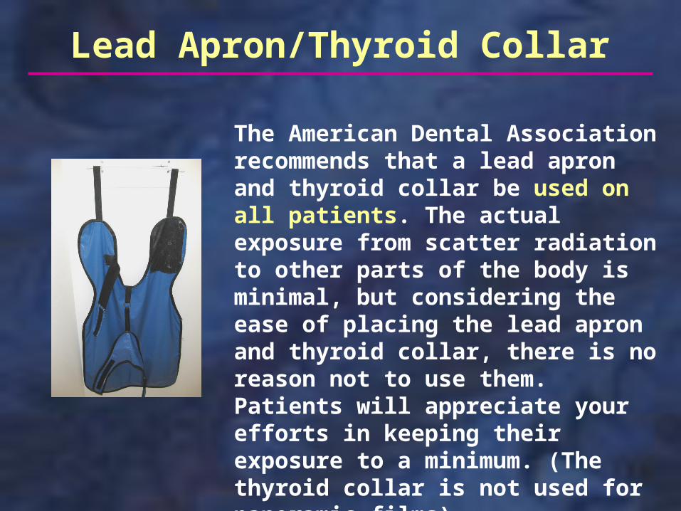
The American Dental Association recommends that a lead apron and thyroid collar be used on all patients. The actual exposure from scatter radiation to other parts of the body is minimal, but considering the ease of placing the lead apron and thyroid collar, there is no reason not to use them. Patients will appreciate your efforts in keeping their exposure to a minimum. (The thyroid collar is not used for panoramic films).
Lead Apron/Thyroid Collar

Good technique in taking films is essential in producing diagnostic radiographs. Proper film placement and selection of the correct exposure factors will maximize the value of the films and will reduce or eliminate the need for retakes, which would increase the patient’s overall exposure.
Technique

Processing
Processing films for the correct amount of time and at the proper temperature produces films of good diagnostic quality, assuming the films were exposed properly. It is necessary to have appropriate safelighting in a light-tight darkroom. Inadequate processing will result in retaking films which will add to the patient’s overall radiation exposure. (See “Self-study: Processing”).

The operator should never hold films in the patient’s mouth during an exposure. Some patient’s, due to physical or mental impairments, may need help in stabilizing the films, but this assistance should be provided by a friend or relative of the patient. This person should wear a lead apron and leaded gloves when holding films in the patient’s mouth.
X-ray Protection for the Operator
The photo at right shows a squamous cell carcinoma on the finger of a dentist who routinely held films for the patient.

The operator should stand behind a protective barrier if available. It has been determined that drywall is adequate protection for this purpose. The operator must be able to observe the patient during the exposure to make sure the patient doesn’t move prior to or during the exposure. If a direct line of sight is not possible, mirrors can be mounted on a wall opposite the doorway to allow visualization of the patient.
If barriers are not available, the operator should follow the position and distance rule (next slide).
X-ray Protection for the Operator

Position and Distance RuleThe operator should stand at least six feet away from the patient at an angle between 90 and 135 degrees. As the tubehead is moved, this safe position will change relative to the patient’s head (see below).
State Requirements
All states have some regulations concerning the operation of x-ray equipment. In Ohio, each dental office must designate a Radiation Safety Officer (any office employee; dentist, dental assistant, hygienist, etc.) who is responsible for maintaining records relating to the x-ray practices. Each office must post a “Notice to Employees”, which briefly lists the responsibilities of the dentist and all employees who take radiographs as detailed in the state’s radiation protection rules. Each office must have a “Safe Operating Procedures” manual and each operator must sign an “Instruction of Individuals” form to indicate that they have read the manual.

Personnel-monitoring devices (film badges) can be used to determine the exposure an operator receives during a given period (often quarterly). Film badges are required in some states if you expect to exceed 25% of the MPD during any calendar quarter (12.5 mSv). Although you should not expect to exceed this dose following normal safe operating procedures, it is beneficial to have a dosimetry service. The cost is minimal and the reports, which hopefully identify the lack of exposure to the operators, reduces any apprehension the office staff may have about radiation exposure.
Film Badges

NCRP Report # 145
The NCRP Report # 145 was released in December, 2003. It listed the following recommendations.
The lead apron is not required. The thyroid collar is required for children and should be used for adults.(The ADA, in response to the National Academy of Science report on low-level radiation effects, recommends lead apron/thyroid collar for all patients).
Rectangular collimation is required for periapicals and should be used, when feasible, for bitewings.

NCRP Report # 145
D-speed film is not to be used. E-speed film or faster is required. (Kodak no longer makes E-speed film; F-speed film, which is faster, is available along with D-speed). Rare earth screens are to be used for pans.
Sight development (dip tanks) is not acceptable.
Shielding design for new or remodeled dental offices is to be done by a qualified expert.
Film badges are required for pregnant personnel.

This concludes the section on Radiobiology, Dosimetry, and Radiation Protection. Additional self-study modules are available at: http://dent.osu.edu/radiology/resources.htm
If you have any questions, you may e-mail me at: [email protected]
Robert M. Jaynes, DDS, MSDirector, Radiology GroupCollege of DentistryOhio State University