but where should they start their search?
TRANSCRIPT


S O U T H W E S T E R N M E D I C I N E 54
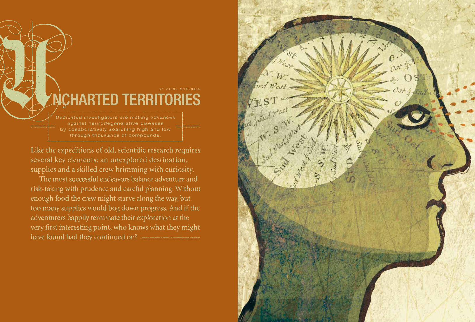
hen Dr. Steven McKnight and his protégé, Dr. Andrew Pieper, set off on their own research expedition at UT Southwestern Medical Center several years ago, they knew they might never arrive at their destination.
They were seeking whether there was a way to enhance the birth or survival of newly formed nerve cells in the adult brain. It was an undertaking that could have profound payoffs. If successful, the research could ultimately lead to new therapeutic strategies for neurodegenerative diseases such as Alzheimer’s and Parkinson’s.
Dr. McKnight, a member of the National Academy of Sciences, is chairman of biochemistry at UT Southwestern and holds the Distinguished Chair in Basic Biomedical Research and the Sam G. Winstead and F. Andrew Bell Distinguished Chair in Biochemistry. Dr. Pieper is an assistant professor of psychiatry and biochemistry who was part of Dr. McKnight’s laboratory team as a post doctoral researcher before joining the UT South western faculty. Together they focused on an area of the mammalian brain called the hippocampus, which plays an important role in learning and memory. The hippocampus is one of the few areas of the brain that makes new nerve cells throughout life in adult rodents and primates, including humans.
But not all newborn nerve cells survive. In a normal winnowingout process, many of the new cells die within a month.
“We asked, ‘What if we could discover a chemical that could enhance the formation or survival of newborn nerve cells?” Dr. McKnight said.
Drs. Pieper and McKnight chose an oldfashioned method to search for such a drug. Spurning robotic
testing systems and highthroughput technologies, they instead leapfrogged over experiments in isolated cells and went directly to screening 1,000 chemical compounds in living mice – an ordeal that took more than two years and identified a handful of promising compounds.
“This is a retro way of chasing after new drug candidates,” Dr. McKnight said. “It’s so slow and lowtech that no one does it anymore.”
Old-fashioned methods
But where should they start their search? No drugs were then known to enhance the birth rate or survival of newborn nerve cells.
Fortuitously, during his time as biochemistry chairman, Dr. McKnight had been laying the groundwork for the department to tackle this sort of problem. The faculty now comprises not only traditional biochemists but also synthetic chemists who specialize in analyzing compounds, finding efficient ways to synthesize them and creating variations that can work more efficiently in biological systems.
“Without access to firstclass capabilities in synthetic chemistry, a venture like this would be doomed from the start,” Dr. McKnight said.
“It took two and a half years just to complete the initial screen,” he said. “More typically, researchers get an idea that takes two, four, five months to get the results.”
The pair had two resources vital to any expedition – a team with a wide variety of skills and enough funding to get them where they hoped to go.
In 2004 Dr. McKnight received a Director’s Pioneer Award from the National Institutes of Health, becoming one of nine U.S. researchers to receive the award in its first year. The NIH initiative was designed to support exceptionally creative investigators, encouraging them to take on unexplored avenues of research that carry a relatively high potential for failure, but that also possess a greater chance for truly groundbreaking discoveries. The award provided Dr. McKnight with $500,000 a year for five years.
Dr. Pieper has received funding from The Hartwell Foundation, a Memphisbased organization that supports innovative research. And multimilliondollar gifts from the A.L. Chilton Foundation have supported biochemistry research programs at UT Southwestern for five decades.
The interdisciplinary collaboration within the biochemistry department was key. “We never could
have done this project had we not, over the last 15 years, built a faculty that includes a halfdozen firstrate chemists,” Dr. McKnight said. “In fact, this could not have been done in almost any other academic setting at all. It would be like an adventurer who says, ‘I want to sail across the ocean,’ but who doesn’t have a boat.”
Teamwork is key
Dr. Andrew Pieper (left) and Dr. Steven McKnight hold a model of the P7C3 molecule.
“This could not have been done in almost any other academic setting at all. It would be like an adventurer who says,
‘I want to sail across the ocean,’ but who doesn’t have a boat.”– Dr. S teven McKn igh t

S O U T H W E S T E R N M E D I C I N E 76
“Science is … quite literally … the final frontier of adventure. The fun of the hunt is more than enough to overcome our recognition
that the road ahead is filled with obstacles.”– D r. S t e v e n M c K n i g h t
To save time, they pooled the 1,000 substances into batches of 10. If any pooled group kept the newborn nerve cells alive, they then tested each compound individually to see which was the most effective. The mice were given not only the test substances, but also a chemical that would stain newly formed cells.
Dr. Pieper – who did the bulk of the lab work – examined the animals’ brain tissue for new cells, which showed up as black dots under a microscope. “We counted every one of those cells,” he said.
The first figure in their published study, which appeared in the journal Cell, shows a bar graph indicating the rate of cell survival for the 100 batches. A few bars extend higher than the rest, indicating compounds that helped protect the new cells. The most promising compound showed up early in the experiment. They named it P7C3, because it was the third compound in the seventh pool.
In elderly rats, which characteristically show a decline in the birth and formation of hippocampal neurons, the researchers found that P7C3 increased both the birth and survival of new neurons, and improved the memory and learning capabilities of the aged rats.
The vessel that carried forward this project was UT Southwestern’s smallmolecule library, a chemical ark containing more than 200,000 compounds chosen for their potential druglike actions. Generally, robotic studies using the library can test hundreds of thousands of chemicals on cultured cells in short order.
To equip its expedition, the research team selected only 1,000 compounds – a balance between what was available and what was doable. They also chose a cache that was chemically diverse and endowed with favorable druglike qualities.
“The length of time it took to test each compound in living animals is what limited the number of substances we could test,” Dr. McKnight said. “It was tedious. We had a library of 200,000 chemicals, but we could only test 1,000.”
But this approach had its advantages, Dr. Pieper said. “The thought was that we’d be able to bypass a lot of the pitfalls that you find when screening in highthroughput testtube assays, such as finding toxicity or side effects when you test compounds later in living animals.”
They also tested P7C3 in a line of genetically engineered mice that lack a gene that protects new brain cells. In these animals, all newly formed hippocampal cells die. These mice have problems with learning and memory and are often used in research as a model for various forms of neurological disease. Humans who bear similar mutations in this gene also have learning disabilities and suffer from mental illnesses such as bipolar disorder and schizophrenia.
When these mice received P7C3, they formed many more properly shaped brain cells.
Even with this early success under their belts, the researchers soldiered on.
“P7C3 was the 73rd compound we tested,” Dr. McKnight said. “It was encouraging that we looked like we had a success. But we wanted to finish screening each and every one of the 1,000 chemicals chosen for study. We found seven other likely ones, although P7C3 remained the best.
“But if we’d stopped at that first success, we’d only have one,” he said. “Years from now, it might turn out that one of the seven other chemicals leads us to a much better starting point for drug discovery.”
Toward the end of the project, they learned that research teams in Russia and the U.S. were studying an antihistamine called Dimebom as a potential antiAlzheimer’s drug. Remarkably, the chemical structure of Dimebom is similar to that of P7C3, Dr. McKnight said.
But instead of viewing this as a setback, “we were very excited because while Dimebom was in Phase 3 clinical trials, no one knew how it works in the brain,” Dr. Pieper said.
Moreover, headtohead studies of Dimebom and P7C3 at UT Southwestern have shown that P7C3 is far more potent in fostering neuron formation.
“The idea that Dimebom promotes new nerve growth in the brain was totally unanticipated,” Dr. McKnight said.
On the heels of their success, the researchers already are peering over the horizon to the next set of challenges. For example, will
P7C3 have any effect on Lou Gehrig’s disease, spinocerebellar atrophy or Parkinson’s disease?
“All these diseases entail the death of existing cells,” Dr. McKnight said. “While we know that P7C3 can block the death of newborn nerve cells, we don’t know whether it might be capable of blocking the death of mature brain cells, as happens in so many forms of neurodegenerative disease.”
Animal models exist for these neurodegenerative diseases, so the researchers are testing whether P7C3 eases symptoms. They also are looking at whether it can help restore brain function after traumatic brain injury.
Dr. Joe Ready, associate professor of biochemistry, led a team that has made hundreds of different versions of P7C3 to improve its effectiveness. “We want to improve the physical characteristics, such as solubility, and make some changes that might help us figure out how it works in the cell,” he said.
Despite the riskiness and tedium of the work, the scientists say it has been satisfying.
“It’s been a wonderful experience,” Dr. Pieper said. “At first there was a lot of doubt, because we could have gone through this whole screen and not found anything. Now I couldn’t be happier. It’s exactly the kind of thing I want to do with my career – something that might be translatable into helping people.”
Dr. Ready said the project was, above all, fun. “It was such an unconventional approach to finding a molecule,” he said. “It’s a detective story.”
Since the publication of the study, the skepticism of other researchers during the early stages has been replaced by optimism, Dr. McKnight said.
“It’s why I get up in the morning,” Dr. McKnight said. “Science is a wonderful occupation. It is – quite literally – the final frontier of adventure. Although it’s too early to tell if we’ve made a fundamental discovery that might really help people, the fun of the hunt is more than enough to overcome our recognition that the road ahead is filled with obstacles.” X
Dr. Joe Ready and colleagues have produced hundreds of versions of the P7C3 molecule to improve its effectiveness.
The joy of science

or Amy Alexander, feeding her family of four rarely involves grabbing the first thing she
spots on the grocery store shelf and throwing together an impromptu meal. n Her 7-year-old
son, Mason, makes that seemingly simple task all but impossible. That’s because Mason is allergic
to peanuts and eggs – two of the most common ingredients in processed foods and two of the
most common food allergies in children. n “It’s been so long since he was diagnosed that it just
seems so normal now, but it was difficult at first,” said the 42-year-old resident of Southlake,
noting that her 5-year-old son, Brandon, does not have any food allergies. “It still impacts whether
or not Mason goes to sleepovers or to camp. And we don’t go out to Chinese restaurants, period.” n
Unfortunately, Mrs. Alexander is not alone in her quest for food that doesn’t endanger her son’s life.
B y K r i s t e n H o l l a n d s H e a r
FoodVS. ME
9S O U T H W E S T E R N M E D I C I N E
F
Food allergies took on Mason’s immune system, but with an assist from UT Southwestern doctors, he was able to keep his opponents at bay.
Mason at age 7

Neither Mrs. Alexander nor her husband has food allergies, so they never imagined that their son – who was 2 years old at the time – would have an allergic reaction to anything, particularly something he hadn’t consumed.
Although Mason’s reaction wasn’t life- threatening, Mrs. Alexander said the incident was a wake-up call.
“Until recently, I didn’t keep anything with peanuts in my house,” she said. “I keep some other nuts, but not peanuts, because that is his most severe allergy.”
Dr. Bird said that a true food allergy – a term used to describe several different diseases – is the immune system’s response to a particular food. Allergies mediated by antibodies called immunoglobin E (IgE), which are generated in large numbers by the immune system, are the most common. Peanut allergy is an IgE-mediated allergy.
Other types of food allergies include celiac disease, a condition in which people suffer an immune reaction if they eat gluten-filled foods such as bread or pasta, and eosinophilic esophagitis (EE), a disease characterized by swelling of the esophagus and weight loss, particularly in children. Although the exact cause of EE remains unclear, some of the most common food triggers include milk, wheat and eggs.
Food allergy symptoms may include hives, difficulty breathing, angioedema (lip swelling), vomiting or loss of consciousness.
A food intolerance, on the other hand, is simply related to the food itself. “It’s more like having too much caffeine or getting food poisoning from eating something that’s contaminated,” Dr. Bird said.
Determining whether someone has a true food allergy depends to a large extent on their initial reaction to the specific food. Simply coming into contact with traces of a product – as Mason did – can cause swelling or a rash-like irritation on the skin, but it is unlikely to progress to a life-threatening reaction.
Dr. Rebecca Gruchalla, chief of allergy and immunology at UT Southwestern, said often people mistakenly believe they have food allergies.
“Many people think they have an allergy if they experience an adverse reaction to a food, but that is often not the case,” said Dr. Gruchalla, holder of the William A. Sellars, M.D., and Joyce M. Sellars Distinguished Chair in Allergy and Immunology. “Because IgE-mediated food allergy is present in only a small percentage of people, it is very important that patients who have adverse food reactions
see a board-certified allergist so that it can be determined whether or not they indeed have a true food allergy.”
One hallmark of allergies – whether they are food-, seasonal-, or drug-related – is the speed at which the reactions occur.
“Most true allergic reactions are going to occur very quickly, often in minutes,” said Dr. David Khan, professor of internal medicine and pediatrics at UT Southwestern and director of the asthma clinic at Parkland Memorial Hospital. “In terms of the nature of the symptoms, in the nose it would be sneezing, itching, stuffy and runny nose; in the lungs, it would be difficulty breathing and wheezing; and on the skin it would be itching and usually hives, whelps or swelling. A more severe reaction might cause someone to lose consciousness.
“You don’t have to have all these things, but to be a true allergy, each organ that’s involved should have the symptoms that sound like an allergy.”
1 0 1 1S O U T H W E S T E R N M E D I C I N E
“Until recently, I didn’t keep anything with peanuts in my house. I keep some other nuts, but not peanuts, because that is his most severe allergy.”
—Amy Alexander
p to 12 million people in the U.S. – including 6 percent to 8 percent of all children under the age of 3 – suffer from food allergies, according to the Food and Drug Administration. And the numbers keep rising. Each year, roughly 30,000 individuals go to the emergency room, and 150 children die as a result of severe food allergies. Cow’s milk, eggs, peanuts and tree nuts are the most common pediatric food allergies, but wheat, soy, fish and shellfish, including lobster and shrimp, are other widespread culprits.
As food manufacturers churn out an increas-ing number of nut- and dairy-free options, researchers at UT Southwestern Medical Center are trying to unravel why certain children are more prone to food allergies and whether those children can effectively beat the allergy by ingesting increasingly larger amounts of the food over a prolonged period of time.
Leading the effort is Dr. Drew Bird, assis- tant professor of pediatrics and internal medicine at UT Southwestern and director of the new Food Allergy Center at Children’s Medical Center Dallas.
The center, which opened on the fourth floor of the Pavilion at Children’s in September 2010, is the only academic-affiliated pediatric food allergy center in North Texas. Clinicians at the center treat infants and children with a broad range of allergic conditions and conduct research expected to be instrumental in the development and treatment of children with food allergies.
“Our primary goals are to serve children and families in the Dallas community with the best and most comprehensive evidence-based care, and to continue learning more about food allergy in order to improve the lives of those affected by such allergies around the world,” said Dr. Bird, a Dedman Family Scholar in Clinical Care who was recruited to UT Southwestern to help establish a clinical and translational research program in pediatric food allergies in conjunction with Children’s. “We exist for the treatment and education of families suffering from food allergies.”
Defining allergies Mrs. Alexander, a stay-at-home mom, said she had no idea Mason was allergic to peanuts until he touched the table at which she had been eating them. After coming in contact with the peanut residue, Mason then touched one of his eyes, and the white of that eye quickly flared up, she said.
U“Our primary goals are to serve children and families in the Dallas community with the best and most comprehensive evidence- based care and to continue learning more about food allergy.”
—Dr. Drew Bird
Mason Alexander (left) and Dr. Drew Bird
Mason Alexander

Cause and effect nlike asthma and allergic rhinitis, which can run in a family, researchers
are uncertain whether food allergies are passed down from generation to generation or if environment affects development of food allergies.
Dr. Bird said there seems to be a genetic predisposition. “Children are at an increased risk of developing a food allergy if both parents have allergies of any kind or if a sibling has a food allergy,” he said. “We don’t know at this point what the association is or what the gene is that’s causing the allergy, but kids who fit that profile do seem to have an increased predisposition.”
Whether the environment plays a role is less clear.
“There have been a lot of theories as to whether the increasing number of pediatric
food allergies can be attributed to changes in the cleanliness of the environment or the amount of infections kids are exposed to at a young age – the sort of things that influence how the immune system develops,” Dr. Bird said. “We hope the theories will help explain why kids are developing more food allergies, but right now the theories are just that: theories.”
One thing’s for sure: Individuals allergic to one food are more likely to be allergic to another, he said.
Mrs. Alexander said she’s grateful that Mason’s egg allergy, which they discovered before he turned 1, isn’t as problematic.
“He doesn’t react to eggs if they’re cooked in something,” she said. “Cookies that have eggs inside don’t cause a reaction.”
Tolerating treatments Treating a food allergy isn’t as straight- forward as dealing with allergies due to pollen, mold or even a drug allergy. Strict avoidance is the only treatment now available.
Dr. Bird said he believes the cure for food allergies might be the culprit itself – the peanut, in particular. His research involves giving children increasing amounts of peanuts in order to build up tolerance.
The technique, known as oral immuno-therapy, should not be undertaken outside a clinical study, but Dr. Bird said the concept has shown promise.
He recently started enrolling patients in a study to determine if incremental doses of peanut protein – in the form of lightly roasted peanut flour – could change how a child’s immune system responds. The doses start as small as 1/1000 of a peanut and gradually increase until a child can eat a small handful without having an allergic reaction.
“We send them home with the flour; they put it in yogurt or some other food vehicle; and they eat it every day,” said Dr. Bird, who began this line of research while at Duke University Medical Center. “Every two weeks we increase the amount they’re getting, so, that over a period of seven to eight months, we have them eating the equivalent of about seven or eight peanuts a day.”
Since many children with peanut allergies dislike the actual taste of peanuts, those who finish the study after three years of therapy often are steered toward things like peanut M&M’s, Reese’s Pieces and other peanut- filled treats.
Mrs. Alexander said it has been extremely liberating to have Mason taking part in the oral immunotherapy study because she and her husband now know exactly how much peanut is needed to trigger Mason’s allergy. “Seeing that he reacted to more than a trace made me more comfortable,” she said.
Dr. Khan said the approach to food allergy research is similar to what has already proven to be a boon for those with other types of allergies.
“With allergens like pollen and dust mites, allergy shots can reduce symptoms dramatically, and in some situations there is almost a cure in that the symptoms go away completely,” he said. “With insect allergies, we’re probably even closer to a cure. About 98 percent of people allergic to bees or fire ants who undergo allergy shots don’t have any reaction the next time they’re stung.
“With food allergies, we’re changing the immune system, with the hope that in the long term these children are no longer allergic to these foods. That’s a very exciting concept.”
Although he admits that an outright cure might be difficult, Dr. Bird said he hopes to understand better why the immune system reacts as it does in order to at least limit the problems associated with food allergies.
“For the majority of kids with food allergies, their parents don’t want them to eat peanut- butter sandwiches,” he said. “They just want to be able to go to a bakery or an ice cream shop and not worry about contamination from other products.
“We’re looking for the safest and most effective therapy we can give that will provide that sort of freedom for these families.”
That day can’t come soon enough for Mrs. Alexander.
“Even now, just having taken part in the study for a few months, I already feel like we gained something from it,” she said. “I’m still not going to let him eat cupcakes from school or baked goods, per Dr. Bird’s directions, but I’m a little less worried about trace amounts of peanuts.” X
For more information about food allergies, please call 214-645-2800.
1 2 1 3S O U T H W E S T E R N M E D I C I N E
U
“Many people think they have an allergy if they experience an adverse reaction to a food, but that is often not the case.” —Dr. Rebecca Gruchalla, with Dr. David Khan
“We’re looking for the safest and most effective therapy we can give that will provide … freedom for these families.” —Dr. Drew Bird

Dr. Jerry Morgan remembers the precise mo-ment and circumstance behind his decision to leave Ohio and move to Texas almost 35 years ago. A snowstorm – big even by Ohio stan-dards – spurred the cross-country trek to the Dallas area, where he has practiced pediatric medicine since 1976.
“We had a 42-inch snowfall, and I thought it was the right time to move away from the cold weather,” Dr. Morgan said. “I’d finished my medical training and was ready for a new chapter in my life.”
Escaping the cold weather proved much easier than escaping a family history rife with prostate cancer. Like many men, Dr. Morgan
didn’t begin thinking about the possibility of developing the disease until his father and uncle were both diagnosed in their early 50s. In a way, he said, it was lucky he knew the risks, which spurred him to get a blood test each year that screens for prostate specific antigen (PSA) – a protein produced by prostate cells that can signal prostate cancer.
“I started getting the test every year because my dad had prostate cancer; my uncle had it; and then, boom – in 2010 I was diagnosed with it,” Dr. Morgan said. “My urologist did a biopsy and called me right before a trip I was taking with my wife to Cozumel.”
LikeFather, Like Son
T h r e e f a c t o r s i n c r e a s e a m a n ’ s
r i s k f o r p r o s t a t e c a n c e r :
e t h n i c i t y , a g e a n d f a m i l y h i s t o r y .
B y K a t h e r i n e M o r a l e s
a n d R a c h e l S k e i D o n i h o o
Patient Dr. Jerry Morgan holds a portrait of his father.
1 5S O U T H W E S T E R N M E D I C I N E

Uncommon risk factors
Prostate cancer exhibits few of the typical risk factors people think of when contemplat-ing their chances of developing cancer in general. This makes treating it a conundrum of sorts.
Smoking and diet, for instance, don’t have a major impact on one’s chances for developing the disease. The three major risk factors currently known are ethnicity, age and having a family history of prostate cancer. African-American men are 60 percent more likely to develop the disease and are substantially more likely to die from it than their Caucasian counterparts.
Prostate cancer is the most common cancer in men, and one in six men can expect to be diagnosed with it.
Dr. Morgan, still in his prime at 62, wasn’t ready to take a wait-and-see approach, so he and his urologist decided surgery was the best option.
The road to success
“I knew about the expertise UT Southwestern had in robotic surgery and that appealed to me,” Dr. Morgan said. “That’s how I met Dr. Roehrborn.”
Dr. Claus Roehrborn, chairman of urology at UT Southwestern Medical Center, has one of the country’s most experienced teams in the surgical treatment of prostate cancer.
That road to success began auspiciously in the early 1980s under the direction of Dr. Paul Peters, former chairman of urology,
who adopted many cutting-edge procedures designed to preserve male sexual and urinary function following prostate removal. The desire to provide patients with the best in surgical and oncologic care continues under the leadership of Dr. Roehrborn, who became chairman in 2002.
Actively recruiting early-career talent and pushing ahead with new technologies available to treat prostate cancer helped establish the department in U.S. News & World Report’s list of best urology programs in the country.
“Because of our reputation, we see a lot of patients with biopsy-proven prostate cancers, and we have to determine the best course of treatment,” said Dr. Roehrborn, who holds the E.E. Fogelson and Greer Garson Fogelson Distinguished Chair in Urology. “There are somewhere between 190,000 and 200,000 cases of prostate cancer diagnosed in the United States every year.”
Determining an individual’s treatment course can be challenging and depends largely on a patient’s health and age at the time of diagnosis. Some prostate cancers are slower growing, and surgery can wait. Others are aggressive and, if diagnosed in a young patient, need to be surgically removed.
Early detection is key
For patients like Dr. Morgan, annual screen-ing is especially important because of his fam-ily history of the disease. A family history of prostate cancer is one of the best predictors of the disease, with double the risk if one family mem-ber is affected and a five- to 11-fold risk increase if two or three first-degree relatives are affected. Families with a history of prostate cancer could benefit from passing information from father to son.
“Fortunately, cure rates for prostate cancer are high if detected early,” Dr. Roehrborn said. “Men who have a family history of prostate cancer should begin annual screenings and be in the care of a physician who can discern the best course of treatment.”
Annual screenings for men typically begin at age 40. These screenings should include a physical exam and a PSA blood test that can detect cancer in its earliest, most treatable stage. Surgery typically remains the best chance of cure for many.
For Dr. Morgan, the promise of a highly effective, minimally invasive procedure seemed the best choice to excise the cancer and minimize recurrence.
“My PSA numbers tilted in favor of not waiting, and I’m relatively young in the scheme of things,” Dr. Morgan said. “So on March 30, 2010, that’s what we did.”
Minimally invasive treatment
Prior to laparoscopic procedures and robotic prostatectomies, open surgery was the only option. The operations typically resulted in sub-stantial blood loss, lengthy recoveries and risk of nerve damage that could lead to side effects such as incontinence and sexual dysfunction.
With the introduction of less-invasive procedures, urologic surgeons at UT South-western have excelled at using the latest technologies to treat patients.
“We use the robotic procedure to remove the entire prostate and sometimes lymph nodes in patients with localized prostate cancer,” Dr. Roehrborn said.
To achieve this, surgeons use a four-armed robot named DaVinci to assist in the delicate procedures. One of the robotic arms holds a miniature camera, while the other arms hold surgical instruments – all of which are manipulated in a console by the surgeon. Since acquiring its first DaVinci robot in 2006, UT Southwestern has been at the forefront of robotic surgery, performing complex urologic, thoracic and bariatric procedures. UT Southwestern has since purchased a newer version of a single-console robot in use at
“Because of our
reputation, we see
a lot of patients with
biopsy-proven prostate
cancers, and we have
to determine the best
course of treatment.”
—Dr. Claus Roehrborn
“Men who have a
family history
of prostate cancer
should begin annual
screenings and be
in the care of a
physician who can
discern the best
course of treatment.”
—Dr. Claus Roehrborn
1 6 1 7S O U T H W E S T E R N M E D I C I N E

UT Southwestern University Hospital - St. Paul. With the addition of these robots, physicians can offer an even wider array of procedures.
In 2010 Dr. Roehrborn and his surgical team completed their 600th prostatectomy using the DaVinci system. Of the 1,200 urologists certified to use the robotic device nationwide, Dr. Roehrborn ranks in the top 5 percent in terms of volume, making his team among the most experienced with robotically assisted surgery to remove diseased prostate glands.
Exploring new techniques
In addition to surgical advances, urologists also are looking into the possibility of microab-lation to treat prostate cancer. One hurdle in treating prostate cancer is that removing the prostate is an all-or-nothing proposition. Por-tions of the prostate cannot be left inside the body, but with new imaging techniques, per-forming cancer ablations on part of the prostate may soon be introduced.
“In the case of the prostate, the dilemma for a surgeon is the organ’s placement in the body. By removing it you have to reconstitute the urinary tract,” said Dr. Jeffrey Cadeddu, profes-sor of urology and radiology and holder of the Ralph C. Smith, M.D., Distinguished Chair in Minimally Invasive Urologic Surgery.
To address this, Dr. Cadeddu is studying how nanotechnology can be used to target the cancer and bypass healthy tissue. He recently published a study on gold nanoshells that can be inserted into the body and heated to kill cancer cells. Using a mouse model in which the gold nanoshells were inserted into the skin, researchers killed 93 percent of cancer cells. Such treatment needs to be coupled with highly effective imaging, which can show doctors where the cancer exists and where to place the nanoparticles.
“Finessing image-guided approaches will offer patients better treatment,” Dr. Cadeddu said. “The next step is working on image-guided particle placement for prostate cancer ablation. These advances would be a major milestone in therapeutic treatment of prostate cancer.”
Biomarkers as predictors
For Dr. Morgan, whose prostate cancer was diagnosed through the PSA test, its effectiveness saved his life. But for many men, some medications can interfere with the results of the PSA, another factor that physicians must be acutely aware of when determining whether to do a biopsy. Some men have abnormally high PSA readings with no concurrent medications, which has spurred research into finding new and more accurate tests for prostate cancer.
Dr. Yair Lotan, associate professor of urology and holder of the Helen J. and Robert S. Strauss Professorship in Urology, studies the distinct biological molecules – biomarkers – that can indicate the presence of prostate and bladder disease.
“Our goal is to identify patients who have a higher chance of developing prostate cancer,” said Dr. Lotan. “This may allow earlier detection. Biomarkers may also identify patients who require treatment versus patients who may be eligible for active surveillance protocols.
“Furthermore, if we can identify key biomarkers that predict an increased risk of recurrence, we might also be able to predict who will benefit from more aggressive therapies.”
Identifying biomarkers could change the treatment patients receive, he explained.
“These discoveries, which are happening at a rapid speed, will allow physicians to individualize treatment and better assess what course to take. As we move forward, we hope that pinpointing biomarkers will eliminate the educated guesswork we have relied on in the past. Biomarkers may also be used to predict the response to novel multimodal therapeutic approaches,” he said.
As UT Southwestern continues to be at the forefront of advances in technology, detection and treatment options, it is the patient who ultimately will benefit.
For Dr. Morgan, the promise of a minimally invasive procedure followed by a shorter recovery was the best option. Although he conceded that even doctors can be apprehensive about surgery, he was confident of a positive outcome.
“It was just a matter of getting it over with,” he said. “Recovery was challenging, at times, and the initial symptoms were more annoying than painful. But with time the symptoms have gotten better and better, and I feel great about the future.” X
For more information about prostate treatments, please call 214-645-4673.
“Finessing image-
guided approaches
will offer patients
better treatment.”
—Dr. Jeffrey Cadeddu
“Our goal is to identify
patients who have
a higher chance of
developing prostate
cancer. This may allow
earlier detection.”
—Dr. Yair Lotan
1 8 1 9S O U T H W E S T E R N M E D I C I N E

2 0 2 1S O U T H W E S T E R N M E D I C I N E
HEART
BUILDINGA
BETTERBy Amanda Siegfried and Katherine Morales

2 3S O U T H W E S T E R N M E D I C I N E 2 2
Cardiology. “Saying we were pleased that this approval came through for Dr. Cox and other patients is an understatement.”
Dr. Cox, retired chairman of philosophy at Centenary College of Louisiana, recognized destination therapy as his best hope for recovery and found out about the LVAD program at UT Southwestern from a family friend.
“This new technology gives patients a new option, when in the past they had none,” said Dr. Dan Meyer, professor of cardiovascular and thoracic surgery and holder of the Sarah M. and Charles E. Seay Distinguished Chair in Thoracic Surgery.
In the future, he said, these pumps should last more than 10 years, truly becoming a viable alternative to transplantation for patients who do not want to manage the risks of the multiple immunosuppressive medications needed after a heart transplant.
Both Dr. Drazner and Dr. Meyer say advances in the field of mechanical devices might help alleviate some of the need for hearts from donors, which are invariably in short supply.
“There is a large unmet need for these patients for whom conventional therapies for heart failure aren’t working,” Dr. Drazner said. “LVADs are a major breakthrough in this respect. We are still making advances in the field of mechanical devices as permanent therapy.”
UT Southwestern doctors currently are participating in a clinical trial to test a third-generation heart pump to see if it works as a permanent therapy for heart-failure patients.
As for Dr. Cox, the challenges of living with a permanently implanted device are far superior to living with heart failure.
“The doctors say I’m right on target in terms of recovery,” said Dr. Cox, who continues to visit the Joyce & Larry Lacerte Heart & Lung Clinic. “I’m grateful to have a good support system at home, and I’m determined to get better and better.”
Sunshine spills into a patient room on the sixth floor of UT Southwestern University Hospital, sharpening the shadow cast
by five research towers that stand across busy Harry Hines Boulevard. With advanced clinical care located on the west side of the street and world-class science on the east, physicians and researchers work in juxtaposition on the same complex puzzle – repairing the human heart.
Clues pointing to more effective treatments for heart failure – the leading cause of death in the Western world – are slowly revealing themselves through painstaking laboratory work, as well as through clinical trials for patients suffering from heart disease.
Only a few decades ago, the possibility of stem-cell therapy and implanted mechanical-assist devices to aid ailing hearts seemed the domain of science fiction. At UT Southwestern Medical Center, physician-scientists are at the forefront of both of these relatively new advancements and, in the case of one small but powerful piece of technology, meeting the needs of patients suffering the devastating effects of heart failure.
IMPLANT ASSISTANCE
The Elaine D. and Charles A. Sammons Heart, Lung and Vascular Comprehensive Center at UT Southwestern is a primary testing site for new technologies, such as permanently implanted devices that assist ailing hearts. Such treatment, called “destination therapy,” is for patients in end-stage heart failure. Recipients undergo surgery to implant a left-ventricular assist device (LVAD), which helps supply oxygen-rich blood through the body.
“Prior to the approval of LVADs for destination therapy, the prognosis for patients with advanced heart failure was not good,” said Dr. Mark Drazner, medical director of the Heart Failure and Cardiac Transplantation Program,
a key component of the Doris and Harry W. Bass Jr. Clinical Center for Heart, Lung and Vascular Disease. “Their quality of life suffers dramatically as well. They are frequently hospitalized; they have fluid retention and can suffer from failure of other organs.”
The pump rests inside the patient’s chest. A thin cable attached to the device exits the body and connects to a controller powered by a battery pack.
“While this does involve a major surgical procedure, most patients are able to go home 14 days after surgery and engage in normal activity, including driving and traveling," Dr. Drazner said.
The Food and Drug Administration signed off on the LVAD for permanent therapy in early 2010 – a critical moment for one UT South-western patient. Dr. Hughes Cox of Shreveport had a quintuple bypass in 2001 after suffering a heart attack. Over the next five years, his heart continued to weaken, and, toward the end of 2009, he said, he knew he would face a tough choice.
Dr. Cox was too weak for a heart transplant. His medications were failing to keep his symptoms from worsening. And, he was losing independence to do the things he and his wife loved: traveling and spending time with their family.
“Cardiac transplantation is the gold-standard therapy for such patients, but its use is constrained by donor organ availability,” said Dr. Drazner, professor of internal medicine and holder of the James M. Wooten Chair in
Dr. Hughes Cox (left) recognized destination therapy as his best hope for recovery and found out about the LVAD program – and Dr. Mark Drazner – from a family friend.
Dr. Dan Meyer says advances in the field of mechanical devices might help
alleviate some of the need for hearts from donors, which are
invariably in short supply.

2 4 2 5S O U T H W E S T E R N M E D I C I N E
as the genetic factors called into play in the adult heart under pathological stress. In 2009 Dr. Olson was awarded the Institut de France’s prestigious Lefoulon-Delalande Foundation Grand Prize for his work on gene regulation in the cardiovascular system. The prize has an international reputation as the most prestigious award in cardiovascular research.
“As heart cells enlarge, they undergo genetic reprogramming,” said Dr. Olson, who holds the Pogue Distinguished Chair in Research on Cardiac Birth Defects, the Robert A. Welch Distinguished Chair in Science, and the Annie and Willie Nelson Professorship in Stem Cell Research.
“Genes that should be active only during fetal heart development are reactivated,” he said. “During this remodeling process, several basic processes are disrupted, including the proteins that control how the heart contracts.
“Our research is directed not at blocking hypertrophy, per se, but rather on the pathological gene program that underlies this remodeling process.”
As it turns out, implanting an LVAD appears to normalize gene activity.
Designing potential drug therapies to return the gene expression pattern back to normal is a challenge, Dr. Olson said, because the biological target has to be very precise to avoid adversely affecting other aspects of normal heart function.
He and his colleagues, however, have made significant inroads in identifying a collection of tiny nucleic acid molecules called microRNAs that function as modifiers of heart disease. Dr. Olson’s research has shown that microRNAs control many aspects of heart disease, such as life and death of heart muscle cells, cardiac contractility and new blood vessel formation.
“We hope to be able to translate these basic discoveries in the laboratory into new therapeutic approaches for improving function of the failing heart,” Dr. Olson said.
To achieve this goal, he co-founded a biotechnology company called miRagen Therapeutics, which is developing new drugs directed against microRNAs. Dr. Olson’s work on microRNAs as regulators of heart disease is supported by grants from the Donald W. Reynolds Foundation and the Leducq Foundation, as well as the National Institutes of Health.
“As heart cells enlarge, they undergo genetic reprogramming. Genes that should be active only during fetal heart development are reactivated.” — Dr. Eric Olson (left), with Dr. Jay Schneider
as coronary artery disease and hypertension and occurs in the aftermath of acute events, including heart attacks.
Heart failure often is preceded by cardiac hypertrophy, a thickening of the heart muscle that occurs in response to increased stress on the heart. For example, in individuals with hypertension, the extra work of pumping blood against the increased pressure of narrowing artery walls thickens the heart muscle, just as lifting weights adds muscle to the body. That extra mass in the heart, however, causes a decrease in size of the heart’s bottom chambers – the left and right ventricles.
This enlargement of the heart – and heart cells – and subsequent decline in cardiac function are triggered by a number of molecular and genetic factors, said Dr. Eric Olson, chairman of molecular biology and director of the Nancy B. and Jake L. Hamon Center for Basic Research in Cancer.
Dr. Olson’s pioneering research has uncovered networks of genes that orchestrate the formation of the embryonic heart, as well
STRONG SCIENCE FOR WEAK HEARTS
Across the street from University Hospital - St. Paul, where Dr. Cox had his LVAD surgery, UT South western scientists diligently work to uncover ways in which the heart might be coaxed to repair itself.
Supported by major research grants from the National Institutes of Health, the American Heart Association and the American Recovery and Reinvestment Act of 2009 – the federal government’s stimulus package – these investigators are advancing the understanding of the role stem cells play in heart disease and repair. Ultimately such studies might lead to new ways to care for patients with heart attacks or congestive heart failure by stimulating heart-cell regeneration.
Heart failure is a complex disorder in which the heart’s pumping action is insufficient to meet the body’s needs. Affecting approximately 5 million Americans, heart failure results from chronic conditions such

2 6 2 7S O U T H W E S T E R N M E D I C I N E
to intercept progenitor cells on their way to becoming scar-forming cells and redirect them toward becoming cardiac muscle or vascular cells.”
Working closely with the group from Massachusetts General, as well as collaborating with leading stem-cell biologists across the country, is “a truly amazing scientific opportunity,” Dr. Schneider said.
A NATIONAL PRIORITYA $2 million grant from the American Heart
Association also supports UT Southwestern studies of the mechanisms that could generate new cardiac muscle cells.
UT Southwestern was one of only three institutions in the country to receive the highly competitive award, which established an American Heart Association-Jon Holden DeHaan Foundation Cardiac Myogenesis Research Center.
Each center conducts a number of basic science research projects to learn more about how cardiac muscle cells develop and work together. The AHA grant provides funding through 2012.
UT Southwestern’s effort combines Dr. Schneider’s and Dr. Olson’s expertise with that of Dr. Joseph Hill, chief of cardiology and director of the Harry S. Moss Heart Center.
Dr. Hill also received a $1.2 million grant through the NIH as part of the American Recovery and Reinvestment Act of 2009. His project, titled “Functional Genomics of Complex Vascular Disease,” will help expand research on the mechanisms of cardiovascular disease and includes support for a new, early-career faculty member.
“More than 200 genetic associations with cardiovascular disease have been identified. Moving forward, we need to know how this information can be harnessed to predict who will develop heart disease and possibly develop new ways to intervene early in at-risk patients,” said Dr. Hill, who also receives support from the Sweetheart Ball Fund for Cardiology Research.
Dr. Hill, who holds the Frank M. Ryburn Jr. Chair in Heart Research and the James T. Willerson, M.D., Distinguished Chair in Cardiovascular Diseases, also is closely involved with studies of molecules called HDAC inhibitors that appear to protect the heart from damage due to stress.
“These molecules are being evaluated in clinical trials for effectiveness against cancer, and two have been approved by the FDA,” Dr. Hill said. “Given this, we are now moving forward with a clinical trial using HDAC inhibitors in patients who have cardiac injury, such as a heart attack.”
As clinicians and laboratory researchers continue to discover valuable pieces to solve the heart-disease puzzle, patients will be the ultimate beneficiaries.
“We have a very active heart-failure program that combines our research and clinical strengths in a way that bridges the two worlds,” Dr. Hill said. “To take something that we study in our labs on the North Campus and translate it into a treatment at University Hospital – that is what we all strive for.” X
For more information about UT South western’s heart failure program, please call 214-645-8000.
COLLABORATIONS MOVE SCIENCE TO CLINIC
Together with Dr. Jay Schneider, assistant professor of internal medicine at UT Southwestern, Dr. Olson also recently established a company called LoneStar Heart to develop potential new drugs for heart failure that work via cardiac stem cells. The company has set up shop at BioCenter at Southwestern Medical District, a 15.5 acre biotech park launched by UT Southwestern to provide state-of-the-art lab space and shared resources for new ventures emerging from medical center scientists.
Dr. Schneider’s clinical expertise in cardiovascular medicine, combined with his research background in human genetics and molecular biology, were the perfect mix for inclusion in an ambitious project led by Dr. Olson and Dr. Steven McKnight, chairman of biochemistry and holder of the Sam G. Winstead and F. Andrew Bell Distinguished Chair in Biochemistry and the Distinguished Chair in Basic Biomedical Research.
The research has involved screening a large library of small molecules made available through the Department of Biochemistry and determining whether any of them have an effect on cardiac stem cells.
“It was a perfect opportunity to combine chemistry and stem cells,” said Dr. Schneider, whose work was originally funded by the Haberecht Wild-Hare Idea Program at UT South western.
Out of hundreds of thousands of potential drug-like molecules in the library, the team
discovered a handful that show promise, and one in particular that could become an important new drug.
“Original studies of that drug were done in cultured stem cells, but we have now shown that it actually works as a drug in the adult heart by targeting a rare population of native cardiac stem cells,” Dr. Schneider said. “It appears to work by activating genes in these cells that transform them into muscle precursor cells.”
He and his colleagues are investigating whether a type of nascent heart cell called a progenitor cell – which is slightly different from an adult stem cell – can repair damage sustained by heart disease or heart attacks.
“We are looking specifically at the role of progenitor cells which, like stem cells, have the ability to form new cells within the body,” Dr. Schneider said.
Dr. Schneider is the primary investigator for a $9 million grant from the National Heart, Lung and Blood Institute (NHLBI) that focuses on the epicardium – the layer of cells that surround the heart. Epicardial cells might be a key source of adult heart progenitor cells capable of repairing damaged muscle, Dr. Schneider said.
His research group works with another team of physician-scientists from Massachusetts General Hospital, which also received $9 million for its portion of research for the study. Together the two institutions form one of nine “hubs” comprising the NHLBI Progenitor Cell Biology Consortium, a $170 million, seven-year initiative aimed at developing the high-potential field of stem- and progenitor-cell biology.
Following a heart attack, the heart does not make new heart cells or makes only a few. Instead, the normal response is to build tough scar tissue, which lacks blood vessels and muscle cells. Scar tissue actually inhibits the heart’s ability to beat efficiently under the enormous pressures needed to pump blood through the body. Moreover, scar tissue can become catastrophically detrimental by predisposing the organ to lethal arrhythmias.
“We have discovered new methods to ‘teach’ native progenitor cells in the epicardium to become cardiac muscle cells or blood-vessel cells rather than scar tissue,” Dr. Schneider said. “When you suffer a heart attack, you lose billions of heart cells in an instant. Our goal is
“We have a very active heart-failure program that combines our research and clinical strengths in a way that bridges the two worlds.”— Dr. Joseph Hill

1 S O U T H W E S T E R N M E D I C I N E 2 9
Even as his fingers flew across the keyboard, Adam Crockett still was in disbelief. As the e-mail
unfolded before him, he retraced the unimaginable details of the past two days.
Only 48 hours earlier, the seemingly healthy 25-year-old had been on a business trip, hang-
ing out with friends in a rowdy bar on the San Antonio Riverwalk. Without warning he passed
out, hitting his head on a table as he crumpled to the floor. Mr. Crockett doesn’t remember how
he got back to his hotel room that night, but he awoke with a nagging headache and a trickle of
blood on his pillow. His friends laughed it off, chalking the incident up to a few too many beers,
but Mr. Crockett had a sinking feeling that there had to be more to it.
When the headache persisted into the second day, he returned home to Austin and found
a local internist. The doctor ordered an MRI. Emerging from the cocoon of the machine, Mr.
Crockett said he had an “eerie feeling” as he watched the technician through the glass partition.
He saw an expression, almost imperceptible, cross the man’s face as he stared at the computer
screen in front of him and then reached for the phone.
The rest is a blur – the additional tests, the frantic phone call to his mother, the endless drive
home. It was the beginning of a complex and often wondrous journey, but that day it felt like
the end of the line.
by Rachel Skei Donihoo
Friends:
A close friend of mine recently told me that everything happens for a
reason. I know most of you have probably heard, but yesterday I found out
I have a “goose-egg”- size tumor on my brain.

S O U T H W E S T E R N M E D I C I N E 3
SAVING LIVES
After the diagnosis and successful treatment of brain cancer, most patients begin measuring their lives in two- to three-month increments. That’s how often an MRI is required to determine whether a malignant brain tumor has returned. Through the weeks, they celebrate family, savor every function and event, and live to the fullest while maintaining a heels-dug-in posture in the face of an insidious foe. In many cases, despite the best treatments currently available, the tumor quickly returns and leads to disability and death.
Some patients, however, thrive for many years after their diagnosis. Physicians and scientists at UT Southwestern Medical Center’s Annette G. Strauss Center in Neuro-Oncology are closely following patients who have survived after developing a brain tumor (glioma), hoping to develop new therapies to treat this devastating disease, which is often resistant to treatment.
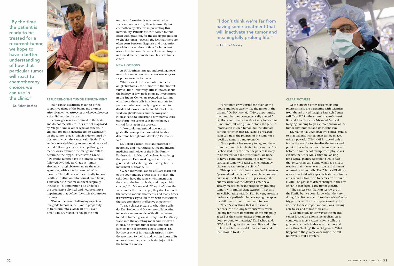
Dr. Bruce Mickey, vice chairman of neurological surgery and holder of the William Kemp Clark Chair in Neurological Surgery, is the leader of an experienced group of physicians and scientists within the Strauss Center, a cornerstone of UT Southwestern’s brain-tumor research effort.
“The sad truth is that right now we don’t have a curative treatment for malignant gliomas, but I don’t think we’re far from having some treatment that will inactivate the tumor and meaningfully prolong life,” Dr. Mickey said. “We meet our responsibility to patients by learning from their experiences – even if we learn that something doesn’t work.”
These discoveries are driven largely by a collaborative spirit and intense sense of urgency across multiple disciplines and centers throughout the UT Southwestern campus, including the Harold C. Simmons Comprehensive Cancer Center, the Advanced Imaging Research Center and Children’s Medical Center Dallas. Experts in imaging and radiology, pathology, physiology, and pediatrics work together on clinical and translational studies aimed at identifying the critical metabolic pathways that drive unregulated tumor-cell growth in the brain.
“The overarching goal of our studies is to identify quickly new therapies,” said Dr. Elizabeth Maher, associate professor
of internal medicine and neurology and neurotherapeutics and holder of the Theodore H. Strauss Professorship in Neuro-Oncology. Dr. Maher recently received a $1 million National Institutes of Health Challenge Grant in Health and Science Research. The support targets specific knowledge gaps, scientific opportunities, new technologies, data generation or research methods that can benefit from an influx of funds to advance disease-specific areas in significant ways.
“We have an intense focus on tumor cells that remain quiet for months after treatment, with the goal of designing therapies that will kill the cells before they start to grow again,” Dr. Maher said. “For a subset of patients who currently are being treated with standard therapy, the idea of a true cure is creeping in as patients live longer than ever before without tumor regrowth.”
Mr. Crockett is one of the long-term survivors who is being tracked by Dr. Maher and her team. In 2006 Mr. Crockett was diagnosed with a low-grade glioma. Dr. Mickey removed as much of the tumor as possible from Mr. Crockett’s brain, but – as is typical of these tumors – malignant cells were left behind. Though currently not dividing, these cells ultimately will start to grow again. Treating patients when their cancer cells are “dormant” is ineffective, explains Dr. Maher, so Mr. Crockett’s brain is monitored with MRI scans every three months. Although his tumor has not returned, he recognizes it will always be a wolf at his door.
“People ask me all the time if I think about my tumor every day,” he said. “The truth is I probably think about it every 10 to 15 seconds. That doesn’t mean that I haven’t gotten on with my life – I have – and I’m a better man for having been through all of this. All those corny lessons about developing true friendships, following your dreams, finding true love – I’ve learned them all.”
In the five years since his diagnosis, Mr. Crockett has done a lot. He “woke up” to win over and marry a college friend; he left an unchallenging job to pursue his passion in business; and he launched his own organization, the Adam Crockett Tumor Fund, to raise money and awareness for brain tumor research. He plans to pursue a Master of Business Administration in the spring and says he is “proud” to tell everyone he meets about the battles he has won.
"I’m a better man for having been through all of this. All those corny lessons about developing true friendships, following your dreams, finding true love – I’ve learned them all.”
— Adam Crockett
3 1
Adam Crockett
3 2 S O U T H W E S T E R N M E D I C I N E 3 3
REPLICATING THE TUMOR ENVIRONMENT
Brain cancer essentially is cancer of the supportive tissue of the brain, and a tumor arises from either astrocytes or oligodendrocytes – the glial cells in the brain.
Because gliomas are confined to the brain and do not metastasize, they are not diagnosed by “stages,” unlike other types of cancers. In gliomas, prognosis depends almost exclusively on the tumor “grade,” which is determined by the rate at which the cancer cells divide. That grade is revealed during an emotional two-week period following surgery, when pathologists meticulously examine the malignant cells to determine their type. Patients with Grade II (low-grade) tumors have the longest survival, followed by Grade III. Grade IV tumors, also known as glioblastomas, are the most aggressive, with a median survival of 14 months. The hallmark of these deadly tumors is diffuse infiltration into normal brain tissue, a characteristic that makes them surgically incurable. This infiltration also underlies the progressive physical and neurocognitive impairment that defines the clinical course for patients.
“One of the most challenging aspects of low-grade tumors is the tumor’s propensity to transform into a Grade III or IV over time,” said Dr. Maher. “Though the time
until transformation is now measured in years and not months, there is currently no chemotherapy effective in preventing this inevitability. Patients are then forced to wait, often with great fear, for the deadly progression to glioblastoma; however, the fact that there are often years between diagnosis and progression provides us a window of time for important research to be done. Patients like Adam inspire us to work harder, smarter and faster to find a cure.”
NEW HORIZONS
At UT Southwestern, groundbreaking novel research is under way to uncover new ways to stop the cancer in its tracks.
While a great deal of attention is focused on glioblastoma – the tumor with the shortest survival time – relatively little is known about the biology of low-grade gliomas. Investigators in the Strauss Center are focused on learning what keeps these cells in a dormant state for years and what eventually triggers them to divide and form a new tumor. Much of their work on glioblastoma and the low-grade gliomas seeks to understand how normal cells transform into cancer cells in the brain, a critical first step in the process.
“If we could understand how normal glial cells develop, then we might be able to determine how gliomas develop,” Dr. Maher said.
Dr. Robert Bachoo, assistant professor of neurology and neurotherapeutics and internal medicine and holder of the Miller Family Professorship in Neuro-Oncology, is studying that process. He is working to identify the genes and molecular signals that regulate the movement of those cells.
“When individual cancer cells are taken out of the body and are grown in a Petri dish, the cells don’t have the same environment that they do in the human body, and they rapidly change,” Dr. Mickey said. “They don’t look the same under the microscope; they don’t respond the same to various treatments; and many times it’s relatively easy to kill them with medications that are completely ineffective in patients.”
To get a clearer picture of what these cells do, Drs. Bachoo and Mickey are collaborating to create a mouse model with all the features found in human gliomas. Every time Dr. Mickey walks into the operating room and removes a glioma, he extracts tumor tissue and calls Dr. Bachoo at his laboratory across campus. Dr. Bachoo or one of his research assistants takes the specimen to the lab and, within hours of its removal from the patient’s brain, injects it into the brain of a mouse.
“The tumor grows inside the brain of the mouse and looks exactly like the tumor in the patient,” Dr. Bachoo said. “More importantly, the tumor has not been genetically altered.” Dr. Bachoo currently has about 40 glioblastoma tumor lines, allowing him to study the genetic information in each tumor. But the ultimate clinical benefit is that Dr. Bachoo’s research team can track the progress of the tumor of a specific patient in a mouse model.
“Say a patient has surgery today, and tissue from the tumor is implanted into a mouse,” Dr. Bachoo said. “By the time the patient is ready to be treated for a recurrent tumor, we hope to have a better understanding of how that particular tumor will react to chemotherapy choices we can use in the clinic.”
This approach falls into a new field known as “personalized medicine.” It can’t be reproduced on a major scale because it is person-specific, but researchers at the Strauss Center have already made significant progress by grouping tumors with similar characteristics. They also are collaborating with Dr. Dan Bowers, associate professor of pediatrics, in developing therapies for children with recurrent brain tumors.
“There’s something that is the same in patients who are long-term survivors. We’re looking for the characteristics of this subgroup as well as the characteristics of tumors that don’t respond to therapies,” Dr. Bachoo said. “We’re looking for the common link and trying to find out how to model it in a mouse and then how to treat it.”
CLEAR PICTURES
At the Strauss Center, researchers and physicians also are partnering with scientists from the Advanced Imaging Research Center (AIRC) in UT Southwestern’s state-of-the-art Bill and Rita Clements Advanced Medical Imaging Building to get a clearer picture of the tumor environment and its metabolism.
Dr. Maher has developed two clinical studies so that patients with gliomas can be imaged using a powerful 7 Tesla MRI – one of only a few in the world – to visualize the tumor and provide researchers clearer pictures than ever before. In routine follow-up when physicians evaluate patients’ MRIs, they are looking for a typical picture resembling white fuzz that researchers call FLAIR, which is a mix of reactive brain tissue, scar tissue, and dormant or growing tumor cells. The 7 Tesla MRI allows researchers to identify specific features of tumor cells, which allow them to be “seen” within the FLAIR. The goal is to detect changes in the area of FLAIR that signal early tumor growth.
“The cancer cells that can regrow are in the FLAIR, but we don’t know what they are doing,” Dr. Bachoo said. “Are they asleep? What triggers them? The first step to knowing the answers to these important questions is being able to see and follow these cells.”
A second study under way at the medical center focuses on glioma metabolism. As is common in most cancers, glioma cells use glucose at a much higher rate than normal cells, thus “fueling” the rapid growth. What happens to the glucose once inside the cell, however, is still a mystery.
“By the time the patient is ready to be treated for a recurrent tumor, we hope to have a better understanding of how that particular tumor will react to chemotherapy choices we can use in the clinic.”
— Dr. Robert Bachoo
“I don’t think we’re far from having some treatment that will inactivate the tumor and meaningfully prolong life.”
— Dr. Bruce Mickey

S O U T H W E S T E R N M E D I C I N E 3 53 4
Dr. Mickey and his colleagues in neurological surgery, along with investigators in the Strauss Center, the AIRC and the Mary Nell and Ralph B. Rogers Magnetic Resonance Imaging Center, use a “tagged” form of glucose to map the path that glucose takes in the cancer cell. In the clinical study, the tagged glucose (on its way to become fuel for new cells) is infused into glioma patients before surgery. The glucose is taken up by the tumor and processed as part of the cell’s metabolism. Once the cancer is surgically removed, researchers can map that tumor’s metabolic pathways using the AIRC’s state-of-the-art technology.
“This process essentially opens the ‘black box’ of glioma cell metabolism and will help identify the critical steps that are needed for tumor growth – steps that may be excellent targets for future treatment,” explained Dr. Maher. “We’re using the imaging and metabolism technology to understand what the malignant cells are doing, so we can get a handle on killing them before they start to grow again. This is particularly important for the low-grade glioma patients who have no treatment options during the entire time the tumor is dormant.
“This new research may one day make an enormous difference in the lives of patients. I’d love to be able to say to a patient, ‘We know what we need to do to kill these cells now,’ instead of waiting for the time when these cells will grow again – often very aggressively. For every one of the MRI appointments, patients
and families hold their breaths, hoping and praying that ‘today is not that day.’”
Dr. Ralph DeBerardinis, assistant professor of pediatrics in the Eugene McDermott Center for Human Growth and Development and a Sowell Family Scholar in Medical Research, recently received a $200,000 grant from the Cancer Prevention and Research Institute of Texas (CPRIT) to study whether glioblastoma growth can be suppressed by targeting its metabolism. He and his team have defined two basic metabolic pathways that allow cells to grow in vivo and are studying which pressure points in those two pathways can be targeted to eliminate tumor growth.
“Glioblastomas are notoriously difficult to treat, so the key question is how to exploit the biological differences between the tumor and the normal tissue in order to intervene clinically,” he said. “Through improved imaging we may be able to get a better sense of which metabolic pathways are active as the tumor is growing, or whether it is responding to therapy. The goal is to treat the cancer in a way that would be well-tolerated by the normal, healthy cells, but toxic to the tumor cells."
The ability to conduct these metabolic studies is possible thanks to AIRC’s active research program in metabolic imaging and spectroscopy of cancer, centered around the 7 Tesla scanner. Progress is limited, however, by commercially available radiofrequency coil technology.
To narrow this gap, Dr. Craig Malloy, medical director of the AIRC and professor of internal medicine and radiology, received a $1.2 million grant from CPRIT to improve imaging technology.
Dr. Malloy has established a partnership between clinicians at UT Southwestern and engineers at Texas A&M University to develop state-of-the-art radiofrequency coils and multitransmit technology for the 7 Tesla.
“The combined effort will allow unprece- dented improvements in the use of MRI and spectroscopy to identify specific metabolic biomarkers in breast cancer and malignancies of the brain,” said Dr. Malloy, who holds the Richard A. Lange Chair in Cardiology.
CHOOSING LIFE
On the first year anniversary of his diagnosis, Mr. Crockett organized a skydiving fundraiser he called “I’d Rather Die Skydiving Than From a Brain Tumor.” The name may have been tongue-in-cheek, but the message was serious.
“I really wanted to show people that you have to choose to live your life in an adventurous, meaningful way and encourage others to come forward to support brain tumor research,” he said. "There are a lot of important treatments on the horizon and, for people like me, the speed with which that research makes it from bench to bedside is critical.”
Mr. Crockett insists on wearing his hair short, proud of the wide scar that zippers across the top of his head. He has lived through things that not many people live through. The scar, he says, is a “badge of honor” and a constant reminder of the joy of life that was found in his darkest moments.
The searching e-mail he wrote five years ago hinted at the meaning of the journey ahead:
For more information on brain cancer, please call 214-645-2300.
“Patients like Adam inspire us to work harder, smarter and faster to find a cure.”
— Dr. Elizabeth Maher
“The combined effort will allow unprecedented improvements in the use of MRI and spectroscopy to identify specific metabolic biomarkers in breast cancer and malignancies of the brain.”
— Dr. Craig Malloy (right),
with Dr. Ralph DeBerardinis
I hope there is a reason for this happening, and I hope it's something
positive. I know that I'm going to find out who my true friends are, and I'll
probably have an entirely new outlook on life itself. I'm going to work less
and play more when I get over this. Life flies by, then something can jump
up and bite you at any minute, so have fun and don't have any regrets.
Your friend, Adam

NCI
SIX YEARS +THREE LITTLE LETTERS =A WORLD OF DIFFERENCEin research and patient care
When Dr. James K.V. Willson arrived in Dallas in the fall
of 2004, he was given a mandate: boost UT Southwestern Medical Center’s cancer programs into a position of national recognition and secure National Cancer In-
stitute (NCI) designation – an elite distinction held by
only the top-tier cancer centers in the U.S.
The plan was an ambitious one, but Dr. Willson had
accomplished the feat once before, at the Case Comprehensive Cancer
Center in Cleveland.
On Aug. 3, 2010, he did it for UT Southwestern.
B y C O N N I e P I l O T O
3 7S O U T H W E S T E R N M E D I C I N E

members to develop programs complementary to existing research and clinical expertise. These specialists are assembled into multidisciplinary teams for each major cancer.
For instance, researchers and clinicians interested in breast cancer previously would gather regularly in the pathology department and review the previous week’s diagnoses.
Now, the groups’ interactions – and meetings – are more structured. Medical and surgical oncologists, radiation oncologists, translational and basic scientists, clinical and research nurses, study coordinators, data specialists, as well as social workers, psychologists and others may take part. The groups discuss clinical scenarios or noteworthy cases, share treatment knowledge, sound out research protocols and grant proposals, and interact “in ways you don’t normally get to interact,” said Dr. David Euhus, associate director for clinical research at the Simmons Cancer Center.
“Ultimately, the immersion of all team members in clinical research issues creates a seamless environment and helps deliver cutting-edge care to patients,” said Dr. Euhus, who directs the Mary L. Brown Breast Cancer Genetics and Risk Assessment Program.
Other building blocks of the Simmons Cancer Center also have been patient-focused. One was the launch of specialized services to help cancer patients navigate their individual treatment programs and find expert supportive and palliative care. Another emphasizes cancer prevention, expanding the clinical framework to accommodate healthy populations and cancer survivors.
These achievements have positioned the Simmons Cancer Center as a leader at a time when both state and federal policymakers are stepping up the fight against cancer. In Texas, voters in 2007 approved a constitutional amendment that authorized the creation of the Cancer Prevention and Research Institute of Texas (CPRIT) to fund cancer research and prevention programs.
In 2010 CPRIT awarded more than $34.8 million to investigators at UT South-western to support cancer-related projects and to recruit a pre-eminent cancer investigator.
BrIDge TO DISCOVerIeSCrucial to securing NCI designation
was the development of new strategies to foster collaborations and interaction across departments that connected and advanced scientific discovery and patient care.
“Historically, we’ve always been an extremely strong basic science institution,” said Dr. Michael White, professor of cell biology and associate director for basic science at the cancer center. “However, the cancer center has really been an organizational magnet to bring together dispersed efforts, each one of which has great value on its own, but which together generate the synergy required to impact human disease.”
Much of that synergy springs from the Simmons Cancer Center’s network of comple-mentary scientific programs: Cancer Cell Networks, Chemistry and Cancer, Development and Cancer, and Molecular Therapeutics of Cancer.
The Harold C. Simmons Cancer Center is the first medical center in North Texas to attain this prestigious status, which the NCI bestows upon the
nation’s top cancer centers in recognition of innovative research and excellence in patient care.
“Over the past six years, we’ve made great strides in bringing together all the elements that collectively comprise an outstanding cancer center,” said Dr. Willson, director of the Simmons Cancer Center and associate dean for oncology programs at UT Southwestern. “NCI designation is the gold standard for elite cancer programs.”
As an NCI-designated center, the Simmons Cancer Center received a $7.5 million support grant covering the next five years, which complements the more than $24 million in NCI grants that currently are active at UT South western.
The National Cancer Institute is the world’s largest organization solely dedicated to cancer research and leads a national effort to eliminate suffering and death due to cancer. It is part of the National Institutes of Health, the primary federal agency responsible for conducting and supporting medical research.
The Simmons Cancer Center at UT South-western provides cancer patients access to promising new cancer treatments available only through the network of NCI-designated centers. There currently are 66 NCI-designated cancer centers; other designated centers in Texas are in Houston and San Antonio.
Clinical care for cancer is provided by UT Southwestern faculty of the Simmons Cancer Center at UT Southwestern University Hospitals
& Clinics, Parkland Memorial Hospital and Children’s Medical Center Dallas, all of which are included in the NCI designation. Faculty also provide cancer services at the Dallas VA Medical Center and Fort Worth’s Moncrief Cancer Institute.
The BlUePrINT fOr grOWThThe foundation for the success of the
Simmons Cancer Center was laid more than two decades ago, with generous support from Dallas philanthropists Harold and Annette Simmons.
To date, the Simmons family has given and pledged more than $100 million to enhance cancer programs at UT Southwestern.
“The continued commitment and encourage ment of the Simmons family really inspired all of us to make the Simmons Cancer Center a national leader in cancer care, research and discovery,” said Dr. Willson, who holds the Lisa K. Simmons Distinguished Chair in Comprehensive Oncology.
In order to elevate the Simmons Cancer Center to national prominence, Dr. Willson and university leaders built upon the success and reputation of several nationally recognized cancer programs that were already in place at UT Southwestern.
A cornerstone for that early success was a robust program in translational lung research, building a bridge from basic laboratory findings to patient care. The lung-cancer program, developed under the leadership of Dr. John Minna, professor of internal medicine and pharmacology, has received continuous funding from a highly competitive NIH initiative known as SPORE – Specialized Program of Research Excellence. Only a handful of centers nationwide receive lung SPORE funding, designed to promote interdisciplinary cancer research and move discoveries from the lab to the clinic.
With that blueprint for collaborative excellence in place, UT Southwestern leaders set about identifying potential resources and opportunities that could be put to work to build the cancer center’s vision for the future.
One of the key components was the recruitment of more than 70 new faculty
“historically, we’ve always been an extremely strong basic science institution. however, the cancer center has really been an organizational magnet to bring together dispersed efforts, each one of which has great value on its own, but which together generate the synergy required to impact human disease.”
— D r . M i c h a e l W h i t e
A cornerstone for ... early success was a robust program in translational lung research ... developed under the leadership of Dr. John Minna.
3 8 3 9S O U T H W E S T E R N M E D I C I N E

PerSONAlIzeD MeDICINe
The approach is a unique collaboration between federal agencies, including the food and Drug Administration and the National Institutes of health, and major pharmaceutical companies to screen promising new drugs being developed for women with high-risk, fast-growing breast cancers.
The Investigational Serial Studies to Predict your Therapeutic response with Imaging and Molecular Analysis, or the I-SPy 2 trial, is being offered at 20 major cancer research centers across the country. Dr. David euhus, professor of surgical oncology, is the principal investigator for the study at UT Southwestern.
The trial was developed to allow the activity of drugs to be assessed much earlier in the research process, potentially enabling drugs to be developed and approved using fewer patients, less time and far fewer resources. The goal is to shave several years and millions of dollars in expensive testing off the current process.
Breast-cancer patients enrolled in the trial will receive chemotherapy to reduce the size of their tumor before surgery. But a distinctive feature of this trial is that physicians will screen multiple drugs from multiple companies – up to 12 different cancer drugs over the course of the trial.
This approach allows physicians to adapt recruitment of patients based on incoming results and to drop ineffective drugs quickly. Drugs that show promising results will be given to more patients with the types of tumors that seem likely to respond.
“It has become apparent that breast cancer is not one disease, but dozens of different diseases,” said Dr. euhus, who holds the Marilyn r. Corrigan Distinguished Chair in Breast Cancer Surgery. “This is a paradigm-changing trial that
starts by understanding the underlying biology of each cancer and then assigning treatments based on this biology. It may very well change the way drugs are tested and go to market.” X
For more information on these trials or other cancer studies, please call 214-648-7097.
— Connie Piloto
A diagnosis of cancer can be a very personal matter. So can its optimal treatment.
researchers at the harold C. Simmons Cancer Center are studying how different one person’s tumor can be from another’s. The goal is to identify tumors’ vulnerabilities and challenges that vary from patient to patient, and then to develop treatments that exploit each cancer’s weaknesses and overcome its strengths.
Two national clinical trials being offered to lung- and breast-cancer patients at the Simmons Cancer Center are using this personalized approach. The aim is to customize treatment for each patient’s tumor and to quickly and efficiently test the most promising drugs in development for these types of cancer.
Dr. Joan Schiller, deputy director of the Simmons Cancer Center, is the principal investigator for UT Southwestern’s commitment to the lung Cancer Mutation Consortium Protocol, a federally funded study coordinated by researchers at the University of Colorado.
“The aim of the project is to identify mutations in malignant lung tumors,” said Dr. Schiller, who holds the Andrea l. Simmons Distinguished Chair in Cancer research. “Ultimately, we hope to use this information to develop therapies that specifically target or disable those mutations, providing an opportunity to customize treatment to each patient’s tumor characteristics. This will lead to more effective and less toxic therapies.”
lung-cancer patients involved in the study have their tumors tested and have access to the results. In addition, medical professionals guide the participants to any current clinical trials of drugs that target specific mutations found in their tumors. researchers at the study sites also will compile a database so that as new therapies are developed, they can contact patients and link them to clinical trials investigating their specific tumor mutations.
for the breast-cancer trial, researchers are rapidly screening new cancer treatments and matching therapies to a patient’s specific cancer characteristics.
Dr. JOAN SChIller
Dr. DAVID eUhUS
“The future of cancer care is the ability to make discoveries that can quickly impact patient care and cancer prevention. We have a discovery engine of
amazing depth that exists in very few places. ”— D r . J a M e s K . V. W i l l s o n
4 0 4 1S O U T H W E S T E R N M E D I C I N E
research advances. Investigators are poised to develop new cancer imaging strategies, based on UT Southwestern’s Advanced Imaging Research Center and metabolism expertise.
Work such as that of Dr. Luis Parada, chairman of developmental biology at UT South western, with nascent types of brain cells known as glial stem cells showcases the Development and Cancer Program’s focus on cancer stem cells and the physiological environment that enables progression to cancer.
And singular work on the role of adiposity, or obesity, in breast cancer is being generated by Dr. Euhus; Dr. Philipp Scherer, director of the Touchstone Center for Diabetes Research and holder of the Gifford O. Touchstone Jr. and Randolph G. Touchstone Distinguished Chair in Diabetes Research; and Dr. Deborah Clegg, assistant professor of internal medicine.
Simultaneously, the continuing success of the lung-cancer effort – and its enhanced clinical reputation with the recruitment of Dr. Joan Schiller, chief of hematology/oncology and an internationally renowned lung cancer expert who joined the cancer center as deputy director in 2006, and radiation oncology chairman Dr. Hak Choy, who holds the Nancy B. and Jake L. Hamon Distinguished Chair in Therapeutic Oncology Research – provides patients with the most promising therapies and laboratory findings.
“The future of cancer care is the ability to make discoveries that can quickly impact patient care and cancer prevention,” Dr. Willson said. “We have a discovery engine of amazing depth that exists in very few places. And we’ve been able to link that engine to our clinical teams so that we can provide not only exquisite patient care and access to the latest treatment and technology, but also the thinking that comes from having a comprehensive-care team for every patient.” X
For more information on the Simmons Cancer Center, please call 214-645-4673.
The Cancer Cell Networks Program, for example, explores how cells normally communicate and how alterations in that system lead to cancer. The Chemistry and Cancer Program then homes in on those alterations as possible targets for new cancer therapeutics.
Development and Cancer Program researchers, meanwhile, study the biochemical pathways important in stem cells and early development, which are key to understanding and treating cancer.
“Here we have a program that has fabulous expertise in modeling human cancer in other organisms,” said Dr. White, who holds the Sherry Wigley Crow Cancer Research Endowed Chair, in Honor of Robert Lewis Kirby, M.D. “We can actually look and see what happens in an authentic tissue environment,” where tumor cells, connective tissue and immune components interact.
Finally, the Molecular Therapeutics of Cancer Program helps tie it all to human cancers through discoveries in genetics, imaging technology and clinical trials.
“Any one laboratory has a very difficult time doing all these things together, and it’s maybe even impossible,” Dr. White said. “What the cancer center does is create a matrix to bring those efforts out of their boxes and leverage their combined capacity to advance cancer research.”
But the scope of the Simmons Cancer Center’s research doesn’t stop with these programs. Two vital additions are emerging: the DNA Repair and Radiation Oncology Program, designed to shed light on DNA repair and cell stress responses to radiation; and the Population Science and Cancer Control Program, which will link the Simmons Cancer Center’s basic discoveries with populations at risk.
SyNergy AIDS reSeArCh, PATIeNT CAre
Synergy among investigators throughout the campus positions the Simmons Cancer Center at the forefront for new discoveries and

B u i l d i n g t h e F u t u r e o f M e d i c i n e
In spring 2011, UT Southwestern will break ground on a new, world-class hospital
melding exceptional comprehensive medical care with forward-looking research.
The facility has been meticulously designed based on input from a dedicated plan-
ning team of physicians, nurses, institutional leaders, patients and community
supporters with one overarching goal – to surpass the needs of patients and their
families. The new University Hospital will combine function and aesthetics to push
the boundaries of design, as well as medical science.
The facility will offer state-of-the-art, specialized care fostered in a patient-centric envi-
ronment that helps to make a stressful situation as comfortable as possible. The
result: A clinical setting that is as innovative as the scientific discoveries that the medical
center is known for around the world.
The new University Hospital will be the only facility in North Texas that places some of
the nation’s pre-eminent clinicians alongside hundreds of internationally prominent
researchers who are working to bring the latest laboratory discoveries to the bedside.
4 2