burkholderia pseudomallei penetrates the brain via ... · is easily isolated from soil and standing...
TRANSCRIPT

Burkholderia pseudomallei Penetrates the Brain via Destruction of theOlfactory and Trigeminal Nerves: Implications for the Pathogenesis ofNeurological Melioidosis
James A. St. John,a Jenny A. K. Ekberg,a,b Samantha J. Dando,c Adrian C. B. Meedeniya,a Rachel E. Horton,c Michael Batzloff,c
Suzzanne J. Owen,c Stephanie Holt,c Ian R. Peak,c,d Glen C. Ulett,c,d Alan Mackay-Sim,a Ifor R. Beachamc
Eskitis Institute for Drug Discovery, Griffith University, Brisbane, Queensland, Australiaa; School of Biomedical Sciences, Queensland University of Technology, Brisbane,Queensland, Australiab; Institute for Glycomics, Griffith University, Gold Coast, Queensland, Australiac; School of Medical Science, Griffith University, Gold Coast,Queensland, Australiad
J.A.S.J. and J.A.K.E. contributed equally to this article.
ABSTRACT Melioidosis is a potentially fatal disease that is endemic to tropical northern Australia and Southeast Asia, with a mor-tality rate of 14 to 50%. The bacterium Burkholderia pseudomallei is the causative agent which infects numerous parts of thehuman body, including the brain, which results in the neurological manifestation of melioidosis. The olfactory nerve constitutesa direct conduit from the nasal cavity into the brain, and we have previously reported that B. pseudomallei can colonize thisnerve in mice. We have now investigated in detail the mechanism by which the bacteria penetrate the olfactory and trigeminalnerves within the nasal cavity and infect the brain. We found that the olfactory epithelium responded to intranasal B. pseu-domallei infection by widespread crenellation followed by disintegration of the neuronal layer to expose the underlying basallayer, which the bacteria then colonized. With the loss of the neuronal cell bodies, olfactory axons also degenerated, and the bac-teria then migrated through the now-open conduit of the olfactory nerves. Using immunohistochemistry, we demonstrated thatB. pseudomallei migrated through the cribriform plate via the olfactory nerves to enter the outer layer of the olfactory bulb inthe brain within 24 h. We also found that the bacteria colonized the thin respiratory epithelium in the nasal cavity and then rap-idly migrated along the underlying trigeminal nerve to penetrate the cranial cavity. These results demonstrate that B. pseu-domallei invasion of the nerves of the nasal cavity leads to direct infection of the brain and bypasses the blood-brain barrier.
IMPORTANCE Melioidosis is a potentially fatal tropical disease that is endemic to northern Australia and Southeast Asia. It iscaused by the bacterium Burkholderia pseudomallei, which can infect many organs of the body, including the brain, and resultsin neurological symptoms. The pathway by which the bacteria can penetrate the brain is unknown, and we have investigated theability of the bacteria to migrate along nerves that innervate the nasal cavity and enter the frontal region of the brain by using amouse model of infection. By generating a mutant strain of B. pseudomallei which is unable to survive in the blood, we show thatthe bacteria rapidly penetrate the cranial cavity using the olfactory (smell) nerve and the trigeminal (sensory) nerve that line thenasal cavity.
Received 9 January 2014 Accepted 14 March 2014 Published 15 April 2014
Citation St. John JA, Ekberg JAK, Dando SJ, Meedeniya ACB, Horton RE, Batzloff M, Owen SJ, Holt S, Peak IR, Ulett GC, Mackay-Sim A, Beacham IR. 2014. Burkholderiapseudomallei penetrates the brain via destruction of the olfactory and trigeminal nerves: implications for the pathogenesis of neurological melioidosis. mBio 5(2):e00025-14.doi:10.1128/mBio.00025-14
Invited Editor Paul Brett, University of South Alabama Editor Patricia Johnson, UCLA
Copyright © 2014 St. John et al. This is an open-access article distributed under the terms of the Creative Commons Attribution-Noncommercial-ShareAlike 3.0 Unportedlicense, which permits unrestricted noncommercial use, distribution, and reproduction in any medium, provided the original author and source are credited.
Address correspondence to Ifor R. Beacham, [email protected].
Melioidosis is a potentially fatal disease endemic to the tropics,particularly northeastern Thailand and northern Australia,
where the mortality rates approach 50% and 14%, respectively (1).Melioidosis is also considered to be underreported and an emerg-ing disease worldwide (1). Clinical presentations of melioidosisvary, ranging from chronic to acute disease and potentially rapiddeath due to systemic infection and septic shock (1–3). Virtuallyany organ of the body may be infected; while skin and soft tissues,including lungs, liver, and spleen, are often involved, the bacteriathat cause melioidosis can also infect the parotid gland, the brain,and bone (1, 3).
Burkholderia pseudomallei, the causative agent of melioidosis,
is easily isolated from soil and standing water in regions of ende-micity. Infection of humans stems from environmental exposureand is closely associated with high rainfall (4). Two commonroutes of inoculation are thought to be percutaneous inoculationand inhalation; inhalation of contaminated water droplets or dust(soil) is probably the most important natural route of infection.Animal studies of melioidosis using inhalational exposure andintranasal inoculation demonstrate infection of the lung and sys-temic infection of internal organs. Such studies have revealed pre-viously unrecognized sites of colonization in the nasal mucosa-associated lymphoid tissue (NALT) and in the nasal mucosa,especially the olfactory epithelium (5). This suggests that respira-
RESEARCH ARTICLE
March/April 2014 Volume 5 Issue 2 e00025-14 ® mbio.asm.org 1
on January 25, 2020 by guesthttp://m
bio.asm.org/
Dow
nloaded from

tory infection and nasal colonization may be a portal of entry tothe brain and blood without necessarily involving the lower respi-ratory tract (5).
Neurological abnormalities in human melioidosis (neurologi-cal melioidosis) are various, and direct invasion of the centralnervous system (brain stem, cerebellum, and spinal cord) occursin at least some cases (6, 7). Clinically, nearly all such cases (5% oftotal cases) in the Royal Darwin Hospital prospective study fea-tured brain stem involvement (brain stem encephalitis) (6, 7),although cases in Singapore appear to present more frequently asmicro- and macroabscess patterns and may represent systemicspread via the blood (8).
It has been suggested previously (6) that classical neurologicalmelioidosis, with involvement of brain stem, cerebellum, and spi-nal cord, is due to direct central nervous system invasion, but theroute of infection is unknown. It also has been suggested (6) thatB. pseudomallei may travel along nerves, and recent evidence in ananimal model provides compelling evidence for direct infection ofthe brain via the nose in the absence of bacteremia (5).
The nasal mucosa comprises the respiratory epithelium, lo-cated in the inferior-anterior nasal cavity, and the olfactory epi-thelium, located in the superior-posterior nasal cavity. The olfac-tory epithelium contains the olfactory receptor neurons, whoseaxons extend all the way from the nasal cavity to the olfactory bulbin the brain (Fig. 1A to D). The olfactory nerve fascicles that liewithin the lamina propria underneath the epithelium coalesce toform larger nerve bundles that project through the bony cribri-form plate to enter the central nervous system where they form thenerve fiber layer of the olfactory bulb (Fig. 1B and D). Thus, theolfactory nerve constitutes a direct conduit from the nasal cavityto the brain. The nasal mucosa is also innervated by the ophthal-mic branch of the trigeminal nerve, a sensory nerve providingtactile, pain, and temperature sensitivity to the nose. Thus, thetrigeminal nerve could also provide a direct route from nasal mu-cosa to the brain and be of particular importance to neurologicalmelioidosis with brain stem encephalitis (1, 7).
Infection of the brain via the olfactory nerve has been demon-strated for many viruses (9–15) and also in the case of certainamoebae which infect the olfactory bulb, causing a necrotizingmeningoencephalitis (11, 16, 17), but it is rarely described forbacteria. There is some evidence that Listeria monocytogenes enterstrigeminal nerve axon terminals and is retrogradely transported tothe brain stem, resulting in brain stem encephalitis (18). There arealso reports that, following intranasal infection of mice, Strepto-coccus pneumoniae may enter the brain via the olfactory nerve (19)and that Neisseria meningitidis can infect the meninges by thisroute (20).
We have previously demonstrated that B. pseudomallei rap-idly infects the olfactory epithelium and the olfactory bulb (5).Our findings further suggested that the olfactory nerve in theolfactory mucosa is a conduit for B. pseudomallei infection ofthe brain. The aims of the current study were (i) to describeexpanded evidence for brain invasion by B. pseudomalleithrough the olfactory nerve, (ii) to gain further insight into themechanisms by which the bacteria invade nasal tissue andtravel along the olfactory nerve, and (iii) to determine if thetrigeminal nerve, which also innervates the olfactory epithe-lium, is also involved in brain infection.
RESULTSBurkholderia pseudomallei destroys and penetrates the olfac-tory epithelium. To determine how B. pseudomallei colonizes theolfactory nerve, we intranasally inoculated adult mice with an iso-genic acapsular mutant strain of B. pseudomallei (MSHR520�cap)for 24 to 48 h and then examined cryostat sections of the infectednasal cavity. The capsule prevents phagocytosis and is essential forbacterial survival in blood; thus, the �cap mutant strain is unableto survive in the bloodstream. As hematogenous brain infection iseliminated, brain infection via nerves extending from the nasalcavity into the brain can be studied in isolation. After 24 to 48 h ofexposure, bacteria were detected by immunohistochemistrywithin the nasal cavity (Fig. 2A). While bacteria were detected inboth the left and the right sides of the nasal cavity, there was oftenone side in which bacteria were more numerous (arrow, Fig. 2A).The healthy olfactory epithelium is a thick uniform stratified layerin the dorsal region of the nasal cavity (Fig. 2B), while the respi-ratory epithelium is a thin layer that lines the ventral and rostralnasal cavity (Fig. 2B). After exposure to intranasally deliveredB. pseudomallei, the olfactory epithelium dramatically alteredmorphology and was crenellated with large swellings adjacent towhere the bacteria were present in high numbers (arrow with tail,Fig. 2C). In comparison, the respiratory epithelium showed nogross morphological changes (Fig. 2C).
FIG 1 The olfactory system is a potential portal of entry into the brain. (A)Schematic sagittal view of the nasal cavity (NC), olfactory bulb (OB), andcerebral cortex (Cx). (B to D) Panels show sagittal sections from S100�-DsRedtransgenic mice immunolabeled with antibodies against olfactory marker pro-tein (OMP; green); olfactory ensheathing cells and chondrocytes expressDsRed, and nuclei are stained with DAPI (blue). (B) Low-power view of thenasal cavity and olfactory bulb; this image is a montage of four fields of viewthat have been spliced together. (C) Within the olfactory mucosa, neurons(green; arrow with tail) reside in the olfactory epithelium (OE) and projecttheir axons in bundles (arrows) through the lamina propria (LP). Olfactoryensheathing cells (red) surround the axon bundles. (D) The axon bundlesmerge together to form a large nerve (arrow) that passes into the nerve fiberlayer (NFL) which lines the outer layer of the olfactory bulb. Bar sizes are in�m.
St. John et al.
2 ® mbio.asm.org March/April 2014 Volume 5 Issue 2 e00025-14
on January 25, 2020 by guesthttp://m
bio.asm.org/
Dow
nloaded from

FIG 2 B. pseudomallei penetrates degraded olfactory and intact respiratory epithelium (RE). All sections were immunolabeled with anti-B. pseudomalleiantibodies (green) and DAPI (blue); some were also labeled with anti-OMP antibodies and UEA1 lectin as indicated. (A) A coronal section of the nasal cavity(NC) shows that one side has extensive infection (arrow) while the other side has little evidence of infection. Boxed areas are shown in panels F to H as indicated.(B) A higher-magnification view of uninfected olfactory epithelium (OE) shows a uniform structure. (C) OE in an inoculated mouse shows an extensive presenceof B. pseudomallei (arrow; green) in the NC at 24 h. The OE is crenellated (arrow with tail); the thin respiratory epithelium (RE) in the ventral NC was not visuallyaffected. The asterisk indicates nonspecific autofluorescence. (D) Bacteria (green) occasionally penetrated relatively intact epithelium, but only in patches whereneurons (immunolabeled with OMP; red) were absent (arrow). (E) In the olfactory epithelium, bacteria (green; arrow) were not associated with Bowman’sglands (labeled with UEA1 lectin; white; arrow with tail); olfactory neurons (red) are labeled with anti-OMP antibodies. (F to H) Higher-magnification views of
(Continued)
Brain Infection via Nasal Mucosa and Olfactory Nerve
March/April 2014 Volume 5 Issue 2 e00025-14 ® mbio.asm.org 3
on January 25, 2020 by guesthttp://m
bio.asm.org/
Dow
nloaded from

We next examined the olfactory epithelium at higher magnifi-cation to determine where the bacteria penetrated it. On a few rareoccasions, we detected B. pseudomallei within relatively intact ep-ithelium, but only in regions where neurons were absent (Fig. 2D).In these regions, we considered that bacteria could have pene-trated Bowman’s glands, which are the mucous secretory ductsthat lie within the epithelium. However, lectin staining withUEA1, which we find is a marker of Bowman’s glands, showed thatthe bacteria were not present in the glands (arrows with tail,Fig. 2E). We then compared the regions of olfactory epithelium inwhich bacteria were in close proximity. In regions where smallnumbers of bacteria were present in the mucous layer that linesthe apical surface of the epithelium, the olfactory epitheliumshowed no gross morphological changes (arrow, Fig. 2F). In areaswhere the epithelium showed signs of ulceration and indentation,bacteria were again present immediately adjacent to but notwithin the olfactory epithelium (Fig. 2G). It was only when thestructural integrity of the epithelium was obviously degraded thatbacteria were able to penetrate the olfactory epithelium (Fig. 2H).In contrast, in the respiratory epithelium, bacteria easily pene-trated the epithelium without any gross morphological changes tothe structural integrity of the epithelium (Fig. 2I). We confirmedthat the structural integrity of the olfactory epithelium had de-graded by immunolabeling with antibodies against olfactorymarker protein (OMP), which is expressed by all primary olfac-tory neurons and their axons. When bacteria were present in themucosal layer on the apical surface of the epithelium (arrow, Fig.2J and K), OMP immunolabeling was uniform throughout theolfactory epithelium (Fig. 2J). In contrast, in areas with partialpenetration of B. pseudomallei into the epithelium, the neuronallayer showed clear signs of disruption and the OMP immunola-beling was not uniform, with some areas showing low levels ofOMP immunoreactivity (Fig. 2L). In regions where the epithe-lium showed extensive degradation, OMP immunolabeling re-vealed that the cells had lost their normal structural morphology(compare Fig. 2N with 2J). Even in these regions of epithelium,however, only a small number of bacteria were detected within theepithelial layer. Some bacteria had penetrated the epithelium,reaching the olfactory nerve bundles within the lamina propria(arrow, Fig. 2N and O). On the few occasions when B. pseudomal-lei was present in the epithelium in which olfactory neurons wereintact, the bacteria were not localized near intact neurons butinstead were in small pockets of degraded epithelium (arrow, Fig.2D and E). It was only when the neuronal layer of the epitheliumhad been lost and shed off into the nasal cavity (arrows with tails,Fig. 2P) that the bacteria penetrated the remaining layer of the
olfactory epithelium and underlying lamina propria (Fig. 2P andQ). In summary, in healthy uninfected mice, the olfactory epithe-lium that lines the rostral-dorsal regions of the nasal cavity is uni-form and the olfactory sensory neurons are distributed through-out the layer (Fig. 2R and S). Infection by B. pseudomallei results inwidespread crenellation of the olfactory epithelium, but the neu-ronal layer generally remains intact and bacteria cannot penetratethe epithelium. It is only when neurons are lost from the epithe-lium that bacteria can penetrate the underlying layers (Fig. 2T).
Burkholderia pseudomallei migrates along olfactory nervefascicles that are devoid of axons. The neurons that reside in theolfactory epithelium project their axons to the olfactory bulb inbundles of axons called fascicles (Fig. 1C). As it was clear thatB. pseudomallei penetrated the olfactory epithelium only when theneuronal layer had been perturbed, we next examined the mor-phology of the axon fascicles in which B. pseudomallei was present.Despite the extensive crenellation of the olfactory epithelium andthe presence of bacteria within the nasal cavity (Fig. 3A), the bac-teria were detected within only a small number of olfactory axonfascicles within the lamina propria (Fig. 3C to E). It should benoted that due to the angle of the cryostat sectioning, the axons infascicles lying within the lamina propria do not necessarily arisefrom neurons in the adjacent epithelium but are likely to arisefrom neurons some distance away. Hence, the infected axon fas-cicles sometimes lay adjacent to regions of relatively intact epithe-lium in which no bacteria were detected (Fig. 3C and E).
In all axon fascicles in which B. pseudomallei was detected, thebacteria were present in areas in which OMP immunoreactivitywas absent, indicating that the axons had degenerated and thatthus only the hollow conduit consisting of glial cells and a fewaxons remained (Fig. 3B, C, E, and F). OMP immunolabeling wasrestricted to the periphery of the axon fascicles, indicating that thefew surviving axons were localized to the periphery (arrows withtails, Fig. 3E). In contrast, in axon fascicles in which bacteria werenot detected and which were adjacent to epithelium that showedloss of morphological integrity, the axon fascicles showed uniformOMP immunoreactivity (arrow with tail, Fig. 3G and H). In axonfascicles in which bacteria were present, we examined the struc-ture of the glia, called the olfactory ensheathing cells (OECs), thatnormally surround the axons. The olfactory ensheathing cells ap-peared intact and showed no gross morphological changes(Fig. 3D; see also Movie S1 in the supplemental material), indicat-ing that it is only the axons that are destroyed by the presence ofthe bacteria. Closer to the olfactory bulb, the axon fascicles co-alesce to form a large nerve bundle (arrow, Fig. 3I). In this region,too, axons were absent from areas in which B. pseudomallei was
Figure Legend Continued
the boxed areas indicated in panel A. (F) B. pseudomallei (arrow) was present on the surface of the OE, but no morphological reaction was apparent. (G)Ulceration of the OE (dashed line) was seen, although the presence of bacteria was limited (arrow). (H) The OE showed extensive destruction and loss of integrity,and bacteria were present (arrows) within the epithelium. Bacteria were not detected in the lamina propria (LP) underlying the OE (G and H). (I) In patches ofrespiratory epithelium, there was widespread infection with B. pseudomallei (arrow), but bacteria did not penetrate the deeper layers. (J and K) OMP immuno-labeling (red) demonstrates that healthy epithelium was not penetrated by bacteria (arrow) despite their presence in the adjacent nasal cavity, but that epitheliumwas penetrated as the neurons partially degraded; panel K shows the same section as that in panel J but with the red channel (OMP) turned off. (L to O) OMPimmunolabeling became patchy with some areas showing low levels of OMP reactivity (arrow with tail). Bacteria penetrated the outer layers and were present innerve bundles in the lamina propria (arrows in panels N and O); panels M and O show the same sections as those in panels L and N, respectively, but with the redchannel (OMP) turned off. (P and Q) Complete loss of the neuronal layer led to colonization of the remaining layer by bacteria (arrows); arrows with tails pointto neurons in the nasal cavity and remaining epithelium. (R to T) Schematics summarizing the infection of the epithelium. (R) Sagittal view of the nasal cavity,olfactory bulbs (OB), and cortex (Cx). (S) In uninfected mice, the olfactory epithelium is uniform and neurons (red) are distributed throughout the epithelium.(T) When B. pseudomallei (green) is present, the majority of epithelium becomes crenellated but neurons remain within the epithelium and bacteria cannotpenetrate. In some regions, the neurons are lost (arrow) and bacteria penetrate the remaining layers. Bar sizes are in �m.
St. John et al.
4 ® mbio.asm.org March/April 2014 Volume 5 Issue 2 e00025-14
on January 25, 2020 by guesthttp://m
bio.asm.org/
Dow
nloaded from

FIG 3 B. pseudomallei travels along olfactory nerve bundles. Sagittal sections were immunolabeled with anti-B. pseudomallei antibodies (green) and stained withDAPI (blue), with some sections also showing neurons (OMP antibodies; red) or glia (S100�-DsRed; red). (A) In regions of OE showing extensive crenellation(arrows with tails), bacteria (arrow) were present in the adjacent NC. (B and C) Double-label fluorescence. (B) Anti-B. pseudomallei (green). (C) Anti-OMP (red)and anti-B. pseudomallei (green). There was no evidence of bacteria penetrating the crenellated OE (arrow with tail), although bacteria were occasionally observedin axon bundles within the LP. Immunolabeling with anti-OMP (red in panel C) showed that axons surrounding the bacteria within the bundles had degradedand left a partially open conduit. (D) A cross section through an olfactory nerve bundle; olfactory ensheathing cells (red) are morphologically intact despite thepresence of B. pseudomallei (arrow) within the nerve bundle; three-dimensional animation is shown in Movie S1 in the supplemental material. (E and F) Whenbacteria (arrow) were present in nerve bundles within the lamina propria, the axons were degraded, with OMP immunolabeling (red) restricted to the peripheryof the bundles (arrows with tails), leaving an open conduit. (G and H) Examples of intact nerve bundles (arrows with tails) that were not infected with bacteriaeven though the epithelium showed signs of crenellation. (I to K) In the large nerve projecting toward the nerve fiber layer of the olfactory bulb (OB), bacteria(arrows) were localized to regions where axons were absent and OMP immunolabeling was restricted to the periphery (arrows with tails); the arrow in panel Ipoints to the region shown in panels J and K, which is a double-label image, with panel K showing the image without OMP immunolabeling. (L and M)Immunolabeling with antibodies against B. pseudomallei (L) and OMP (M). (L) In addition to labeling the rod-like bacteria, the anti-B. pseudomallei antibodiesalso specifically labeled numerous vesicles in the vicinity of the bacteria. (N to Q) Schematics summarizing the infection of the axon fascicles. (N) Sagittal viewof uninfected olfactory region with axons (red) projecting from the epithelium toward the OB. (O) Close-up of the healthy olfactory epithelium (OE) and laminapropria (LP) with OECs surrounding the axons in the LP. (P) In uninfected mice, some axons are degraded (arrow) and bacteria (green) migrate along the emptynerve fascicles. (Q) Close-up view demonstrating how the bacteria migrate along the empty nerve fascicles within the confines of the surrounding OECs. Bar sizesare in �m.
Brain Infection via Nasal Mucosa and Olfactory Nerve
March/April 2014 Volume 5 Issue 2 e00025-14 ® mbio.asm.org 5
on January 25, 2020 by guesthttp://m
bio.asm.org/
Dow
nloaded from

localized (arrows, Fig. 3J and K) and the OMP immunolabelingwas restricted to the periphery of the fascicles (arrows with tails,Fig. 3J). It should be noted that the intact bacteria within theolfactory epithelium and axon fascicles were immunoreactive forthe OMP antibodies (Fig. 3L and M), whereas bacteria within thenasal cavity and not in contact with the olfactory neurons were notimmunoreactive for OMP antibodies (Fig. 2M to P). The antibod-ies against B. pseudomallei also specifically labeled tiny vesiclesthat were in the immediate vicinity of the rod-like bacteria (ar-rows, Fig. 3L; vesicles can also be seen dispersed around the bac-teria in Fig. 3K); immunolabeling in regions where bacteria werenot present did not result in detection of any vesicles (data notshown). The small vesicles which were immunoreactive to theanti-B. pseudomallei antibodies (arrows, Fig. 3L) were not labeledby the anti-OMP antibodies (Fig. 3M). In summary, in healthyuninfected animals, the olfactory sensory neurons project axonsin fascicles through the lamina propria toward the olfactory bulb(Fig. 3N) with the fascicles surrounded by OECs (Fig. 3O). Wheninfection causes loss of neurons from the epithelium, the axons arealso degraded, resulting in open conduits through which the bac-teria can migrate (Fig. 3P) within the confines of the surroundingOECs (Fig. 3Q).
Burkholderia pseudomallei enters the nerve fiber layer of theolfactory bulb by migrating along fascicles that are devoid ofaxons. We next determined whether or not B. pseudomallei couldenter the olfactory bulb in the central nervous system through theolfactory nerves. We found that the bacteria penetrated the nervefiber layer, which is the outer layer of the olfactory bulb within thecranial cavity. The peripheral olfactory nerve fascicles pass throughthe bony cribriform plate to form the nerve fiber layer which iswithin the cranial cavity (Fig. 4A). In uninfected animals, the areain which olfactory axon fascicles merge together and enter thenerve fiber layer of the olfactory bulb showed uniform structuralintegrity (Fig. 4B). In infected animals, bacteria were presentwithin the large nerve fascicles that projected toward the nervefiber layer (Fig. 4C to E). At the region spanning the cribriformplate, bacteria were clearly present on both sides: within the axonfascicles of the lamina propria (peripheral nervous system), withinthe fascicles as they crossed through the cribriform plate (arrows,Fig. 4G and H), and in the nerve bundles within the nerve fiberlayer (central nervous system) (Fig. 4F to H). We next examinedthe distribution of axons in regions where B. pseudomallei waspresent within the nerve fiber layer in the olfactory bulb usingimmunolabeling for OMP. We found that the distributions ofbacteria in the nerve fiber layer were the same using wild-type and�cap mutant strains of B. pseudomallei. In animals inoculated withwild-type B. pseudomallei, in all regions where bacteria were pres-ent, immunoreactivity for OMP was low and scattered, indicatingthat the primary olfactory axons had degenerated and were absent(Fig. 4I to L). In contrast, in regions where bacteria were not pres-ent, OMP immunoreactivity was strong and uniform (Fig. 4I andM). Within the olfactory bulb of animals at 24 to 48 h after inoc-ulation, bacteria were detected only within the nerve fiber layerand were not usually detected in the glomerular layer or deeperlayers of the olfactory bulb (Fig. 4I and J). In summary, in healthyuninfected animals, axons from the olfactory sensory neuronspass through the cribriform plate and enter the nerve fiber layer,which is the outer layer of the olfactory bulb (Fig. 4N and O). Ininfected animals where the axons are lost, the bacteria can migratealong the axon fascicles, pass through the cribriform plate, and
enter the nerve fiber layer within the olfactory bulb (Fig. 4P andQ).
Burkholderia pseudomallei infects the trigeminal nerve ofthe nasal cavity and travels along axon conduits devoid of axons.Neurological melioidosis often presents with involvement ofbrain stem (1); one possible route of transmission to the brainstem is via the trigeminal nerve that innervates the nasal cavity andprojects to the brain stem. We therefore examined the trigeminalnerve for the presence of B. pseudomallei. We cut serial coronalsections through the nasal cavity and olfactory bulbs (Fig. 5A) ofS100�-DsRed transgenic mice which had been inoculated withthe �cap mutant strain of B. pseudomallei. This fluorescent re-porter transgenic mouse strain was used as it allows for the easyidentification of the trigeminal nerve, since the glia that surroundthe nerve express the fluorescent protein DsRed. One branch ofthe trigeminal nerve innervates the rostral region of the respira-tory epithelium (Fig. 5B); in this region of the nasal cavity, there isno olfactory epithelium. In contrast to invasion of the olfactoryepithelium, which required degradation of the olfactory epithe-lium, the bacteria easily traversed the thin respiratory epithelium.Bacteria were detected within the respiratory epithelium lining thedorsal and septal surfaces (Fig. 5C and D) and were present withinthe trigeminal nerve underlying the respiratory epithelium(Fig. 5E). In adjacent sections, immunolabeling for the axonalmarker �-tubulin III showed that axons were absent from theareas of the nerve in which bacteria were present (arrow, Fig. 5F).In contrast, in uninfected trigeminal nerves, �-tubulin III immu-nolabeling (trigeminal neurons do not express OMP) showed thattrigeminal axons were uniformly distributed throughout thenerve (Fig. 5G). We traced the trigeminal nerve as it projectedcaudally along the dorsal nasal cavity. In all locations, B. pseu-domallei was detected within the trigeminal nerve (Fig. 5H and I).In the more caudal regions of the nasal cavity, the trigeminal nerveprojected alongside the olfactory nerve bundles. Anti-�-tubulinIII labeled both the olfactory and trigeminal nerves (Fig. 5J). Todistinguish between olfactory and trigeminal axons, we used im-munolabeling with anti-OMP to selectively label olfactory axons(arrows, Fig. 5K), whereas the trigeminal axons were not labeled(dashed oval, Fig. 5K).
The trigeminal nerve passes from the nasal cavity into the fron-tal cranial cavity and traverses from the medial to the lateral side ofthe rostral olfactory bulb (Fig. 5N to Q). We were easily able todistinguish trigeminal axons from olfactory axons, as only theolfactory axons express OMP (Fig. 5P); further, trigeminal axonsexhibited lower levels of �-tubulin III immunolabeling than didolfactory axons (Fig. 5Q). B. pseudomallei was present in the tri-geminal nerve (arrow, Fig. 5O), but in this series of experiments,bacteria were not detected within the olfactory bulb. In more cau-dal positions, a branch of the trigeminal nerve projected along thelateral wall of the cranial cavity and bacteria were detected withinat least one axon bundle encased by Schwann cells (Fig. 5R to U).Overall, B. pseudomallei had successfully penetrated the respira-tory epithelium in the rostral nasal cavity, migrated along at leastone Schwann cell-encased trigeminal nerve bundle that was de-void of axons, and passed into the cranial cavity, covering a totaldistance of over 9,000 �m in 48 h.
DISCUSSION
We show here extensive immunohistological evidence for braininfection by B. pseudomallei from olfactory epithelium and respi-
St. John et al.
6 ® mbio.asm.org March/April 2014 Volume 5 Issue 2 e00025-14
on January 25, 2020 by guesthttp://m
bio.asm.org/
Dow
nloaded from

FIG 4 B. pseudomallei travels along the olfactory nerve to enter the olfactory bulb. (A) The olfactory nerve bundles (green; arrow) offer a direct route from thenasal olfactory epithelium (OE) to the nerve fiber layer (NFL) of the olfactory bulb. Panel A shows a sagittal section of an uninfected OMP-ZsGreen andS100�-DsRed transgenic mouse in which neurons are green and glia and chondrocytes are red. Panel B shows a sagittal section of an uninfected animal, andpanels C to H show sagittal sections of animals infected with the �cap strain; sections are labeled with anti-B. pseudomallei antibodies (green) and DAPI (blue).(B) Uninfected olfactory nerve and olfactory bulb (OB). (C to E) Localized infection of the olfactory nerve projecting to the OB; the arrow points to B. pseu-domallei (green); higher-magnification views of panel C are shown in panels D and E. (F) B. pseudomallei (green) infection was present in the lamina propria (LP)and ventral NFL (arrow). CP, cribriform plate. (G and H) Bacteria were present in the nerve bundles (arrow) passing through the cribriform plate (CP). (I to L)In animals infected with the wild-type (WT) strain of B. pseudomallei, the presence of bacteria (green) colocalized with regions of low OMP immunoreactivity(red). (I) Low-power sagittal view of the olfactory bulb; boxed areas are shown in panels J and M. (J) Bacteria (arrows) were present in the nerve bundles ventralto the CP and in the NFL. (K and L) Higher-magnification view of regions of panel J; OMP immunolabeling (arrow with tail) was high in regions where bacteriawere absent but was low in regions where bacteria (green; arrows) were present. Panel L shows the same section as that in panel K but without OMPimmunolabeling. (M) Higher-magnification view of boxed area shown in panel I at the rostral region of the NFL where no bacteria were present; the OMPimmunolabeling is uniform, unlike that shown in panels J and K. (N to Q) Schematics summarizing the infection of the nerve fiber layer. (N) Sagittal view ofuninfected olfactory region with axons (red) projecting into the olfactory bulb (OB). (O) Close-up view of axons passing through the cribriform plate (CP) andentering the NFL. (P) When axons are lost, B. pseudomallei migrates into the outer layer of the OB. (Q) Close-up view demonstrating how the bacteria migratealong the empty fascicles into the NFL. Bar sizes are in �m.
Brain Infection via Nasal Mucosa and Olfactory Nerve
March/April 2014 Volume 5 Issue 2 e00025-14 ® mbio.asm.org 7
on January 25, 2020 by guesthttp://m
bio.asm.org/
Dow
nloaded from
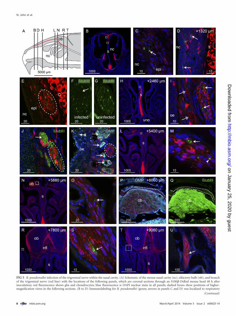
FIG 5 B. pseudomallei infection of the trigeminal nerve within the nasal cavity. (A) Schematic of the mouse nasal cavity (nc), olfactory bulb (ob), and branchof the trigeminal nerve (red line) with the locations of the following panels, which are coronal sections through an S100�-DsRed mouse head 48 h afterinoculation; red fluorescence shows glia and chondrocytes; blue fluorescence is DAPI nuclear stain in all panels; dashed boxes show positions of higher-magnification views in the following sections. (B to D) Immunolabeling for B. pseudomallei (green; arrows in panels C and D) was localized to respiratory
(Continued)
St. John et al.
8 ® mbio.asm.org March/April 2014 Volume 5 Issue 2 e00025-14
on January 25, 2020 by guesthttp://m
bio.asm.org/
Dow
nloaded from

ratory epithelia in the nasal cavity. The bacteria were able to mi-grate along olfactory and trigeminal nerve bundles and traversethe cribriform plate to enter the central nervous system within thecranial cavity within 24 h. We also show that bacterial coloniza-tion of the nasal lumen mucus by B. pseudomallei resulted in wide-spread crenellation of the olfactory epithelial surface followed bysloughing of the epithelial sheet, after which the bacteria were ableto penetrate the remaining layers of the epithelium (Fig. 6).
The surface of the olfactory epithelium consists of homoge-neously distributed supporting cells with dendrites of mature ol-factory sensory neurons interspersed between, and protruding
above, the supporting cells into the mucus layer of the nasal lumen(21, 22). We did not detect any evidence of bacteria selectivelyentering either the supporting cells or sensory dendrites. Nor wasthere evidence of the bacteria invading Bowman’s glands, whichsecrete mucus through ducts and could offer a potential conduitfrom the nasal lumen through to the underlying lamina propria.Not even crenellation of the olfactory epithelium led to consistentcolonization of the epithelium by the bacteria. Indeed, extensivebacterial invasion of the olfactory epithelium was detected onlywhen the neuronal layer was sloughed off into the nasal cavity,exposing the underlying layers. These findings demonstrate that
Figure Legend Continued
epithelium (epi) on the dorsal portion of the septum (s; chondrocytes in the septum are red); the white box in panel B is enlarged in panel C; the yellow box inpanel B shows the location of panel D in a section that is 1,320 �m more caudal; red fluorescence within the epithelium is localized within glia. (E) The trigeminalnerve that projects along the upper septum is infected; B. pseudomallei (green) appears within the trigeminal nerve (dashed oval). (F) A section adjacent to panelE shows �-tubulin III immunolabeling of the trigeminal axons (green); there is a noticeable loss of �-tubulin III in the area corresponding to where B. pseu-domallei was localized in panel E. (G) In comparison, the trigeminal nerve in an uninfected animal shows uniform �-tubulin III immunolabeling across the nerve.(H to K) At a location 2,460 �m more caudal than that in panel B and in the location shown by the dotted box in panel H, bacteria (green; arrows in panel I) werepresent within the trigeminal nerve, which is distinguished by the tube-like Schwann cells; adjacent olfactory nerve bundles are indicated by double-headedarrows. (J and K) �-Tubulin III immunolabeling (green) (J) and olfactory marker protein (OMP) immunolabeling (cyan) (K) verified the identity of thetrigeminal nerve (dashed ovals) in comparison to the adjacent olfactory nerve bundles (arrows). (K) The olfactory epithelium (oe) that was infected by bacteria(green; arrow with tail) was degenerated. (L to P) At locations 5,400 to 6,060 �m more caudal than that in panel B, the trigeminal nerve with degenerated particlesof bacteria (arrows) or intact bacteria (arrowhead in panel M) entered the cranial cavity and passed over the rostral-dorsal region of the olfactory bulb. (P andQ) OMP (cyan) (P) and �-tubulin III (green) (Q) immunolabeling confirmed the identity of the trigeminal nerve in comparison to the olfactory axons thatstrongly express both OMP and �-tubulin III; dashed lines demarcate the trigeminal nerve. (R to U) The trigeminal nerve projected to the lateral portions of thecranial cavity, and immunolabeling for B. pseudomallei (green; arrows) was present in locations up to 9,060 �m caudal from panel B; boxed regions in panels Rand T are shown at higher magnification in panels S and U, respectively. Bar sizes are in �m.
FIG 6 B. pseudomallei enters the central nervous system (CNS) via open channels. (A) Schematic sagittal view of the nasal cavity and olfactory system. Thetrigeminal nerve (red) is represented in panel B; the yellow boxed area is represented in panels C to E. (B) Some trigeminal nerve axons (blue) surrounded bySchwann cells degenerate as a result of the infection, and B. pseudomallei (green) migrates through the empty Schwann cell canals. (C) In healthy animals, primaryolfactory neurons (blue) reside in the olfactory epithelium (OE) and project axons (blue lines) in nerve bundles through the cribriform plate (CP) into theolfactory bulb (OB) within the central nervous system. OECs (red) surround the nerve bundles. (D) Exposure to B. pseudomallei (green rods) results in neuronsdegrading (dark blue ovals) in some regions of the epithelium; the loss of the neuron cell bodies (brown ovals) from the epithelium leads to the degradation oftheir axons (dashed lines); OECs remain intact. (E) After the epithelium is sloughed off into the nasal cavity, the bacteria penetrate the remaining layers of themucosa and migrate along the open channels left by the degraded axons and thereby enter the central nervous system.
Brain Infection via Nasal Mucosa and Olfactory Nerve
March/April 2014 Volume 5 Issue 2 e00025-14 ® mbio.asm.org 9
on January 25, 2020 by guesthttp://m
bio.asm.org/
Dow
nloaded from

the olfactory nerve constitutes a direct conduit by which patho-gens can rapidly invade the brain. This is consistent with a previ-ous study which demonstrated that in healthy mice, the olfactoryepithelium is an effective barrier against Staphylococcus aureus in-vasion; however, following damage to the olfactory epithelium bymild detergent treatment, S. aureus was detected in the olfactoryepithelium and the olfactory bulb (23). Combined, these data sug-gest that significant damage to the olfactory epithelium may be aprerequisite for bacterial invasion of the olfactory nerves and ol-factory bulb.
As olfactory axons project from the olfactory epithelium in-ward to the olfactory bulb, they group together to form nervefascicles which are surrounded by olfactory ensheathing cells.When neuron cell bodies are lost, the axons rapidly die and arephagocytosed by the olfactory ensheathing cells (24), which main-tain their positions and the overall integrity of the fascicles. Thus,the loss of axons from the fascicles results in open conduits ex-tending from the nasal epithelium directly into the brain. Weshow clear evidence that the bacteria rapidly exploited this routeand migrated along the open olfactory conduits that were devoidof axons so that within 24 h the bacteria had penetrated the outerlayer of the olfactory bulb (Fig. 6C and D).
It is unknown whether B. pseudomallei virulence factors play arole in the cellular disruption and bacterial invasion of the olfac-tory epithelium and nerves. Recently, Cruz-Migoni et al. (25) de-scribed a B. pseudomallei toxin, structurally similar to Esche-richia coli cytotoxic necrotizing factor 1, which demonstratedcytotoxic activity against J774.2 macrophages in vitro. However,an isogenic B. pseudomallei mutant deficient in this toxin stillcauses crenellation of the olfactory epithelium and destruction ofolfactory neurons (data not shown). Alternatively, it is possiblethat crenellation and sloughing of the olfactory epithelium mayoccur as part of a host response against B. pseudomallei infectionof the nasal cavity. Indeed, we have shown that B. pseudomalleiinduces a strong cytokine response in many tissues, including theolfactory epithelium (unpublished data). Further research to de-termine the role of B. pseudomallei virulence factors in the inva-sion of the olfactory pathway is warranted.
It is noteworthy that a feature of the bacterial infection was thepresence of spherical particles which were selectively immunola-beled with the bacterial antisera. The size of these particles wasapproximately 200 nm, consistent with B. pseudomallei outermembrane vesicles (26). Outer membrane vesicles have a numberof physiological functions, including the delivery of specific mol-ecules to other cells through cell fusion (27). We speculate that thecontinual production of the vesicles may contribute to the de-struction of the axons and hence facilitate the migration of thebacteria along the nerves. Thus, investigation of a possible role forB. pseudomallei outer membrane vesicles in infection is merited.Interestingly, the intact bacteria were reactive to the anti-OMPantibodies while the tiny vesicles were not. It has been previouslyreported that Neisseria meningitidis within the olfactory nerve co-localized with OMP immunolabeling (20). The reason for the bac-terial immunoreactivity to OMP antibodies is unknown and wor-thy of further exploration.
The brain stem is a major site of signs and symptoms arising inneurological melioidosis, and the trigeminal nerve constitutes adirect pathway from the nasal cavity to the trigeminal nucleus inthe brain stem (7). We clearly demonstrated that B. pseudomalleicould easily penetrate the thin respiratory epithelium in the nasal
cavity and colonize the trigeminal nerve. Bacteria were able tocross the intact respiratory epithelium, in contrast to the olfactoryepithelium, which had to be damaged for bacteria to traverse it.Again, it was clear that the presence of the bacteria resulted in theloss of axons from within individual Schwann cell bundles, whichlikely facilitated the migration of bacteria along the trigeminalnerve into the cranial cavity. Thus, the trigeminal nerve bundles(Fig. 6B) from the nasal cavity provide a potential route to thebrain stem in a manner essentially homologous to that of infectionof the olfactory bulb. While we did not detect bacteria in the brainstem in this study, we could trace the bacteria a considerable dis-tance along the trigeminal nerve within the cranial cavity, and it ispossible that the bacteria could migrate even further and reach thebrain stem or other brain areas if given more time.
In summary, we have provided evidence that B. pseudomalleipenetrates the thin respiratory epithelium but penetrates the ol-factory epithelium only once the neuronal layer has been sloughedoff. The bacteria then migrate along nerves and pass through thecribriform plate to enter the cranial cavity. Thus, both the olfac-tory and trigeminal nerve routes offer a pathway into the brain bywhich the bacteria can bypass the blood-brain barrier.
MATERIALS AND METHODSBacterial strains and growth conditions. B. pseudomallei strainMSHR520 (previously “08” [28]) is a clinical isolate from a human case ofmelioidosis, donated by Bart Currie (Menzies School of Health Research,Darwin, Australia). An allele replacement capsule-deficient mutant ofMSHR520 (MSHR520�cap) has been previously described (5). B. pseu-domallei was grown in liquid Luria broth (LB) medium with shaking.
Mice and experimental infection. Unanesthetized female BALB/cmice (age, 5 to 10 weeks) and S100�-DsRed (29) mice were infected in-tranasally by placement of 10 �l of bacteria onto the nostrils (5 �l pernostril). The inoculum contained 3 � 105 stationary-phase cells resus-pended in phosphate-buffered saline (PBS). Crosses of two transgenicreporter lines of mice, OMP-ZsGreen (30) and S100�-DsRed (29), wereused to demonstrate the anatomy of the olfactory system. All protocolswere approved by the animal ethics committee of Griffith University.
Immunohistochemistry and lectin chemistry. Animal tissue was pre-pared and cryostat sections were cut as previously described (31), andimmunochemistry was performed as previously described (32). The pri-mary antibodies used were rabbit anti-B. pseudomallei (1:2,000) (33),polyclonal goat anti-olfactory marker protein (OMP; 1:1,000; Wako), andpolyclonal rabbit anti-�-tubulin III (1:500; Abcam) followed by the sec-ondary antibody anti-rabbit Alexa Fluor 488 or anti-goat Alexa Fluor 594(1:400; Invitrogen). The biotinylated lectin UEA1 (1:100; Dako) was in-cubated similarly to the antibodies, followed by streptavidin-Alexa Fluor647 (1:400; Invitrogen). Cell nuclei were stained with 4=,6-diamidino-2-phenylindole (DAPI). Images were captured using an Olympus FV1000microscope, and image panels were created using Adobe Illustrator CS3.
SUPPLEMENTAL MATERIALSupplemental material for this article may be found at http://mbio.asm.org/lookup/suppl/doi:10.1128/mBio.00025-14/-/DCSupplemental.
Movie S1, AVI file, 1.8 MB.
ACKNOWLEDGMENTS
This work was supported by a grant from the National Health and MedicalResearch Council, no. 1020394, to I.R.B., M.B., and G.U. and an Austra-lian Research Council Fellowship to J.A.K.E., grant no. DP0986294.
REFERENCES1. Currie BJ, Ward L, Cheng AC. 2010. The epidemiology and clinical
spectrum of melioidosis: 540 cases from the 20 year Darwin prospective
St. John et al.
10 ® mbio.asm.org March/April 2014 Volume 5 Issue 2 e00025-14
on January 25, 2020 by guesthttp://m
bio.asm.org/
Dow
nloaded from

study. PLoS Negl. Trop. Dis. 4:e900. http://dx.doi.org/10.1371/journal.pntd.0000900.
2. Cheng AC, Hanna JN, Norton R, Hills SL, Davis J, Krause VL, DowseG, Inglis TJ, Currie BJ. 2003. Melioidosis in northern Australia, 2001-02.Commun. Dis. Intell. Q. Rep. 27:272–277.
3. White NJ. 2003. Melioidosis. Lancet 361:1715–1722. http://dx.doi.org/10.1016/S0140-6736(03)13374-0.
4. Currie BJ, Jacups SP. 2003. Intensity of rainfall and severity of melioid-osis, Australia. Emerg. Infect. Dis. 9:1538 –1542. http://dx.doi.org/10.3201/eid0912.020750.
5. Owen SJ, Batzloff M, Chehrehasa F, Meedeniya A, Casart Y, Logue CA,Hirst RG, Peak IR, Mackay-Sim A, Beacham IR. 2009. Nasal-associatedlymphoid tissue and olfactory epithelium as portals of entry for Burkhold-eria pseudomallei in murine melioidosis. J. Infect. Dis. 199:1761–1770.http://dx.doi.org/10.1086/599210.
6. Currie BJ, Fisher DA, Howard DM, Burrow JN. 2000. Neurologicalmelioidosis. Acta Trop. 74:145–151. http://dx.doi.org/10.1016/S0001-706X(99)00064-9.
7. Koszyca B, Currie BJ, Blumbergs PC. 2004. The neuropathology ofmelioidosis: two cases and a review of the literature. Clin. Neuropathol.23:195–203.
8. Chadwick DR, Ang B, Sitoh YY, Lee CC. 2002. Cerebral melioidosis inSingapore: a review of five cases. Trans. R. Soc. Trop. Med. Hyg. 96:-72–76. http://dx.doi.org/10.1016/S0035-9203(02)90248-8.
9. Aronsson F, Robertson B, Ljunggren HG, Kristensson K. 2003. Invasionand persistence of the neuroadapted influenza virus A/WSN/33 in themouse olfactory system. Viral Immunol. 16:415– 423. http://dx.doi.org/10.1089/088282403322396208.
10. Barnett EM, Perlman S. 1993. The olfactory nerve and not the trigeminalnerve is the major site of CNS entry for mouse hepatitis virus, strain JHM.Virology 194:185–191. http://dx.doi.org/10.1006/viro.1993.1248.
11. Kristensson K. 2011. Microbes’ roadmap to neurons. Nat. Rev. Neurosci.12:345–357. http://dx.doi.org/10.1038/nrn3029.
12. Lundh B, Kristensson K, Norrby E. 1987. Selective infections of olfactoryand respiratory epithelium by vesicular stomatitis and Sendai viruses.Neuropathol. Appl. Neurobiol. 13:111–122. http://dx.doi.org/10.1111/j.1365-2990.1987.tb00175.x.
13. Mori I, Goshima F, Ito H, Koide N, Yoshida T, Yokochi T, Kimura Y,Nishiyama Y. 2005. The vomeronasal chemosensory system as a route ofneuroinvasion by herpes simplex virus. Virology 334:51–58. http://dx.doi.org/10.1016/j.virol.2005.01.023.
14. Plakhov IV, Arlund EE, Aoki C, Reiss CS. 1995. The earliest events invesicular stomatitis virus infection of the murine olfactory neuroepithe-lium and entry of the central nervous system. Virology 209:257–262.http://dx.doi.org/10.1006/viro.1995.1252.
15. Salinas S, Schiavo G, Kremer EJ. 2010. A hitchhiker’s guide to thenervous system: the complex journey of viruses and toxins. Nat. Rev.Microbiol. 8:645– 655. http://dx.doi.org/10.1038/nrmicro2395.
16. Jarolim KL, McCosh JK, Howard MJ, John DT. 2000. A light microscopystudy of the migration of Naegleria fowleri from the nasal submucosa tothe central nervous system during the early stage of primary amebic me-ningoencephalitis in mice. J. Parasitol. 86:50 –55. http://dx.doi.org/10.2307/3284907.
17. Martinez J, Duma RJ, Nelson EC, Moretta FL. 1973. ExperimentalNaegleria meningoencephalitis in mice. Penetration of the olfactory mu-cosal epithelium by Naegleria and pathologic changes produced: a lightand electron microscope study. Lab. Invest. 29:121–133.
18. Dons L, Jin Y, Kristensson K, Rottenberg ME. 2007. Axonal transport ofListeria monocytogenes and nerve-cell-induced bacterial killing. J. Neuro-sci. Res. 85:2529 –2537. http://dx.doi.org/10.1002/jnr.21256.
19. van Ginkel FW, McGhee JR, Watt JM, Campos-Torres A, Parish LA,Briles DE. 2003. Pneumococcal carriage results in ganglioside-mediated
olfactory tissue infection. Proc. Natl. Acad. Sci. U. S. A. 100:14363–14367.http://dx.doi.org/10.1073/pnas.2235844100.
20. Sjölinder H, Jonsson AB. 2010. Olfactory nerve—a novel invasion routeof Neisseria meningitidis to reach the meninges. PLoS One 5:e14034.http://dx.doi.org/10.1371/journal.pone.0014034.
21. Danciger E, Mettling C, Vidal M, Morris R, Margolis F. 1989. Olfactorymarker protein gene: its structure and olfactory neuron-specific expres-sion in transgenic mice. Proc. Natl. Acad. Sci. U. S. A. 86:8565– 8569.http://dx.doi.org/10.1073/pnas.86.21.8565.
22. Hempstead JL, Morgan JI. 1983. Monoclonal antibodies to the rat olfac-tory sustentacular cell. Brain Res. 288:289 –295. http://dx.doi.org/10.1016/0006-8993(83)90105-1.
23. Herbert RP, Harris J, Chong KP, Chapman J, West AK, Chuah MI.2012. Cytokines and olfactory bulb microglia in response to bacterial chal-lenge in the compromised primary olfactory pathway. J. Neuroinflamma-tion 9:109. http://dx.doi.org/10.1186/1742-2094-9-109.
24. Su Z, Chen J, Qiu Y, Yuan Y, Zhu F, Zhu Y, Liu X, Pu Y, He C. 2013.Olfactory ensheathing cells: the primary innate immunocytes in the olfac-tory pathway to engulf apoptotic olfactory nerve debris. Glia 61:490 –503.http://dx.doi.org/10.1002/glia.22450.
25. Cruz-Migoni A, Hautbergue GM, Artymiuk PJ, Baker PJ, Bokori-Brown M, Chang CT, Dickman MJ, Essex-Lopresti A, Harding SV,Mahadi NM, Marshall LE, Mobbs GW, Mohamed R, Nathan S, NgugiSA, Ong C, Ooi WF, Partridge LJ, Phillips HL, Raih MF, RuzheinikovS, Sarkar-Tyson M, Sedelnikova SE, Smither SJ, Tan P, Titball RW,Wilson SA, Rice DW. 2011. A Burkholderia pseudomallei toxin inhibitshelicase activity of translation factor eIF4A. Science 334:821– 824. http://dx.doi.org/10.1126/science.1211915.
26. Nieves W, Asakrah S, Qazi O, Brown KA, Kurtz J, Aucoin DP,McLachlan JB, Roy CJ, Morici LA. 2011. A naturally derived outer-membrane vesicle vaccine protects against lethal pulmonary Burkholderiapseudomallei infection. Vaccine 29:8381– 8389. http://dx.doi.org/10.1016/j.vaccine.2011.08.058.
27. Kulp A, Kuehn MJ. 2010. Biological functions and biogenesis of secretedbacterial outer membrane vesicles. Annu. Rev. Microbiol. 64:163–184.http://dx.doi.org/10.1146/annurev.micro.091208.073413.
28. Brown NF, Beacham IR. 2000. Cloning and analysis of genomic differ-ences unique to Burkholderia pseudomallei by comparison with B. thailan-densis. J. Med. Microbiol. 49:993–1001.
29. Windus LC, Claxton C, Allen CL, Key B, St. John JA. 2007. Motilemembrane protrusions regulate cell-cell adhesion and migration of olfac-tory ensheathing glia. Glia 55:-1708 –1719. http://dx.doi.org/10.1002/glia.20586.
30. Ekberg JA, Amaya D, Chehrehasa F, Lineburg K, Claxton C, WindusLC, Key B, Mackay-Sim A, St. John JA. 2011. OMP-ZsGreen fluorescentprotein transgenic mice for visualisation of olfactory sensory neurons invivo and in vitro. J. Neurosci. Methods 196:88 –98. http://dx.doi.org/10.1016/j.jneumeth.2011.01.008.
31. Chehrehasa F, Windus LC, Ekberg JA, Scott SE, Amaya D, Mackay-SimA, St. John JA. 2010. Olfactory glia enhance neonatal axon regeneration.Mol. Cell. Neurosci. 45:277–288. http://dx.doi.org/10.1016/j.mcn.2010.07.002.
32. Chehrehasa F, Ekberg JA, Lineburg K, Amaya D, Mackay-Sim A, St.John JA. 2012. Two phases of replacement replenish the olfactory en-sheathing cell population after injury in postnatal mice. Glia 60:322–332.http://dx.doi.org/10.1002/glia.22267.
33. Boddey JA, Day CJ, Flegg CP, Ulrich RL, Stephens SR, Beacham IR,Morrison NA, Peak IR. 2007. The bacterial gene lfpA influences thepotent induction of calcitonin receptor and osteoclast-related genes inBurkholderia pseudomallei-induced TRAP-positive multinucleated giantcells. Cell. Microbiol. 9:514 –531. http://dx.doi.org/10.1111/j.1462-5822.2006.00807.x.
Brain Infection via Nasal Mucosa and Olfactory Nerve
March/April 2014 Volume 5 Issue 2 e00025-14 ® mbio.asm.org 11
on January 25, 2020 by guesthttp://m
bio.asm.org/
Dow
nloaded from