bronchoscopy in the diagnosis and treatment of bronchiectasis
TRANSCRIPT

BRONCHOSCOPY IN THE DIAGNOSIS AND TREATMENT OF BRONCHIECTASIS
PAUL M . MOORE, JR. , M . D .
Bronchiectasis is a chronic, debilitating disease characterized by dilatation of the smaller bronchi and copious, foul, purulent sputum. It robs the patient of his appetite, strength, and social contacts. In its advanced stage it is incurable. When less advanced it is curable only by lobectomy. If recognized sufficiently early it may be cured or at least its progress may be arrested by less radical means.
Certain definite factors have been quite well established in the etiology of bronchiectasis:
(1 ) . Bronchopneumonia, sometimes unrecognized in infancy, may result in contracting scar tissue which produces dilatation of the bronchi1'2.
(2 ) . Localized atelectasis due to a mucous plug or aspirated foreign body in a bronchus may produce a dilatation of the bronchi in the affected area3'4'5'6'7'8. If the obstruction is removed early and the atelectatic areas become aerated, the dilatations of the bronchi may disappear9'10. If the atelectasis is overlooked and the obstruction is not removed the dilatations will remain11.
( 3 ) . Watson and Kibler12 have recently stated that the most common cause is allergy, the sequence of events being basal allergic bronchitis followed by atelectasis which in turn is followed by dilatation of the bronchi.
(4 ) . There is also a congenital form which may go unrecognized until it becomes infected.
Attempts have been made to establish a single infecting organism as the principal cause of the disease. Those most frequently accused are the Bacillus influenzae, the fusospirillum, and Treponema micro-dentium and Treponema macrodentium13. That these organisms play an important part in some cases is without question but they have not been found in a sufficient percentage of the reported cases to assign an all-important role to them. A rather large unreported series of cases which I observed some years ago yielded a variety of organisms in the cultures secured by bronchoscopy. The one organism which appeared in almost every culture was the nonhemolytic streptococcus. These findings agree with those reported by Marvin14 in 1932.
Because these cases are chronic and resist efforts to cure them, the unfortunate victims are too often given inadequate treatment. But even if a cure cannot be obtained their lives can be made easier by proper care.
140
uses require permission. on January 17, 2022. For personal use only. All otherwww.ccjm.orgDownloaded from

BRONCHOSCOPY IN BRONCHIECTASIS
The first objective is an early diagnosis. A patient with a chronic cough which produces small quantities of sputum should be suspected of having bronchiectasis, especially if the sputum is foul. The cough and sputum are more marked when the patient lies down. Ordinary examination of the lungs may yield nothing more than some sticky rales at the base. Roentgen examination may or may not show the dilated bronchi. Bronchoscopy should be done, especially if there is any evidence of atelectasis. Secretions can be sucked out for culture and an autogenous vaccine can be made if required. If atelectasis is present, the cause of the obstruction can be determined and removed. This may be an unrecognized foreign body, a mucous plug, granulations, or an early carcinoma. After removal of the obstruction the lung distal to it will usually fill with air. If the obstruction has not been present for too long a time the bronchi and lung tissue should return to normal unless a carcinoma is present. Following the bronchoscopy, a lung mapping should be done to outline the bronchi. This will definitely establish whether or not bronchiectasis is present.
The lung mapping is best done after the secretions have been removed by suction through the bronchoscope. The most satisfactory mapping is obtained by passing a rubber catheter through the glottis under the guidance of the laryngeal mirror. This is performed in the fluoroscopy room and with the patient under local anesthesia. After the catheter is in place the lipiodol is injected. By using the fluoroscope, the progress of the oil can be watched and directed where it is wanted by changing the position of the patient.
The majority of patients with bronchiectasis have a suppurative in-volvement of one or more of the paranasal sinuses. Many men have felt that the sinus infection was primarily responsible for the bron-chiectasis. Mullin15 has shown that the infection can reach the lung either by way of the lymphatics or by direct drainage. Others have claimed that neither infection is primary but that they are merely due to extensive co-existing infection of the respiratory tract. It is certain that they do exist together and that it is necessary to treat all localized centers of infection if the best results are to be secured.
Once the diagnosis of bronchiectasis is established, the sinuses should be examined carefully. Those sinuses found to have a suppurative con-dition in them should be treated and every effort made to clear up the infection. Radical sinus surgery should be done if more conservative measures fail to produce the desired results. I do not regard a hyper-plastic membrane without actual suppuration as a condition requiring radical treatment in these cases. Whether the sinus infection is the primary cause or not, it must cleared up if only to take one more load from the patient's health and thereby improve his general condition.
141
uses require permission. on January 17, 2022. For personal use only. All otherwww.ccjm.orgDownloaded from

PAUL M. MOORE, JR.
General measures directed toward building up the patient's resistance and general health should be followed. The diet should be checked to ascertain that there are no important omissions. He should have plenty of vitamins, rest, and fresh air.
Treatment of the lung condition per se must of necessity follow one of two courses: one is directed toward keeping the infected cavities as clean as possible and the other toward eradicating the diseased tissue. The latter course is the one of choice in selected cases because it is the only one which holds forth the certainty of a cure. It should be used only in well-established and not too extensive cases, however. A few years ago the mortality rate for lobectomy was very high. Through steady improvement in the technic, it has been decreasing until now it is not of serious moment. Other surgical procedures such as pneumothorax, oleothorax, phrenicotomy, etc., have been of little value because they have compressed the lung tissue while the dilated bronchi have remained unaffected.
The treatment directed toward keeping the infected cavities as clean as possible must be used in all cases. Those who are to be operated upon can be put in better condition to withstand the operation by a thorough course of treatment first. This treatment consists of two im-portant parts:
Postural drainage should be done by the patient both morning and evening. The patient must be instructed in the proper way to do this or he will not obtain satisfactory results. One of the best methods is to have him lie on his face over the side of the bed with the hips and legs on the bed and the shoulders on the floor. A basin should be available to receive the sputum. He should maintain this position for from 20 to 30 minutes. In some cases this may be sufficient to completely drain the cavities. Usually, however, only a partial cleaning results and this makes the second part of the treatment desirable.
The second part is the bronchoscopic aspiration of the secretions at regular intervals. These intervals may vary from once a week to once a month or longer, depending upon the individual case. Some bron-choscopists advise instilling a few cubic centimeters of sterile solution into the bronchi at times and aspirating it out, thus washing out the cavities. Others feel that such a procedure may tend to spread the in-fection and therefore they depend upon aspiration alone. Following a good cleaning out of this sort the patient may be almost sputum-free for a few days. His general health improves, his appetite increases, and there is a decrease in the odor of the secretions.
Various antiseptics have been employed but I have yet to find one that I felt contributed anything to the treatment. If it is strong enough
142
uses require permission. on January 17, 2022. For personal use only. All otherwww.ccjm.orgDownloaded from

BRONCHOSCOPY IN BRONCHIECTASIS
to really affect the bacteria it is too strong for the normal mucous mem-branes. If it is mild enough that it does not damage the normal mucosa it is useless as a sterilizing agent.
Figures 1 and 2 show the progress of a far-advanced case of bron-chiectasis. When this patient was first seen she was about eleven years old and her body was merely skin and bones. The roentgen examination showed suppuration in the lung throughout the entire right side. It was
FIGURE 1 : Roentgenogram showing extensive involvement of the lung on the right side.
143
uses require permission. on January 17, 2022. For personal use only. All otherwww.ccjm.orgDownloaded from

PAUL M. MOORE, JR.
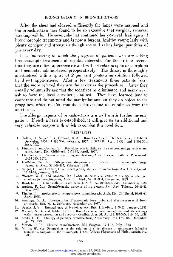
FIGURE 2 : Following bronchoscopy, the right lung is clear except for bronchiectasis.
felt that chest surgery would be necessary to drain this extensive suppura-tive process but that she was in too poor condition to withstand any opera-lion at that time. She was referred to the nose and throat department for bronchoscopic treatments preparatory for operation. This produced such marked improvement that it was decided to continue with her treat-ments rather than operate at that time.
144
uses require permission. on January 17, 2022. For personal use only. All otherwww.ccjm.orgDownloaded from
BRONCHOSCOPY IN BRONCHIECTASIS
After the chest had cleared sufficiently the lungs were mapped and the bronchiectasis was found to be so extensive that surgical removal was impossible. However, she has continued her postural drainage and bronchoscopic treatments and is now a buxom, healthy young lady with plenty of vigor and strength although she still raises large quantities of pus every day.
It is interesting to watch the progress of patients who are taking bronchoscopic treatments at regular intervals. For the first or second time they are rather apprehensive and will not relax in spite of morphine and nembutal administered preoperatively. The throat is thoroughly anesthetized with a spray of 2 per cent pontocaine solution followed by direct applications. After a few treatments these patients learn that the more relaxed they are the easier is the procedure. Later they usually voluntarily ask that the sedatives be eliminated and many even ask to have the local anesthetic omitted. They have learned how to cooperate and do not mind the manipulations but they do object to the grogginess which results from the sedatives and the numbness from the anesthesia.
The allergic aspects of bronchiectasis are well worth further investi-gation. If such a basis is established, it will give us an additional and very valuable weapon with which to combat this condition.
REFERENCES
1. Ballon, H., Singer, J. J., Graham, E. A . : Bronchiectasis, I . Thoracic Surg., 1:154-193, December, 1931; 1:296-326, February, 1932; 1:397-431, April, 1932; and 1:502-561, June, 1932.
2. Findlay, L. and Graham, S.: Bronchiectasis in children; its symptomatology, course and cause, Arch. Dis. Childhood, 2:71-96, April, 1927.
3. Lichtheim, L. : Versuche über lungenatelektase, Arch. f. exper. Path. u. Pharmakol., 10:54-100, 1879.
4. Hedblom, Carl A . : Pathogenesis, diagnosis and treatment of bronchiectasis, Surg.. Gynec. & Obst., 52:406-417, February, 1931.
5. Singer, J. J. and Graham, E. A . : Roentgen-ray study of bronchiectasis, Am. J. Roentgenol., 15:54-58, January, 1926.
6. Warner, W . P. and Graham, D. : Lobar atelectasis as cause of triangular roentgen shadows in bronchiectasis, Arch. Int. Med., 52:888-904, December, 1933.
7. Boyd, G. L. : Lobar collapse in children, J. A. M. A., 105:1832-1835, December 7, 1935. 8. Andrus, P. M. : Bronchiectasis, analysis of its causes, Am. Rev. Tuberc., 36:46-81,
July, 1937. 9. Findlay, L. : Atelectatic or compensatory bronchiectasis, Arch. Dis. Childhood, 10:61-64,
April, 1935. 10. Jennings, G. H . : Re-expansion of atelectatic lower lobe and disappearance of bron-
chiectasis, Brit. M. J., 2:963-965, November 13, 1937. 11. Sparks, J. V. : Unusual case of bronchiectasis, Brit. J. Radiol., 4:30-31, January, 1931. 12. Watson, S. H. and Kibler, C. S.: Bronchiectasis; new conception and its etiology
which makes prevention and recovery possible, J. A. M. A., 111:394-395, July 30, 1938. 13. Smith, D. T. : Etiology of primary bronchiectasis, Arch. Surg., 21:1173-1187, December,
(pt. 2 ) , 1930. 14. Marvin, H. P. : Chronic bronchiectasis, Mil. Surgeon, 17:1-15, July, 1932. 15. Mullin, W . V . : Symposium on the relation of sinus disease to pulmonary infection
from the standpoint of the rhinologist, Trans. College Physicians of Phila., 52:245-247, 1930.
145
uses require permission. on January 17, 2022. For personal use only. All otherwww.ccjm.orgDownloaded from