brock genetic exchange in bacteria
TRANSCRIPT
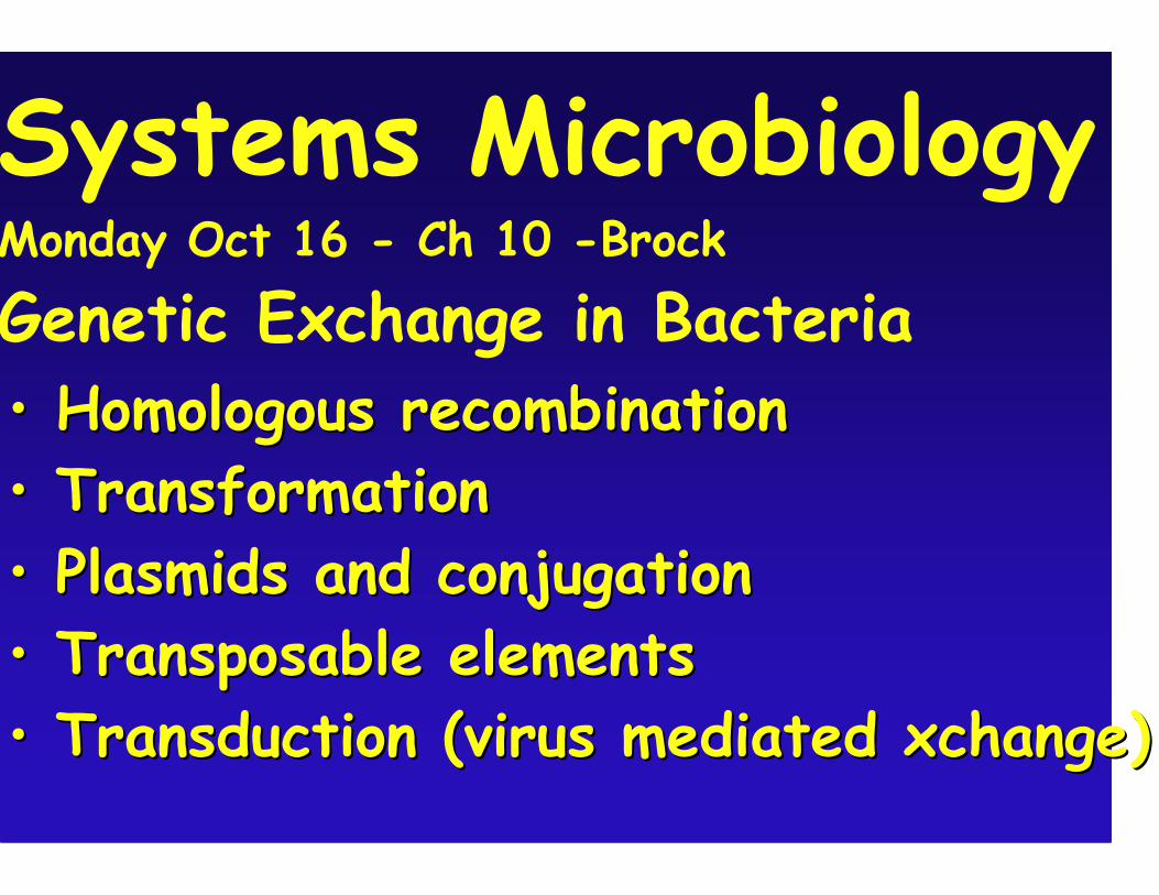
Systems Microbiology Monday Oct 16 - Ch 10 -Brock
Genetic Exchange in Bacteria •• Homologous recombinationHomologous recombination•• TransformationTransformation •• Plasmids and conjugationPlasmids and conjugation•• Transposable elementsTransposable elements•• Transduction (virus mediatedTransduction (virus mediated xchangexchange))
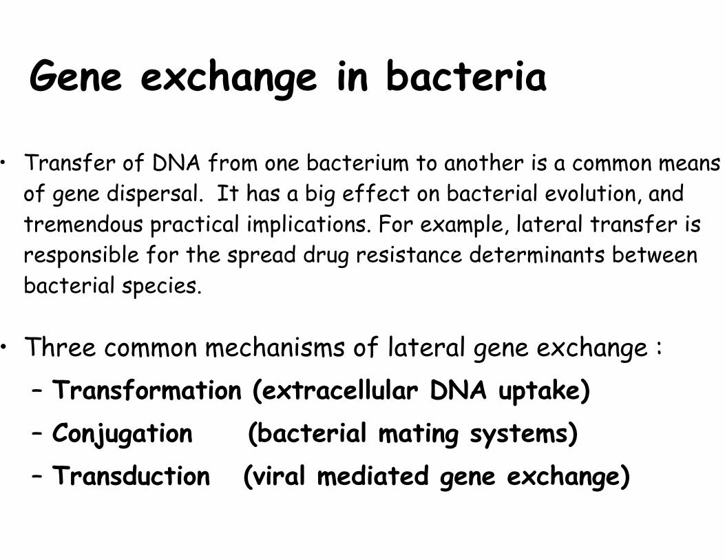
Gene exchange in bacteria
• Transfer of DNA from one bacterium to another is a common means of gene dispersal. It has a big effect on bacterial evolution, and tremendous practical implications. For example, lateral transfer is responsible for the spread drug resistance determinants between bacterial species.
• Three common mechanisms of lateral gene exchange : –Transformation (extracellular DNA uptake) –Conjugation (bacterial mating systems) –Transduction (viral mediated gene exchange)
RecA mediated Homologous recombination
Images removed due to copyright restrictions. See Figures 10-9 and 10-10 in Madigan, Michael, and John Martinko. Brock Biology of Microorganisms.11th ed. Upper Saddle River, NJ: Pearson Prentice Hall, 2006. ISBN: 0131443291.
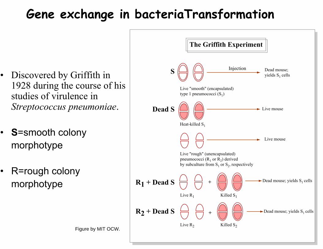
Gene exchange in bacteriaTransformation
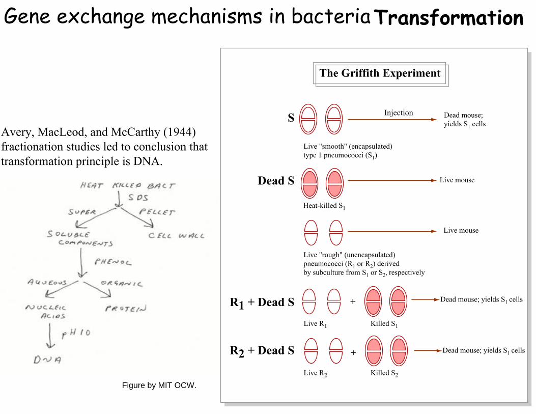
• Discovered by Griffith in 1928 during the course of his studies of virulence in Streptococcus pneumoniae.
• S=smooth colony morphotype
• R=rough colony morphotype
Injection
Live "smooth" (encapsulated)type 1 pneumococci (S1)
Dead mouse; yields S1 cells
Heat-killed S1
Live mouse
Live mouse
Live "rough" (unencapsulated)pneumococci (R1 or R2) derived by subculture from S1 or S2, respectively
Live R1
Live R2
Killed S1
Killed S2
Dead mouse; yields S1 cells
Dead mouse; yields S1 cells
+
+
S
Dead S
R1 + Dead S
R2 + Dead S
The Griffith Experiment
Figure by MIT OCW.
Gene exchange mechanisms in bacteriaTransformation
Avery, MacLeod, and McCarthy (1944) fractionation studies led to conclusion thattransformation principle is DNA.
Injection
Live "smooth" (encapsulated)type 1 pneumococci (S1)
Dead mouse; yields S1 cells
Heat-killed S1
Live mouse
Live mouse
Live "rough" (unencapsulated)pneumococci (R1 or R2) derived by subculture from S1 or S2, respectively
Live R1
Live R2
Killed S1
Killed S2
Dead mouse; yields S1 cells
Dead mouse; yields S1 cells
+
+
S
Dead S
R1 + Dead S
R2 + Dead S
The Griffith Experiment
Figure by MIT OCW.
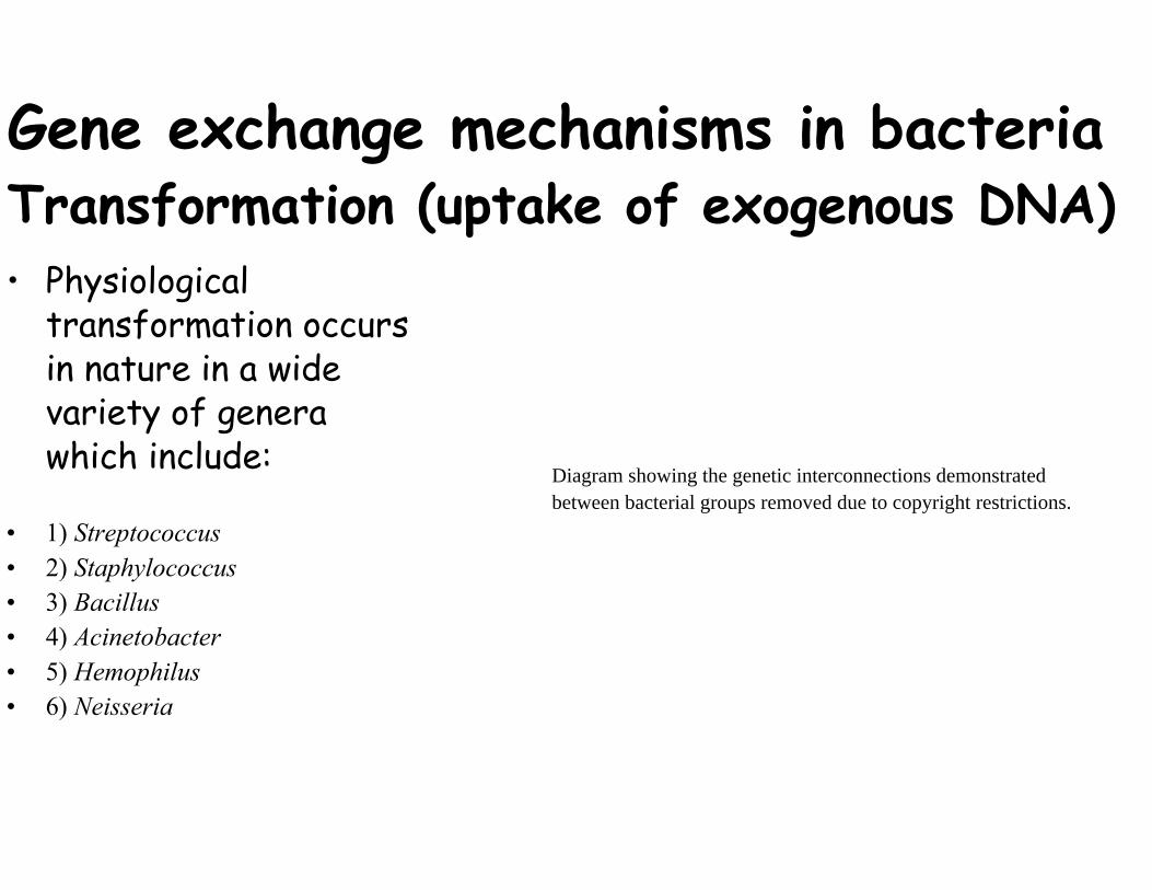
Gene exchange mechanisms in bacteriaTransformation (uptake of exogenous DNA)• Physiological
transformation occurs in nature in a wide variety of genera which include:
Diagram showing the genetic interconnections demonstrated between bacterial groups removed due to copyright restrictions.
• 1) Streptococcus • 2) Staphylococcus • 3) Bacillus • 4) Acinetobacter • 5) Hemophilus • 6) Neisseria
Natural Bacterial Transformation
Image removed due to copyright restrictions.
Closely Linked Genes will Tend to Transform Together More Frequentlythan More Distal Genes
Gene exchange mechanisms in bacteria Transformation
• Competence. The ability to take up DNA varies regularly during the cell cycle. In Image removed due to copyright restrictions. Streptococcus competence is See Figure 10-14 in Madigan, Michael, and John Martinko. Brock Biology of
highest shortly after cell Microorganisms.11th ed. Upper Saddle River, NJ: Pearson Prentice Hall, 2006. ISBN: 0131443291.
division. • Entry & integration. Cell
components required foruptake.
• Heteroduplex formation withhomologous recipient DNA.
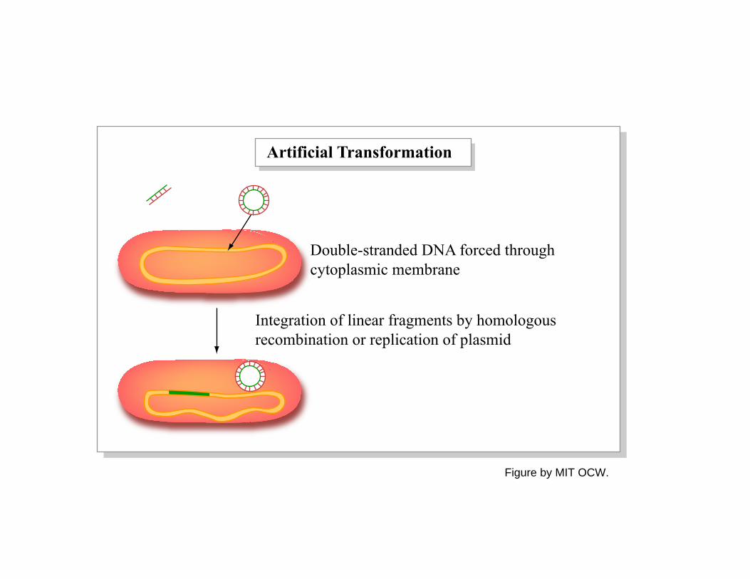
Artificial Transformation
Double-stranded DNA forced through cytoplasmic membrane
Integration of linear fragments by homologous recombination or replication of plasmid
Figure by MIT OCW.
BacterialConjugation
-
Image of bacterial conjugation, showing the donor (F+), pilus, and recipient (F-), removed due to copyright restrictions.
PLASMIDS
• Extrachromosomal DNA, usually circular
• Usually encode ancillary functions for in vitro growth
• Can be essential for specific environments: virulence, antibiotics resistance, use of unusual nutrients, production of bacteriocins (colicins)
• Must be a replicon - self-replicating genetic unit
Plasmid Replication
• Plasmid DNA must replicate each time cell divides or it will be lost
• Host cells do not “spit out” plasmid DNA
• Two functions required in replication DNA replication Partitioning (distributing plasmid to progeny cells)
• High copy (>20) and low copy (<5) plasmids
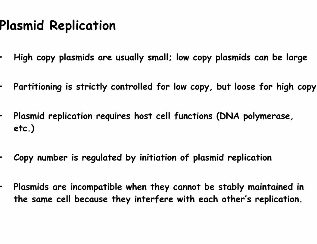
Plasmid Replication
• High copy plasmids are usually small; low copy plasmids can be large
• Partitioning is strictly controlled for low copy, but loose for high copy
• Plasmid replication requires host cell functions (DNA polymerase, etc.)
• Copy number is regulated by initiation of plasmid replication
• Plasmids are incompatible when they cannot be stably maintained in the same cell because they interfere with each other’s replication.
Confers resistance : sulfonamide chloramphenicol mercury ions streptomycin tetracycline Image removed due to copyright restrictions.
See Figure 10-20 in Madigan, Michael, and John Martinko. Brock Biology of Microorganisms. 11th ed. Upper Saddle River, NJ: Pearson Prentice Hall, 2006. ISBN: 0131443291.
Table of some phenotypes conferred by plasmids in prokaryotes removed due to copyright restrictions. See Figure 10-3 in Madigan, Michael, and John Martinko. Brock Biology of Microorganisms. 11th ed. Upper Saddle River, NJ: Pearson Prentice Hall, 2006. ISBN: 0131443291.
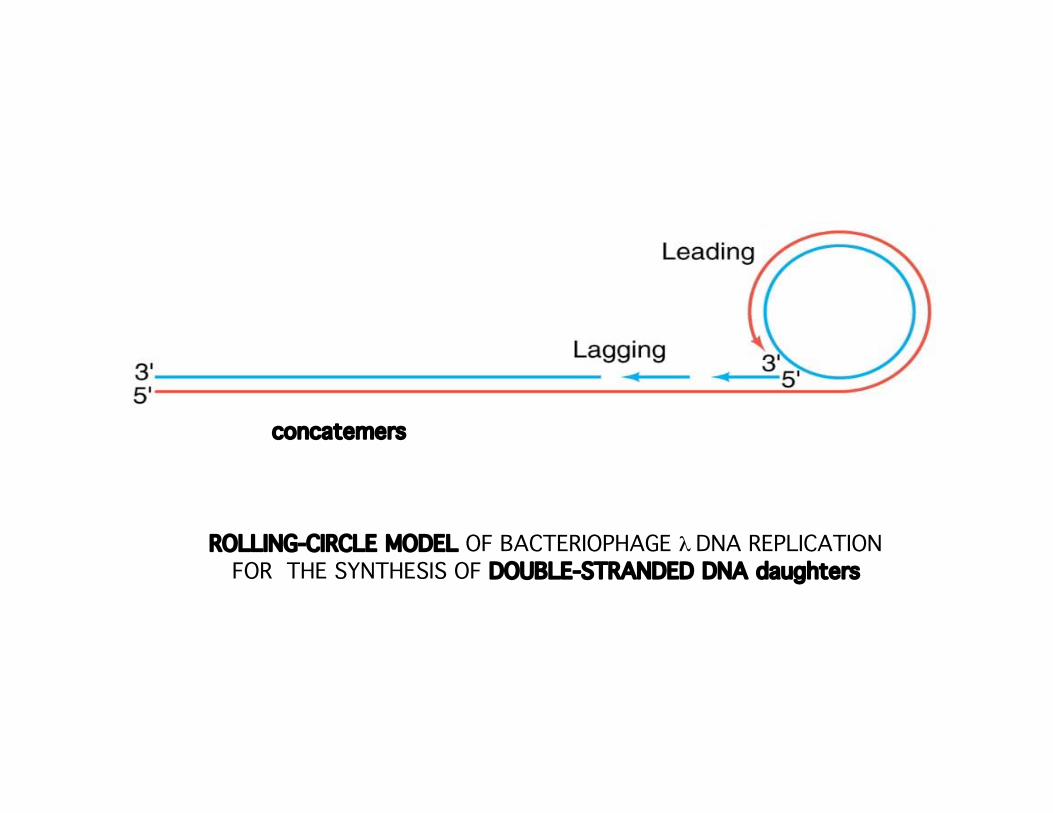
concatemers
ROLLING-CIRCLE MODEL OF BACTERIOPHAGE λ DNA REPLICATION FOR THE SYNTHESIS OF DOUBLE-STRANDED DNA daughters
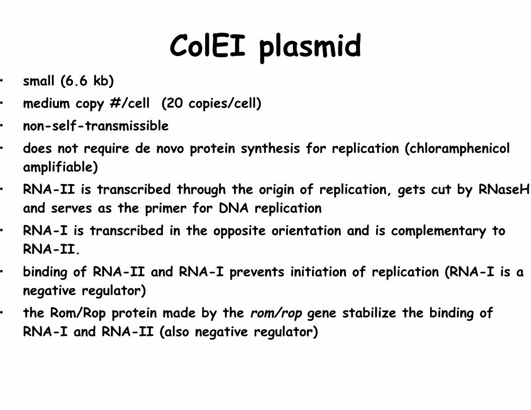
ColEI plasmid• small (6.6 kb) • medium copy #/cell (20 copies/cell) • non-self-transmissible • does not require de novo protein synthesis for replication (chloramphenicol
amplifiable) • RNA-II is transcribed through the origin of replication, gets cut by RNaseH
and serves as the primer for DNA replication • RNA-I is transcribed in the opposite orientation and is complementary to
RNA-II. • binding of RNA-II and RNA-I prevents initiation of replication (RNA-I is a
negative regulator) • the Rom/Rop protein made by the rom/rop gene stabilize the binding of
RNA-I and RNA-II (also negative regulator)
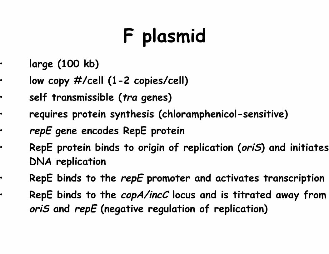
F plasmid• large (100 kb) • low copy #/cell (1-2 copies/cell) • self transmissible (tra genes) • requires protein synthesis (chloramphenicol-sensitive) • repE gene encodes RepE protein • RepE protein binds to origin of replication (oriS) and initiates
DNA replication • RepE binds to the repE promoter and activates transcription
• RepE binds to the copA/incC locus and is titrated away from oriS and repE (negative regulation of replication)
Image removed due to copyright restrictions. See Figure 10-18 in Madigan, Michael, and John Martinko. Brock Biology of Microorganisms. 11th ed. Upper Saddle River, NJ: Pearson Prentice Hall, 2006. ISBN: 0131443291.
Bacterial Conjugation
Diagram showing the process of bacterial conjugation removed due to copyright restrictions. See Figure 10-22 in Madigan, Michael, and John Martinko. Brock Biology of Microorganisms. 11th ed. Upper Saddle River, NJ: Pearson Prentice Hall, 2006. ISBN: 0131443291.
Image removed due to copyright restrictions. See Figure 10-23 in Madigan, Michael, and John Martinko. Brock Biology of Microorganisms. 11th ed. Upper Saddle River, NJ: Pearson Prentice Hall, 2006. ISBN: 0131443291.
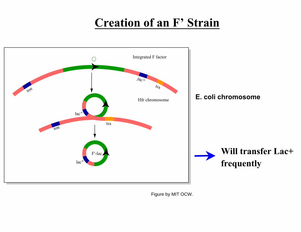
Creation of an F’ Strain
E. coli chromosome
Will transfer Lac+frequently
Integrated F factor
ton
ton
jac+tsx
tsx
lac+
lac+
F'-lac
Hfr chromosome
Figure by MIT OCW.
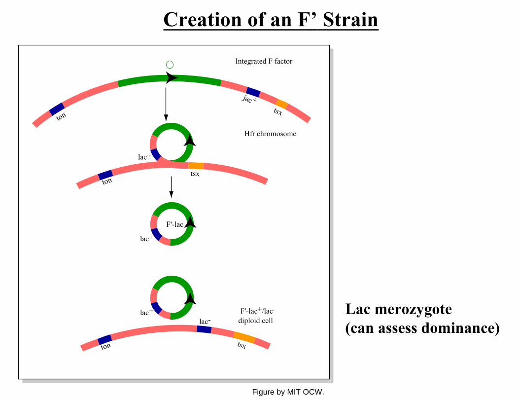
Creation of an F’ Strain
Lac merozygote (can assess dominance)
Integrated F factor
ton
ton
ton
jac+tsx
tsx
tsx
lac+
lac+
lac+lac-
F'-lac
F'-lac+/lac-diploid cell
Hfr chromosome
Figure by MIT OCW.
Hfr Strains
• The F plasmid can integrate into the chromosome (many sites – directed by transposon homology). This creates a high frequency of recombination (Hfr) strain.
• The integrated F plasmid directs transfer of the chromosome, starting from the origin. Genes close to the site of integration will be transferred first.
• Transfer continues, with the order of transfer matching the order of genes along the chromosome, until it is interrupted.
(interrupted mating experiments for chromosomal mapping…)
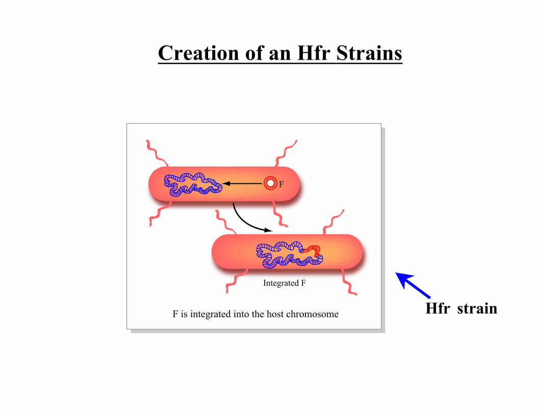
Creation of an Hfr Strains
Hfr strain
Integrated F
F
F is integrated into the host chromosome
DNA Transfer in an Hfr Strain
Diagram removed due to copyright restrictions.
Image removed due to copyright restrictions. See Figure 10-24 in Madigan, Michael, and John Martinko. Brock Biology of Microorganisms. 11th ed. Upper Saddle River, NJ: Pearson Prentice Hall, 2006. ISBN: 0131443291.
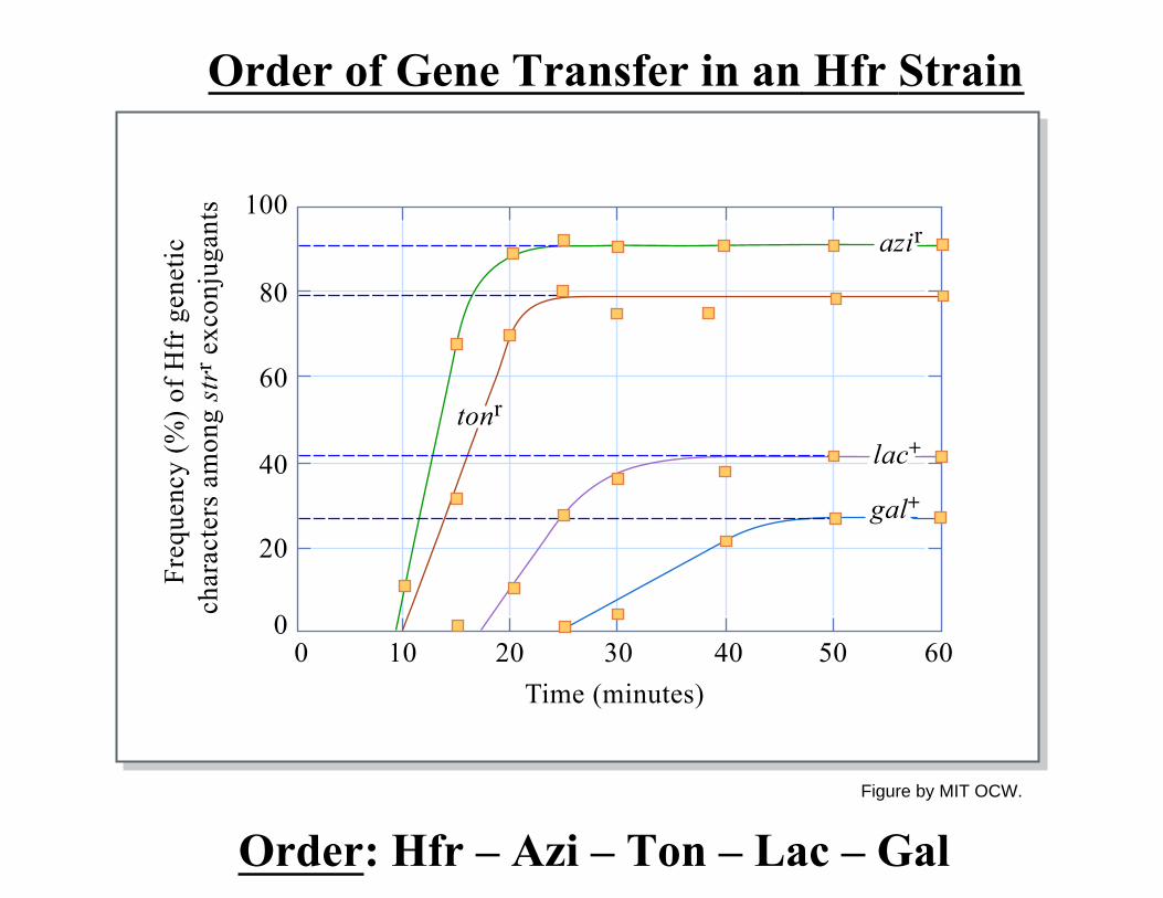
Order of Gene Transfer in an Hfr Strain
Order: Hfr – Azi – Ton – Lac – Gal
0
20
0
Freq
uenc
y (%
) of H
fr g
enet
ic
char
acte
rs a
mon
g st
rr e
xcon
juga
nts
10 20 30Time (minutes)
40 50 60
40
60
80
azir
tonr
lac+
gal+
100
Figure by MIT OCW.
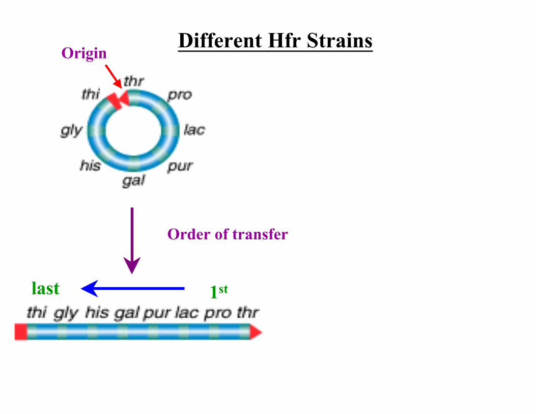
Different Hfr StrainsOrigin
1stlast
Order of transfer
1stlast

High Resolution Mapping Using Hfr Strain
Image removed due to copyright restrictions.
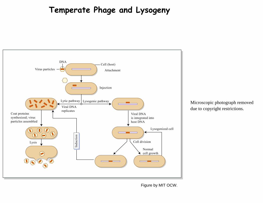
Temperate Phage and Lysogeny
Microscopic photograph removeddue to copyright restrictions.
DNA
Virus particles
Viral DNAreplicates
Viral DNAis integrated intohost DNA
Lysogenized cell
Cell division
Normal cell growth
Cell (host)
Lysogenic pathwayLytic pathway
Coat proteins synthesized; virus particles assembled
Lysis
Attachment
Injection
Indu
ctio
n
Figure by MIT OCW.
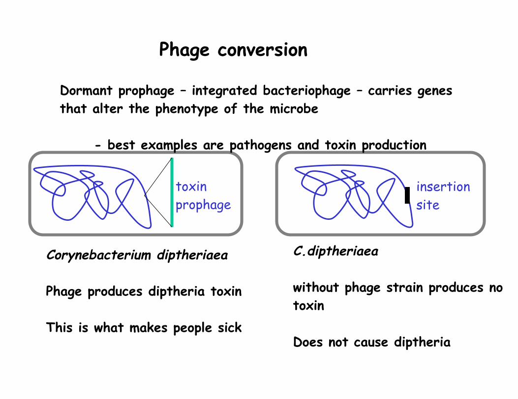
Phage conversion
Dormant prophage – integrated bacteriophage – carries genes that alter the phenotype of the microbe
- best examples are pathogens and toxin production
toxin insertion prophage site
Corynebacterium diptheriaea C.diptheriaea
Phage produces diptheria toxin without phage strain produces no toxin
This is what makes people sick Does not cause diptheria
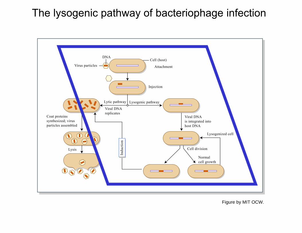
The lysogenic pathway of bacteriophage infection
DNA
Virus particles
Viral DNAreplicates
Viral DNAis integrated intohost DNA
Lysogenized cell
Cell division
Normal cell growth
Cell (host)
Lysogenic pathwayLytic pathway
Coat proteins synthesized; virus particles assembled
Lysis
Attachment
Injection
Indu
ctio
n
Figure by MIT OCW.
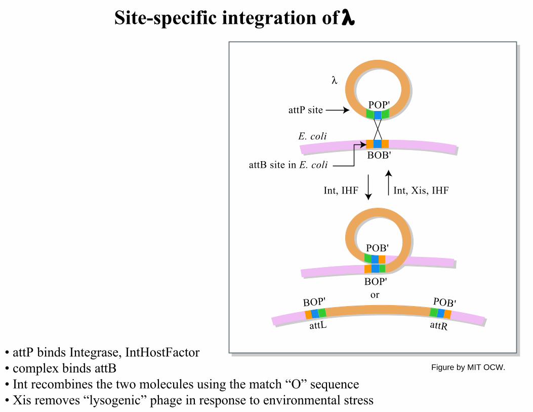
Site-specific integration of λ
• attP binds Integrase, IntHostFactor • complex binds attB • Int recombines the two molecules using the match “O” sequence • Xis removes “lysogenic” phage in response to environmental stress
POP'
λ
BOB'
POB'
BOP'
BOP' POB'
attL attR
or
Int, IHF
attP site
attB site in E. coli
Int, Xis, IHF
E. coli
Figure by MIT OCW.
Invitrogen image of Phage lambda recombination in E. coli removed due to copyright restrictions.
Mechanism of Integrase action
Diagram showing the mechanism of integrase action removed due to copyright restrictions.
• ATP independent process • 5’ OH and 3’ phosphates • Covalent enzyme-tyrosine-integrase attachment -akin to topoisomerases
Specialized transduction (in phage lambda)
Specialized transduction is site specific, and so results in transfer only of specific genes.
Eg, genes next to the attB site for lambda Infecting E. coli
Diagram removed due to copyright restrictions. See Figure 10-16 in Madigan, Michael, and John Martinko. Brock Biology of Microorganisms. 11th ed. Upper Saddle River, NJ: Pearson Prentice Hall, 2006. ISBN: 0131443291.
Visible effects on DNA during viral infection
Images showing DNA and T4 phage in a pre-infection and post-infection cell removed due to copyright restrictions.
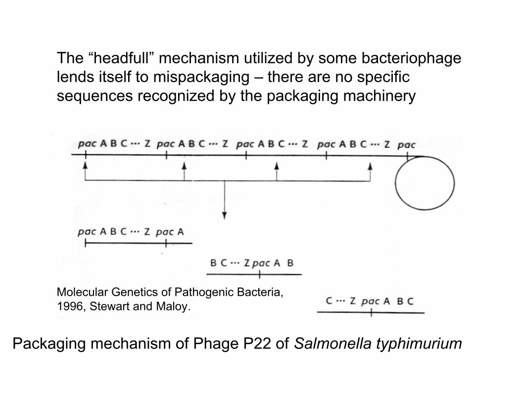
The “headfull” mechanism utilized by some bacteriophage lends itself to mispackaging – there are no specific sequences recognized by the packaging machinery
Molecular Genetics of Pathogenic Bacteria, 1996, Stewart and Maloy.
Packaging mechanism of Phage P22 of Salmonella typhimurium
Generalized transduction This happens when host DNA, instead of phage DNA is accidentally packaged.
Generalized transduction is more or less random, and so can result in the transfer of almost any gene.
Image removed due to copyright restrictions. See Figure 10-15 in Madigan, Michael, and John Martinko. Brock Biology of Microorganisms. 11th ed. Upper Saddle River, NJ: Pearson Prentice Hall, 2006. ISBN: 0131443291.
An example – P22 phage transduction of Salmonella typhimurium
P22 HT is a efficient generalized transducer - its sloppy – 50% of the viral particles contain host
cell DNA (ie are transducing particles or TPs)
Each transducing particle (TP) carries 44 kb of DNA – the Salmonella genome is app. 4400 kb in size
Therefore, if the process is random 100 different transducing particles should represent the entire genome.
(0.5)(1011 viruses/ml)/(100 TP [1 genome]) = 5x10⁸ copies of the genome/ml of lysate
Generalized transduction is a useful way to exchange genes between bacteria
Also extremely Image removed due to copyright restrictions.
useful for mapping of genetic markers relative to each other
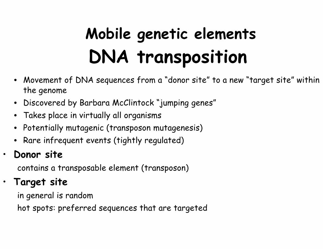
Mobile genetic elementsDNA transposition
• Movement of DNA sequences from a “donor site” to a new “target site” within the genome
• Discovered by Barbara McClintock “jumping genes” • Takes place in virtually all organisms • Potentially mutagenic (transposon mutagenesis) • Rare infrequent events (tightly regulated)
• Donor site contains a transposable element (transposon)
• Target site in general is random hot spots: preferred sequences that are targeted
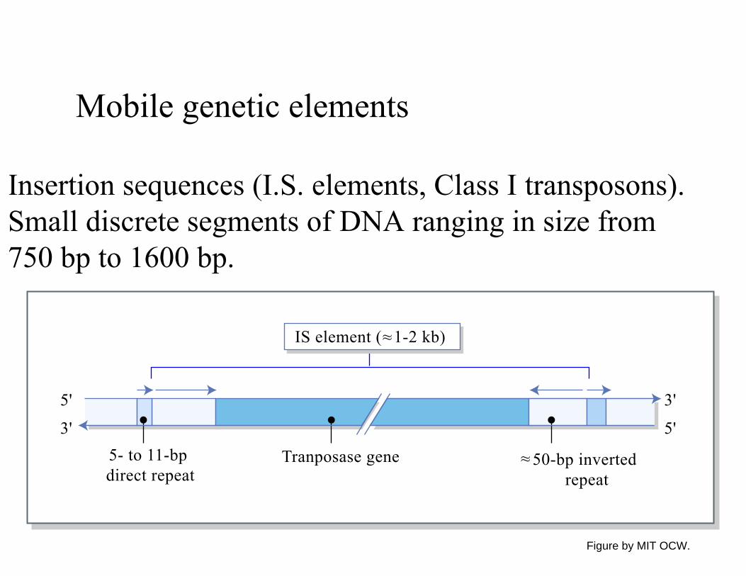
Mobile genetic elements
Insertion sequences (I.S. elements, Class I transposons). Small discrete segments of DNA ranging in size from 750 bp to 1600 bp.
5'
3'5- to 11-bp direct repeat
50-bp inverted repeat
3'
5'
~~Tranposase gene
IS element ( 1-2 kb)~~
Figure by MIT OCW.
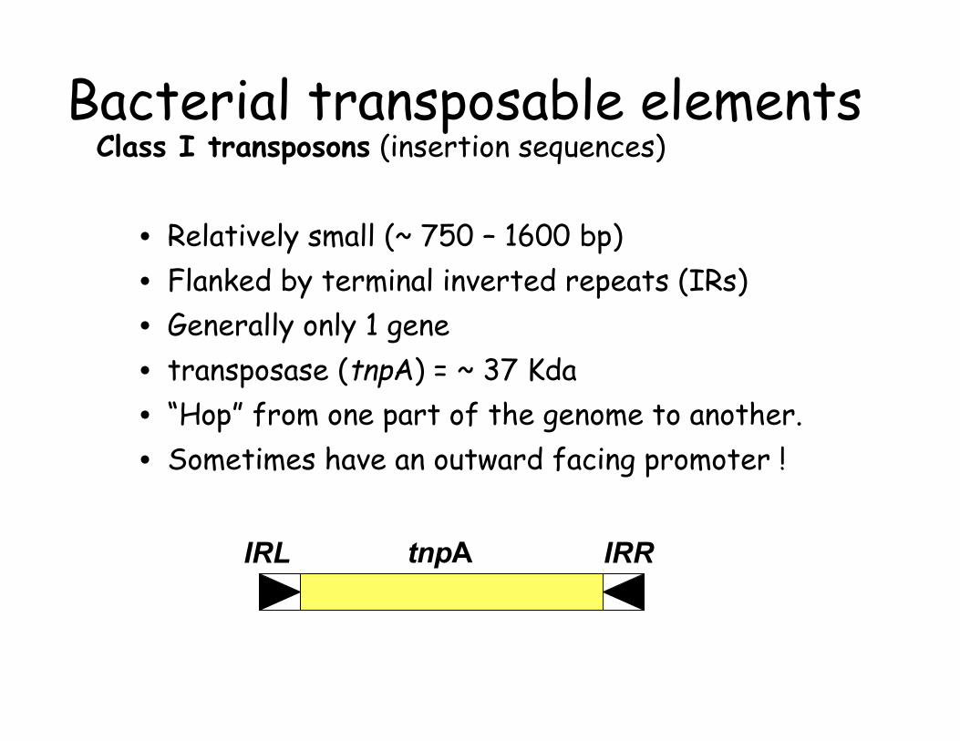
Bacterial transposable elementsClass I transposons (insertion sequences)
• Relatively small (~ 750 – 1600 bp) • Flanked by terminal inverted repeats (IRs) • Generally only 1 gene • transposase (tnpA) = ~ 37 Kda • “Hop” from one part of the genome to another.• Sometimes have an outward facing promoter !
IRL tnpA IRR
Mobile genetic elements
I.S. elements can act in pairs to mobilize intervening DNA.
I.S. elements can mobilize important determinants such as antibiotic resistance genes, genes for lactose utilization, or genes for bacterial enterotoxins.
In E. coli the ST enterotoxin gene is encoded by a transposon and is sometimes found on plasmids and sometimes on temperate phages.
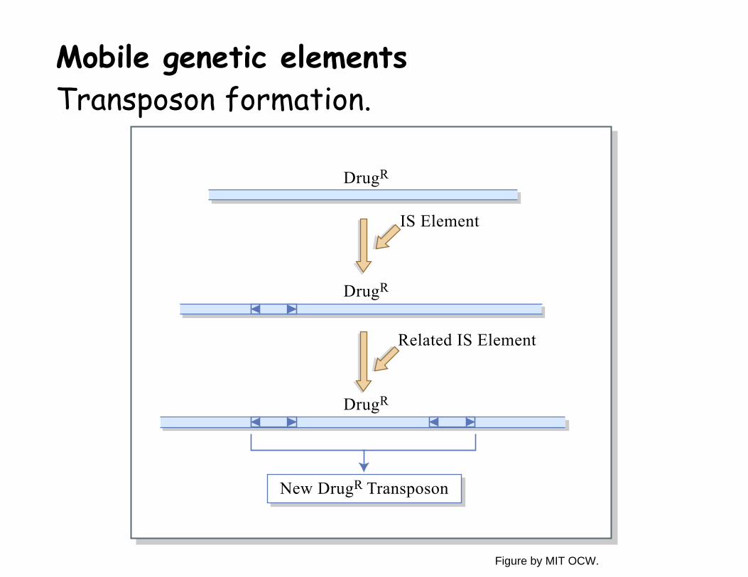
Mobile genetic elementsTransposon formation.
DrugR
DrugR
IS Element
DrugR
Related IS Element
New DrugR Transposon
Figure by MIT OCW.
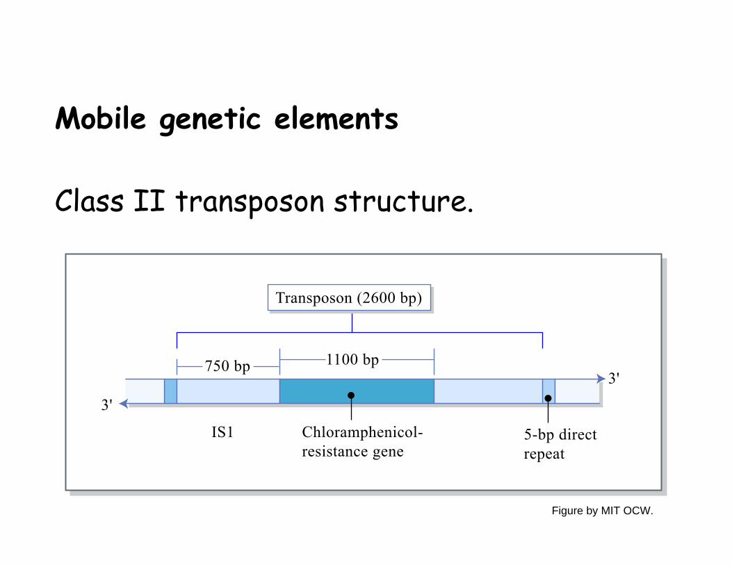
Mobile genetic elements
Class II transposon structure.
750 bp
3'
3'1100 bp
Chloramphenicol-resistance gene
IS1 5-bp direct repeat
Transposon (2600 bp)
Figure by MIT OCW.
Image removed due to copyright restrictions. See Figure 10-29b in Madigan, Michael, and John Martinko. Brock Biology of Microorganisms. 11th ed. Upper Saddle River, NJ: Pearson Prentice Hall, 2006. ISBN: 0131443291.
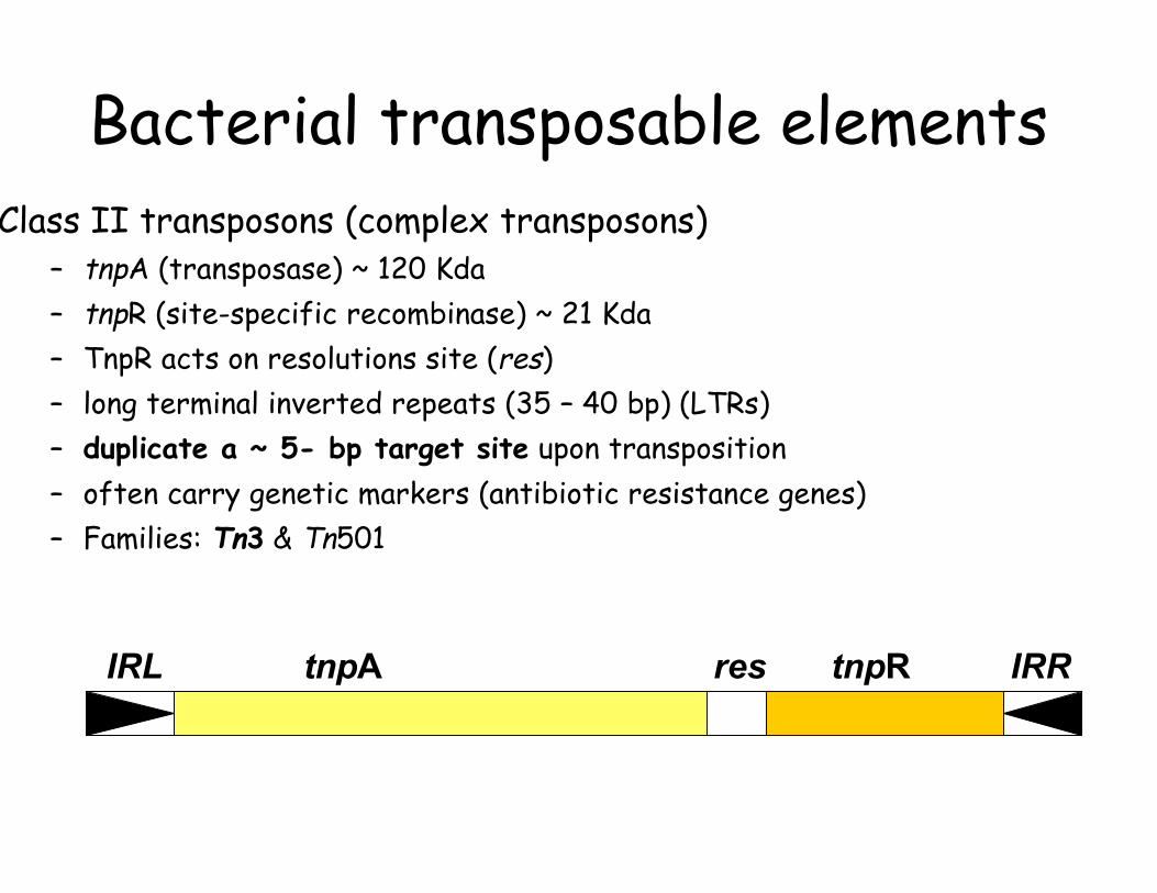
Bacterial transposable elements Class II transposons (complex transposons)
– tnpA (transposase) ~ 120 Kda – tnpR (site-specific recombinase) ~ 21 Kda – TnpR acts on resolutions site (res) – long terminal inverted repeats (35 – 40 bp) (LTRs) – duplicate a ~ 5- bp target site upon transposition– often carry genetic markers (antibiotic resistance genes)– Families: Tn3 & Tn501
IRL tnpA res tnpR IRR
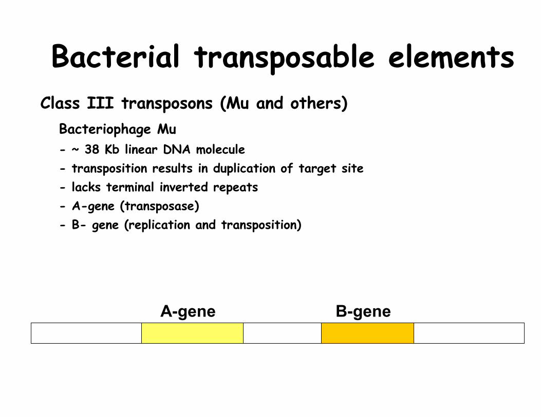
Bacterial transposable elementsClass III transposons (Mu and others)
Bacteriophage Mu - ~ 38 Kb linear DNA molecule - transposition results in duplication of target site - lacks terminal inverted repeats - A-gene (transposase) - B- gene (replication and transposition)
A-gene B-gene
Image removed due to copyright restrictions. See Figure 10-31 in Madigan, Michael, and John Martinko. Brock Biology of Microorganisms. 11th ed. Upper Saddle River, NJ: Pearson Prentice Hall, 2006. ISBN: 0131443291.
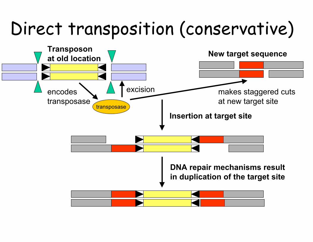
Direct transposition (conservative)Transposon New target sequence at old location
encodes excision makes staggered cuts transposase at new target site
Insertion at target site transposase
DNA repair mechanisms result in duplication of the target site
Image removed due to copyright restrictions. See Figure 10-32 in Madigan, Michael, and John Martinko. Brock Biology of Microorganisms. 11th ed. Upper Saddle River, NJ: Pearson Prentice Hall, 2006. ISBN: 0131443291.
Replication-dependent transposition
Image removed due to copyright restrictions.

Strategy for transposon mutagenesis
Diagram showing the process of transposon mutagenesis removed due to copyright restrictions.
In vivo Tn mutation Generating mutantstemplates in vitro
Mutagenesis of bacteria
Diagrams removed due to copyright restrictions.
Epicentre Biotechnologies Website
Gene expression on cloned operons from environmental librariesProteorhodopsin/retinal + β -carotene/retinal/proteorhodopsin operon
X X X X X X XIpp blh crtY crtB crtI crtE PR
Images removed due to copyright restrictions.