briefcommunications the ink4a/arf … ·...
TRANSCRIPT
Brief Communications
The Ink4a/Arf Locus Is a Barrier to Direct NeuronalTransdifferentiation
James D. Price,1,2,3* Ki-Youb Park,1,3* Jiadong Chen,1,4 Ryan D. Salinas,1,3 Mathew J. Cho,1,3 Arnold R. Kriegstein,1,4
and Daniel A. Lim1,2,3,5
1Eli and Edythe Broad Center of Regeneration Medicine and Stem Cell Research, 2Developmental and Stem Cell Biology Graduate Program, 3Department ofNeurological Surgery, 4Department of Neurology, and 5San Francisco Veterans Affairs Medical Center, University of California, San Francisco, SanFrancisco, California 94143
Non-neurogenic cell types, such as cortical astroglia and fibroblasts, can be directly converted into neurons by the overexpression ofdefined transcription factors. Normally, the cellular phenotype of such differentiated cells is remarkably stable and resists direct celltransdifferentiation. Here we show that the Ink4a/Arf (also known as Cdkn2a) locus is a developmental barrier to direct neuronaltransdifferentiation induced by transcription factor overexpression. With serial passage in vitro, wild-type postnatal cortical astrogliabecome progressively resistant to Dlx2-induced neuronal transdifferentiation. In contrast, the neurogenic competence of Ink4a/Arf-deficient astroglia is both greatly increased and does not diminish through serial cell culture passage. Electrophysiological analysisfurther demonstrates the neuronal identity of cells induced from Ink4a/Arf-null astroglia, and short hairpin RNA-mediated acuteknockdown of p16Ink4a and p19Arf p16 Ink4a and p19 Arf indicates that these gene products function postnatally as a barrier to cellulartransdifferentiation. Finally, we found that mouse fibroblasts deficient for Ink4a/Arf also exhibit greatly enhanced transcription factor-induced neuronal induction. These data indicate that Ink4a/Arf is a potent barrier to direct neuronal transdifferentiation and furthersuggest that this locus functions normally in the progressive developmental restriction of postnatal astrocytes.
Key words: astroglia; induced neuron; Ink4a/Arf; transcription factor; transdifferentiation
IntroductionNeurons can be directly converted from non-neuronal cell typesvia the enforced expression of specific transcription factors (Vi-erbuchen and Wernig, 2012). Other than tissue-specific stem cellpopulations, cells in the adult mammal generally do not changetheir cellular identities, and this inherent phenotypic stabilityrepresents a barrier to cell transdifferentiation (Zhou andMelton, 2008). Thus, discovering the molecular-genetic mecha-nisms that facilitate direct neuronal transdifferentiation providesinsight into how cell fates normally become restricted, and mayalso inform strategies that enhance cell fate conversion for thepurpose of disease-modeling and cell-based therapeutics.
The Ink4a/Arf tumor suppressor locus encodes the p16 Ink4a
and p19 Arf cell cycle inhibitors, which are expressed at basal levelsin differentiated cell types. In addition to their function in tumor
suppression (Kim and Sharpless, 2006), Ink4a/Arf gene productsimpede the generation of induced pluripotent stem cells (iPSCs)from fibroblasts: Ink4a/Arf is rapidly silenced during iPSC pro-duction, and Ink4a/Arf-deficient fibroblasts generate iPSCs moreefficiently (Banito et al., 2009; Li et al., 2009; Utikal et al., 2009).Whether Ink4a/Arf also constitutes a barrier to direct neuronalreprogramming is not known.
During postnatal development, astrocytes in the mouse cere-bral cortex divide symmetrically, greatly expanding their num-bers (Ge et al., 2012); and when isolated from postnatal day 7 (P7)mice, astroglial cells still exhibit developmental plasticity: theseyoung astroglia can give rise to multipotent neurospheres(Palmer et al., 1999; Laywell et al., 2000) and can also be con-verted into mature neurons via enforced expression of neuro-genic transcription factors, such as Dlx2 (Heins et al., 2002;Berninger et al., 2007; Heinrich et al., 2010). However, corticalastrocytes from P15 mice no longer give rise to neurospheres(Laywell et al., 2000), and transcription factor-mediated neu-ronal transdifferentiation is greatly hindered in cells isolatedfrom later postnatal mice (Heinrich et al., 2011; Robel et al.,2011). Thus, during this postnatal period of symmetric glialcell proliferation, cortical astrocytes appear to lose their neu-rogenic “competence.”
Here, we used transcription factor-mediated neuronal trans-differentiation to gain insight into the molecular-genetic mecha-nisms that normally restrict neurogenic competence. We foundthat Ink4a/Arf is a potent barrier to direct neuronal transdiffer-
Received July 24, 2013; revised Aug. 1, 2014; accepted Aug. 6, 2014.Author contributions: J.D.P., K.-Y.P., and D.A.L. designed research; J.D.P., K.-Y.P., J.C., R.D.S., and M.J.C. per-
formed research; J.D.P., K.-Y.P., J.C., R.D.S., M.J.C., A.R.K., and D.A.L. analyzed data; J.D.P., K.-Y.P., and D.A.L. wrotethe paper.
This work was supported by National Institutes of Health DP2-OD006505-01, the Sontag Foundation, a NorthernCalifornia Institute for Research and Education/Department of Defense subaward to D.A.L., the Shurl and Kay CurciFoundation, and the San Francisco Veterans Affairs Medical Center for resources.
The authors declare no competing financial interests.*J.D.P. and K.-Y.P. contributed equally to this work.Correspondence should be addressed to Dr. Daniel A. Lim, University of California, San Francisco, 35 Medical
Center Way, San Francisco, CA 94143. E-mail: [email protected]:10.1523/JNEUROSCI.3159-13.2014
Copyright © 2014 the authors 0270-6474/14/3412560-08$15.00/0
12560 • The Journal of Neuroscience, September 10, 2014 • 34(37):12560 –12567
entiation of mouse astroglia isolated from non-neurogenic brainregions as well as mouse fibroblasts. Our findings suggest a gen-eralizable approach to enhance direct cell conversion methodol-ogies and further indicate a role for Ink4a/Arf in the postnatalglial-fate restriction of parenchymal astrocytes.
Materials and MethodsAnimals. Ink4a/Arf-null mice were maintained and genotyped as in Ser-rano et al. (1996). All experiments were performed with mice of either sexand in accordance to protocols approved by the Institutional AnimalCare and Use Committee at University of California, San Francisco.
Cell culture and neuronal transdifferentiation. Postnatal cortical astro-glial cultures were established essentially as in Berninger et al. (2007).Briefly, cortical tissue above the corpus callosum was microdissectedfrom P5–P7 mouse brain sections, dissociated by treatment with 0.25%trypsin 0.5 mM EDTA as in Lim et al. (2009), and plated into 6-wellculture plates (Corning) coated with 0.1 mg/ml poly-D-lysine (Sigma-Aldrich) in astrocyte growth medium described by Heinrich et al. (2010).Glial cultures were replated 5–7 d later into 16-well poly-D-lysine-coatedNunc Lab-Tek chamber slides (Thermo Scientific) at a density of 50,000cells per well (passage 1) or passaged into culture plates for continuedculture in astrocyte growth medium. Cells in chamber slides were in-fected with lentivirus and switched to astrocyte differentiation medium(DMEM/F12, B27, glutamine, and penicillin/streptomycin) (Heinrich etal., 2010) 24 h later.
Mouse embryonic fibroblasts (MEFs) were isolated and cultured in N3medium (DMEM/F12, apotransferrin, insulin, sodium selenite, proges-terone, putrescine, andpenicillin/streptomycin) (Vierbuchen et al.,2010). Passage 3 MEFs were transduced with lentivirus expressing reversetetracycline transactivator (FUW-M2rtTA (Hockemeyer et al., 2008) andthe transcription factors Myt1l and Brn2 (Vierbuchen et al., 2010), andrat Ascl1 expressed from the doxycycline-inducible lentiviral vectortetO-FUW. At 16 –20 h after infection, cells were switched to fresh MEFmedium containing doxycycline (2 �g/ml, Sigma). At 48 h after theaddition of doxycycline, cells were switched to N3 medium containingdoxycycline, which was replaced every 3 d. Neuronal differentiation wasanalyzed by immunocytochemistry 20 d after infection.
Immunocytochemistry. Immunocytochemistry was performed as inLim et al. (2009) with the following primary antibodies: mouse anti-Tuj1(Covance), chicken anti-GFP (Aves), rabbit anti-dsRed (Clontech), rab-bit anti-Nestin (Millipore), guinea pig anti-GLAST (Millipore), rabbitanti-S100� (Sigma), chicken anti-GFAP (Millipore), chicken anti-vimentin(Millipore), mouse anti-phospho-histone H3 (Millipore), rabbit anti-Pax6(Covance), and rabbit anti-Sox2 (Santa Cruz Biotechnology). DAPI (Sigma)was used for nuclear staining.
Microscopy, cell counting, and statistical analysis. For quantification ofcell cultures, at least 6 random, nonoverlapping fields of view were digi-tally acquired at 100 –200� magnification (CMI 4000B, Leica), and cellswere counted manually with aid of ImageJ (National Institutes of Health)or the Leica Application Suite Advanced Fluorescence software (Leica).Significance was calculated with Student’s t test using Excel (Microsoft).
Electrophysiology. Cells were analyzed 14 –30 d after infection. Thepatch electrodes were made from borosilicate glass capillaries (SutterInstruments) with a resistance in the range of 5–7 M�. The pipettes weretip-filled with internal solution containing the following (in mM): 125K-gluconate, 15 KCl, 10 HEPES, 4 MgCl2, 4 Na2ATP, 0.3 Na3GTP, 10Tris-phosphocreatine, 0.2 EGTA. The bath was constantly perfused withfresh recording medium containing the following (in mM): 145 NaCl, 3KCl, 3 CaCl2, 2 MgCl2, 10 HEPES, 8 glucose, at room temperature.Recordings were made with an Axon 700B patch-clamp amplifier and1320A interface (Molecular Devices). Signals were filtered at 2 kHz usingamplifier circuitry, sampled at 10 kHz, and analyzed using Clampex 10.2(Molecular Devices).
Microarray and qRT-PCR analysis. Analysis by qPCR was performed asin Ramos et al. (2013). For microarray analysis, samples from three rep-licate cultures for each cell type were prepared as in Ramos et al. (2013)and hybridized to MouseRef-8 v2.0 Expression BeadChip arrays (Illu-mina). Array data were processed and analyzed as in Park et al. (2014).
Lentiviral production. To acutely knock-down Ink4a/Arf, one shorthairpin RNA (shRNA) sequence against Ink4a/Arf (targeting sequence:AATGGCTGGATTGTTTAAA) (Fasano et al., 2007) and one controlsequence targeting luciferase described in targeting sequence: GAGCT-GTTTCTGAGGAGCC (Ventura et al., 2004) was cloned into the lenti-viral vector pSicoR-mCh (a gift from Dr. Miguel Ramalho-Santos). Theefficiency of viral transduction ranged from 95 to 98%, and no significantdifferences were observed between wild-type (WT) and null glia. Brn2,Ascl1, and Myt1l lentiviral vectors have been described previously (Vier-buchen et al., 2010) and were obtained through Addgene. VSV-G andEnvA pseudotyped lentiviruses were produced in HEK 293T cells as inLewis et al. (2001).
ResultsLoss of neurogenic competence of cortical astroglia throughserial passage in vitroP5–P7 cortical astroglial cultures infected with lentivirus express-ing Dlx2 and the GFP marker (LV-Dlx2-GFP) but not GFP alone(LV-GFP) produced GFP-positive neuronal cells immunoposi-tive for �III Tubulin (Tuj1-positive) after 7 d of differentiation(Fig. 1A,B). We investigated whether this “neurogenic compe-tence” is maintained through serial passage in vitro. Cortical as-troglia were serially passaged 5 times over 15 d, infected withLV-Dlx2-GFP or LV-GFP at passages 1, 2, 3, and 5, and neuronaldifferentiation was quantified 8 d after infection. While the effi-ciency of LV-Dlx2-GFP induced neuronal transdifferentiationwas �7% at passage 1 (Fig. 1B, Pass 1), the percentage of Tuj1-positive LV-Dlx2-GFP transduced cells was reduced 2.7-fold bypassage 2 (Fig. 1B, Pass 2), becoming 8.5-fold reduced (0.8%efficiency) at passage 5 (Fig. 1B, Pass 5). Thus, with serial in vitropassage, cortical astroglia derived from P5–P7 mice progressivelylose their neurogenic competence.
Ink4a/Arf deficiency enhances the neurogenic competence ofpostnatal astrogliaInk4a/Arf is expressed in cortical astroglial cultures but not neu-rogenic neural precursors (Bachoo et al., 2002). To determinewhether Ink4a/Arf is a barrier to neuronal transdifferentiation,we produced cultures from Ink4a/Arf-null mice and WT litter-mates. Interestingly, at passage 1, Ink4a/Arf-null astroglia exhib-ited low levels of neurogenesis even without enforced expressionof Dlx2 (Fig. 1B), possibly because of low levels of neural stemcell-like behavior in response to EGF signaling, as previously re-ported (Bachoo et al., 2002). However, by passage 3, very little ofthis neural stem cell-like behavior remained (Fig. 1B). In con-trast, at passage 1, LV-Dlx2-GFP induced neuronal differentia-tion in 25.17% (SD � 1.14%, n � 4 independent experiments) ofinfected Ink4a/Arf-null cells (Fig. 1B, Pass 1), resulting in �4-foldmore neurons than in WT cultures. Furthermore, the neurogeniccompetence of Ink4a/Arf-null cells did not diminish with contin-ued in vitro passage (Fig. 1B, Pass1–5); by passage 5, transdiffer-entiation of Ink4a/Arf-null astroglial cells was �30-fold moreefficient than WT cells (Fig. 1B). Thus, Ink4a/Arf deficiency bothenhances and maintains the neurogenic competence of postnatalcortical astroglia.
In typical neural cell growth media, Ink4a/Arf-null astrogliagrow faster than WT cultures, and cellular reprogramming, suchas that of iPSCs, can be promoted by high rates of proliferation(Hanna et al., 2009). To address the possibility that the increasedproliferation rate of Ink4a/Arf-null astroglia relates to their en-hanced transdifferentiation potential, we reduced the rate of cellproliferation by omitting recombinant EGF and bFGF from thegrowth medium. Ink4a/Arf-null astroglia grown without exoge-nous EGF and bFGF incorporated the thymidine analog ethynyl
Price, Park et al. • Ink4a/Arf Inhibits Neuronal Transdifferentiation J. Neurosci., September 10, 2014 • 34(37):12560 –12567 • 12561

deoxyuridine (EdU) at a rate similar toWT astroglia cultured with EGF andbFGF (Fig. 1C); despite a nearly sixfoldreduction in EdU incorporation, Inka/Arf-null astroglia cultured in growthfactor-deficient conditions still exhibited20-fold greater LV-Dlx2-GFP-inducedneuronal transdifferentiation, comparedwith WT astroglia grown with EGF andbFGF (Fig. 1D). Thus, independent of therate of cell proliferation, Ink4a/Arf defi-ciency promotes neuronal transdifferen-tiation in cortical astroglia.
Because parenchymal astrocytes can beregionally distinct (Zhang and Barres,2010), we next asked whether Ink4a/Arf-null astroglia from other non-neurogenicbrain regions also exhibit increased effi-ciencies of neuronal transdifferentiation.Whereas astroglia derived from the cere-bellum and brainstem did not grow wellunder these culture conditions and there-fore could not be evaluated, striatal astro-glia did propagate efficiently. LV-Dlx2-GFPinduced neuronal differentiation of astro-glial cells in Ink4a/Arf-null striatal cultureswith �5-fold greater efficiency than WTcontrols (Fig. 1D), even when cultured inmedia without EGF and bFGF, indicatingthat Ink4a/Arf deficiency enhances neuronaltransdifferentiation in regionally distinctpopulations of astroglia.
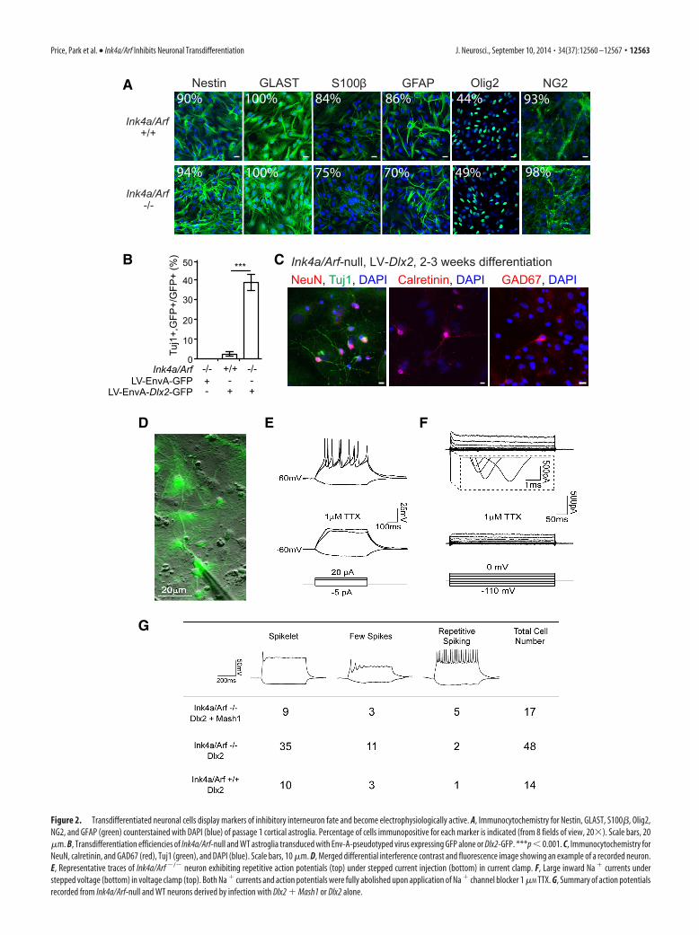
GFAP-positive, Ink4a/Arf-null astrogliaexhibit increased neurogeniccompetenceAlthough astroglial cultures from bothInk4a/Arf-null and WT cortex containedsimilar proportions of cells expressing Nes-tin, GLAST, OLIG2, and NG2 (Fig. 2A),Ink4a/Arf-null cultures contained �20%fewer cells expressing GFAP, a markerof astrocyte identity. We therefore investi-gated whether Ink4a/Arf deficiency inGFAP-positive astroglia enhances neuronaltransdifferentiation.
We restricted LV-Dlx2-GFP infectionto cells expressing GFAP. EnvA pseu-dotyped lentiviruses (LV/EnvA) cannotinfect mammalian cells unless they ex-press the avian viral receptor gene, tv-a(Lewis et al., 2001). The Gtv-a mousetransgene expresses tv-a from the GFAPpromoter, enabling LV/EnvA infection of
A
B
C
Ink4a/Arf +/+
LV-Dlx2-GFP, Pass 1GFP Tuj1 DAPI merge
Ink4a/Arf -/-
0
5
10
15
20
0
Pass 1Pass 2Pass 3Pass 5
0
10
20
40
30
Ink4a/Arf
Tuj1
+,G
FP+/
GFP
+ (%
)
-/-+/+LV-GFP
-/-+/+LV-Dlx2
***
D
-EGF, FGF+EGF, FGF
n.s.
Striatum
Striatum
Ink4a/Arf +/+ -/-
0
10
20
30
40
0
-EGF, FGF+EGF, FGF
Ink4a/Arf +/+ -/-
-EGF, FGF+EGF, FGF
n.s.
Cortex
Cortex
+/+ -/-
+/+ -/-
EdU
/DA
PI (
%)
-EGF, FGF+EGF, FGF
*****
Tuj1
+,G
FP+/
GFP
+ (%
)
60
40
20
60
40
20
Ink4a/Arf
Tuj1
+,G
FP+/
GFP
+ (%
)
Ink4a/Arf
EdU
/DA
PI (
%)
Figure 1. Ink4a/Arf deficiency enhances neuronal transdifferentiation. A, Immunocytochemistry for GFP (green) and Tuj1 (red)of Ink4a/Arf-null and WT astroglia infected at passage 1 (Pass 1) with LV-Dlx2-GFP. B, Transdifferentiation efficiencies of astroglia
4
serially cultured from passage 1 (bright red) through 5 (lightpink). C, Quantification of EdU-positive cells in Ink4a/Arf-nulland WT cortical (left) and striatal (right) astroglial cultures,with and without exogenous EGF and FGF. D, Efficiencies ofneuronal conversion of cells from C with and without exoge-nous EGF and FGF. Error bars indicate SEM. **p � 0.01.***p � 0.001. n.s., Not significant. Scale bars, 10 �m.
12562 • J. Neurosci., September 10, 2014 • 34(37):12560 –12567 Price, Park et al. • Ink4a/Arf Inhibits Neuronal Transdifferentiation
-/- -/-+/++
+ +
Ink4a/ArfLV-EnvA-GFP
LV-EnvA-Dlx2-GFP
Ink4a/Arf +/+
Ink4a/Arf -/-
A
B
D E F
Tuj1
+,G
FP+/
GFP
+ (%
)
G
C
Nestin
GLAST
GFAP
S100β 84%
75% 70%
86%100%
100%94%
90% Olig2
NG2
49%
44% 93%
98%
- --
***
0
10
20
30
40
50
NeuN, Tuj1, DAPI Calretinin, DAPI GAD67, DAPIInk4a/Arf-null, LV-Dlx2, 2-3 weeks differentiation
Figure 2. Transdifferentiated neuronal cells display markers of inhibitory interneuron fate and become electrophysiologically active. A, Immunocytochemistry for Nestin, GLAST, S100�, Olig2,NG2, and GFAP (green) counterstained with DAPI (blue) of passage 1 cortical astroglia. Percentage of cells immunopositive for each marker is indicated (from 8 fields of view, 20�). Scale bars, 20�m. B, Transdifferentiation efficiencies of Ink4a/Arf-null and WT astroglia transduced with Env-A-pseudotyped virus expressing GFP alone or Dlx2-GFP. ***p � 0.001. C, Immunocytochemistry forNeuN, calretinin, and GAD67 (red), Tuj1 (green), and DAPI (blue). Scale bars, 10 �m. D, Merged differential interference contrast and fluorescence image showing an example of a recorded neuron.E, Representative traces of Ink4a/Arf �/� neuron exhibiting repetitive action potentials (top) under stepped current injection (bottom) in current clamp. F, Large inward Na currents understepped voltage (bottom) in voltage clamp (top). Both Na currents and action potentials were fully abolished upon application of Na channel blocker 1 �M TTX. G, Summary of action potentialsrecorded from Ink4a/Arf-null and WT neurons derived by infection with Dlx2 Mash1 or Dlx2 alone.
Price, Park et al. • Ink4a/Arf Inhibits Neuronal Transdifferentiation J. Neurosci., September 10, 2014 • 34(37):12560 –12567 • 12563

0
5
10
15
sh-Ink4a/Arfsh-Luc
Ink4a/Arf +/+
LV-GFP LV-Dlx2-GFP
Tuj1
+,G
FP+,
RFP
+/
GFP
+, R
FP+
(%) **
0
10
20
30
LV-Dlx2-GFP+/+ -/-Ink4a/Arf
**
p16/p19Control
Tuj1
+,G
FP+,
RFP
+/
GFP
+, R
FP+
(%)
A
C
B
D
0.00
0.05
0.10
0.15
0.20
0.25
(nor
mal
ized
to IR
ES
-Dlx
2)
Dlx1 Dlx5 Dlx6
after diff.
mR
NA
leve
l
Dlx1 Dlx5 Dlx60.00
0.01
0.02
0.03
0.04
0.05
mR
NA
leve
l(n
orm
aliz
ed to
IRE
S-D
lx2)
before diff.
Ink4a/Arf +/+Ink4a/Arf -/-
Ink4a/Arf +/+Ink4a/Arf -/-
*
Gene ID Gene Symbol Description Fold Change in Null p-Value11834 AQR aquarius intron-binding spliceosomal factor 2.2723 1.495E-05100042493 CCL21A chemokine (C-C motif) ligand 21 2.1578 2.998E-0411537 CFD complement factor D (adipsin) 2.2943 2.489E-0267529 FGFR1OP2 FGFR1 oncogene partner 2 2.1752 2.138E-0566606 LRRC57 leucine rich repeat containing 57 2.7283 1.314E-0567809 RMDN3 regulator of microtubule dynamics 3 2.0634 2.233E-06100213 RUSC2 RUN and SH3 domain containing 2 2.2669 6.806E-0616651 SSPN sarcospan 2.1520 2.529E-0421754 TESK1 testis specific protein kinase 1 2.6011 1.409E-0512331 CAP1 CAP, adenylate cyclase-associated protein 1 -2.2045 1.916E-0712450 CCNG1 cyclin G1 -2.4338 5.856E-0512505 CD44 CD44 antigen -2.1051 2.220E-0812575 CDKN1A cyclin-dependent kinase inhibitor 1A -4.4689 1.762E-0512578 CDKN2A cyclin-dependent kinase inhibitor 2A -3.4479 6.798E-12333182 COX6B2 cytochrome c oxidase subunit VIb polypeptide 2 -2.4131 3.499E-0567880 DCXR dicarbonyl/L-xylulose reductase -2.5716 2.829E-0513849 EPHX1 epoxide hydrolase 1, microsomal -2.4240 2.762E-0714456 GAS6 growth arrest specific 6 -2.0353 1.079E-0493695 GPNMB glycoprotein (transmembrane) nmb -2.0055 5.207E-0529870 GTSE1 G-2 and S-phase expressed 1 -2.2249 2.334E-0427280 PHLDA3 pleckstrin homology-like domain, family A, member 3 -3.6802 2.108E-06217430 PQLC3 PQ loop repeat containing 3 -2.7262 4.491E-0520280 SCP2 sterol carrier protein 2 -2.0279 6.908E-10116914 SLC19A2 solute carrier family 19 (thiamine transporter), member 2 -2.0600 4.861E-0568666 SVOP SV2 related protein homolog -3.4871 2.069E-04211499 TMEM87A transmembrane protein 87A -2.2476 2.366E-05
E
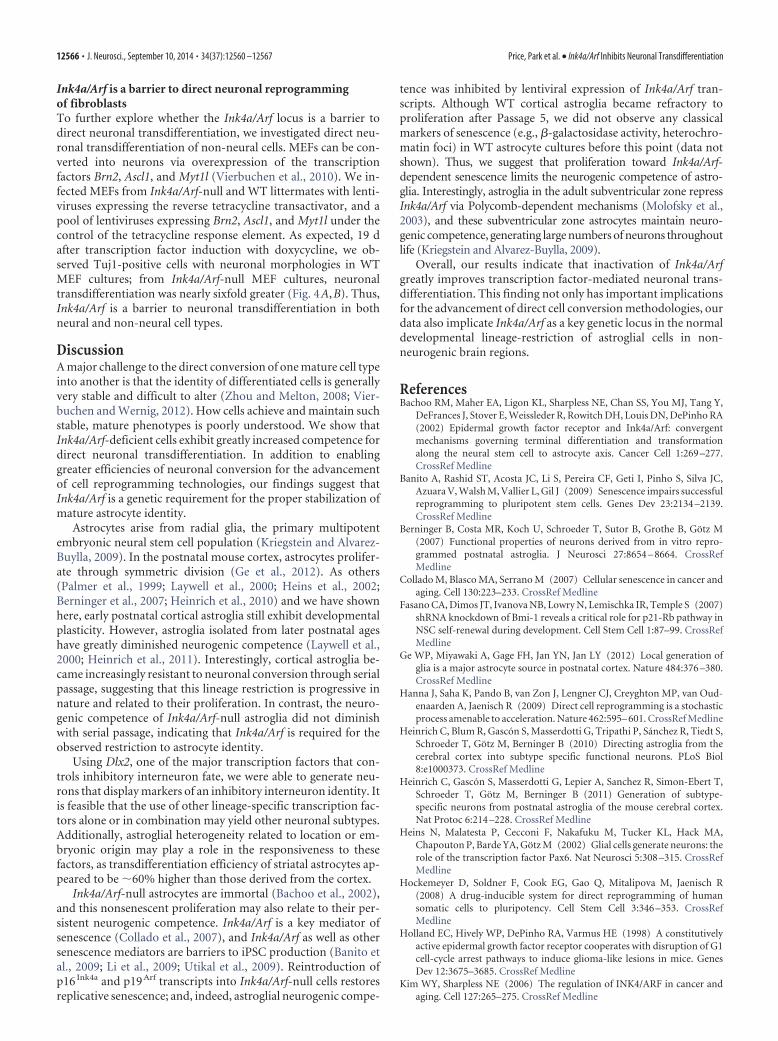
Figure 3. Acute changes to Ink4a/Arf affect transdifferentiation efficiency. A, Trandifferentiation efficiencies of Ink4a/Arf WT astroglia infected with lentivirus expressing RFP and an shRNAtargeting Ink4a/Arf or control. Luc, Luciferase. B, Transdifferentiation efficiencies of Ink4a/Arf WT or null glia infected with virus overexpressing RFP both p16Ink4a and p19Arf or control (alkalinephosphatase). C, qRT-PCR expression analysis of Dlx1, Dlx5, and Dlx6 expression after LV-Dlx2-GFP infection after differentiation. D, qRT-PCR expression analysis of Dlx1, Dlx5, and Dlx6 expressionafter LV-Dlx2-GFP infection before differentiation. *p � 0.05. **p � 0.01. Error bars indicate SD of triplicate reactions. E, Summary of microarray expression data for genes calculated to be greaterthan twofold different between Ink4a/Arf WT or null glial cultures.
12564 • J. Neurosci., September 10, 2014 • 34(37):12560 –12567 Price, Park et al. • Ink4a/Arf Inhibits Neuronal Transdifferentiation

GFAP-positive astroglia (Holland et al., 1998). When infectedwith LV/EnvA-Dlx2-GFP, Gtv-a;Ink4a/Arf-null astroglial cul-tures exhibited �10-fold higher neuronal differentiation effi-ciency compared with Gtv-a;Ink4a/Arf/ control cultures (Fig.2B). When infected with the LV/EnvA-GFP control virus, Gtv-a;Ink4a/Arf-null cells did not give rise to neurons, indicating thatthe low levels of neurogenesis observed in Ink4a/Arf-null corticalcultures without enforced Dlx2 expression (Fig. 1B) likely origi-nated from a GFAP-negative cell population. Together, thesedata indicate that Ink4a/Arf deficiency facilitates neuronal trans-differentiation from GFAP-positive astroglia.
Neurons induced from Ink4a/Arf-deficient cells fireaction potentialsNeuronal cells induced from Ink4a/Arf-null cells by LV-Dlx2-GFP were immunopositive for neuronal antigen NeuN (399 of857 Tuj1 cells; 46.6%) as well as calretinin (40 of 169 Tuj1
cells; 23.7%) and GAD67 (45 of 169 Tuj1 cells; 26.6%) (Fig.2C). To further characterize their neuronal identity, we usedelectrophysiological analysis (Fig. 2D–G). As in WT astroglia,Dlx2 alone was sufficient to generate neurons in Ink4a/Arf-null cultures that were able to fire action potentials; however,after 2–3 weeks of differentiation, a large proportion of trans-differentiated cells exhibited immature action potential firingpatterns (few spikes, Fig. 2E). In WT postnatal astroglia, co-expression of proneural Mash1 (Ascl1) with Dlx2 promotesfurther neuronal maturation (Heinrich et al., 2010). We there-fore coexpressed Mash1 and Dlx2 from lentiviral vectors inInk4a/Arf-null cells and found that GFP-positive neuronalcells exhibited repetitive action potential firing patterns undercurrent clamp, as well as large inward Na currents undervoltage clamp, and both were sensitive to the Na channelblocker TTX (Fig. 2 E, F ). Indeed, Ink4a/Arf-null astroglialcultures transduced with both Mash1 and Dlx2 lentiviral vec-tors produced a greater proportion of neuronal cells with re-petitive spiking than Dlx2 alone (Fig. 2G).
Acute changes to Ink4a/Arf expression regulateneurogenic competenceWe next investigated whether acute inhibition of Ink4a/Arfexpression can increase astroglial neurogenic competence.Lentiviruses encoding shRNAs targeting both Ink4a and Arf(LV-sh-Ink4a/Arf-mCherry) reduced their transcript levels in
passage 1 cortical astroglial cells by �80% after 2 d relative tocontrol LV-sh-luciferase-mCherry; these cultures were nextinfected with LV-Dlx2-GFP or LV-GFP, and the cellular phe-notype of double-infected cells (mCherry/GFP-positive) wasquantified after differentiation. Cells transduced with LV-sh-Ink4a/Arf-mCherry exhibited �3-fold greater neuronal differentia-tion compared with LV-sh-luciferase-mCherry control (Fig. 3A).
To determine whether enforced expression of Ink4a and Arfwould conversely inhibit neuronal transdifferentiation in Ink4a/Arf-null astroglia, we infected Ink4a/Arf-null and WT passage 1cortical astroglial cultures with lentivirus encoding p16 Ink4a andp19 Arf cDNAs (LV-Ink4a-P2A-Arf-mCherry) or LV-mCherrycontrol vectors and assessed the efficiency of neuronal transdif-ferentiation induced by LV-Dlx2-GFP. Reexpression of Ink4a/Arfreduced the number of Tuj1-positive neuronal cells by �2.5-foldin both Ink4a/Arf-null and WT cells (Fig. 3B). Thus, acutechanges to Ink4a and Arf transcript levels can rapidly alter theneurogenic competence of astroglia.
Ink4a/Arf-null glia cultures more efficiently activateDlx2-dependent transcriptionTo begin to investigate why Ink4a/Arf deficiency enhances theneurogenic competence of postnatal astroglia, we compared theInk4a/Arf-null transcriptome to that of WT controls. Surpris-ingly, in Ink4a/Arf-null cultures, only 27 genes were differentiallyexpressed by �2-fold (Fig. 3E).
In normal development, DLX2 upregulates expression of boththe Dlx1/2 and Dlx5/6 bigene clusters, which are key regulators ofinterneuron development (Panganiban and Rubenstein, 2002).To determine whether Dlx2 overexpression can activate the tran-scription of these DLX2 target genes, we analyzed gene expressionduring LV-Dlx2-GFP-induced neuronal transdifferentiation.Three days after differentiation with LV-Dlx2-GFP, Ink4a/Arf-null cultures exhibited �10-fold higher levels of Dlx1, Dlx5, andDlx6 (Fig. 3C). To analyze gene expression related to the enforcedexpression of Dlx2 rather than the resultant neuronal differenti-ation, we analyzed cultures before the onset of neurogenesis.Even before the emergence of Tuj1-positive cells, LV-Dlx2-GFPinduced nearly sixfold higher levels of Dlx1 and Dlx5/6 expres-sion in Ink4a/Arf-null cultures (Fig. 3D). Thus, Ink4a/Arf-deficient astroglia are more competent to upregulate DLX2downstream targets.
0.0
0.5
1.0
2.0***
-/-+/+- + +
Ink4a/ArfDoxycycline
-/-+/+-
GFP Tuj1 Merge
LV-Brn2, Myt1l, Ascl1A B
Ink4a/Arf +/+
Ink4a/Arf -/-
1.5%
Tuj
1+, D
AP
I+ /
DA
PI
Figure 4. Ink4a/Arf is a barrier to fibroblast neuronal reprogramming. A, Transdifferentiation efficiencies from Ink4a/Arf-null and WT MEFs infected with lentivirus expressing Brn2, Myt1l, andAscl1 with or without induction by doxycycline. B, Immunocytochemistry for Tuj1 and GFP from MEF cultures 7 d after infection (B). ***p � 0.001. Scale bars, 40 �m.
Price, Park et al. • Ink4a/Arf Inhibits Neuronal Transdifferentiation J. Neurosci., September 10, 2014 • 34(37):12560 –12567 • 12565
Ink4a/Arf is a barrier to direct neuronal reprogrammingof fibroblastsTo further explore whether the Ink4a/Arf locus is a barrier todirect neuronal transdifferentiation, we investigated direct neu-ronal transdifferentiation of non-neural cells. MEFs can be con-verted into neurons via overexpression of the transcriptionfactors Brn2, Ascl1, and Myt1l (Vierbuchen et al., 2010). We in-fected MEFs from Ink4a/Arf-null and WT littermates with lenti-viruses expressing the reverse tetracycline transactivator, and apool of lentiviruses expressing Brn2, Ascl1, and Myt1l under thecontrol of the tetracycline response element. As expected, 19 dafter transcription factor induction with doxycycline, we ob-served Tuj1-positive cells with neuronal morphologies in WTMEF cultures; from Ink4a/Arf-null MEF cultures, neuronaltransdifferentiation was nearly sixfold greater (Fig. 4A,B). Thus,Ink4a/Arf is a barrier to neuronal transdifferentiation in bothneural and non-neural cell types.
DiscussionA major challenge to the direct conversion of one mature cell typeinto another is that the identity of differentiated cells is generallyvery stable and difficult to alter (Zhou and Melton, 2008; Vier-buchen and Wernig, 2012). How cells achieve and maintain suchstable, mature phenotypes is poorly understood. We show thatInk4a/Arf-deficient cells exhibit greatly increased competence fordirect neuronal transdifferentiation. In addition to enablinggreater efficiencies of neuronal conversion for the advancementof cell reprogramming technologies, our findings suggest thatInk4a/Arf is a genetic requirement for the proper stabilization ofmature astrocyte identity.
Astrocytes arise from radial glia, the primary multipotentembryonic neural stem cell population (Kriegstein and Alvarez-Buylla, 2009). In the postnatal mouse cortex, astrocytes prolifer-ate through symmetric division (Ge et al., 2012). As others(Palmer et al., 1999; Laywell et al., 2000; Heins et al., 2002;Berninger et al., 2007; Heinrich et al., 2010) and we have shownhere, early postnatal cortical astroglia still exhibit developmentalplasticity. However, astroglia isolated from later postnatal ageshave greatly diminished neurogenic competence (Laywell et al.,2000; Heinrich et al., 2011). Interestingly, cortical astroglia be-came increasingly resistant to neuronal conversion through serialpassage, suggesting that this lineage restriction is progressive innature and related to their proliferation. In contrast, the neuro-genic competence of Ink4a/Arf-null astroglia did not diminishwith serial passage, indicating that Ink4a/Arf is required for theobserved restriction to astrocyte identity.
Using Dlx2, one of the major transcription factors that con-trols inhibitory interneuron fate, we were able to generate neu-rons that display markers of an inhibitory interneuron identity. Itis feasible that the use of other lineage-specific transcription fac-tors alone or in combination may yield other neuronal subtypes.Additionally, astroglial heterogeneity related to location or em-bryonic origin may play a role in the responsiveness to thesefactors, as transdifferentiation efficiency of striatal astrocytes ap-peared to be �60% higher than those derived from the cortex.
Ink4a/Arf-null astrocytes are immortal (Bachoo et al., 2002),and this nonsenescent proliferation may also relate to their per-sistent neurogenic competence. Ink4a/Arf is a key mediator ofsenescence (Collado et al., 2007), and Ink4a/Arf as well as othersenescence mediators are barriers to iPSC production (Banito etal., 2009; Li et al., 2009; Utikal et al., 2009). Reintroduction ofp16 Ink4a and p19 Arf transcripts into Ink4a/Arf-null cells restoresreplicative senescence; and, indeed, astroglial neurogenic compe-
tence was inhibited by lentiviral expression of Ink4a/Arf tran-scripts. Although WT cortical astroglia became refractory toproliferation after Passage 5, we did not observe any classicalmarkers of senescence (e.g., �-galactosidase activity, heterochro-matin foci) in WT astrocyte cultures before this point (data notshown). Thus, we suggest that proliferation toward Ink4a/Arf-dependent senescence limits the neurogenic competence of astro-glia. Interestingly, astroglia in the adult subventricular zone repressInk4a/Arf via Polycomb-dependent mechanisms (Molofsky et al.,2003), and these subventricular zone astrocytes maintain neuro-genic competence, generating large numbers of neurons throughoutlife (Kriegstein and Alvarez-Buylla, 2009).
Overall, our results indicate that inactivation of Ink4a/Arfgreatly improves transcription factor-mediated neuronal trans-differentiation. This finding not only has important implicationsfor the advancement of direct cell conversion methodologies, ourdata also implicate Ink4a/Arf as a key genetic locus in the normaldevelopmental lineage-restriction of astroglial cells in non-neurogenic brain regions.
ReferencesBachoo RM, Maher EA, Ligon KL, Sharpless NE, Chan SS, You MJ, Tang Y,
DeFrances J, Stover E, Weissleder R, Rowitch DH, Louis DN, DePinho RA(2002) Epidermal growth factor receptor and Ink4a/Arf: convergentmechanisms governing terminal differentiation and transformationalong the neural stem cell to astrocyte axis. Cancer Cell 1:269 –277.CrossRef Medline
Banito A, Rashid ST, Acosta JC, Li S, Pereira CF, Geti I, Pinho S, Silva JC,Azuara V, Walsh M, Vallier L, Gil J (2009) Senescence impairs successfulreprogramming to pluripotent stem cells. Genes Dev 23:2134 –2139.CrossRef Medline
Berninger B, Costa MR, Koch U, Schroeder T, Sutor B, Grothe B, Gotz M(2007) Functional properties of neurons derived from in vitro repro-grammed postnatal astroglia. J Neurosci 27:8654 – 8664. CrossRefMedline
Collado M, Blasco MA, Serrano M (2007) Cellular senescence in cancer andaging. Cell 130:223–233. CrossRef Medline
Fasano CA, Dimos JT, Ivanova NB, Lowry N, Lemischka IR, Temple S (2007)shRNA knockdown of Bmi-1 reveals a critical role for p21-Rb pathway inNSC self-renewal during development. Cell Stem Cell 1:87–99. CrossRefMedline
Ge WP, Miyawaki A, Gage FH, Jan YN, Jan LY (2012) Local generation ofglia is a major astrocyte source in postnatal cortex. Nature 484:376 –380.CrossRef Medline
Hanna J, Saha K, Pando B, van Zon J, Lengner CJ, Creyghton MP, van Oud-enaarden A, Jaenisch R (2009) Direct cell reprogramming is a stochasticprocess amenable to acceleration. Nature 462:595– 601. CrossRef Medline
Heinrich C, Blum R, Gascon S, Masserdotti G, Tripathi P, Sanchez R, Tiedt S,Schroeder T, Gotz M, Berninger B (2010) Directing astroglia from thecerebral cortex into subtype specific functional neurons. PLoS Biol8:e1000373. CrossRef Medline
Heinrich C, Gascon S, Masserdotti G, Lepier A, Sanchez R, Simon-Ebert T,Schroeder T, Gotz M, Berninger B (2011) Generation of subtype-specific neurons from postnatal astroglia of the mouse cerebral cortex.Nat Protoc 6:214 –228. CrossRef Medline
Heins N, Malatesta P, Cecconi F, Nakafuku M, Tucker KL, Hack MA,Chapouton P, Barde YA, Gotz M (2002) Glial cells generate neurons: therole of the transcription factor Pax6. Nat Neurosci 5:308 –315. CrossRefMedline
Hockemeyer D, Soldner F, Cook EG, Gao Q, Mitalipova M, Jaenisch R(2008) A drug-inducible system for direct reprogramming of humansomatic cells to pluripotency. Cell Stem Cell 3:346 –353. CrossRefMedline
Holland EC, Hively WP, DePinho RA, Varmus HE (1998) A constitutivelyactive epidermal growth factor receptor cooperates with disruption of G1cell-cycle arrest pathways to induce glioma-like lesions in mice. GenesDev 12:3675–3685. CrossRef Medline
Kim WY, Sharpless NE (2006) The regulation of INK4/ARF in cancer andaging. Cell 127:265–275. CrossRef Medline
12566 • J. Neurosci., September 10, 2014 • 34(37):12560 –12567 Price, Park et al. • Ink4a/Arf Inhibits Neuronal Transdifferentiation

Kriegstein A, Alvarez-Buylla A (2009) The glial nature of embryonic andadult neural stem cells. Annu Rev Neurosci 32:149 –184. CrossRefMedline
Laywell ED, Rakic P, Kukekov VG, Holland EC, Steindler DA (2000) Iden-tification of a multipotent astrocytic stem cell in the immature and adultmouse brain. Proc Natl Acad Sci U S A 97:13883–13888. CrossRefMedline
Lewis BC, Chinnasamy N, Morgan RA, Varmus HE (2001) Development ofan avian leukosis-sarcoma virus subgroup A pseudotyped lentiviral vec-tor. J Virol 75:9339 –9344. CrossRef Medline
Li H, Collado M, Villasante A, Strati K, Ortega S, Canamero M, Blasco MA,Serrano M (2009) The Ink4/Arf locus is a barrier for iPS cell reprogram-ming. Nature 460:1136 –1139. CrossRef Medline
Lim DA, Huang YC, Swigut T, Mirick AL, Garcia-Verdugo JM, Wysocka J,Ernst P, Alvarez-Buylla A (2009) Chromatin remodelling factor Mll1 isessential for neurogenesis from postnatal neural stem cells. Nature 458:529 –533. CrossRef Medline
Molofsky AV, Pardal R, Iwashita T, Park IK, Clarke MF, Morrison SJ (2003)Bmi-1 dependence distinguishes neural stem cell self-renewal from pro-genitor proliferation. Nature 425:962–967. CrossRef Medline
Palmer TD, Markakis EA, Willhoite AR, Safar F, Gage FH (1999) Fibro-blast growth factor-2 activates a latent neurogenic program in neuralstem cells from diverse regions of the adult CNS. J Neurosci 19:8487–8497. Medline
Panganiban G, Rubenstein JL (2002) Developmental functions of the Dis-tal-less/Dlx homeobox genes. Development 129:4371– 4386. Medline
Park DH, Hong SJ, Salinas RD, Liu SJ, Sun SW, Sgualdino J, Testa G, MatzukMM, Iwamori N, Lim DA (2014) Activation of neuronal gene expres-
sion by the JMJD3 demethylase is required for postnatal and adult brainneurogenesis. Cell Rep. In press. CrossRef
Ramos AD, Diaz A, Nellore A, Delgado RN, Park KY, Gonzales-Roybal G,Oldham MC, Song JS, Lim DA (2013) Integration of genome-wide ap-proaches identifies lncRNAs of adult neural stem cells and their progenyin vivo. Cell Stem Cell 12:616 – 628. CrossRef Medline
Robel S, Berninger B, Gotz M (2011) The stem cell potential of glia: lessonsfrom reactive gliosis. Nat Rev 12:88 –104. CrossRef Medline
Serrano M, Lee H, Chin L, Cordon-Cardo C, Beach D, DePinho RA (1996)Role of the INK4a locus in tumor suppression and cell mortality. Cell85:27–37. CrossRef Medline
Utikal J, Polo JM, Stadtfeld M, Maherali N, Kulalert W, Walsh RM, Khalil A,Rheinwald JG, Hochedlinger K (2009) Immortalization eliminates aroadblock during cellular reprogramming into iPS cells. Nature 460:1145–1148. CrossRef Medline
Ventura A, Meissner A, Dillon CP, McManus M, Sharp PA, Van Parijs L,Jaenisch R, Jacks T (2004) Cre-lox-regulated conditional RNA interfer-ence from transgenes. Proc Natl Acad Sci U S A 101:10380 –10385.CrossRef Medline
Vierbuchen T, Wernig M (2012) Molecular roadblocks for cellular repro-gramming. Mol Cell 47:827– 838. CrossRef Medline
Vierbuchen T, Ostermeier A, Pang ZP, Kokubu Y, Sudhof TC, Wernig M(2010) Direct conversion of fibroblasts to functional neurons by definedfactors. Nature 463:1035–1041. CrossRef Medline
Zhang Y, Barres BA (2010) Astrocyte heterogeneity: an underappreciated topicin neurobiology. Curr Opin Neurobiol 20:588–594. CrossRef Medline
Zhou Q, Melton DA (2008) Extreme makeover: converting one cell intoanother. Cell Stem Cell 3:382–388. CrossRef Medline
Price, Park et al. • Ink4a/Arf Inhibits Neuronal Transdifferentiation J. Neurosci., September 10, 2014 • 34(37):12560 –12567 • 12567