brief report xenografted islet cell clusters from...
TRANSCRIPT
Xenografted Islet Cell Clusters From INSLEA29YTransgenic Pigs Rescue Diabetes and Prevent ImmuneRejection in Humanized MiceNikolai Klymiuk,
1Lelia van Buerck,
2Andrea Bähr,
1Monika Offers,
2Barbara Kessler,
1
Annegret Wuensch,1Mayuko Kurome,
1Michael Thormann,
3Katharina Lochner,
2Hiroshi Nagashima,
4
Nadja Herbach,5Rüdiger Wanke,
5Jochen Seissler,
2and Eckhard Wolf
1
Islet transplantation is a potential treatment for type 1 diabetes,but the shortage of donor organs limits its routine application. Aspotential donor animals, we generated transgenic pigs expressingLEA29Y, a high-affinity variant of the T-cell costimulation inhibitorCTLA-4Ig, under the control of the porcine insulin gene promoter.Neonatal islet cell clusters (ICCs) from INSLEA29Y transgenic(LEA-tg) pigs and wild-type controls were transplanted intostreptozotocin-induced hyperglycemic NOD-scid IL2Rgnull mice.Cloned LEA-tg pigs are healthy and exhibit a strong b-cell–specifictransgene expression. LEA-tg ICCs displayed the same potential tonormalize glucose homeostasis as wild-type ICCs after transplan-tation. After adoptive transfer of human peripheral blood mononu-clear cells, transplanted LEA-tg ICCs were completely protectedfrom rejection, whereas reoccurrence of hyperglycemia was ob-served in 80% of mice transplanted with wild-type ICCs. In thecurrent study, we provide the first proof-of-principle report on trans-genic pigs with b-cell–specific expression of LEA29Y and their suc-cessful application as donors in a xenotransplantation model. Thisapproach may represent a major step toward the development of anovel strategy for pig-to-human islet transplantation without side ef-fects of systemic immunosuppression.Diabetes 61:1527–1532, 2012
Type 1 diabetes is a chronic metabolic disease as-sociated with development of severe complica-tions (1). It has been shown that type 1 diabetescan be cured by the transplantation of the pancreas
or isolated islets of Langerhans. Nonetheless, the success ofpancreas and islet transplantation is limited by the shortageof organ donors and the need for systemic immunosuppres-sive therapy (2) and is therefore restricted to few patients (3).
Limited availability of human donor organs may beovercome by the use of pigs as organ donors. Pig-to-human
xenotransplantation faces the problem of strong rejection,predominantly by direct T-cell recognition of pig majorhistocompatibility complex and indirect T-cell response toxenogeneic antigens presented by the recipient antigen-presenting cells (4).
Recent advances in immunosuppressive therapies pro-vided evidence that transplanted porcine islets can promotethe long-lasting cure of diabetes in nonhuman primates(5–7). However, the currently used intensive immunosup-pressive regimen in pig islet transplantation may have severeside effects in humans and cannot be transferred into clini-cal practice. Blockade of the B7/CD28 costimulatory path-way by LEA29Y, a high-affinity variant of the CTLA-4Igfusion protein (8), has been shown to be effective in clinicaltrials following kidney transplantation (9,10) and in porcineislet transplantation studies (5,7,11). Thus, local expressionof LEA29Y restricted to the transplantation site may repre-sent an innovative approach to protect grafted islets fromxenogeneic immune rejection without the side effects ofsystemic immunosuppression.
Therefore, we chose to generate transgenic pigs express-ing LEA29Y specifically in pancreatic b-cells. We demon-strate for the first time the potential of neonatal INSLEA29Ytransgenic (LEA-tg) islet clusters to normalize blood glucoselevels and evaluate the inhibition of human–anti-pig rejectionin a humanized NOD-scid IL2Rgnull (NSG) model.
RESEARCH DESIGN AND METHODS
All experiments were approved by the local animal welfare authority. NSG micewere obtained from The Jackson Laboratory. For generation of INSLEA-tg pigs,the coding sequence for LEA29Y was cloned into a b-cell–specific expressionvector (12) with 1.3-kb upstream regions, exon 1 and intron 1 of the porcineinsulin gene, and a poly-adenylation cassette of the bovine growth hormonegene. The vector was completed by linking the INSLEA construct to a floxedneomycin resistance cassette (13). Porcine fetal fibroblasts (PFF#14; 13 106)were nucleofected (Nucleofector Technology, Lonza, Germany). Stably nucle-ofected cell clones were used as donors for somatic cell nuclear transfer (14).Embryo transfer was carried out laparoscopically (15). Integration and ex-pression of the transgene was analyzed by Southern blot and immunohisto-chemistry. Donor piglets for transplantation experiments were generated byrecloning, as described previously (13).Isolation and transplantation of neonatal islet cell clusters into
hyperglycemic NSG mice. Islet cell clusters (ICCs) from 1- to 2-day-oldrecloned LEA-tg and wild-type pigs were isolated as previously described (16)and cultured for 6 days at 37°C in RPMI (Biochrom) with 2% human serum al-bumin (Octapharm), 1% antibiotic-antimycotic, 10 mmol/L nicotinamide, and 20nmol/L exendine-4 (Sigma). Insulin content in ICCs was determined by enzyme-linked immunosorbent assay (ELISA) (Millipore) (17). A total of 2,500 clustersper mouse were transplanted under the kidney capsule of streptozotocin-induced diabetic (180 mg/kg; Sigma) NSG mice (blood glucose .350 mg/dL).Characterization of graft function. Neonatal ICCs require a 6- to 8-week invivo maturation period until physiological glucose-dependent insulin secretionhas developed. Animals with blood glucose levels .300 mg/dL received exog-enous insulin subcutaneously (0.5 IU glargine per day). Mice displaying blood
From the 1Chair for Molecular Animal Breeding and Biotechnology, and Lab-oratory for Functional Genome Analysis (LAFUGA), Gene Center, Ludwig-Maximilians-Universität, Munich, Germany; the 2Diabetes Zentrum,Medizinische Klinik Campus Innenstadt, Klinikum der Ludwig-Maximilians-Universität, Munich, Germany; the 3Department of Cardiac Surgery, Ludwig-Maximilians-Universität, Munich, Germany; the 4Laboratory of DevelopmentalEngineering, Meiji University, Kawasaki, Japan; and the 5Institute of VeterinaryPathology, Center for Clinical Veterinary Medicine, Ludwig-Maximilians-Universität, Munich, Germany.
Corresponding authors: Eckhard Wolf, [email protected], andJochen Seissler, [email protected].
Received 23 September 2011 and accepted 28 January 2012.DOI: 10.2337/db11-1325This article contains Supplementary Data online at http://diabetes
.diabetesjournals.org/lookup/suppl/doi:10.2337/db11-1325/-/DC1.N.K., L.v.B., J.S., and E.W. contributed equally to this work.� 2012 by the American Diabetes Association. Readers may use this article as
long as the work is properly cited, the use is educational and not for profit,and the work is not altered. See http://creativecommons.org/licenses/by-nc-nd/3.0/ for details.
See accompanying commentary, p. 1348.
diabetes.diabetesjournals.org DIABETES, VOL. 61, JUNE 2012 1527
BRIEF REPORT
glucose levels ,150 mg/dL for a period of 5 days were considered normogly-cemic. Intraperitoneal glucose tolerance testing (IPGTT) was performed 10 dayslater using 2 g glucose/kg body weight (18). Porcine serum insulin was de-termined by ELISA (Mercodia) that displayed no cross-reactivity with mouseinsulin. Serum LEA29Y concentrations were determined by sandwich ELISAusing 1 mg/mL monoclonal anti–human-CTLA-4 antibody (Beckmann Coulter)and horseradish peroxidase–conjugated polyclonal rabbit anti-human IgG (Dako).Analyses in humanized mice. To analyze human anti-pig immune response,20 3 106 human peripheral blood mononuclear cells (hPBMCs) from onedonor were transferred intraperitoneally into transplanted normoglycemicNSG mice, as described previously (19). The remaining PBMCs were culturedin X-VIVO 20 medium (CambrexBio Science) supplemented with porcinesplenocyte cell lysate (cytosolic fraction corresponding to 15 3 106 spleno-cytes) added at day 0 and day 3 to activate T cells directed against porcineantigens. After 6 days of culture, 2.5 3 106 primed hPBMCs were injectedintravenously.
Reconstituted mice were monitored daily for the reoccurrence of hypergly-cemia. The investigation period was limited to 29 days because of the de-velopment of graft-versus-host disease. Animals displaying severe hyperglycemia(blood glucose levels .350 mg/dL in two consecutive measurements) werekilled ahead of schedule, whereas normoglycemic mice underwent IPGTT atday 27. To exclude endogenous b-cell regeneration, the graft of normoglycemicmice was removed at day 28 by uninephrectomy. From each animal, blood andspecimen for fluorescence-activated cell sorter (FACS) analysis (FACS Canto;BD Biosciences) and immunohistochemistry (kidney and liver) were taken.Spleen and bone marrow cells were stained using the following fluorochrome-labeled monoclonal antibodies: mouse CD45-FITC, human CD45-APC, ormatched isotype antibodies (eBioscience).Immunohistochemical analyses. Pig organs and graft-bearing kidneys wereprocessed as described previously (20). Serial paraffin sections were stainedwith guinea pig anti-insulin (1:500), rabbit anti-human CD3 (1:100), rabbit anti-human IgG (recognizing the COOH-terminal part of LEA29Y; 1:50), mouse anti-human CD4 (1:20) (Dako), rabbit anti-human CD8 (1:80; Vector), and rabbitanti-human CD45 (1:400; antibodies-online Inc.). As secondary antibodies,horseradish peroxidase–conjugated anti-guinea pig IgG, anti-rabbit IgG, bio-tinylated anti-rabbit IgG or anti-mouse IgG (Dako), and alkaline phosphatase–conjugated anti-guinea pig IgG (Southern Biotech) were used. Chromogens
included Fuchsin+Substrate Chromogen (Dako) or 3,39-diaminobenzidine(DAB) (Kem-En-Tec Diagnostics) (20).Data presentation and statistical analysis. Data represent means and SEM.Statistical analyses were performed using the Student t test or Log-rank test(diabetes reoccurrence). P values ,0.05 were considered significant.
RESULTS
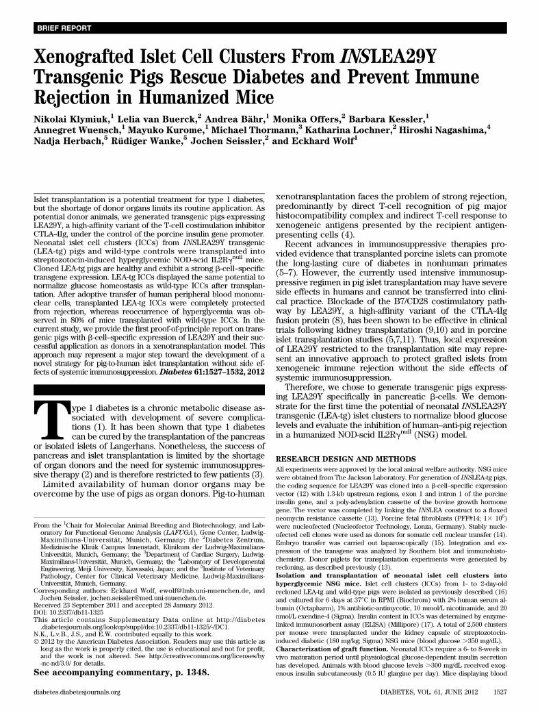
Generation of INSLEA29Y transgenic (LEA-tg) pigs.The INSLEA29Y expression construct (Fig. 1A) was usedfor nucleofection of porcine fetal fibroblasts, and stablecell clones were pooled for nuclear transfer (13). Clonedembryos (n = 216) were transferred to three synchronizedgilts, resulting in two pregnancies with nine born pigletsincluding two stillborn animals. Seven of eight genotypedpiglets were transgenic, each representing a unique founder,as demonstrated by Southern blot analysis (Fig. 1B). Four ofthese animals were killed at the age of 3 months for im-munohistochemical staining of different organs (Fig. 1C).Transgenic pigs displayed a strong LEA29Y staining in thepancreatic islets (Fig. 1C). Recloned animals served as isletdonors or were raised for future breeding purposes. Thesepigs are fertile, have no signs of opportunistic infections, andexhibit normal blood glucose levels.Differentiation and maturation of transplanted ICCs.Mice transplanted with wild-type ICCs (Tx, wt; n = 5) andmice transplanted with ICCs from LEA-tg pigs (Tx, LEA-tg;n = 5) (insulin content 3.2 6 0.9 ng/mg protein) returned tonormoglycemia after 51 6 7 and 43 6 7 days, respectively(Fig. 2A). Normoglycemic mice of both transplantationgroups exhibited a comparable restored glucose tolerance(area under the curve of glucose during IPGTT: Tx, wt:12,121 6 1,303; Tx, LEA-tg: 11,310 6 719) with similar
FIG. 1. Generation of INSLEA29Y transgenic (LEA-tg) pigs. A: The vector consisted of a 1.3-kb regulatory sequence from the porcine INS gene, theLEA29Y coding sequence, and the poly-adenylation box from the bovine GH gene. Regulatory sequences are depicted as lines, whereas exonicstructures are boxed. Untranslated regions are shaded. The selection cassette provides resistance to neomycin. Binding sites for primers areindicated as arrows, and the probe for Southern blot hybridization is shown as a bold line. B: Southern blotting of seven founders was performed onXbaI-digested genomic DNA with a probe binding to the neomycin resistance cassette. C: Immunohistochemical staining for LEA29Y on tissuesections from a neonatal transgenic pig (aged 2 days, pancreas, C2), an adult founder animal (age 3 months; pancreas, liver, lung, kidney, andspleen, C4, 5–8), and from age-matched wild-type control pigs (pancreas, C1, 3). Scale bar: 100 mm. (A high-quality digital representation of thisfigure is available in the online issue.)
XENOGRAFTED INSLEA29Y TRANSGENIC ISLETS
1528 DIABETES, VOL. 61, JUNE 2012 diabetes.diabetesjournals.org

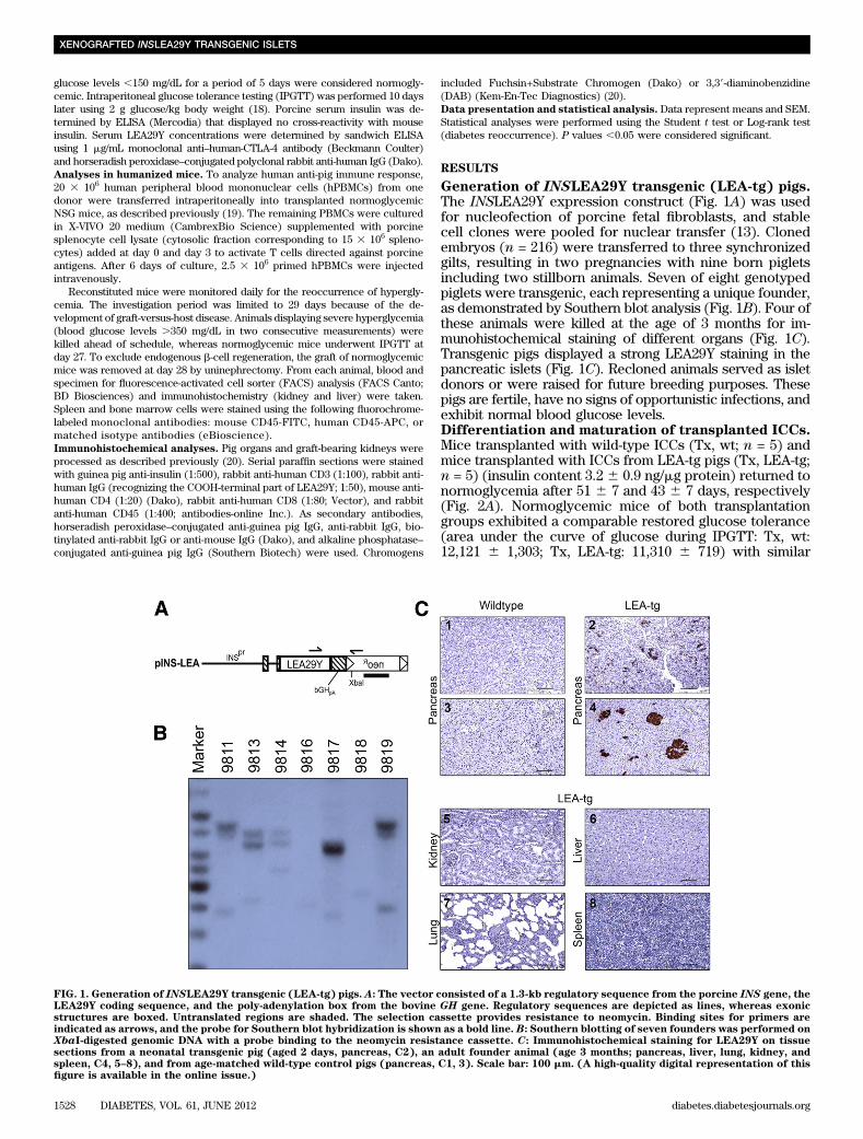
glucose-responsive porcine insulin secretion (Fig. 2B).Immunohistochemical staining of the subcapsular graftrevealed maturation of the transplanted ICCs toward astrongly insulin-expressing endocrine tissue in both trans-plantation groups with LEA29Y transgene expression re-stricted to the grafts of Tx, LEA-tg mice (Fig. 2C and D).LEA29Y concentrations in the plasma of normoglycemic Tx,LEA-tg mice were 270 6 24 ng/mL.INSLEA29Y expression prevents reoccurrence ofhyperglycemia. The finding that transgenic ICCs hadstrong LEA29Y expression and were able to normalizeblood glucose levels raised the question whether these ICCswere protected from graft rejection after reconstitution withhuman PBMCs. The proportion of human CD45+ cells in thespleen and bone marrow was comparable in both trans-plantation groups (Fig. 3A), and histological examinationrevealed identical mononuclear cell infiltration in varioustissues reflecting graft-versus-host disease in both trans-plantation groups (Supplementary Fig. 1). However, at day14 after adoptive transfer of PBMCs, blood glucose levelswere significantly elevated in Tx, wt as compared with Tx,LEA-tg mice (204 6 63 mg/dL vs. 58 6 2 mg/dL; P , 0.05).
Life-table analysis revealed that reoccurrence of hypergly-cemia, defined by blood glucose levels .250 mg/dL, wasabsent in Tx, LEA-tg mice (n = 5), whereas four of fiveanimals (80%) transplanted with wild-type ICCs becamehyperglycemic within the observation period of 29 days(P , 0.05) (Fig. 3B and C). One Tx, wt mouse remainednormoglycemic throughout the observation period, withpreserved glucose tolerance at day 27. In three of four hy-perglycemic mice, porcine serum insulin was below thedetection limit. In contrast, in all mice transplanted withLEA29Y transgenic ICCs, the area under the glucose andinsulin curve during IPGTT was comparable before and 27 dafter transfer of human PBMCs (Fig. 3D). After removal ofthe graft-bearing kidney, all of the diabetes-free animalsreturned to hyperglycemia (Fig. 3B). Histological examina-tion of the graft bearing kidney from wild-type mice whichdeveloped hyperglycemia revealed a massive infiltration ofICCs with human mononuclear cells (CD3+, CD45+, CD4+,and CD8+ cells) and reduced insulin staining. In contrast, inall Tx, LEA-tg mice, ICCs appeared preserved and T-cellinfiltration was restricted to the surrounding tissue and al-most absent within islet clusters (Fig. 4).
FIG. 2. Grafted LEA-tg ICCs display physiological b-cell function. Course of blood glucose levels after transplantation (A), IPGTT (performed 10days after the development of normoglycemia (B), and immunohistochemistry of grafted ICCs (7–9 days after transplantation [C] and 4.0–4.5months after transplantation [normoglycemic animals] [D]) in mice transplanted with wild-type (Tx, wt) and in mice transplanted with LEA-tg (Tx,LEA-tg) ICCs. Mice of both transplantation groups developed stable normoglycemia (A) and restored glucose tolerance (B, bottom), by porcineinsulin secretion (B, top). The area under the curve (AUC) for glucose and insulin (B) during IPGTT was comparable in both transplantationgroups. C: Immunohistochemical staining of serial sections from the transplantation site against insulin and IgG revealed insulin/LEA29Y ex-pression in a minor proportion of ICCs a few days after transplantation. D: In contrast, after the development of normoglycemia, the transplantedcells have differentiated into a widespread insulin-positive stained tissue in both transplantation groups with LEA29Y transgene expression re-stricted to the grafted ICCs from transgenic pigs. Scale bar: 100 mm. n = 5 for each transplantation group. (A high-quality digital representation ofthis figure is available in the online issue.)
N. KLYMIUK AND ASSOCIATES
diabetes.diabetesjournals.org DIABETES, VOL. 61, JUNE 2012 1529

DISCUSSION
Major obstacles in pig-to-human islet transplantation arethe strong xenogeneic immune response and the severeadverse effects of the required intensive immunosuppressiveregimen. To overcome these limitations, we developed anislet donor animal that provides a local immunosuppressiveenvironment within transplanted islets of Langerhans.
The LEA-tg pigs generated in this study express highlevels of LEA29Y, specifically in the b-cells, with no signsof b-cell dysfunction or systemic immunosuppression, suchas increased susceptibility to opportunistic infections. Thisis in contrast to transgenic pigs with ubiquitous porcineCTLA-4Ig expression, which were immune compromisedand died of infections (21). To assess the in vivo b-cellfunction and the immunomodulatory potential of LEA-tgislets, ICCs were transplanted into NSG mice, an estab-lished model for studying human immunity (22,23). After anin vivo maturation period, which is required for immatureICCs to develop physiological insulin secretion (16), miceof both transplantation groups developed complete restora-tion of glucose homeostasis. These findings, togetherwith the strong, colocalized graft staining for insulin andLEA29Y, indicate that LEA29Y expression in b-cells does not
interfere with b-cell development and function. Previoustransplantation studies in rats and nonhuman primates usinghigh doses of belatacept for systemic immunosuppressionalso have shown that costimulatory blockade by LEA29Ydoes not exert any adverse effects on b-cell function (5,11).
After adoptive transfer of a human immune system, weobserved that 80% of Tx, wt animals developed hyperglyce-mia, whereas all Tx, LEA-tg mice were protected from graftrejection and showed preserved b-cell function. The deve-lopment of hyperglycemia after xenograft removal indicatedthat glucose homeostasis was completely maintained bygraft-derived porcine insulin secretion, excluding thepossibility of endogenous b-cell regeneration. In Tx, LEA-tg mice, human lymphocyte accumulation was observed inthe periphery of the transplantation site and in the kidney,but LEA29Y-tg ICCs were protected from infiltration. Thus,our study shows for the first time that local expression ofLEA29Y results in a prolonged islet xenograft function,supporting the hypothesis that inhibition of costimulationis able to modulate allo- and xenoimmunity (6,7). Thesedata are in line with findings from Zhai et al. (24), de-monstrating a prolonged survival of adenoviral vector–transduced pig islets expressing porcine CTLA-4Ig. LEA29Y
FIG. 3. LEA29Y expression prevents reoccurrence of hyperglycemia after transfer of human PBMCs. A: Engraftment of human PBMCs (as in-dicated by FACS staining for human CD45
+cells in both spleen and bone marrow cells) did not significantly differ between mice transplanted with
wild-type (Tx, wt) and in mice transplanted with LEA-tg (Tx, LEA-tg) ICCs. B: Four of five Tx, wt mice, but no Tx, LEA-tg mice, developed hy-perglycemia within 28 days after human PBMC transfer. After removal of the graft-bearing kidney (uninephrectomy, Unx) of normoglycemicanimals, all mice returned to severe hyperglycemia, indicating the absence of endogenous b-cell regeneration. C: Life-table analysis revealeda significantly (P = 0.016) higher proportion of hyperglycemia reoccurrence in Tx, wt as compared with Tx, LEA-tg mice. D: Furthermore, the areaunder the curve (AUC) of glucose and insulin during the IPGTT was unchanged before and 27 days after the transfer of human PBMCs in Tx,LEA-tg mice. n = 4–5 animals for each transplantation group. †One animal died at day 26 as a result of graft-versus-host disease.
XENOGRAFTED INSLEA29Y TRANSGENIC ISLETS
1530 DIABETES, VOL. 61, JUNE 2012 diabetes.diabetesjournals.org

serum concentrations in recipients of LEA-tg ICCs were~100–150 times lower as compared with systemic LEA29Ytreatment in clinical trials (belatacept, BMS-224818), sug-gesting that graft protection is primarily mediated by localand not systemic LEA29Y immunomodulatory effects.
In conclusion, the present proof-of-principle study de-monstrates that the availability of transgenic pigs ex-pressing LEA29Y in b-cells may represent a major stepforward to overcome the immunological barrier to isletxenotransplantation. Additional transplantation studies usinglarger groups of mice with a stably transferred human im-mune system (22) will be conducted to investigate the long-term effects of LEA29Y transgenic islets on xenogeneic graftrejection.
ACKNOWLEDGMENTS
This study was supported by the Deutsche Forschungs-gemeinschaft (DFG Transregio Research Unit FOR 535“Xenotransplantation” [SE 725/5-3 and WO 685/10-3])and BMBF (01GN0949).
No potential conflicts of interest relevant to this articlewere reported.
N.K. and E.W. researched data (generation and character-ization of transgenic pigs) and wrote the manuscript. L.v.B.and J.S. researched data (isolation of ICCs, transplantation,FACS, and immunohistochemical analyses) and wrote themanuscript. A.B. and B.K. researched data (characterizationof transgenic pigs and pig pancreas extraction) and re-viewed and edited the manuscript. M.O. and K.L. researcheddata (ELISA, animal monitoring, and FACS analyses) andreviewed and edited the manuscript. M.K. researcheddata (cloning and recloning of transgenic pigs and SCNT)and reviewed and edited the manuscript. M.T. researcheddata (large animal surgery) and reviewed and edited themanuscript. H.N. researched data (SCNT) and reviewedand edited the manuscript. N.H. and R.W. researched data
(immunohistochemical analyses) and reviewed and editedthe manuscript. J.S. is the guarantor of this work and, assuch, had full access to all the data in the study and takesresponsibility for the integrity of the data and the accuracyof the data analysis.
The authors thank the staff of the animal facilities forthe excellent assistance and J. Postrach (Department ofCardiac Surgery, University of Munich) for his dedicatedsupport.
REFERENCES
1. Nathan DM, Zinman B, Cleary PA, et al.; Diabetes Control and Complica-tions Trial/Epidemiology of Diabetes Interventions and Complications(DCCT/EDIC) Research Group. Modern-day clinical course of type 1 di-abetes mellitus after 30 years’ duration: the Diabetes Control and Com-plications Trial/Epidemiology of Diabetes Interventions and Complicationsand Pittsburgh Epidemiology of Diabetes Complications Experience(1983–2005). Arch Intern Med 2009;169:1307–1316
2. Robertson RP. Islet transplantation a decade later and strategies for fillinga half-full glass. Diabetes 2010;59:1285–1291
3. CITR Research Group. 2007 update on allogeneic islet transplantationfrom the Collaborative Islet Transplant Registry (CITR). Cell Transplant2009;18:753–767
4. Clarkson MR, Sayegh MH. T-cell costimulatory pathways in allograft re-jection and tolerance. Transplantation 2005;80:555–563
5. Cardona K, Korbutt GS, Milas Z, et al. Long-term survival of neonatalporcine islets in nonhuman primates by targeting costimulation pathways.Nat Med 2006;12:304–306
6. Hering BJ, Wijkstrom M, Graham ML, et al. Prolonged diabetes reversalafter intraportal xenotransplantation of wild-type porcine islets in im-munosuppressed nonhuman primates. Nat Med 2006;12:301–303
7. Cardona K, Milas Z, Strobert E, et al. Engraftment of adult porcine isletxenografts in diabetic nonhuman primates through targeting of costimulationpathways. Am J Transplant 2007;7:2260–2268
8. Larsen CP, Pearson TC, Adams AB, et al. Rational development of LEA29Y(belatacept), a high-affinity variant of CTLA4-Ig with potent immunosup-pressive properties. Am J Transplant 2005;5:443–453
9. Durrbach A, Pestana JM, Pearson T, et al. A phase III study of belataceptversus cyclosporine in kidney transplants from extended criteria donors(BENEFIT-EXT study). Am J Transplant 2010;10:547–557
FIG. 4. LEA29Y expressing ICCs are almost completely preserved from mononuclear cell infiltration. Characteristic insulin (red) and CD3+,
CD45+, CD4
+, and CD8
+cell (brown) staining pattern of serial sections from the transplantation sites of a mouse transplanted with wild-type ICCs
(Tx, wt; rejection at day 12 after PBMC transfer) vs. an animal with LEA29Y transgenic ICCs (Tx, LEA-tg, day 29 post PBMC transfer). In Tx,wt only few ICCs were detectable with vast T-cell (CD3
+, CD4
+, and CD8
+) and CD45
+cell infiltration in the graft region. In contrast, Tx LEA-tg
ICCs appeared completely preserved with T-cell and leukocyte accumulation restricted to the subcapsular area (day 29 after Tx). The localizationof tissue sections shown in the insets is marked by an asterisk. Scale bar: 100 mm, insets: scale bar 20 mm. (A high-quality digital representation ofthis figure is available in the online issue.)
N. KLYMIUK AND ASSOCIATES
diabetes.diabetesjournals.org DIABETES, VOL. 61, JUNE 2012 1531
10. Vincenti F, Charpentier B, Vanrenterghem Y, et al. A phase III study ofbelatacept-based immunosuppression regimens versus cyclosporine in renaltransplant recipients (BENEFIT study). Am J Transplant 2010;10:535–546
11. Tchorsh-Yutsis D, Hecht G, Aronovich A, et al. Pig embryonic pancreatictissue as a source for transplantation in diabetes: transient treatment withanti-LFA1, anti-CD48, and FTY720 enables long-term graft maintenance inmice with only mild ongoing immunosuppression. Diabetes 2009;58:1585–1594
12. Grzech M, Dahlhoff M, Herbach N, et al. Specific transgene expression inmouse pancreatic beta-cells under the control of the porcine insulin pro-moter. Mol Cell Endocrinol 2010;315:219–224
13. Aigner B, Renner S, Kessler B, et al. Transgenic pigs as models fortranslational biomedical research. J Mol Med (Berl) 2010;88:653–664
14. Kurome M, Ueda H, Tomii R, Naruse K, Nagashima H. Production oftransgenic-clone pigs by the combination of ICSI-mediated gene transferwith somatic cell nuclear transfer. Transgenic Res 2006;15:229–240
15. Besenfelder U, Mödl J, Müller M, Brem G. Endoscopic embryo collectionand embryo transfer into the oviduct and the uterus of pigs. Theriogenology1997;47:1051–1060
16. Korbutt GS, Elliott JF, Ao Z, Smith DK, Warnock GL, Rajotte RV. Largescale isolation, growth, and function of porcine neonatal islet cells. J ClinInvest 1996;97:2119–2129
17. Pamir N, Lynn FC, Buchan AMJ, et al. Glucose-dependent in-sulinotropic polypeptide receptor null mice exhibit compensatory
changes in the enteroinsular axis. Am J Physiol Endocrinol Metab 2003;284:E931–E939
18. Ayala JE, Samuel VT, Morton GJ, et al.; NIH Mouse Metabolic PhenotypingCenter Consortium. Standard operating procedures for describing andperforming metabolic tests of glucose homeostasis in mice. Dis ModelMech 2010;3:525–534
19. Hesselton RM, Koup RA, Cromwell MA, Graham BS, Johns M, Sullivan JL.Human peripheral blood xenografts in the SCID mouse: characterization ofimmunologic reconstitution. J Infect Dis 1993;168:630–640
20. Renner S, Fehlings C, Herbach N, et al. Glucose intolerance and reducedproliferation of pancreatic beta-cells in transgenic pigs with impairedglucose-dependent insulinotropic polypeptide function. Diabetes 2010;59:1228–1238
21. Phelps CJ, Ball SF, Vaught TD, et al. Production and characterization oftransgenic pigs expressing porcine CTLA4-Ig. Xenotransplantation 2009;16:477–485
22. Brehm MA, Shultz LD, Greiner DL. Humanized mouse models to studyhuman diseases. Curr Opin Endocrinol Diabetes Obes 2010;17:120–125
23. King M, Pearson T, Rossini AA, Shultz LD, Greiner DL. Humanized mice forthe study of type 1 diabetes and beta cell function. Ann N Y Acad Sci 2008;1150:46–53
24. Zhai C, Yu L, Zhu H, Tian M, Xiaogang Z, Bo W. Porcine CTLA4-Ig prolongislet xenografts in rats by downregulating the direct pathway of T-cellactivation. Xenotransplantation 2011;18:40–45
XENOGRAFTED INSLEA29Y TRANSGENIC ISLETS
1532 DIABETES, VOL. 61, JUNE 2012 diabetes.diabetesjournals.org