breed associated eye diseases · pdf filebreed associated eye diseases ... the posterior...
TRANSCRIPT

Animal Eye Centerof New Jersey
Little Falls, New Jersey888.722.7200
BreedAssociated Eye DiseasesQuick Reference Guide

1
Foreword
Inherited or breed-related ocular diseases of dogs and cats have been andcontinue to be intensively studied by numerous investigators. Genetic,pathological, and clinical investigations of these conditions havecontributed invaluably to the current body of knowledge in veterinaryophthalmology. In addition, the efforts of the Canine Eye RegistryFoundation (CERF) have provided clinical data to better document andmonitor the prevalence of these diseases among the canine population.
The following reference is intended to familiarize the veterinarypractitioner with breed-related eye diseases. The information included,however, is not exhaustive and cannot be considered a replacement for athorough anamnesis and ophthalmic examination. While certain breedsare strongly predisposed to specific conditions, it is important to bear inmind that the entire list of ocular diseases can be diagnosed in any breed(or breed combination).
Included at the end of this reference is a brief glossary for a number ofthe listed conditions. I urge the reader to consult the provided referencesas they can be invaluable clinical resources for the small animal practitioner.
Animal Eye Center of New Jersey
Contents
Foreword . . . . . . . . . . . . . . . . . . . . . . . . . . . . . . . . . . . . . . . . . . . 1
Canines . . . . . . . . . . . . . . . . . . . . . . . . . . . . . . . . . . . . . . . . . . . . 2
Felines . . . . . . . . . . . . . . . . . . . . . . . . . . . . . . . . . . . . . . . . . . . . . 8
Glossary. . . . . . . . . . . . . . . . . . . . . . . . . . . . . . . . . . . . . . . . . . . . 9
References . . . . . . . . . . . . . . . . . . . . . . . . . . . . . . . . . . . . . . . . . 10
Meet Our Ophthalmologists . . . . . . . . . . . . . . . . . . . . . . . . . . . 11
Monday – Friday 8:00 AM – 5:00 PM
Local: 973.890.4430Toll free: 888.722.7200Fax: 973.890.4876
animaleyesofnj.comanimalerc.com
32
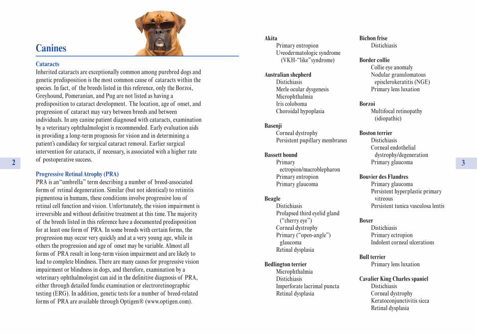
CaninesCataractsInherited cataracts are exceptionally common among purebred dogs andgenetic predisposition is the most common cause of cataracts within thespecies. In fact, of the breeds listed in this reference, only the Borzoi,Greyhound, Pomeranian, and Pug are not listed as having apredisposition to cataract development. The location, age of onset, andprogression of cataract may vary between breeds and betweenindividuals. In any canine patient diagnosed with cataracts, examinationby a veterinary ophthalmologist is recommended. Early evaluation aidsin providing a long-term prognosis for vision and in determining apatient’s candidacy for surgical cataract removal. Earlier surgicalintervention for cataracts, if necessary, is associated with a higher rateof postoperative success.
Progressive Retinal Atrophy (PRA)PRA is an “umbrella” term describing a number of breed-associatedforms of retinal degeneration. Similar (but not identical) to retinitispigmentosa in humans, these conditions involve progressive loss ofretinal cell function and vision. Unfortunately, the vision impairment isirreversible and without definitive treatment at this time. The majorityof the breeds listed in this reference have a documented predispositionfor at least one form of PRA. In some breeds with certain forms, theprogression may occur very quickly and at a very young age, while inothers the progression and age of onset may be variable. Almost allforms of PRA result in long-term vision impairment and are likely tolead to complete blindness. There are many causes for progressive visionimpairment or blindness in dogs, and therefore, examination by aveterinary ophthalmologist can aid in the definitive diagnosis of PRA,either through detailed fundic examination or electroretinographictesting (ERG). In addition, genetic tests for a number of breed-relatedforms of PRA are available through Optigen® (www.optigen.com).
AkitaPrimary entropionUveodermatologic syndrome
(VKH-“like” syndrome)
Australian shepherdDistichiasisMerle ocular dysgenesis MicrophthalmiaIris colobomaChoroidal hypoplasia
BasenjiCorneal dystrophyPersistent pupillary membranes
Bassett houndPrimary
ectropion/macroblepharonPrimary entropionPrimary glaucoma
BeagleDistichiasisProlapsed third eyelid gland
(“cherry eye”)Corneal dystrophyPrimary (“open-angle”)
glaucomaRetinal dysplasia
Bedlington terrierMicrophthalmiaDistichiasisImperforate lacrimal punctaRetinal dysplasia
Bichon friseDistichiasis
Border collieCollie eye anomalyNodular granulomatous
episclerokeratitis (NGE)Primary lens luxation
BorzoiMultifocal retinopathy
(idiopathic)
Boston terrierDistichiasisCorneal endothelial
dystrophy/degenerationPrimary glaucoma
Bouvier des FlandresPrimary glaucomaPersistent hyperplastic primary
vitreousPersistent tunica vasculosa lentis
BoxerDistichiasisPrimary ectropionIndolent corneal ulcerations
Bull terrierPrimary lens luxation
Cavalier King Charles spanielDistichiasisCorneal dystrophyKeratoconjunctivitis siccaRetinal dysplasia
54
German shepherdChronic superficial keratitis
(“pannus”)Corneal dystrophyMedial canthal erosion syndromeOptic nerve hypoplasia
Golden retrieverDistichiasisEctopic ciliaPrimary entropionIris cystsPigmentary
(immune-mediated) uveitisRetinal dysplasiaProgressive retinal atrophy (PRA)
Great daneMicrophthalmiaPrimary entropion/ectropionEverted third eyelid
(“scrolled” cartilage)Ciliary body cystsPrimary glaucoma
GreyhoundChronic superficial keratitis
(“pannus”)Persistent hyperplastic
primary vitreous
HavaneseDistichiasisVitreous degeneration
Italian greyhoundVitreous degeneration
Jack Russell terrierPrimary lens luxationPrimary glaucomaVitreous degeneration
Japanese ChinPrimary medial entropionPigmentary keratitis/exposure
keratopathy syndrome
Labrador retrieverDistichiasisPrimary entropionPrimary ectropionIris melanomaPersistent hyaloidPersistent hyperplastic
primary vitreousPersistent tunica vasculosa lentisRetinal dysplasiaProgressive retinal atrophy (PRA)
Lhasa apsoDistichiasisEctopic ciliaProlapsed third eyelid gland
(“cherry eye”)Imperforate lacrimal punctaKeratoconjunctivitis siccaPigmentary keratitis/exposure
keratopathy syndrome
Alaskan malamutePrimary glaucomaRetinal dysplasiaProgressive retinal atrophy (PRA)
ChihuahuaCorneal endothelial
dystrophy/degenerationVitreous degeneration
Chow chowPrimary entropionPrimary glaucoma
Cocker spanielDistichiasisEctopic ciliaPrimary entropion/ectropionImperforate lacrimal punctaProlapsed third eyelid glandKeratoconjunctivitis siccaCorneal dystrophyRetinal dysplasiaPrimary glaucomaProgressive retinal atrophy (PRA)
CollieMicrophthalmiaNodular granulomatous
episclerokeratitis (NGE)Collie eye anomalyProgressive retinal atrophy (PRA)
DachshundMicrophthalmiaDistichiasisDermoidChronic superficial keratitis
(“pannus”)Punctate superficial keratitisCorneal dystrophy
Uveodermatologic syndrome (VKH-“like” syndrome)
Progressive retinal atrophyMicropapilla (small optic nerve)Optic nerve coloboma
Doberman pinscherMicrophthalmiaLigneous conjunctivitisPersistent hyperplastic primary
vitreousPersistent tunica vasculosa lentis
English bulldogDistichiasisEctopic ciliaProlapsed gland of the
third eyelidPrimary entropion/ectropionKeratoconjunctivitis sicca
English springer spanielPrimary entropionCorneal dystrophyPrimary glaucomaRetinal dysplasiaProgressive retinal atrophy (PRA)
Fox terrierPrimary lens luxationPrimary glaucoma
French bulldogDistichiasis
7
Saint BernardMicrophthalmiaPrimary entropion/ectropionDermoidEverted third eyelid
(“scrolled” cartilage)
SamoyedCorneal dystrophyUveodermatologic syndrome
(VKH-“like” syndrome)Primary glaucomaRetinal dysplasiaProgressive retinal atrophy
Shar peiPrimary entropionProlapsed third eyelid gland
(“cherry eye”)Primary glaucomaPrimary lens luxationCongenital esotropia
Shetland sheepdogCorneal dystrophyPunctate superficial keratitisUveodermatologic syndrome
(VKH-“like” syndrome)Collie eye anomaly
Shiba inuPrimary glaucoma
Shih tzuDistichiasisEctopic cilia
Primary medial entropionPigmentary keratitis/exposure
keratopathy syndromeKeratoconjunctivitis siccaVitreous degenerationProgressive retinal atrophy (PRA)Optic nerve hypoplasia
Siberian huskyCorneal dystrophyUveodermatologic syndrome
(VKH-“like” syndrome)Primary glaucomaProgressive retinal atrophy (PRA)
WeimeranerPrimary entropionEverted third eyelid
(“scrolled” cartilage)
Welsh corgiIndolent corneal ulcerationRetinal dysplasia
West Highland white terrierKeratoconjunctivitis sicca
Yorkshire terrierCongenital alacrima
(absolute KCS)Corneal dystrophyRetinal dysplasiaProgressive retinal atrophy (PRA)
6
MaltesePersistent hyaloid arteryProgressive retinal atrophy (PRA)
MastiffPrimary entropion/ectropionProlapsed third eyelid gland
(“cherry eye”)Retinal dysplasiaProgressive retinal atrophy (PRA)
Miniature SchnauzerMicrophthalmia
(with congenital cataract)Keratoconjunctivitis siccaPersistent hyaloid arteryProgressive retinal atrophy (PRA)
Neapolitan mastiffPrimary entropion/ectropionProlapsed third eyelid gland
NewfoundlandPrimary entropion/ectropionProlapsed third eyelid gland
Norwegian elkhoundPrimary glaucomaProgressive retinal atrophy
PekingeseDistichiasisPrimary medial entropionPigmentary keratitis/exposure
keratopathy syndromeFacial fold trichiasisKeratoconjunctivitis sicca
Pointer (German short-haired)Everted third eyelid cartilage
(“scrolled” cartilage)Persistent hyperplastic
primary vitreousPersistent tunica vasculosa lentisProgressive retinal atrophy (PRA)
PomeranianDistichiasis
PoodleMicrophthalmiaDistichiasisEctopic ciliaImperforate lacrimal punctaPrimary glaucomaVitreous degenerationRetinal dysplasiaProgressive retinal atrophy (PRA)Optic nerve hypoplasiaMicropapilla
PugDistichiasisPrimary medial entropionPigmentary keratitis/exposure
keratopathy syndromeKeratoconjunctivitis sicca
RottweilerPrimary entropionCorneal dystrophyIris cystsRetinal dysplasia

98
Glossary
Anterior uveitis: inflammation of the ciliary body and/or iris
Cataract: opacity of the lens and/or lens capsule
Choroid: the posterior aspect of the uveal tract immediately external to theretina
Chronic superficial keratitis: immune-mediated disease of the conjunctivaand cornea of dogs; also known as pannus
Collie eye anomaly: inherited developmental defect of collies and relatedbreeds characterized by choroidal hypoplasia, with or without colobomas,and retinal detachment
Coloboma: congenital absence of any ocular tissue
Corneal dystrophy: progressive and bilateral hereditary corneal disease,unassociated with inflammation
Corneal sequestrum: condition unique to the cat cornea in which a regionof corneal stroma acquires an amber to black discoloration and undergoesdegeneration; corneal ulceration may or may not be concurrent
Dermoid: a congenital choristomatous tumor consisting of skin and itsappendages
Distichiasis: condition in which cilia (eyeliashes) emerge abnormally fromone or more meibomian gland orifices
Ectopic cilia: abnormal hair/cilia protruding through the palpebralconjunctiva
Ectropion: eversion or outward rolling of the eyelid
Entropion: introversion or inward rolling of the eyelid
Episcleritis: inflammation of the connective tissue immediately exterior tothe sclera
BirmanDermoid
BurmeseProlapsed third eyelid gland
(“cherry eye”)
Persian/HimalayanEntropionExposure keratopathy syndromeCorneal sequestrum
SiameseCongenital nystagmus
(pendular)
Felines

1110
References
Maggs DJ, Miller PE, and Ofri R, eds. Slatter’s Fundamentals ofVeterinary Ophthalmology. 4th ed. St. Louis: Elsevier, 2008.
Gelatt KN, ed. Veterinary Ophthalmology. 4th ed. Wiley and Sons, 2007.
Ocular Disorders Presumed to be Inherited in Purebred Dogs. 3rd ed.1999, American College of Veterinary Ophthalmologists.
Lens luxation: disinsertion of the lens zoules from the complete lensequator such that the lens displaces into the anterior chamber (anteriorluxation) or the vitreous chamber (posterior luxation)
Macropalpebral fissure: horizontally enlarged palpebral fissure due toexcessive eyelid length
Microphthalmos: congenitally small globe
Nodular granulomatous episclerokeratoconjunctivitis (NGE): a diseasecharacterized by a raised tan-pink mass or masses, arising from the episclerausually at the dorsolateral corneoscleral limbus; suspected to be immune-mediated
Persistent pupillary membranes: congenital defect in which persistentstrands of fetal vascular tissue extend from the iris collarette to otherregions of the iris, to the anterior lens capsule, or to the cornealendothelium
Retinal dysplasia: abnormal differentiation of the retinal layers
Staphyloma: protrusion of uveal tissue into a bulging area of corneaand/or sclera due to thinning or rupture of the eye wall
Uveodermatologic syndrome: autoimmune destruction of melanocytescausing marked panuveitis, retinitis, and dermatitis seen in dogs; caninecounterpart to human Vogt-Koyanagi-Harada (VKH) syndrome
What the AEC of NJ is all about:
Dr. Michael Brown has been providingcutting edge ophthalmology services inLittle Falls since 1996. He was thedriving force behind establishing thisarea’s first ophthalmology-dedicatedspecialty center. The Animal Eye Centerof NJ, a partner of Animal Emergency& Referral Associates in Fairfield, was
the first veterinary practice in the world to use the Whitestar SignaturePhacoemulsification Unit, a sophisticated and successful cataractremoval treatment modality.
Meet Our Ophthalmologists
Michael H. Brown, DVM, MS, Diplomate ACVODr. Brown received his Doctorate of VeterinaryMedicine from Kansas State University and thencompleted a small animal internship at the AnimalMedical Center in New York City. After returning toKansas State University for a comparativeophthalmology residency, he received a Master of Science degree for hisbiochemical study of animal tears.
Dr. Brown became a Diplomate of the American College of VeterinaryOphthalmologists in 1996. His special interests include diseases of thecornea, corneal surgery, intraocular surgery, and diseases of the retina.He has written scientific papers and is a noted lecturer throughout thecountry.

Bradford J. Holmberg, DVM, MS, PhD, Diplomate ACVODr. Holmberg received his Doctorate of VeterinaryMedicine from the University of Missouri. He completeda small animal internship at the University of Floridaand then pursued a comparative ophthalmology residencyat the University of California – Davis.
In addition, Dr. Holmberg received his Master of Sciencein neuroscience from Purdue University and his
Doctorate of Philosophy with a concentration in neuroendocrinologyfrom the University of Missouri. Dr. Holmberg became a Diplomate ofthe American College of Veterinary Ophthalmologists in 2005.
His special interests include exotic animal ophthalmology and all aspectsof ophthalmic surgery. Dr. Holmberg has been awarded severalprestigious research grants, has written numerous scientific papers, andhas contributed chapters to several veterinary textbooks. He joinedAnimal Eye Center in August 2006.
J. Seth Eaton, VMD, Diplomate ACVODr. Eaton graduated magna cum laude from the University ofPennsylvania, School of Veterinary Medicine in 2004. He thencompleted internships in general medicine/surgery and ophthalmologyat the Animal Medical Center in New York City. Hejoined the ophthalmology service at the University ofCalifornia - Davis as an ophthalmology resident inAugust, 2006.
His clinical interests include corneal therapeutics, neuro-ophthalmology and intraocular surgery. He joinedAnimal Eye Center in September 2009.
12
DIRECTIONS to AEC48 Notch Road, Little Falls, NJ 07424
From Points NorthTake the Garden State Parkway (GSP) south to exit 154 (Rt. 46/Clifton). Follow signs for Rt.46 west. Take the Great Notch/Cedar Grove exit. Make a left at the stop sign onto Notch road.The Animal Eye Center is the first building ahead on your left immediately after crossing overRt. 46.
From Points SouthTake the GSP north to exit 153B (Rts. 3 and 46). Follow signs for Rt 46 west. Take the GreatNotch/Cedar Grove exit. Make a left at the stop sign onto Notch road. The Animal Eye Centeris the first building ahead on your left immediately after crossing over Rt. 46.
From Points West (via Rt. 46)Follow Rt. 46 east. Take the Great Notch/Little Falls exit. (after the Lower Notch exit) Bearright onto the off ramp. The Animal Eye Center is across the street on your left.
From Points West (via Route 80)Follow Route 80 east to exit 56A (Squirrelwood Road/West Paterson). You will merge ontoSquirrelwood Road. Follow this road (the name will change to Rifle Camp Road) forapproximately 3.5 miles. The Animal Eye Center is the first building ahead on your leftimmediately after crossing over Rt. 46.
From Points East (Lincoln Tunnel)Follow signs for Route 3 west. Route 3 will merge with Route 46 west. Take the GreatNotch/Cedar Grove exit. Make a left at the stop sign onto Notch road. The Animal Eye Centeris the first building ahead on your left immediately after crossing over Rt. 46.
From Points East (Holland Tunnel)Take Route 78 west to the GSP. Follow directions above from points south.
From Points East (George Washington Bridge)Take Route 80 west to exit 56 (Squirrelwood Road/West Paterson). Make a left after the offramp onto Squirrelwood Road. Follow this road (the name will change to Rifle Camp Road)for approximately 3.5 miles. The Animal Eye Center is the first building ahead on your leftimmediately after crossing over Rt. 46.
animalerc.com • animaleyesofnj.com888.722.7200 • 973.890.4430
48 Notch Road, Little Falls, NJ 07424
Animal Emergency
ANIMAL EYE CENTER OF NJ
& referral associates