breathin’ and breathin’
TRANSCRIPT

Copyright © 2020 Care Center. All rights reserved.
Just Keep Breathin’ and
Breathin’ and Breathin’:
Respiratory Monitors –
Monitoring Ventilation
Brandon Wahler, DVM, MS, Practice Limited to Anesthesia

Copyright © 2020 Care Center. All rights reserved.
Goals
Identify what the difference between capnometry,
capnometer, capnography, capnograph, and a
capnogram is
Identify and describe the different types of
capnometry/capnography used in modern anesthetic
practice. What is the benefit of one over the other?
Identify the different parts of the capnogram
Interpret several common capnogram waveforms and
list potential differentials for them
Discuss relevant literature involving capnometry and capnography in veterinary
practice

Copyright © 2020 Care Center. All rights reserved.
Ventilation
• Part of respiration that is associated with bulk movement of air into and out of the lungs
• Commonly use carbon dioxide to measure ventilation
• Measure of metabolic activity in the body
• CO2 is readily diffusible and can be measured both invasively and non-invasively (approximately 20-40x more diffusible than O2 in water)
• Ventilation is a product of tidal volume and respiratory rate
• Determines the minute ventilation
• Average tidal volume is 6-15mL/kg
• Normal total minute ventilation is 150-250mL/kg/min
https://www.austincc.edu/apreview/PhysText/Respiratory.html

Copyright © 2020 Care Center. All rights reserved.
Dead Space
• Portions of the large airways, equipment, or alveoli that don’t participate in gas exchange
• The larger the portion of dead space the less efficient respiration and ventilation is
• We have innate control over mechanical dead space
https://www.criticalcarepractitioner.co.uk/mechanical-ventilation-physiologic-effects-mechanical-ventilation/

Copyright © 2020 Care Center. All rights reserved.
Mechanical Dead Space• The portion of the endotracheal tube which extends out of the trachea (from the mouth to the breathing circuit)
• The elbow on the breathing circuit
• Any connector used between the end of the tube and the breathing circuit (e.g., CO2 adapters, apnea alarm adapters, etc.)
• The Y piece at the end of a Y circuit
• Tube Size does not play a role in dead space
https://www.dispomed.com/dead-space-in-breathing-circuit/

Copyright © 2020 Care Center. All rights reserved.
Dead Space
• High measured total minute ventilation in combination with normal PaCO2 shows increased dead space ventilation
• Positive pressure ventilation
• Some volume is taken up by compression of gases within the circuit
• Breathing circuit expansion
• Must be subtracted to determine the amount of compliance in the system and to determine the compliance and true tidal volume of the patient
• Modern ventilators do this
• Must be calibrated between each patient
https://www.mindraynorthamerica.com/anesthesia-systems/anesthesia-machines/a5-anesthesia-machine/

Copyright © 2020 Care Center. All rights reserved.
Measuring Ventilation
• End tidal CO2• 3-6mmHg lower of PaCO2
• Arterial blood gas• PaCO2 >60mmHg normally indicates
need for ventilation• PaCO2 <20mmHg is associated with
respiratory alkalosis and decreased cerebral blood flow
• Venous blood gas• PvCO2 is normally within 3 to
6mmHg of PaCO2
• PvCO2 is higher in transition states and during hypovolemia, poor cardiac output, anemia
Copyright © 2020 Care Center. All rights reserved.
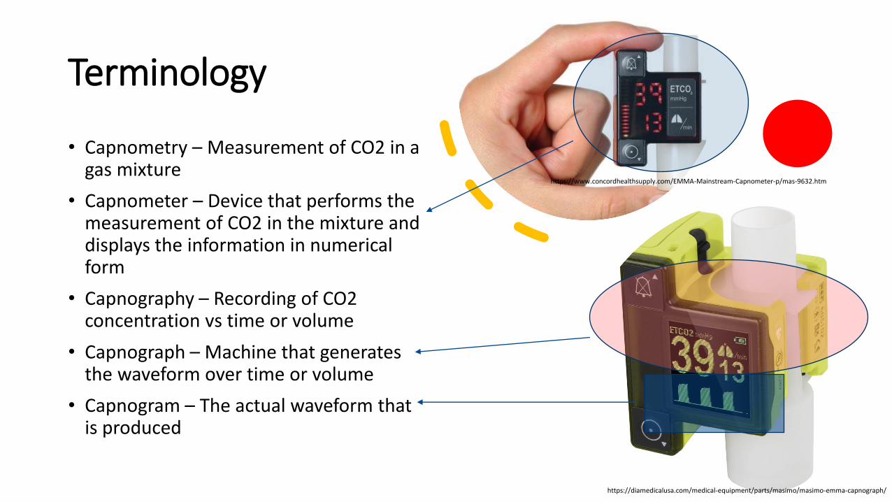
Terminology
• Capnometry – Measurement of CO2 in a gas mixture
• Capnometer – Device that performs the measurement of CO2 in the mixture and displays the information in numerical form
• Capnography – Recording of CO2 concentration vs time or volume
• Capnograph – Machine that generates the waveform over time or volume
• Capnogram – The actual waveform that is produced
https://diamedicalusa.com/medical-equipment/parts/masimo/masimo-emma-capnograph/
https://www.concordhealthsupply.com/EMMA-Mainstream-Capnometer-p/mas-9632.htm

Copyright © 2020 Care Center. All rights reserved.
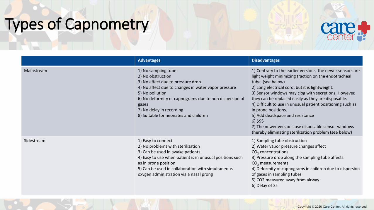
Types of Capnometry
• Primarily use infrared light absorption with absorption being proportional to the partial pressure of the gas
• Mainstream capnometers
• Breathes through cell or cuvette surrounded by infrared transmitter and photosensor
• Sidestream
• Aspirate gas samples (variable rates 50-200mL/min) and analyzed at the module
Copyright © 2020 Care Center. All rights reserved.
Types of Capnometry
Advantages Disadvantages
Mainstream 1) No sampling tube2) No obstruction3) No affect due to pressure drop4) No affect due to changes in water vapor pressure5) No pollution6) No deformity of capnograms due to non dispersion of gases7) No delay in recording8) Suitable for neonates and children
1) Contrary to the earlier versions, the newer sensors are light weight minimizing traction on the endotracheal tube. (see below)2) Long electrical cord, but it is lightweight.3) Sensor windows may clog with secretions. However, they can be replaced easily as they are disposable.4) Difficult to use in unusual patient positioning such as in prone positions.5) Add deadspace and resistance6) $$$7) The newer versions use disposable sensor windows thereby eliminating sterilization problem (see below)
Sidestream 1) Easy to connect2) No problems with sterilization3) Can be used in awake patients4) Easy to use when patient is in unusual positions such as in prone position5) Can be used in collaboration with simultaneous oxygen administration via a nasal prong
1) Sampling tube obstruction2) Water vapor pressure changes affect CO2 concentrations3) Pressure drop along the sampling tube affects CO2 measurements4) Deformity of capnograms in children due to dispersion of gases in sampling tubes5) CO2 measured away from airway6) Delay of 3s
Copyright © 2020 Care Center. All rights reserved.
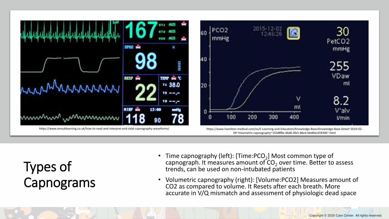
Types of Capnograms
• Time capnography (left): [Time:PCO2] Most common type of capnograph. It measures amount of CO2 over time. Better to assess trends, can be used on non-intubated patients
• Volumetric capnography (right): [Volume:PCO2] Measures amount of CO2 as compared to volume. It Resets after each breath. More accurate in V/Q mismatch and assessment of physiologic dead space
https://www.emsuklearning.co.uk/how-to-read-and-interpret-end-tidal-capnography-waveforms/ https://www.hamilton-medical.com/es/E-Learning-and-Education/Knowledge-Base/Knowledge-Base-Detail~2019-02-04~Volumetric-capnography~1f2d8f8e-46d6-4fe5-86e4-bbd6bcd34306~.html

Copyright © 2020 Care Center. All rights reserved.
Literature
Both work fine, but mainstream may be betterBoth are inaccurate in severe hypercapnia
Copyright © 2020 Care Center. All rights reserved.
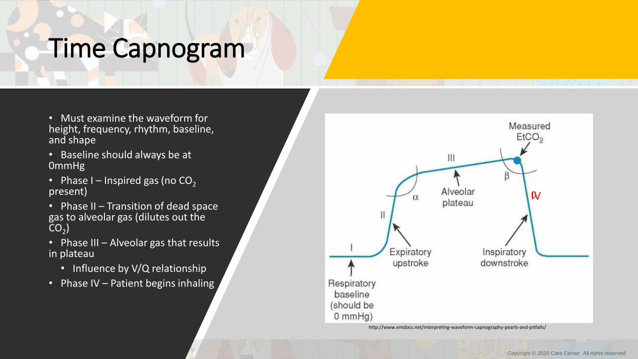
Time Capnogram
• Must examine the waveform for height, frequency, rhythm, baseline, and shape
• Baseline should always be at 0mmHg
• Phase I – Inspired gas (no CO2present)
• Phase II – Transition of dead space gas to alveolar gas (dilutes out the CO2)
• Phase III – Alveolar gas that results in plateau
• Influence by V/Q relationship
• Phase IV – Patient begins inhaling
http://www.emdocs.net/interpreting-waveform-capnography-pearls-and-pitfalls/
IV

Copyright © 2020 Care Center. All rights reserved.
Time Capnogram
• α (takeoff, elevation) angle –Normally between 100-110°
• Increased in obstructive lung disease
• Influenced by capnometer response time, sweep speed, respiratory cycle time
• β angle – Approximately 90°
• Increased with rebreathing or prolonged response time compared with respiratory cycle time
http://www.emdocs.net/interpreting-waveform-capnography-pearls-and-pitfalls/
IV
Copyright © 2020 Care Center. All rights reserved.
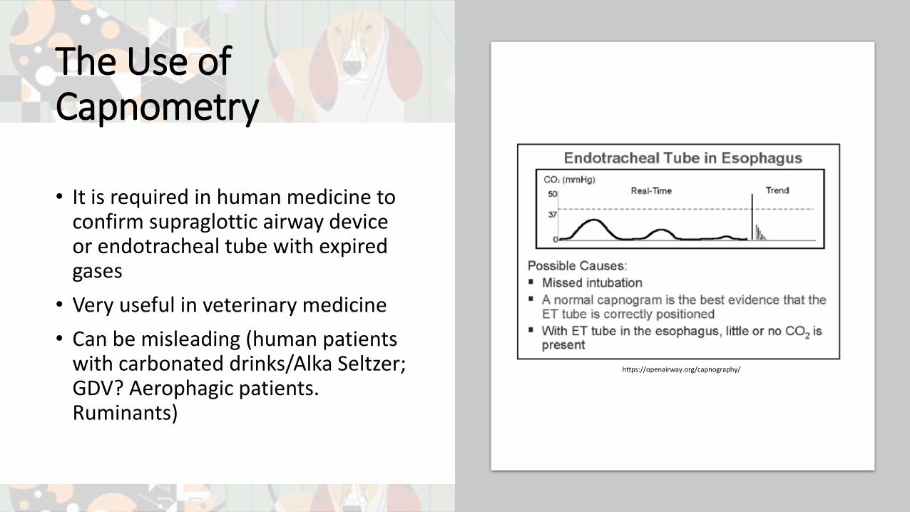
The Use of Capnometry
• It is required in human medicine to confirm supraglottic airway device or endotracheal tube with expired gases
• Very useful in veterinary medicine
• Can be misleading (human patients with carbonated drinks/Alka Seltzer; GDV? Aerophagic patients. Ruminants)
https://openairway.org/capnography/

Copyright © 2020 Care Center. All rights reserved.
Literature

Copyright © 2020 Care Center. All rights reserved.
Uses of Capnometry• Used as an early indicator of
malignant hyperthermia (super rare!)
• ETCO2 will increase dramatically before any other sign
• Metabolically active tissues create CO2 more
https://www.sciencedirect.com/science/article/pii/B9780323429740000410

Copyright © 2020 Care Center. All rights reserved.
Uses of Capnometry
• Metabolic changes• Must be monitored in a mechanically
ventilated patient
• Spontaneous ventilating patients may increase minute ventilation in response to increased metabolic production
• Malignant hyperthermia would also increase through an increase in metabolic production of carbon dioxide
Dorsch and Dorsch. Understanding Anesthesia Equipment 5th Ed.

Copyright © 2020 Care Center. All rights reserved.
Uses of Capnometry
• Circulation changes• Decreases in end-tidal carbon dioxide coincide with decreases in cardiac output
• Must be monitored in mechanically ventilated patients
• Can detect air emboli in the lungs
• ROSC – will discuss later• Epinephrine
• Bicarbonate
Dorsch and Dorsch. Understanding Anesthesia Equipment 5th Ed.

Copyright © 2020 Care Center. All rights reserved.
Uses of Capnometry
• Respiration• Important to determine if your patient is no longer connected to the tube
• Able to determine esophageal intubation, but not necessarily endobronchial intubation
• Can monitor ventilation in non-intubated patients (easier with sidestream up the nares)

Copyright © 2020 Care Center. All rights reserved.
Uses of Capnometry
• Breathing system• Oftentimes these are specific to
the breathing system
• Important to understand increased mechanical dead space
• Unidirectional valve capnographs should also be learned
Dorsch and Dorsch. Understanding Anesthesia Equipment 5th Ed.

Copyright © 2020 Care Center. All rights reserved.
Valve Malfunction
• The top two are inspiratory valve malfunction. Note the normal plateau in the top image, but the increasing inspired CO2 and the change in phase 0
• The bottom clip is of the expiratory valve incompetence with increase in phase II, slanting descending limb, and increased inspired CO2
Poor inspiratory valve seal
Incompetent inspiratory valve
Incompetent expiratory valve
Copyright © 2020 Care Center. All rights reserved.
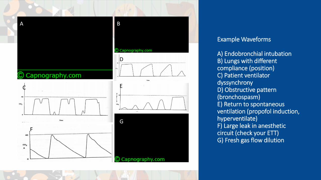
Example Waveforms
A) Endobronchial intubationB) Lungs with different compliance (position)C) Patient ventilator dyssynchronyD) Obstructive pattern (bronchospasm)E) Return to spontaneous ventilation (propofol induction, hyperventilate)F) Large leak in anesthetic circuit (check your ETT)G) Fresh gas flow dilution
A B
C
D
E
F
G

Copyright © 2020 Care Center. All rights reserved.
Problems with Capnography
• In normal healthy patients PaCO2 and ETCO2 are very close together• Gradient reduced in obese or pregnant patients (reductions in functional residual capacity),
and in rebreathing
• Influenced by sampling volumes (too much may dilute it, too slow may not give accurate measure)
• Influenced by poor connections
• Not accurate in all non-rebreathing systems
• Dramatic changes in V/Q will alter capnography
• Changes in body position (lateral, dorsal recumbency)
• Patients with pulmonary disease
• Patients on acetazolamide
• Patients who are hypothermic

Copyright © 2020 Care Center. All rights reserved.
The Use of Capnometry
• Strong evidence for its use in CPCR• Not influenced by motion• Not influenced by random
electrical activity• Indirect indicator of how
good your compressions are (you will never be as good as the body…)
• May be associated with prognosis for ROSC https://www.onlinejets.org/article.asp?issn=0974-2700;year=2014;volume=7;issue=4;spage=332;epage=340;aulast=Kodali

Copyright © 2020 Care Center. All rights reserved.
Literature
• Found that a reasonable descriminator between animals who had ROSC and those who did not was ETCO2:• Dogs >= 15mmHg• Cats >=20mmHg

Copyright © 2020 Care Center. All rights reserved.
Literature
• Similarly found that there was a “cut off” that would make ROSC likely:• 18mmHg ETCO2
• The authors discuss likely don’t need that high to continue CPCR, but that 10mmHg may represent a good cutoff point to stop CPCR• If below 10mmHg then it is unlikely that
ROSC will be achieved

Copyright © 2020 Care Center. All rights reserved.
Practice
Oscillation with ventilation, but not representative of
PaCO2 and doesn’t return to baseline = Tachypnea
Cardiac oscillations of airway pressure
Rises with exhalation, but does not plateau and
returns to phase I before patient inhalation = Sample
port too close to FGF
Small inspiratory effort prior to proper inspiration
(patient-ventilator dysynchrony)
Shape is normal, but plateau is very low = Excessive
alveolar dead space (hypovolemia, PTE)
Shape is normal, but the baseline never returns to 0 = Dead space rebreathing
Lumb and Jones Veterinary Anesthesia and Analgesia. 5th Ed.

Copyright © 2020 Care Center. All rights reserved.
Summary
• Identify what the difference between capnometry, capnometer, capnography, capnograph, and a capnogram is• -try or –phy is the measure of carbon dioxide and either a resultant number or graphical
representation, the –meter or –graph is the actual unit, the capnogram is the picture
• Identify and describe the different types of capnometry/capnography used in modern anesthetic practice. What is the benefit of one over the other?• Mainstream versus sidestream – One is faster, maybe more accurate, but can get
contaminated, damaged, etc. Sidestream is normally sufficient for most veterinary practices and is very acceptable for anesthetic use. Time capnography versus volumetric capnography – Time capnography is the standard and is used for trends, but likely can’t give you as much subtle information as volumetric
• Identify the different parts of the capnogram• Know the 4 phases. Know the angles. It is most important that you understand where
inspiration is occurring, and where expiration is occurring (phase 0 and I vs. II and III)

Copyright © 2020 Care Center. All rights reserved.
Summary
• Interpret several common capnogram waveforms and list potential differentials for them• Practice, practice, practice• Capnography.com• Books on ventilation and monitoring
• Discuss relevant literature involving capnometry and capnography in veterinary practice• Literature is also sparse, and mostly old looking if it was applicable in veterinary
medicine (it is)• More accurate in small patients than large patients• Likely useful in CPCR (ROSC >10-20mmHg)• Likely not accurate in severe hypoventilation (PaCO2 >60mmHg)

Copyright © 2020 Care Center. All rights reserved.
Questions?
https://www.machinedesign.com/community/article/21836908/what-questions-should-you-ask-during-the-product-lifecycle

Copyright © 2020 Care Center. All rights reserved.
Care Center Cincinnati6995 East Kemper Rd.Cincinnati, OH 45249PH: 513.530.0911 FAX: 513.530.0811
Care Center Dayton6421 Clyo Rd.Centerville, OH 45459PH: 937.428.0911FAX: 937.428.6667
carecentervets.com
carecenterbloodbank.com