breast cancer awareness kristen deep jennifer d’onofrio, bs, fnp-s kelly strine bs, rn, fnp-s
TRANSCRIPT

Breast Cancer AwarenessKristen Deep
Jennifer D’Onofrio, BS, FNP-S
Kelly Strine BS, RN, FNP-S

Objectives
• To develop an adequate understanding of screening guidelines according to the U.S. Preventive Service Task Force (USPTF) and the American Cancer Society.
• Distinguish difference of benign versus malignant breast conditions.
• Identify patients that should be screened for genetic testing
• Identify three community resources available for breast cancer patients
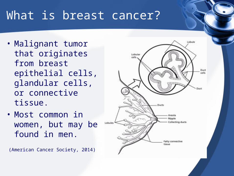
What is breast cancer?
• Malignant tumor that originates from breast epithelial cells, glandular cells, or connective tissue.
• Most common in women, but may be found in men.
(American Cancer Society, 2014)
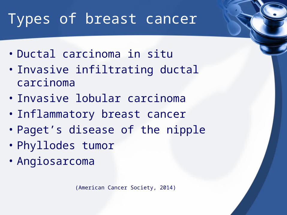
Types of breast cancer
• Ductal carcinoma in situ• Invasive infiltrating ductal carcinoma• Invasive lobular carcinoma• Inflammatory breast cancer• Paget’s disease of the nipple• Phyllodes tumor• Angiosarcoma
(American Cancer Society, 2014)
Ductal carcinoma in situ
• Non-invasive or pre-invasive cells that line the ducts have changed to look like cancer cells.
• Cells have not spread through the walls of the ducts into surrounding breast tissue.
(American Cancer Society, 2014)
Invasive/Infiltrating ductal carcinoma
• Most common type of breast cancer. About 8/10 breast cancers are this type.
• Starts in the milk duct of the breast. Tumor breaks through this milk duct and invades the fatty tissue of the breast.
• Able to spread to other parts of the body via the lymphatic system or bloodstream.
(American Cancer Society, 2014)

Invasive lobular carcinoma
• Begins in the milk producing glands, also known as lobules.
• This can also spread to other areas of the body.
• May be harder to detect through mammogram.
(American Cancer Society, 2014)
Inflammatory breast cancer
• Invasive
• Uncommon, 1-3% of all breast cancers
• No single lump or tumor
• Caused by cancer cells blocking lymph vessels on skin.
• Makes skin on the breast look red and feel warm.
• Breasts may be larger, firmer, tender, or itchy.
• Skin may also be thick and have peau d’orange texture.
(American Cancer Society, 2014)
Paget’s disease of the nipple
• Rare, 1% of all breast cancer cases
• Starts in the breast ducts and spreads to the skin of the nipple and then to the areola.
• Nipples and areolae may appear crusty, scaly, red, and have areas or bleeding or oozing.
(American Cancer Society, 2014)
Phyllodes tumor
• Develops in the stroma, which is the connective tissue of the breast.
• Usually benign, but it may turn cancerous.
(American Cancer Society, 2014)

Angiosarcoma
• High grade tumor that is aggressive and fast growing.• Starts in cells that line the blood or lymph vessels.• Rarely occurs in the breast, but when it does it develops due to
previous radiation treatments.• May develop 5-10 years after radiation therapy.• Can look like a skin infection, a bruise or lesion that will not heal.• May also present as a soft lump that can be felt or seen.
(American Cancer Society, 2014, Tunstall, 2012)
(American Cancer Society, 2014)
Clinical Presentation
• Breast lump or area that feels denser (may or may not have pain)
• Tenderness
• Dimpling
• Nipple retraction
• Nipple ulceration
• Erythema• Peau d’orange• Change in breast shape• Breast enlargement• Alteration in vein pattern
of breast tissue• Nipple discharge• One or more palpable
enlarged axillary lymph nodes
(Hollier & Hensley, 2011)

Non cancerous breast conditions
• Fibrocystic breast disease
• Intraductal papilloma
• Fibroadenoma
• Mastitis
• Hyperplasia
• Adenosis
• Duct ectasia(American Cancer Society, 2014; Hollier & Hensley, 2011)
Fibrocystic breast disease
• Benign breast disorder
• Assessment findings– Palpation of smooth, moveable masses– Breast pain or tenderness that diminishes after
menses.– Breast engorgement– Breast thickening– Worsening of symptoms premenstrually– Nipple discharge of varying color and
consistency.(Hollier & Hensley, 2011)
Intraductal Papilloma
• Benign tumor within the ductal system of the breast
• Assessment findings– Bloody or serous nipple discharge– Usually unilateral unless multiple ducts are
involved.
(Hollier & Hensley, 2011)
Fibroadenoma
• Benign tumor containing fibrous tissue
• Commonly occurs in women in 20’s-30’s
• Use of birth control pills before age 20 liked to risk of fibroadenomas
• Assessment findings– Single, nontender, firm mass– Multiple lesions in 10-15% of cases– Freely moveable– No change in mass with menstrual cycle– No nipple discharge
(American Cancer Society, 2014; Hollier & Hensley, 2011)

Adenosis
• Breast lobules are enlarged and contain more glands than usual.
(American Cancer Society, 2014)
Mastitis
• Plugged milk ducts, which lead to infection• Most common in women who are
breastfeeding• Assessment findings
– Breast tenderness and pain– Lump in one or both breasts– Fever, chills– Erythema overlying sore area– Flu-like symptoms– Fatigue
(Hollier & Hensley, 2011)
Hyperplasia
• Also known as proliferative breast disease
• Overgrowth of cells that line the ducts or lobules
• Can be usual or atypical
(American Cancer Society, 2014)

Duct Ectasia
• Common in women over 50
• Occurs when breast ducts widen and thicken. This leads to blockage, which leads to fluid build-up.
(American Cancer Society, 2014)
Incidence
• The American Cancer Society estimated in 2012 that 226,870 new cases of invasive breast cancer and 63, 300 in situ cases would have been diagnosed.
• The second leading cause of death among women
• Lifetime risk of developing of breast cancer in the U.S. is 12.15% or 1 in 8 women.
Magnus, Ping, Shen, Bourgeoi, & Magnus, 2011;Mahon, 2012
Incidence
• Women greater than 65 years of age have a six times greater risk of diagnosis
• If rate remains unchanged an estimated 80 million women will be diagnosed with breast cancer by the year 2050
• Increase of 72 percent
Ravert & Huffaker, 2010
Early Detection
• Early detection is crucial for decreasing mortality rates.
• Routine screening is fundamental in detecting breast cancer at an early stage, thus decreasing overall mortality rate.
• Estimated a 25-30 percent reduction in women aged 50-69 and 17 percent reduction in women aged 40-49 mortality rate
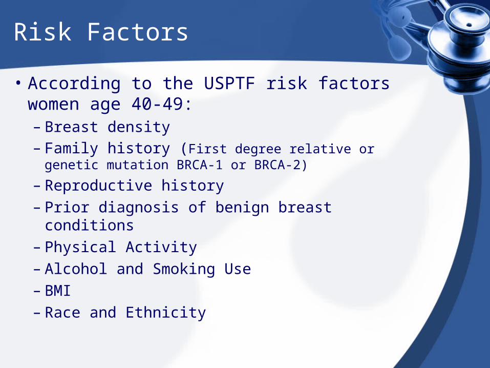
Risk Factors
• According to the USPTF risk factors women age 40-49:– Breast density– Family history (First degree relative or genetic
mutation BRCA-1 or BRCA-2)
– Reproductive history– Prior diagnosis of benign breast conditions– Physical Activity– Alcohol and Smoking Use– BMI– Race and Ethnicity
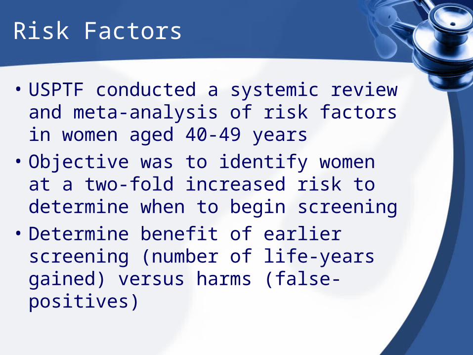
Risk Factors
• USPTF conducted a systemic review and meta-analysis of risk factors in women aged 40-49 years
• Objective was to identify women at a two-fold increased risk to determine when to begin screening
• Determine benefit of earlier screening (number of life-years gained) versus harms (false-positives)
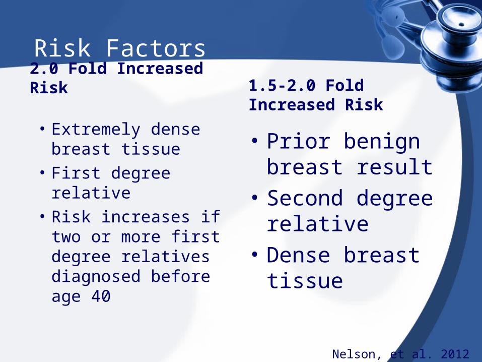
Risk Factors
2.0 Fold Increased Risk
• Extremely dense breast tissue
• First degree relative• Risk increases if two
or more first degree relatives diagnosed before age 40
1.5-2.0 Fold Increased Risk
• Prior benign breast result
• Second degree relative
• Dense breast tissue
Nelson, et al. 2012
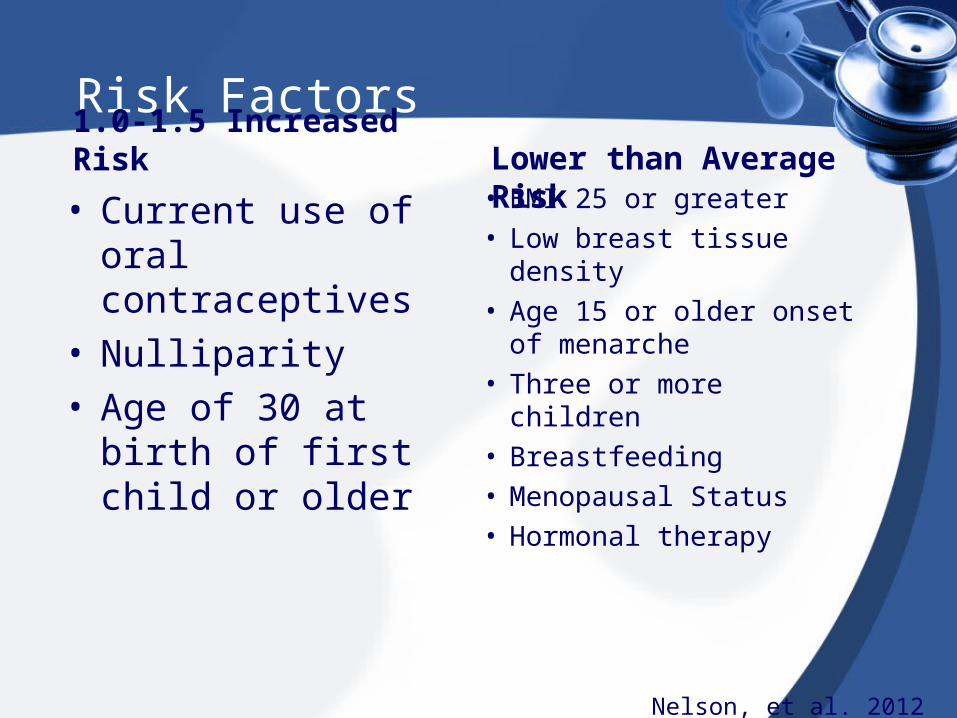
Risk Factors1.0-1.5 Increased Risk
• Current use of oral contraceptives
• Nulliparity• Age of 30 at birth of
first child or older
Lower than Average Risk• BMI 25 or greater• Low breast tissue density• Age 15 or older onset of
menarche• Three or more children• Breastfeeding• Menopausal Status• Hormonal therapy
Nelson, et al. 2012

Risk Factors
• A positive family history is essential in determining if genetic counseling and mutation testing need to occur– Consider medication to reduce risk
• Increasing age is an important risk factor
• There is a risk of diagnosis even when no potential risk factors are identified
Nelson, et al. 2012; USPTF, 2012

Screening Guidelines
Screening
• Clinical breast examinations
• Self-breast examinations
• Mammography
• MRI
Breast Examinations
• Lack of reliability in using only clinical and self breast examinations as detection methods
Mammography
• Considered the GOLD standard for early detection of breast cancer
• An x-ray of the breast• Screening: asymptomatic
– 2 view
• Diagnostic: symptoms or abnormal screening.– More views
Mammography
• False-positives occur more in younger women compared to older women related to breast density.– Increased anxiety/psychological harm– Additional medical visits– Additional imaging– Biopsies– Un-necessary treatment– Radiation Exposure
USPTF, 2012
USPTF: Mammography
• According to the USPTF Recommendation:– Women aged 40-49: Individualized decision to
initiate biennial screening (Grade C)
– Women aged 50-74: Every 2 years (Grade B)
– Women aged 75 or greater: No recommendation
USPTF: Mammography
• For women greater than 40 years of age who have no known genetic mutation or positive family history.
• Biennial screening is optional.
• Greater reduction in mortality women aged 50-74 than younger women.
• False-positives are higher in younger women
• Over-diagnosis with increasing age
Digital Mammography
• Increase of accuracy for pre-menopausal or perimenopausal women
• Increase of accuracy for dense breast tissue
• Uncertain if over-diagnosis occurs more with digital mammography versus film mammography
• More expensive than film mammography
Mahon, 2012; USPTF, 2012
American Cancer Society
• Beginning in 20’s, women should be educated on performing self-breast examinations and to report any new symptoms
• Clinical breast examinations: Every 1-3 years for women aged 20-39 and annual for women 40 and greater, prior to mammography
• Mammography: annually at age 40 with no specific age to stop. Benefits versus risk need to be considered to determine age when to discontinue regular screening
• MRI: 20-25% or higher lifetime risk associated with positive family history (breast or ovarian cancer), genetic mutation, or chest radiation. Begin at age 30– In addition to yearly mammogram
Summary
• USPTF: Recommendation for biennial mammograms for women greater than 40 years old based on individual.
• USPTF: Mammograms every two years for women greater than 50 years
• American cancer society recommends annual screening for women 40 years and greater
• American cancer society offers educational information on web-site for patients
Genetic Testing
Genetic Testing
• What are BRCA 1 and BRCA 2?– Genes that produce tumor suppressor
proteins.
• It is estimated that 2-7% of breast cancers and 10-15% of ovarian cancers are results of BRCA1 and BRCA 2 mutations.
Breast Cancer Risks
• In the general population, 12% of women will develop breast cancer in their lives.
• If a harmful BRCA1 mutations is inherited that risk increases to 55-65%.
• If a harmful BRCA2 mutation is inherited, that risk increases to 45%.
Ovarian Cancer
• In the general population, 1.4% of women will develop Ovarian Caner in their lives.
• In those with an inherited BRCA1 mutation this risk is increased to 39%.
• In those with an inherited BRCA2 mutation, this risk is increased to 11-17%.
Who Should Have Genetic Testing
• BRCA 1 & 2 gene mutations are relatively rare in the general population.
• Testing should be performed only when the family history suggests possible harmful mutations.
• BRST Screening Tool www.breastcancergenescreen.org
Negative Test Results
• If a close first or second degree relative is known to have a harmful BRCA 1 or 2 mutation and the patient is negative, this means the test is negative and it cannot be passed along to their children.
• If there is a possibility of a harmful mutation, but no mutation has been identified in the family, the results are less clear.
Positive Test Results
• A positive results indicates that a known harmful gene mutation of BRCA1 or 2 is present and there is an increased risk of developing cancer.
• The genetic mutation has a 50% chance of being passed down to sons or daughters.
• Siblings of BRCA 1 or 2 known gene mutations have a 50% chance of being positive for the mutation.
Ambiguous Test Results
• Tests results described as ambiguous have been identified as genetic variant of uncertain significance.
Other Considerations
• What would I do if I knew I was positive for the gene that could cause Breast or Ovarian Cancer?
• What would I do if my results were found to be negative?
• What would I do if my results were found to be ambiguous?
Case Study
• Mandy, a 24 year old Caucasian female arrives to the office inquiring about BRCA testing. She tells you that her aunt who is 49, is currently battling breast cancer and she is concerned about her risks for developing breast cancer. There is no family history of ovarian cancer. No other family members have been tested previously for BRCA 1 or 2. Her age of menarche is 14. She is married with 1 child. How should you counsel this patient?
Case Study
• Sydnie is an 18 year old female who is starting college in the fall. When Sydnie was a couple of months old, her mother was diagnosed with Breast Cancer at the age of 26. Her mother underwent a unilateral mastectomy with lymph node removal, chemotherapy and radiation. Five years later she was in remission. Syndie’s maternal grandmother was also diagnosed with cancer around this time. She underwent treatment as well. Sydnie’s mother had another child when she was 8 years old. Within a year, her mother was diagnosed with recurrent breast cancer. When Sydnie was 13, her mother passed away at the age of 39. One month later, her Grandmother passed away. Sydnie’s first age of menarche was 14. There is no known history of ovarian cancer. Sydnie’s Aunt, her mother’s sister, is alive and well and her uncle, her mother’s brother, is alive and well. What type of counseling would you provider this patient?

Referrals
• Patients with several risk factors for BRCA1 or BRCA2 mutations should be referred to a Genetic Counselor. – https://www.health.ny.gov/diseases/cancer
/genetics/genetic_counselors.htm– https://www.breastcancergenescreen.org/
definitions.aspx– Bonnie R. Braddock
Senior Certified Genetic CounselorUpstate Medical University750 E Adams St Syracuse, NY, US 13210-2342(315) 464-6395 (phone)
Resources
• Susan G. Komen http://ww5.komen.org/Default.aspx
• American Cancer Society http://www.cancer.org/
• New York State Cancer Services Program https://www.health.ny.gov/diseases/cancer/services/
References
American Cancer Society (2014). Breast cancer: Early detection. Retrieved from www.cancer/org/cancer/breastcancer/moreinformation/breastcancerearlydetection/index
American Cancer Society (2014). Types of breast cancer. Retrieved from http://www.cancer.org/cancer/breastcancer/detailedguide/breast-cancer-breast-cancer-types
Bellcross C 2014 Breast Cancer Genetics Referral Screening Tool. Retrieved from https://www.breastcancergenescreen.org/201403012213071011244655
Centers for Disease Control and Prevention (2013). Geenetic Counseling and Evaluation for BRCA1/2 Testing. Retrieved from http://www.cdc.gov/genomics/resources/diseases/breast_ovarian_cancer/counseling.htm
Hollier, A. & Hensley, R. (2011). Clinical guidelines in primary care: A reference and review book. Lafayette, LA: Advanced Practice Education Associates, Inc.
Magnus, M., Ping, M., Shen, M.M., Bourgeois, J., & Magnus, J. (2011). Effectiveness of mammography screening in reducing breast cancer mortality in women 39-49 years: A meta-analysis. Journal of Women’s Health, 20(6), 845-852.
DOI:10.1089/jwh.2010.2098
Mahon, S. (2012). Screening for breast cancer: Evidence and recommendations. Clinical Journal of Oncology Nursing, 16(6),
567-570. DOI: 10.1188/12.CJON.567-571.
National Cancer Institute at the National Institutes of Health (2014) BRCA1 and BRCA2: Cancer risk and genetic testing Retrieved from http://www.cancer.gov/cancertopics/factsheet/Risk/BRCA201403012220201474938393
National Society of Genetic Counselors (n.d.). Find a genetic counselor. Retrieved from http://nsgc.org/p/cm/ld/fid=164201403012214251193767309
Nelson, H., Zakher, B., Cantor, A., Fu, R., Griffin, J…Miglioretti, D. (2012). Risk factors for breast cancer for women aged 40-49 years. Annals of Internal Medicine, 156 (9), 635-648.
New York State Department of Health (2013) New York State Clinical Geneticists and Genetic Counselors: Caner Genetics. Retrieved from https://www.health.ny.gov/diseases/cancer/genetics/genetic_counselors.htm201403012210041167124391
Ravert, P. & Huffaker, C. (2010). Breast cancer screening in women: An integrative literature review. Journal of the American Academy of Nurse Practitioners, 22, 668-673. DOI:10.1111/j.1745-7599.2010.00564.x
Susan G Komen Retrieved from http://ww5.komen.org/BreastCancer/RecommendationsforWomenwithHigherRisk.html#BRCA20140301221749116994023
• Tunstall, B. (2012). What is angiosarcoma? Retrieved from http://sarcomahelp.org/angiosarcoma.html
• US Preventive Services Task Force 2013 Risk assessment, genetic counseling, and genetic testing for BRCA-related cancer in women Retrieved from http://www.uspreventiveservicestaskforce.org/uspstf12/brcatest/brcatestfinalrs.htm201403012211231969841481