branemark booklet new v2 - dental implants · th e implants used for retention of the baha hearing...
TRANSCRIPT

Maxillofacial Concept™
help your most challenged patients face the world again

2
IndexI: Historical perspective 4
II: Anatomical considerations 5–6
III: Armamentarium 7
IV: Surgical considerations 8–9
V: Prosthetic considerations 10
VI: Healing period 11
VII: Management of radiated patients 12
VIII: Long term maintenance 13
IX: Multicenter treatment outcomes 14
X: Clinical case 1–4 16–19
Professor PI Brånemark
Steven Parel, DDS Diplomate, American Board of Prosthodontists
Marcelo Ferraz de Oliveira, DDS Maxillofacial Prosthodontist
Patrick Henry, DDS Diplomate, American Board of Prosthodontists
Kenji Higuchi, DDS Diplomate, American Board of Oral & Maxillofacial surgeons
Contribution by:
Edmond Bedrossian, DDS Diplomate, American Board of Oral & Maxillofacial Surgeons
Edited by:Forwarded by:

3
“Osseointegration has the same principles and needs irrespective
of the severity of the tissue damage. Using the most subtile
surgical techniques and precision made titanium components, it
is possible to satisfy the demands of the human frame.”
P-I Brånemark

Th e treatment of patients with Maxillofacial defects is
pioneered by Professor PI Brånemark.
Th e extraordinary lifetime achievements and humanitarian
activities of Professor Per-Ingvar Brånemark has been
the reason for the development of procedures to treat
patients with Maxillofacial defects.
Using the creative genius, intuitive empathy, and relentless
determination, Professor Brånemark has improved the
quality of life for patients throughout the world. In turn,
he has provided the rest of us in the healthcare profession
the understanding and ability to care for our fellow human
beings better.
Th e Brånemark Institute in Bauru Brazil continues
Professor Brånemark’s vision of treating the patients with
Maxillofacial defects with its philanthropic programs.
Clinicians from around the world volunteer their time
and skills to treat these patients under the supervision
of Professor Brånemark.
Isura was one of the early patient’s treated in Brazil by
Professor Brånemark and his team in 1992. She is now
able to interact and take care of her grandchildren every
afternoon. Th e only image the children have of their
grand-mother is one of a smiling and a happy woman.
Historical perspective
4 section: I
Bildtext lorem ipsum dolor sit amet, consectetuer adipiscing elit, sed.

Anatomical considerations
section: II 5
Orbital defect ready for the maxillofacial reconstruction Transition line of the prosthesis with the cutaneous tissue is very suttle
Having adequate residual boney volume for placement
of osseointegrated implants is obviously paramount.
However, as important is the presence of healthy overlying
soft tissues. Whether cutaneous or mucosal tissue, the
clinicians must be very aware of the thickness of the
tissues in order to allow a maintainable environment
around the prosthetic substructures by the patient and
the Maxillofacial Prosthodontist.
Planning with the oncological surgeons prior to resection
of patients with maxillary tumors is essential in order to
maximize the residual volume of Zygomatic bone remaining
after the tumor ablation surgery. Th e Zygomatic bone
serves as an essential boney land mark in treatment
planning anchorage positions for implants during the
treatment planning with the Maxillofacial Prosthodontist.
Th e transition line of the prosthesis with the patient’s
skin should be as subtle as possible to allow a life like
appearance. In order to allow adequate room for the
emergence profi le of the abutments as well as the
retaining components of the Maxillofacial prosthesis,
considerations of the “depth’ of the defect is critical.
In cases of orbital exenteraton, the free fl ap reconstruction
must allow for a residual “concave” defect. If the depth of the
defect is not adequate, pre-prosthetic soft tissue surgery
may be necessary in consultation with the Maxillofacial
Prosthodontist and/or the Anaplastologist.

6 section: II
Complex cases including maxillectomies and orbital
exenteraton, free fl aps are not necessary in order to allow
fabrication of the prosthetic substructure with bilateral
anchorage. Implants are placed in residual supra-orbital
rim on the eff ected side. Bilateral anchorage is possible by
placing additional implants in the contralateral Zygomatic
bone. Th e fi nal prosthetic substructure will have bilateral
anchorage and adequate depth for a life like transition
margins of the maxillofacial prosthesis.
Independent treatment planning of each patient is
absolutely necessary to individualize the anchorage sites
as well as the substructure design and the prosthetic
needs prior to initiation of the surgical procedures.
Anatomic considerations
Frontal view of Maxillectomy and orbital exentration Lateral view post-cancer resection; pre-maxillofacial reconstruction

section: III 7
Armamentarium
Extra-oral implants:
Th e implants used are the regular platform implants. Th e
extra oral implants used to date are the machined surface
regular platform, 3.75 or 4.0 mm implants. Note; extra
oral implants used for the BAHA appliance or to support
a prosthetic ear, are not the regular platform Brånemark
implants. Th e implants used for ear prosthesis are specially
designed implants and are not a part of the scope of this
brochure.
Intra-oral implants:
Th e intraoral implants are also the machined surface,
regular platform implants. In cases where the Zygomatic
bone was used for anchoring implants several approaches
were considered. For patients having had partial or total
maxilectomies, regular platform implants were used in
the reaming portion of the Zygomatic body to allow for
contra- lateral point stabilization of the prosthetic frame-
work. In cases where the maxillary sinus and the maxil-
lary residual arch were intact, the Brånemark Zygomatic
implant with the 45 degree angulated platform was used.
Preparing the osteotomy:
Th e drilling sequence to prepare the osteotomy is the
same as for conventional intraoral implants. Th e surgeon
has to judge clinically the quality of bone while preparing
the osteotomy.

8 section: IV
Th e Maxillofacial Concept™ Software allows collaboration
between the surgeon, the Maxillofacial Prosthodontist
as well as the Anaplastologist in treatment planning the
patient with the Maxillofacial defects prior to initiation
of the treatment. Th e 2-dimensional DICOM fi les of the
patients are converted into 3-dimentional format allowing
better visualization of the remaining osseous tissues by
the surgical team. Th e patient’s soft tissues can also be
reformatted and superimposed onto the reconfi gured
3-dimentional boney volume showing the topography of
the patients remaining facial and or intra-oral architecture.
Th e Maxillofacial Prosthodontist and the Anaplastologists
can asses the thickness of the soft tissues and guide the
surgical team in the best location for the implants and
consequently the abutments and the prosthetic frame
work design.
By using the information provided by the Maxillofacial
Concept™ Software, the entire team can plan the position
of the implants which best complies with the surgical as
well as the prosthetic principles.
Surgical considerations
Bildtext lorem ipsum dolor sit amet, consectetuer adipiscing elit, sed.

section: IV 9
Surgical considerations
Orbits:
Placement of three implants is generally adequate for
support of the substructure and the orbital prosthesis.
Th e preferred site is the supraorbital rim, if it has not
been resected. However, implants may be placed in the
residual periorbital boney rim.
Noses:
Placement of three implants is considered. One implant
is placed in the nasal bone followed by two additional
implants opposing each other in the fl oor of the nasal
cavity. Th e implants are placed with their axis paralleling
the plane of the hard palate when possible.
Ears:
Th e implants used for retention of the BAHA hearing aid
device or a prosthetic ears, are specially designed implants
and should not be treated using the Brånemark System®.
Maxilectomies:
Implants are placed in the remaining residual bone. Th e
Zygomatic bone is considered when possible. Complex cases
including combination of intraoral and extra oral defects:
Individual planning of this patient group is necessary. the
quality of bone while preparing the osteotomy.
10 section: VI
Prosthetic considerations
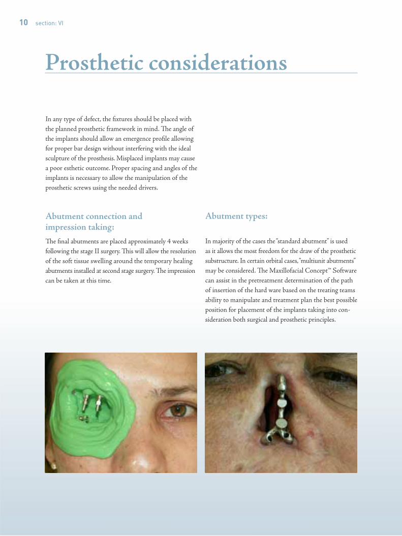
In any type of defect, the fi xtures should be placed with
the planned prosthetic framework in mind. Th e angle of
the implants should allow an emergence profi le allowing
for proper bar design without interfering with the ideal
sculpture of the prosthesis. Misplaced implants may cause
a poor esthetic outcome. Proper spacing and angles of the
implants is necessary to allow the manipulation of the
prosthetic screws using the needed drivers.
Abutment connection and impression taking:
Th e fi nal abutments are placed approximately 4 weeks
following the stage II surgery. Th is will allow the resolution
of the soft tissue swelling around the temporary healing
abutments installed at second stage surgery. Th e impression
can be taken at this time.
Abutment types:
In majority of the cases the “standard abutment” is used
as it allows the most freedom for the draw of the prosthetic
substructure. In certain orbital cases, “multiunit abutments”
may be considered. Th e Maxillofacial Concept™ Software
can assist in the pretreatment determination of the path
of insertion of the hard ware based on the treating teams
ability to manipulate and treatment plan the best possible
position for placement of the implants taking into con-
sideration both surgical and prosthetic principles.

section: VI 11
Healing period
Th e intra oral as well as the extra oral implant surgery is
performed in the traditional 2 stage manner. After instal-
lation of the implants, cover screws are placed and the
implants are submerged. All attempts are made not to
load the surgical sites during the osseointegration period.
Stage II, for intra-oral implants, uncovering of the implants
through the intra-oral mucosal tissue is performed 7 months
after installation of the fi xtures. Exposure of the extra-oral
implants through the cutaneous tissue is performed 4
months following the stage I surgery.
6 months post implant placement. Inflammation free soft tissue adaptation ready for impression taking.

12 section: VII
In cases of the irradiated patient planned for placement
of intra oral and or extra oral implants, hypobaric oxygen
treatments (HBO) should be considered.
Th e general protocol for HBO treatment in this group
includes; 20 dives prior to the fi xture placement followed
by 10 additional dives after the placement of the fi xtures.
Th e osseointegration period for the extra-oral implants is
extended to 6 months in the irradiated patients.
Management of the irradiated patient
Hyperbaric Oxygen Chamber for irradiated patients Patient radiated with 7500 RADS, ready for 20 pre-operative HBO dives

section: VIII 13
Long term maintenance
Extra-oral application:
In order to increase the long term success of the extra-oral
implants, maintenance of healthy cutaneous cuff around
the extra-oral abutments is crucial. Daily cleansing of the
percutaneous abutments using cotton swabs moistened
with peroxide followed by application of chlorhexadine
has shown to be suffi cient. A soft bristled brush may also
help in removal of debris. It is important to avoid the use
of sharp or metal instruments around the percutaneous
abutments. Th e prosthesis should also be cleansed daily
using soap and water. Care should be taken to maintain a
clean surface in order to avoid damage to the prosthesis.
Bacteria and or fungal colonization on the surface of the
prosthetic may cause skin irritation too. Sleeping with
the prosthesis removed allows ventilation around the
percutaneous abutments and should be encouraged.
Classification of skin Condition:
0 = No irritation
1 = Slight redness
2 = Red and slightly moist tissue.
3 = Red and slightly red tissue with presence of
granulation issue around he percutaneous cuff .
4 = Overt signs of infection with fi xture mobility.
Type 1 skin condition

14 section: VIII
Management of skin conditions:
• For grade 1 conditions: Application of Terracortil
with Polymyxin B (An ointment with hydro cortisone
acetate, oxytetracycline hydro chloride and Polymyxin
n B sulphsate).
• For grade 2 conditions: Terracortil
• For grade 3 conditions: Local soft tissue revision should
be performed.
• For grade 4 conditions: Removal of the eff ected fi xture.
Management of the Skin tissues around the extra-oral abutments:
Minimal soft tissue trauma during the surgical phase
of the reconstruction in conjunction with the thinning
of the subcutaneous tissues is critical for the long term
maintenance of the extra-oral implants. Minimizing
motion around the abutments by application of a pressure
dressing will also help in stabilizing the soft tissue
adaptation around the extra-oral abutments. Th e use
of antibiotics and hydrocortisone applied to the pressure
dressing will aid in reducing the infl ammation as well as
prevent bacterial colonization post operatively.
Longterm maintenance

section: IX 15
Epidemiology
Non-radiated Patient:
Treatment of patients with Maxillofacial defect using
osseointegrated implants have been studied in multiple
International centers. Success rates of 94.4%, 96.3%
and 97% have been reported by these centers in the
non-radiated patients.
• San Antonio, Sweden, 13 centers Parel S,
Tjellstrom A, IJOMI, 1991
• Canada (6), Sweden, US, Wolfaardt J, et al,
IJOMI, 1993
• FDA Study, 24 Centers, Tolman D, Taylor P,
IJOMI, 1996
Irradiated Patient:
Success of maxillofacial implants in the irradiated patients
range from 57.9–64.7% as reported by Dr Parel.
A later study confi rmed these outcomes by reporting a
success rate of 62%.
Parel,et al, IJOMI,1991;6:75-79.
Granstrom G,etal,IJOMI,1994;9:653-662
“Through our broad based co operation with International clinical teams, we can continue to spread the knowledge about osseointegration. At the same time we are, ourselves, enriched by meeting new and competent co-workers.”
PI Brånemark

16 section: X
Clinical case 1
1. Necrotic midface secondary to neoplastic lesion. 2. Post debridment surgery.
3. Placement of Quad zygoma implants.
4. Implant retained prosthetic bar, Dental prosthesis and facial 5. 6 years post reconstruction.

section: X 17
Clinical case 2
1. Pre-operative frontal view
4. Implant and prosthetic substructure
2. Patient is missing her maxilla, nasalseptum and nose.
3. Prosthetic substructure in place 5. Patient with her grandchildren, 15 years post reconstruction

Clinical case 3
1. Pre-op showing orbital-nasal communication 2. Lateral orbital fixtures and obturator sealing the orbital-nasal communication
3. Orbital prosthesis; Magnet retained
5. Symmetrical orbital profile
18 section: X
4. Post-operative frontal view

Clinical case 4
1. Pre-operative frontal view 2. Post-operative film showing 3 fixtures placed
3. Retaining bar secured to fixtures 4. Clip retained nasal prosthesis
section: X 19

20
Craniofacial Prostheses. Anaplastology and Osseointe-
gration. Eds: P-I Brånemark, M. Ferraz de Oliveira,
Quintessence Publ. Co. Inc, Chicago, IL, USA (1997).
Osseointegration in Craniofacial Reconstruction. Eds:
P-I Brånemark,DE Tolman, Quintessence Publ.Co.Inc,
Chicago, IL USA. (1998).
Rehabilitation of Complex Cleft Palate and Craniomaxillo-
facial Defects. Th e Challenge of Bauru. Eds: P-I Brånemark,
K. Higuchi, M. Ferraz de Oliveria, Quintessence Publ.
Co.Inc. Chicago,IL,USA (1999).
Th e Osseointegration Book, From Calvarium To
Calcaneus. Eds: Brånemark P-I, Chien S, Gröndahl H-G
and Robinson K. Quintessenz Verlag-GmbH, Berlin,
Germany (2005).
References
