brain waves - home - le bonheur children's hospital
TRANSCRIPT

Summer 2013
Brain WavesNeuroscience Institute
Referrals: 888-890-0818
www.lebonheur.org/neuroscience
A pediatric partner
with The University
of Tennessee Health
Science Center/College
of Medicine and
St. Jude Children’s
Research Hospital
Memphis, Tennessee
Michael Boyd had a seizure at school. At
the Emergency Department radiologists found an abnormal spot, and the MRI confirmed the 14-year-old from Lebanon, Va., had a mass on his left frontal lobe. Physicians referred Michael to Niswonger’s Children’s Hospital in Johnson City, Tenn. There, doctors recommended Michael fly immediately 500 miles across the state to Le Bonheur Children’s Hospital.
“They told us that Dr. Frederick Boop was the best. He had done many surgeries like this before,” said mother Amy Boyd.
When the family arrived in Memphis, Michael’s room was waiting. The doctors and nurses began to explain what was going to happen. Michael underwent a series of diagnostic tests – MRI, functional MRI, tractography, spectroscopy, magnetoencephalography (MEG) and transcranial magnetic stimulation (TMS) — all completed on his first day at Le Bonheur.
“Before they took him for each test, they spoke to all of us. They explained what each test was for and how it was going to help the doctors see and understand the tumor. Our biggest fear was not knowing how the tumor was attached,” said Amy.
Michael had a fibrillary astrocytoma, a low-grade tumor that is not likely to spread to other parts of the body. Michael’s tumor was located in the left frontal lobe, in an area which typically controls language functions. The tumor had an infiltrating appearance and might possibly impair functions as it grew.
Because of the tumor’s high-risk location, the wide range of diagnostic tools available
at Le Bonheur gave Michael’s surgical team important information.
“The goal is maximum lesion resection with minimal functional deficit,” said Neuroradiologist Asim Choudhri, MD. “These images served as a road map and helped Dr. Boop remove as much of the tumor’s margins
without impairing functions.”The surgical team used the intraoperative MRI. “There came a margin in which the tissue of the
tumor began to blend with the tissue of the normal brain and once we were unable to tell brain from tumor we felt it best to stop. If this small area starts to grow down the road he has several treatment options but for now we will just follow him by MRI,” said Rick Boop, MD, chairman of the Department of Neurosurgery.
Four days after surgery, Michael was ready to go home. He experienced slurred speech and occasional headaches for a few weeks after surgery, but he’s now returned to feeling like himself.
“He’s doing great now. He told us that he felt he was doing better in school now since the tumor is gone,” said Amy.
Michael will be monitored by phy-sicians at St. Jude Children’s Research Hospital, Niswonger’s Children’s Hospital and Le Bonheur.
Surgeons remove fibrillary astrocytoma in 14-year-old, preserve language functions
Michael Boyd sees Dr. Frederick Boopfive months after his brain tumor surgery.
1. Axial FLAIR MRI image2. Axial FLAIR with Diffusion Tensor Imaging (DTI) overlay to show impact of the mass on adjacent nerve bundles3. Axial FLAIR image with functional MRI (fMRI), with pink showing areas of expressive language (Broca’s area)4. Axial T2 image from intraoperative MRI (iMRI) showing resection cavity corresponding to the previously seen left inferior frontal lobe mass. A thin rim of lesion was left at the posterior rim due to the adjacent language activation centers. The remainder of the margins show no residual tumor.
1. 2. 3. 4.
SCANS OF MICHAEL BOYD’S BRAIN TUMOR
Watch a video case study with Neuroradiologist Asim Choudhri, MD, at www.lebonheur.org/boyd
Dr. Frederick Boop and Dr. Asim Choudhri review Michael Boyd’s MRI scan.
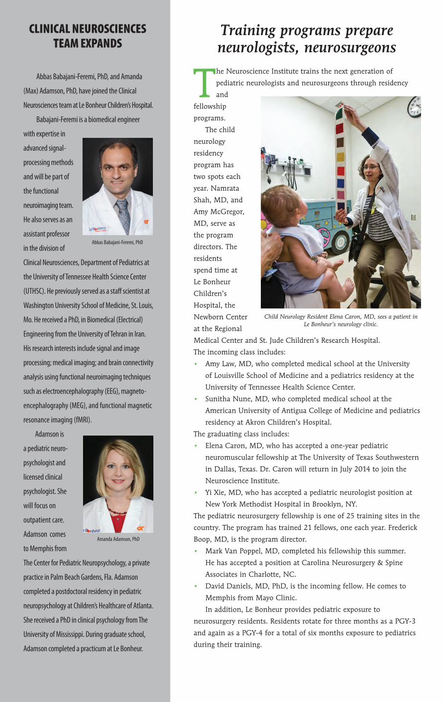
Training programs prepare neurologists, neurosurgeons
The Neuroscience Institute trains the next generation of
pediatric neurologists and neurosurgeons through residency
and
fellowship
programs.
The child
neurology
residency
program has
two spots each
year. Namrata
Shah, MD, and
Amy McGregor,
MD, serve as
the program
directors. The
residents
spend time at
Le Bonheur
Children’s
Hospital, the
Newborn Center
at the Regional
Medical Center and St. Jude Children’s Research Hospital.
The incoming class includes:
• Amy Law, MD, who completed medical school at the University
of Louisville School of Medicine and a pediatrics residency at the
University of Tennessee Health Science Center.
• Sunitha Nune, MD, who completed medical school at the
American University of Antigua College of Medicine and pediatrics
residency at Akron Children’s Hospital.
The graduating class includes:
• Elena Caron, MD, who has accepted a one-year pediatric
neuromuscular fellowship at The University of Texas Southwestern
in Dallas, Texas. Dr. Caron will return in July 2014 to join the
Neuroscience Institute.
• Yi Xie, MD, who has accepted a pediatric neurologist position at
New York Methodist Hospital in Brooklyn, NY.
The pediatric neurosurgery fellowship is one of 25 training sites in the
country. The program has trained 21 fellows, one each year. Frederick
Boop, MD, is the program director.
• Mark Van Poppel, MD, completed his fellowship this summer.
He has accepted a position at Carolina Neurosurgery & Spine
Associates in Charlotte, NC.
• David Daniels, MD, PhD, is the incoming fellow. He comes to
Memphis from Mayo Clinic.
In addition, Le Bonheur provides pediatric exposure to
neurosurgery residents. Residents rotate for three months as a PGY-3
and again as a PGY-4 for a total of six months exposure to pediatrics
during their training.
Child Neurology Resident Elena Caron, MD, sees a patient in Le Bonheur’s neurology clinic.
CliniCal neurosCienCes team expands
Abbas Babajani-Feremi, PhD, and Amanda
(Max) Adamson, PhD, have joined the Clinical
Neurosciences team at Le Bonheur Children’s Hospital.
Babajani-Feremi is a biomedical engineer
with expertise in
advanced signal-
processing methods
and will be part of
the functional
neuroimaging team.
He also serves as an
assistant professor
in the division of
Clinical Neurosciences, Department of Pediatrics at
the University of Tennessee Health Science Center
(UTHSC). He previously served as a staff scientist at
Washington University School of Medicine, St. Louis,
Mo. He received a PhD, in Biomedical (Electrical)
Engineering from the University of Tehran in Iran.
His research interests include signal and image
processing; medical imaging; and brain connectivity
analysis using functional neuroimaging techniques
such as electroencephalography (EEG), magneto-
encephalography (MEG), and functional magnetic
resonance imaging (fMRI).
Adamson is
a pediatric neuro-
psychologist and
licensed clinical
psychologist. She
will focus on
outpatient care.
Adamson comes
to Memphis from
The Center for Pediatric Neuropsychology, a private
practice in Palm Beach Gardens, Fla. Adamson
completed a postdoctoral residency in pediatric
neuropsychology at Children’s Healthcare of Atlanta.
She received a PhD in clinical psychology from The
University of Mississippi. During graduate school,
Adamson completed a practicum at Le Bonheur.
Abbas Babajani-Feremi, PhD
Amanda Adamson, PhD

Neurosurgeon named secretary of AANS
The American Association of Neurological Surgeons has named Frederick Boop, MD, FAANS, FACS, as its secretary. Boop is a professor and chairman of the neurosurgery department at the University of Tennessee Health Science Center and co-director of the Neuroscience Institute.
Neuroradiologists share expertise
Neuroradiologists Asim F. Choudhri, MD, and Matthew T. Whitehead, MD, presented research at the American Society of Neuroradiology Annual Meeting. Choudhri made six presentations and served on the exhibit award committee; Whitehead made three presentations. Their collaborators included: Harris L. Cohen, MD, James Wheless, MD, Amy McGregor, MD, Frederick A. Boop, MD, Paul Klimo Jr, MD, Stephanie Einhaus, MD, and C. Bruce MacDonald, MD.
Choudhri, also presented lectures, grand rounds and board reviews at Johns Hopkins University, University of Washington and Naval Medical Center San Diego.
Physicians influence concussion legislation
Tennessee Gov. Bill Haslam has signed legislation that requires Tennessee schools and youth athletic organizations to adopt concussion policies. The legislation is designed to ensure that parents, coaches and students are educated about the symptoms and implications of concussion and defines what health providers are authorized to release a youth athlete to resume exercise and play.
Dr. Paul Klimo, chief of the Division of Pediatric Neurosurgery at Le Bonheur Children’s and the University of Tennessee Health Science Center, and Dr. Trey Eubanks, pediatric surgeon and medical director of Trauma at Le Bonheur, worked with other advocates and groups including TMA, TNAAP, CHAT and Tennessee Disability Coalition to write the legislation. Klimo also served as a panelist on the University of Memphis’ Papasan Public Policy Forum to educate lawmakers in Nashville on this important topic.
The legislation was overwhelmingly approved 93-3 in the House of Representatives and unanimously 30-0 in the state Senate. The law will go into effect on Jan. 1, 2014.
Experts share TSC knowledge Research from James Wheless, MD, and Roozbeh
Rezaie, PhD, was presented at the 2013 International Tuberous Sclerosis Complex Research Conference held in Washington, DC, this summer.
Wheless’ poster presentation, “A Novel Topical Rapamycin Cream for Treatment of Facial Angiofibromas in Tuberous Sclerosis Complex,” is also described in this newsletter on page 4.
Other presentations include the economic burden on families, humanistic burden in caregivers , health care resource utilization and patient-reported physical and mental health burden.
Pediatric Neurology Update held, 2014 event planned The seventh annual Greater Mid-South Pediatric
Neurology Update was held in May. More than 70 neurologists and other pediatric caregivers attended the conference. Attendees represented 20 different institutions from 15 states. Also, seven organizations were represented in the exhibition area.
Guest faculty included Jonathan W. Mink, MD, PhD, and Raman Sankar, MD. Mink is a professor of Neurology, Neurobiology & Anatomy, Brain & Cognitive Sciences, and Pediatrics and chief of the Division of Child Neurology at the University of Rochester Medical Center. Sankar is chief of Pediatric Neurology at University of California Los Angeles.
The eighth annual Update will be held in May 2014. For more information, visit www.methodistmd.org or call (901) 516-8933.
IN BrIEf
Orthopaedics and Neurosurgery teams partner to remove spinal tumors
At the age of 10, Mary Kaitlyn “Katie” Myers of Madison, Miss., was diagnosed with astrocytoma – an intramedullary
tumor growing from inside her spinal cord. “We noticed she was walking crooked,” said her mother,
Donna Myers. A local orthopaedic surgeon ordered an MRI to find the cause of her atypical scoliosis. Scans revealed a spinal tumor.
Katie was sent to Le Bonheur Children’s Hospital in Memphis, Tenn., where the Myers family met with William Warner, MD, a Le Bonheur orthopaedic surgeon, and Frederick Boop, MD, chair of the Department of Neurosurgery at the University of Tennessee Health Science Center and co-director of the Neuroscience Institute.
Le Bonheur’s orthopaedic and neurosurgery teams provide comprehensive surgical care for pediatric spinal tumor patients.
“Our spinal cord tumor program, a combined program with St. Jude Children’s Research Hospital, is one of the busiest spinal tumor programs in the country. Working as a team with the neurosurgeons of Semmes-Murphey Clinic and the orthopaedic surgeons at Campbell’s Clinic, the program brings an unparalleled level of expertise to children with this rare disorder,” said Boop.
Katie needed surgery to resect the tumor and determine her prognosis.
“I didn’t believe it was happening,” said Katie, now 17, of her diagnosis.
“From the delicate micro-neurosurgery required to remove an intra-spinal tumor to the complexities of reconstructing a growing spine afterwards, the management of complex tumors
of the spine requires a multidisciplinary team, not just for the surgery but for the post-operative care and rehabilitation needed for these children,” Boop said.
The surgery was a success, and doctors were able to remove all of the tumor. Katie remained at Le Bonheur for 5 days as she recovered from surgery and continued therapy at home to regain her strength and mobility.
Now a senior in high school, Katie continues to see Warner annually for checkups and is doing well. She loves to swim, ride bikes and go to the beach. She’s had two surgeries since her initial operation in 2006. The first one in 2007 was to stabilize a spondlyolisthesis of the fifth lumbar vertebrae — a condition Warner detected during
her initial operation. The second in 2008 was to stabilize her spine due to progressive instability of her cervical spine after the tumor had been removed.
“I had the best care team at Le Bonheur,” said Katie. “The doctors and nurses have become my favorite people. They saved my life.”
Mary Kaitlyn “Katie” Myers of Madison, Miss., 17, was diagnosed with astrocytoma – an
intramedullary tumor growing from inside her spinal cord. Now a senior in high school,
Katie enjoys swimming and riding bikes.
Frederick Boop, MD
Asim F. Choudhri, MD
Matthew T. Whitehead, MD
Roozbeh Rezaie, PhDJames Wheless, MD
Paul Klimo, MD

Non-Profit Org.
US POSTAGEPAID
Memphis, TNPermit No. 3093
848 Adams AvenueMemphis Tennessee 38103
Patient at baseline
/ lebonheurchildrens@LeBonheurChild /lebonheurchildrens
Topical cream demonstrates positive results for TSC patients
researchers at Le Bonheur and the University
of Tennessee Health Science Center have
developed a topical cream to treat facial lesions
associated with Tuberous Sclerosis Complex (TSC).
The initial study follows the treatment of two
patients and was published online in the Journal
of Child Neurology.
TSC is a neurocutaneous disorder characterized
by excess cell growth and proliferation, resulting in
multi-organ hamartomatosis. Skin lesions occur in
more than 90 percent of TSC patients and are more
common in late childhood or adolescence.
Historically, the skin lesions have been resistant
to medical and surgical treatments. Oral rapamycin
has been used in TSC patients, but the side effects
prevent its routine use without significant internal
involvement.
Neuroscience Institute Co-director James
Wheless, MD, collaborated with Hassan Almoazen,
PhD, to create a novel rapamycin cream. The cream
is easy to compound and apply, does not cause local
or systemic side effects, and results in a dramatic
improvement of facial angiofibromas. Patient at 12 months of treatment
Memphis team to study new VNS device
Researchers at Le Bonheur are launching a new study of the latest Vagus Nerve Stimulation (VNS) device. The Memphis hospital is one of a few sites studying the Model 106 Generator by Cyberonics.
VNS is an adjunctive therapy in reducing the frequency of seizures in adults and adolescents with partial onset seizures not controlled by medication or who experience intolerable side effects.
The Model 106 Generator has a new feature that allows the generator to sense changes in the heart rate and deliver stimulation in response to those changes, automatically. This device uses cardiac-based seizure detection and Automatic Magnet Mode (AMM) technology.
The information obtained from patient self-reports (seizure frequency, severity, duration, intensity and post-ictal duration) during the course of study will be assessed to help establish the best clinical outcomes for the Model 106 Generator.
For more information, contact Clinical Research Coordinator Tracee Ridley-Pryor, RN, MSN, CCRC, at (901) 287-5338 or [email protected].
Brain Waves is a quarterly publication of the Neuroscience Institute at Le Bonheur Children’s Hospital. The institute is a nationally recognized center for evaluation and treatment of nervous system disorders in children and adolescents, ranging from birth defects and learning and behavioral disorders to brain tumors, epilepsy and traumatic injuries.
Institute co-directorsFrederick Boop, MDAndrew Papanicolaou, PhDJames W. Wheless, MD Amanda Adamson, PhD Adam Arthur, MDAbbas Babajani-Feremi, PhD Paras Bhattarai, MD Asim F. Choudhri, MD Nancy Clanton, PhDStephanie Einhaus, MDLucas Elijovich, MDJulius Fernandez, MDStephen Fulton, MD Masanori Igarashi, MDPaul Klimo, MDAmy McGregor, MDKathryn McVicar, MDRobin L. Morgan, MDBasanagoud Mudigoudar, MDMichael S. Muhlbauer, MDShalini Narayana, MBBS, PhDBrian Potter, Psy DRoozbeh Rezaie, PhD Robert Sanford, MDNamrata Shah, MDMatthew Whitehead, MD
Scan to learn more about our Neuroscience Institute.