brain computer interface to enhance episodic memory in
TRANSCRIPT

Frontiers in Human Neuroscience Research Article8 December 2014
1
Brain computer interface to enhance episodicmemory in human participantsJohn F. Burke 1, Maxwell B. Merkow 2, Joshua Jacobs 3 Michael J. Kahana 4,∗ andKareem A. Zaghloul 5,∗1Perelman School of Medicine, University of Pennsylvania, Philadelphia, PA, USA2Department of Neurosurgery, University of Pennsylvania, Philadelphia, PA, USA3Biomedical Engineering, Science & Health Systems, Drexel University, Philadelphia, PA, USA4Department of Psychology, University of Pennsylvania, Philadelphia, PA, USA5Surgical Neurology Branch, NINDS, National Institutes of Health, Bethesda, MD, USA
Correspondence*:Kareem A. ZaghloulSurgical Neurology Branch, NINDS, National Institutes of Health, Bethesda, MA, 20894, USA,[email protected] J. KahanaDepartment of Psychology, University of Pennsylvania, Philadelphia, PA, 19104, USA,[email protected]
ABSTRACT2
Recent research has revealed that neural oscillations in the theta (4-8 Hz) and alpha (9-143Hz) bands are predictive of future success in memory encoding. Because these signals occur4before the presentation of an upcoming stimulus, they are considered stimulus-independent in5that they correlate with enhanced memory encoding independent of the item being encoded.6Thus, such stimulus-independent activity has important implications for the neural mechanisms7underlying episodic memory as well as the development of cognitive neural prosthetics. Here,8we developed a brain computer interface (BCI) to test the ability of such pre-stimulus activity9to modulate subsequent memory encoding. We recorded intracranial electroencephalography10(iEEG) in neurosurgical patients as they performed a free recall memory task, and detected11iEEG theta and alpha oscillations that correlated with optimal memory encoding. We then used12these detected oscillatory changes to trigger the presentation of items in the free recall task.13We found that item presentation contingent upon the presence of prestimulus theta and alpha14oscillations modulated memory performance in more sessions than expected by chance. Our15results suggest that an electrophysiological signal may be causally linked to a specific behavioral16condition, and contingent stimulus presentation has the potential to modulate human memory17encoding.18
Keywords: BCI Episodic Memory ECoG Theta19
1 INTRODUCTION
In the laboratory setting, episodic memory is commonly studied by presenting participants with a list of20items and then asking them to later recall those items. For over a century [1], analysis of behavioral data21from these tasks has highlighted many intriguing facets of the memory system [2]. Recently, the ability22to record electrophysiological data from participants engaging in a memory task has begun to reveal23the neural correlates of these behavioral phenomena. While important electrophysiological hallmarks of24
1

Burke et al. episodic memory BCI
encoding and retrieval are evident in the time domain [3], many lines of evidence have suggested that25neural oscillations have a unique functional role in the memory system [4, 5, 6].26
In particular, data from electroencephalography (EEG) [7], magnetoencephalography [8], and27electrocorticography (ECoG) [9, 10] have revealed that changes in theta (4-8 Hz) and alpha (10-1428Hz) oscillations correlate with successful episodic memory encoding and retrieval. In terms of spatial29specificity, theta activity has been most commonly observed in the medial temporal lobe and the prefrontal30cortex [11, 5]. Indeed, the degree of theta synchronization between these structures has been shown to31predict the degree of memory formation [12, 13, 14]. Similarly, alpha activity has been shown to play32a role in memory processing, although the precise direction and meaning of such alpha activity is less33certain [15, 16]. Nonetheless, studies using trans-cranial magnetic stimulation have suggested that such34alpha activity also synchronizates across large regions of cortex during memory processing [17].35
These theta and alpha electrophysiological correlates of episodic memory have traditionally been observed36after stimulus presentation, and are thus interpreted to reflect the act of forming or retrieving a memory.37Recent research has extended these findings to the time interval before stimulus presentation. Specifically,38spectral activity in the theta and alpha frequency bands has been reported to increase prior to successful39memory encoding [18, 19, 20, 21, 22] and retrieval [23]. The observation that on-going neural activity can40predict future memory performance is consistent with observations from both scalp EEG and functional41imaging studies [24, 25, 26, 27]. More specifically, although human surface recordings have suggested that42theta power in particular is elevated before successful encoding [19, 20, 21], human intracranial recordings43from the medial temporal love have also consistently identified an alpha component to this pre-stimulus44activity [18, 22]. Thus, empirically, there is data to support that both theta and alpha oscillations play a45role in human pre-stimulus memory processing.46
These results place episodic memory into the larger context of higher order cognitive functions that are47enhanced by ongoing oscillatory activity [28, 29]. The functional role of such oscillations in relation to the48cognitive event of interest remains speculative; possible mechanisms include increased attention [30, 31],49phase reorganization to optimize encoding or retrieval [32, 33], or the evolution of temporal context50[34, 35]. It is clear, however, that pre-stimulus oscillations, especially in the theta and alpha frequency51bands, are correlated with a heightened ability to both encode and retrieve memories. Therefore, if devices52could be designed to induce these signals, it may be possible to selectively enhance the episodic memory53system [36].54
Before devices can be engineered using these pre-stimulus signals, however, it is necessary to establish55their causal role, if any, during memory encoding. In particular, the presence of an oscillation before the56successful encoding of an episodic memory does not necessarily suggest that inducing that oscillation will57enhance successful encoding. Brain computer interface (BCI) experimental paradigms offer an attractive58methodology to test this set of issues. Using real-time feedback, a particular electrophysiological event59of interest can be used to trigger the presentation of an item to be remembered, and the corresponding60behavioral response can subsequently be investigated [37, 38, 39]. This reverses the traditional dependent61and independent variables of the experiment: instead of analyzing electrophysiological correlates of62memory, we can analyze the mnemonic correlates of electrophysiology. If the neural oscillation plays63a mechanistic role in memory encoding, then a modulation of the electrophysiology should cause an64analogous modulation of the behavioral response. Studies using this BCI approach have established that65pre-stimulus theta oscillations in the rabbit hippocampus are sufficient to double the learning rate in an66associative learning task [40, 41]. Here, we implement a similar approach in humans participants to67investigate the role of pre-stimulus theta oscillations in episodic memory.68
This is a provisional file, not the final typeset article 2

Burke et al. episodic memory BCI
2 MATERIAL & METHODS
2.1 PARTICIPANTS
Participants with medication-resistant epilepsy underwent a surgical procedure in which grid, strip,69and depth electrodes were implanted for seizure localization. Data were collected at Thomas Jefferson70University Hospital and the Hospital of the University of Pennsylvania. Our research protocol was71approved by the institutional review board at each hospital and informed consent was obtained from72the participants and their guardians. Our final participant pool consisted of 14 patients (5 Female) left-73language dominant). Language dominance was assessed by either the patients’ handedness, a clinically74administered intracarotid injection of sodium amobarbital (Wada test), or fMRI using a verb generation75task (Thomas Jefferson Hospital).76
2.2 RECORDINGS
Clinical indications alone determined electrode number and placement. Subdural (grids and strips) and77depth contacts were spaced 10 mm and 8 mm apart, respectively. Depth electrodes are placed using a78frameless stereotactic approach. We recorded intracranial EEG (iEEG) using a Nicolet, Grass Telefactor,79or Nihon-Kohden EEG system. Depending on the amplifier and the discretion of the clinical team, the80signals were sampled at 400 Hz (Grass), 512 Hz (Nicolet) 500 Hz, 1000 Hz, or 2000 Hz (Nihon Khoden).81Signals were referenced to a common contact placed subcutaneously, on the scalp, or on the mastoid82process. The testing laptop sent +/-5 V analog pulses via an optical isolator into a pair of open lines on83the clinical recording system to synchronize the electrophysiological recordings with behavioral events.84
For post-hoc analyses, all recorded traces were resampled at 256 Hz, and a fourth order 2 Hz stopband85butterworth notch filter was applied at 60 Hz to eliminate electrical line noise. In addition, signals were86converted to a bipolar montage by differencing the signals between each pair of immediately adjacent87contacts on grid, strip, and depth electrodes. We defined the bipolar montage in our data-set based on the88geometry of ECoG electrode arrangements. For every grid, strip and depth probe, we isolated all pairs89of contacts that were positioned immediately adjacent to one another; bipolar signals were then found by90differencing the signals between each pair of immediately adjacent contacts [13]. The resulting bipolar91signals were treated as new virtual electrodes (henceforth referred to as electrodes throughout the text),92originating from the mid-point between each contact pair [14]. All subsequent analyses were performed93using these derived bipolar signals.94
Contact localization was accomplished by co-registering the post-op CTs with the post-op MRIs using95both FSL Brain Extraction Tool (BET) and FLIRT software packages and mapped to both MNI and96Talairach space using an indirect stereotactic technique and OsiriX Imaging Software DICOM viewer97package. Pre-op MRI’s were used when post-op MR images were were not available.98
2.3 FREE RECALL TASK
Standard version Each patient participated in both standard and BCI versions of a free recall99episodic memory task. Tasks were administered at the patient’s bedside using the python experimental100programming language (PyEPL) [42]. In each version, participants were shown lists of common nouns101chosen at random and without replacement from a pool of high-frequency nouns. Each word was visually102presented to the patient on a laptop computer screen placed at an arm’s length from the patient. Each103experimental session of the standard version contained up to 20 lists, and each list contained 15 words.104At the start of each trial, a plus sign appeared at the center of the screen to alert patients to the upcoming105word presentation and to encourage them to fixate on the center of the screen. The plus sign appeared for1061600 msec, followed by an inter-stimulus interval (ISI). The length of the ISI depended on each version107of the task. In the standard version of the task, the ISI was 800 ms followed by a randomly jittered 0108to 400 ms blank interval. The random ISI served to decorrelate physiological responses from successive109word presentations. In the BCI version of the task, lists were composed of 10 words that also appeared110
Frontiers in Human Neuroscience 3

Burke et al. episodic memory BCI
on the screen for 1600 ms. However, in the BCI version of the task, the length of the ISI depended on the111electrophysiologic data (see below).112
Immediately after each list presentation, patients were given a series of simple arithmetic problems. This113end-of-list distractor task served to reduce the large advantage accorded to end-of-list items during recall114[43]. Each problem took the form of A+B +C =??, where A, B, and C were randomly chosen positive115integers from the set one through nine. After patients solved arithmetic problems for at least 20 sec, we116presented a row of asterisks accompanied by a 300 msec tone signaling the start of the recall period.117Patients were given 45 sec to recall list items in any order (standard free-recall instructions). After each118session, vocal responses, digitally recorded during the trial, were scored for analysis. Words recalled from119the most recent list were considered correct recalls.120
Brain Computer Interface version In the BCI version of the task, the timing of word presentation121depended on the detection of a pre-determined neural oscillation captured from an intracranial contact.122To control for variability in ISI’s, each experimental session was composed of twenty lists of words123divided into two blocks of ten. Lists were composed of 10 study items. In the first block, lists randomly124alternated between a contingent condition and a control condition. In the contingent condition (half the125lists in the block), presentation of words, and hence the ISI, were contingent on whether a calculated126index of power exceeded a pre-determined high threshold. We recorded all ISIs used in the contingent127condition, and the first list in the block was always a contingent condition. In the control condition (the128remaining half of the lists in the first block), we presented words using the same sequence of ISIs used129in one of the contingent conditions, regardless of neuronal oscillatory activity. By using an identical ISI130sequence, equal amounts of time are allocated for stimulus encoding in both conditions. In the second131block of the contingent condition, we used an identical procedure for determining ISIs, but in this case132word presentation during the contingent condition was determined by whether the calculated index of133power decreased below a pre-determined low threshold. We compared behavioral performance between134the high and low contingent conditions and each condition’s respective control condition. Thus, items in135the contingent condition were presented based on the amount of power in during the 600 ms preceding136each item. In contrast, the items in the control condition, items were presented at random periods with137respect to the on-going electrophysiological activity.138
2.4 BRAIN COMPUTER INTERFACE
The closed loop experimental procedure used to present oscillatory contingent word items is shown in139Figure 1. We used a Y-splitter to provide a copy of the recorded iEEG signals (Figure 1A) to a research140recording system (Neuralynx, Inc. Digital Lynx data acquisition system, Bozeman, MT; Figure 1B).141We amplified iEEG signals, sampled at 32 kHz, and bandpass filtered between 0.3 and 300 Hz. We142temporarily stored the iEEG signal in a Matlab R© (The Mathworks, Inc., Natick, MA) readable buffer143using the MatCom software package (Neuralynx, Inc.) before it was written to disk to enable real-time144data processing. Data from each electrode stored in this buffer was immediately downsampled to 256145Hz and then used to update and fill a 600 ms sliding window every 50 ms. We extracted theta or alpha146oscillatory power from this 600 ms window using a Fast-Fourier Transform (Figure 1C; see results). We147visualized the intracranial EEG signal from the chosen electrode, and the resulting calculated index of148power, in real-time using a custom Matlab GUI (Figure 1D). To calculate an index of oscillatory power,149we normalized the power in the frequency band of interest by dividing power in this band over the power of150equal bandwidths immediately above and below the frequency bandwidth of interest [40]. The calculated151index of power was used to determine the timing of word presentation during the BCI version of the free152recall task (Figure 1E).153
This is a provisional file, not the final typeset article 4

Burke et al. episodic memory BCI
2.5 DATA ANALYSIS AND SPECTRAL POWER
Each participant performed a standard version of the task first in order to quantify memory related changes154in spectral power and to identify an optimal oscillation to be used to trigger word presentations in the155subsequent BCI version. Downsampled bipolar iEEG signals captured during the standard version of the156task were convolved with complex valued Morlet wavelets (wavelet number 7) to obtain magnitude and157phase information [44]. We used 50 wavelets with center frequencies logarithmically spaced between 2 Hz158and 100 Hz. We convolved each wavelet with 3500 ms of iEEG data surrounding each word presentation,159from 1000 ms before word onset to 2500 ms after word onset (a 1000 ms buffer was included on both160sides of the clipped data). We squared and log-transformed the magnitude of the continuous time wavelet161transform to generate a continuous measure of instantaneous power. We averaged these continuous power162spectra into a two time intervals: a pre-stimulus window (1000-0 ms before word presentation) and a163post-stimulus window (300-1500 ms after word presentation).164
We then z-transformed power values separately for each session using the mean and standard deviation165of each electrode’s power values sampled every 60 +/- 10 sec throughout the duration of the session166[45]. This method allowed us to estimate the mean and standard deviation of each session separately, and167corrects for any changes in impedance that occurred during that session.168
To assess memory related changes in spectral power within theta (4-8 Hz) and alpha (10-14 Hz)169frequencies, we averaged the instantaneous power across each time epoch, and calculated the average170power separately across theta and alpha frequencies. To account for changes in power across experimental171sessions, we z-transformed power values separately for each frequency and for each session using the172mean and standard deviation of 1000 ms epochs spaced every 60 ± 10 seconds during that session173[14, 46]. For every electrode and for every temporal epoch, we assessed the difference in z-scored spectral174power in the theta and alpha frequency bands during memory formation by calculating a t-statistic on the175distribution of power values during successful and unsuccessful encoding. We averaged these t-statistics176across electrodes for each patient. To generate a p value for changes in spectral power across patients, we177performed a one sample t test comparing these across patient distributions to zero [47].178
We also identified the electrodes that exhibited the most reliable change in theta and alpha power between179successful and unsuccessful encoding for each participant and for each experimental session of the180standard task. Specifically, we calculated a t-statistic (and a p-value) on the distribution of power values181during successful and unsuccessful encoding as above, however we focused on the pre-stimulus window182for this analysis. We selected the most reliable electrode and frequency band to use in the contingent183condition (as measured by the p-value), with the stipulation that the p-value must be below the 0.05 level184to be used as a trigger during the BCI version of the task.185
Once the electrode and frequency band were selected, we ran the BCI version of the task. To determine186if the oscillatory contingent presentation of study items significantly modulated memory performance,187we compared the rate of correct recall between the contingent and control conditions for both the high188and the low trigger conditions using a χ2-test. The χ2-test was generated using a 2x2 table with trigger189and control blocks as rows, and number of recalled and number of not recalled words as columns. We190determined whether an individual session demonstrated a significant modulation of memory performance191by identifying sessions that exhibited a significant difference in recall rate between the contingent and192control conditions (p < 0.05). To correct for multiple comparisons across sessions, we use a false193discovery rate procedure at the Q = 0.10 level [48]. Specifically, each of the 29 experimental BCI194sessions in this study provided two p-values using the chi-square tests, the p-values for the low and the195high-trigger conditions (Table 1). We applied the FDR correction across all of these statistical tests to196correct for multiple comparisons.197
Frontiers in Human Neuroscience 5

Burke et al. episodic memory BCI
3 RESULTS
Fourteen neurosurgical patients with medication resistant epilepsy underwent a surgical procedure in198which intracranial electrodes were placed for seizure monitoring. After the surgery, the patients were199monitored outside of the operating room with the intracranial electrodes in place for a period of 1-3200weeks. During this period, the participants agreed to run in two different tasks: a standard version of free201recall (stFR) followed by an oscillatory contingent, or brain computer interface (BCI), version (bciFR; see202Methods). In both versions of the task, participants were instructed to study a list of words and were then203asked to freely recall as many words as possible. However, the amount of time between successive word204presentations, or the interstimulus interval (ISI), differed in each version of the task. In the stFR task, the205ISI was set at a fixed interval of 800 ms with a 400 ms uniformly distributed jitter. In the bciFR task, the ISI206was determined by the amount of spectral power recorded from one of the patient’s intracranial electrodes207(Figure 1). We selected the electrode and the spectral frequency band based on the data collected in the208stFR task.209
The stFR task is part of a much larger, multi-center study that has been reported on previously [35, 14,21046, 22]. Here, however, we only report on the subset of patients that also participated in the bciFR task;211the BCI data are completely novel data and have not been previously reported in any study.212
Behaviorally, during the stFR task, the 14 participants studied 701.8 ± 382.2 words over 46.8 ± 25.5213lists, and successfully recalled 27.6 ± 7.6% of all words with a mean response time of 10,989.5 ± 3,415.8214msec (all values represent across patient averages and standard deviations).215
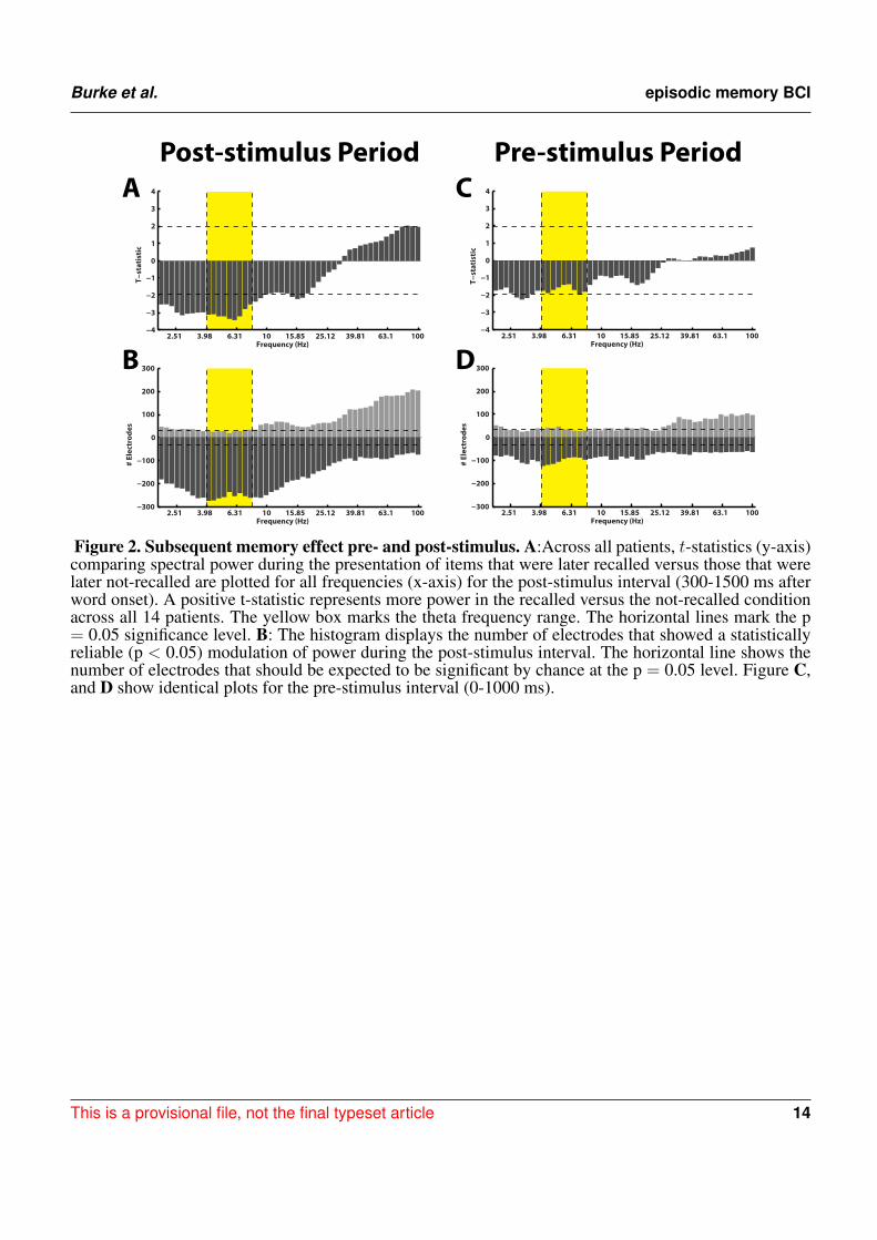
Electrophysiologically, we separated the stFR task in two time windows: the post-stimulus and the pre-216stimulus intervals. In the post-stimulus time interval (300-1500 ms after word presentation), we calculated217spectral power values for all word presentation periods, for each frequency, electrode, and patient (see218Methods). We compared these power values for words that were subsequently recalled and words that were219not recalled [49]. We found that, independent of anatomical location, successful encoding is associated220with an overall increase in high-frequency activity and a decrease in low-frequency activity (Figure 2A).221This result actually reflects a highly dynamic modulation of spectral power that occurs during successful222encoding, which was shown using a larger number of patients in the stFR task [46]. Next, we showed this223effect by counting the number of electrodes showing a modulation of spectral power (p < 0.05) during224successful encoding.225
The results in Figures 2A–B are consistent with previous reports, and show that the memory results from226the subset of patients in this dataset generalize to the overall population [46, 47]. However, we were227primarily interested in the pre-stimulus interval (0-1000 ms before word presentation), because activity228from the pre-stimulus interval could be used to drive the BCI version of the task. In Figures 2C–D, we229repeated the analyses described above for the pre-stimulus interval. We find that, first, there is not an230overall modulation of spectral power that correlated with successful encoding in the pre-stimulus interval231(Figure 2C). Second, although there was not an overall effect across all patients and brain regions in the232pre-stimulus interval, we did find that there were a few electrodes that showed a significant modulation of233theta/alpha power (Figure 2D).234
Even though there was not a reliable uni-variate spectral modulation in the pre-stimulus interval that235correlated with successful memory encoding, Figure 2D shows that there were certain electrodes that236showed a modulation of theta/alpha power during the pre-stimulus period. We therefore used the stFR237task to isolate individual electrodes that displayed the most reliable modulation of theta/alpha power in238the pre-stimulus interval. Figure 3 displays two such examples, one of which shows an overall positive239theta effect (more theta power during successful encoding; Figure 3A) and the other of which shows an240overall negative theta effect (less theta power during successful encoding; Figure 3B).241
Our goal was to identify if these individual pre-stimulus electrode fluctuations could be used to modulate242memory performance. To do that, we identified an electrode in each participant that exhibited the largest243difference (t-statistic) in theta or alpha oscillatory power between successful and unsuccessful encoding244during the pre-stimulus period. Table 1 gives a list of the patients who participated in the bciFR task, and245
This is a provisional file, not the final typeset article 6

Burke et al. episodic memory BCI
the electrodes (including the location) for each patient that we identified as the most reliable increases246in theta/alpha power for each patient. The table lists whether the theta or the alpha band was the most247reliable pre-stimulus modulation and the direction of the effect.248
Having identified a pre-stimulus marker for memory encoding, participants next performed the bciFR249task in which we integrated real-time data acquisition and analysis into the experiment (Figure 1). During250the task, we acquired iEEG signals in real-time from the identified electrode and stored this signal in a251600 msec sliding window, updated every 50 msec. We calculated an index of oscillatory power in the252identified frequency band of interest by comparing the ratio of the power within the identified band to253the power in adjacent frequency bands (see Methods) [40]. We used this index to trigger subsequent254word presentation in the oscillatory contingent condition of the task. In one block of each experimental255session, we triggered word presentation when this index exceeded a pre-determined threshold during the256contingent condition (see Methods). In the second block of each session, we triggered word presentation257when this index decreased below this threshold. During control conditions in each experimental block,258we used the recorded ISIs during the contingent condition to present word items, regardless of oscillatory259power recorded in that electrode. This controlled for the variable timing between word presentations.260
To confirm that our real-time system triggered word presentation only during the presence of the261identified oscillatory marker in the contingent conditions, we examined the average z-scored oscillatory262power during all word presentations in the contingent condition. In one participant, we triggered word263presentation off of alpha oscillatory power (Patient 12 in Table 1). We used this marker to trigger264word presentation during the contingent conditions of the BCI version of the task. Post-hoc analysis265of the average oscillatory power in the contingent conditions indeed revealed that word presentation was266preceded by increases or decreases in alpha oscillatory power when increases or decreases, respectively, of267the calculated index of power were used to trigger word presentation (Figures 4A). In a second participant,268we identified an electrode that demonstrated significant increases in theta oscillatory power during the269pre-stimulus encoding period in the standard version of the task (Patient 10; Table 1). Similarly, post-hoc270analysis of the average z-scored oscillatory power surrounding word presentation during the contingent271conditions revealed increases or decreases in theta oscillatory power preceding word presentation in the272contingent conditions of each experimental block (Figures 4B).273
If increases in pre-stimulus theta and alpha oscillatory activity identified in the standard version of the task274are causally related to memory, then we hypothesized that words presented during the presence of these275oscillations should be remembered more frequently than words presented at random times. We captured276behavioral data from 29 sessions of the BCI version of the task across 14 participants.277
Our goal was to modulate memory performance in all patients by first recording activity that correlated278with memory encoding, and then use that activity to trigger the presentation of words in a BCI task.279Upon implementation, the BCI that we constructed did not modulate memory performance in every280patient. However, we did find that the number of experimental sessions in which we elicited a relibale281difference (p<0.05; χ2-test; see methods) in memory performance using the contingent presentation282of stimuli was significantly greater than the number of sessions expected by chance (Table 1). In the283table, the orange boxes represent four sessions that displayed modulation of behavioral performance after284correcting for multiple comparisons using an FDR correction (q=0.10). The green boxes represents a285session that trended toward significance (p < 0.05), but did not survive multiple comparison correction.286In total, 10 sessions showed modulation of memory performance at the p = 0.05 level, which was more287than expected by chance at this significance level. In summary, although our results did not reliably288demonstrate improvement in memory performance, they do support the claim that pre-stimulus iEEG289confers information about the memory encoding state in some subjects.290
Frontiers in Human Neuroscience 7

Burke et al. episodic memory BCI
4 DISCUSSION
Neural oscillations have been hypothesized to play a mechanistic role in episodic memory formation,291however the link between oscillations and memory formation has been largely established by correlational292studies. In such studies, memory performance is recorded and used to partition electrophysiological293activity into high and low mnemonic states; then the activity in each state is compared to find oscillations294that co-vary with memory function. Using this approach, theta/alpha activity in the pre-stimulus interval295has been linked to memory formation. If such activity plays a mechanistic role in memory formation, then296it could be induced to give an individual a “memory boost” for an arbitrary set of items. For example,297an elderly patient could give themselves a boost before they encode where they parked their car, or when298their physician tells them how to use their medications. Such technology would have a major impact on299the ability of a patient with pathological memory loss to perform activities of daily living.300
A key step in the development of such technology is to investigate whether pre-stimulus theta/alpha301activity plays a mechanistic role in memory function, or whether such activity is a mere epiphenomenon.302In order to accomplish this first step, we constructed the BCI in Figure 1 to poll for theta/alpha oscillations303in real-time and link them to an episodic memory task. We found that pre-stimulus theta/alpha oscillations304were able to boost memory encoding reliably in a limited number of patients/sessions. The fact that a few305sessions were successfully modulated by spectral activity in the theta/alpha bands is an important proof-306of-principle that, in select cases, the BCI approach to enhance memory formation is feasible. Of note, 6/10307sessions that exhibited a modulation of memory performance (p < 0.05) were triggered off of contacts308in the medial temporal lobe (MTL; Table 1). More research is needed to assess whether the MTL has a309greater capacity for memory modulation then other regions.310
Across all patients, the effect was too variable to be implemented as a mnemonic device. Understanding311and reducing this variability represents the main hurdle in the realization of a mnemonic BCI to enhance312memory formation, and should be the focus of future research. One source of variability is that participants313likely have more than one strategy to form memories. This is especially true during free recall in which314memories are retrieved using a set of internal memory cues. Because these internal memory cues are315unconstrained, a variety of factors during encoding can influence the probability of later free recall. As316a result, memory encoding in free recall is very complex [43]; there is a well documented encoding317advantage for early-list items [50], late-list items [51], items nearby in list position [52], and items nearby318in semantic meaning [53].319
Behavioral studies have shown that participants use a combination of these encoding strategies to320remember the items, and such strategies are antagonistic. For example, the more a person recalls words321using a temporal encoding strategy, the less likely they are to use a semantic encoding strategy [54]. In322addition, many of these behavioral effects have different neurophysiological correlates [55, 56, 57, 58].323For example, if theta activity reflects a temporal encoding strategy [58], then reducing theta activity could324simply force the individual to rely on semantic encoding strategies, leaving the overall rate of recall intact.325This may explain why triggering off of theta power may not impact overall recall ability. Another example326of variability is the issue of whether theta increases or decreases predict memory formation. The amount327that decreases in theta power actually enhance human memory encoding may ultimately explain much of328the variance in these data, and future research should definitely link how theta oscillations relate to human329memory [59].330
Finally, we note that although this study used intracranial EEG to trigger word presentation, we recognize331that other studies have found non-invasively recorded medial temporal lobe theta activity is increased332before the presentation of items that are later successfully encoded (Guderian et al, 2009). Furthermore,333scalp EEG studies, which non-invasively record spatially correlated activity at the surface of the scalp,334have also detected changes in activity prior to encoding that influence subsequent memory encoding.335However, EEG is limited in its spatial resolution. Using iEEG, we can identify predictive markers for336subsequent encoding with good spatial specificity. Furthermore, depth electrodes enable the identification337of prestimulus activity within structures such as the hippocampus, which may be more effective as a338target for prediction during contingent conditions. Future studies would be well served to explore these339
This is a provisional file, not the final typeset article 8
Burke et al. episodic memory BCI
possibilities using surface recordings, but it still remains unclear whether the limited spatial resolution340offered by these recordings will afford sufficient specificity to predict subsequent memory encoding, and341may involve source localization procedures to target markers of encoding with greater fidelity.342
In conclusion, here we have linked pre-stimulus theta/alpha oscillations, which have been previously343correlated with the ability to encode memories, to the act of forming a memory. If such oscillations344play a mechanistic role in encoding, then their presence should boost memory formation. We found that,345although theta oscillations were able to improve memory in a few sessions, the result was not consistently346observed across all participants. The main utility of this work is that it is the first device, to our knowledge,347to use intracranial EEG in a BCI to enhance memory. This provides a proof-of-principle that a BCI driven348off of chronically implanted electrodes could serve as a “memory boosting” device.349
AUTHOR CONTRIBUTIONS
J.F.B., K.A.Z., J.J., and M.J.K. designed research; J.F.B. and M.B.M. performed research; J.F.B., K.A.Z.,350and M.B.M. analyzed data; J.F.B., K.A.Z., J.J., M.B.M. and M.J.K. wrote the paper.351
ACKNOWLEDGEMENT
We thank Dale H. Wyeth and Edmund Wyeth for technical assistance at Thomas Jefferson Hospital;352Ashwin G. Ramayya, Jeremy R. Manning, Emily A. Rosenberg, and Ryan B. Williams for helpful353discussion and input. The authors declare no competing financial interests. We are indebted to all patients354who have selflessly volunteered their time to participate in our study.355
Funding: This work was supported by National Institutes of Health grants MH055687, MH061975,356NS067316, MH017168 and the Dana Foundation.357
REFERENCES[1] Ebbinghaus H. On Memory: A contribution to experimental psychology (New York: Teachers358
College, Columbia University) (1885/1913).359[2] Kahana MJ. Foundations of Human Memory (New York, NY: Oxford University Press) (2012).360[3] Rugg, Wilding. Retrieval processing and episodic memory. Trends Cogn Sci 4 (2000) 108–115.361[4] Kahana MJ. The cognitive correlates of human brain oscillations. Journal of Neuroscience 26 (2006)362
1669–1672. doi:10.1523/JNEUROSCI.3737-05c.2006.363[5] Nyhus E, Curran T. Functional role of gamma and theta oscillations in episodic memory.364
Neuroscience & Biobehavioral Reviews 34 (2010) 1023–1035.365[6] Basar E, Basar-Eroglu C, Karakas S, Schurmann M. Are cognitive processes manifested in event-366
related gamma, alpha, theta, and delta oscillations in the EEG. Neurosci. Lett. 259 (1999) 165–168.367[7] Klimesch W, Doppelmayr M, Schimke H, Ripper B. Theta synchronization and alpha368
desynchronization in a memory task. Psychophysiology 34 (1997) 169–176.369[8] Osipova D, Takashima A, Oostenveld R, Fernndez G, Maris E, Jensen O. Theta and gamma370
oscillations predict encoding and retrieval of declarative memory. J Neurosci 26 (2006) 7523–7531.371doi:10.1523/JNEUROSCI.1948-06.2006.372
[9] Fell J, Klaver P, Lehnertz K, Grunwald T, Schaller C, Elger CE, et al. Human memory formation373is accompanied by rhinal-hippocampal coupling and decoupling. Nature Neuroscience 4 (2001)3741259–1264.375
[10] Sederberg PB, Schulze-Bonhage A, Madsen JR, Bromfield EB, McCarthy DC, Brandt A, et al.376Hippocampal and neocortical gamma oscillations predict memory formation in humans. Cerebral377Cortex 17 (2007) 1190–1196.378
Frontiers in Human Neuroscience 9

Burke et al. episodic memory BCI
[11] Lega B, Jacobs J, Kahana M. Human hippocampal theta oscillations and the formation of episodic379memories. Hippocampus 22 (2011) 748–761.380
[12] Fell J, Klaver P, Elfadil H, Schaller C, Elger CE, Fernandez G. Rhinal-hippocampal theta coherence381during declarative memory formation: interaction with gamma synchronization? European Journal382of Neuroscience 17 (2003) 1082–1088.383
[13] Anderson KL, Rajagovindan R, Ghacibeh G, Meador KJ, Ding M. Theta oscillations mediate384interaction between prefrontal cortex and medial temporal lobe in human memory. Cerebral Cortex38520 (2010) 1604–1612.386
[14] Burke JF, Zaghloul KA, Jacobs J, Williams RB, Sperling MR, Sharan AD, et al. Synchronous and387asynchronous theta and gamma activity during episodic memory formation. Journal of Neuroscience38833 (2013) 292–304.389
[15] Waldhauser GT, Johansson M, Hanslmayr S. Alpha/beta oscillations indicate inhibition of interfering390visual memories. The Journal of Neuroscience 32 (2012) 1953–1961.391
[16] Hanslmayr S, Staudigl T, Fellner M. Oscillatory power decreases and long-term memory: the392information via desynchronization hypothesis. Frontiers in Human Neuroscience 6 (2012).393
[17] Zanto TP, Rubens MT, Thangavel A, Gazzaley A. Causal role of the prefrontal cortex in top-down394modulation of visual processing and working memory. Nature neuroscience 14 (2011) 656–661.395
[18] Fell J, Ludowig E, Staresina B, Wagner T, Kranz T, Elger CE, et al. Medial temporal theta/alpha396power enhancement precedes successful memory encoding: evidence based on intracranial eeg.397Journal of Neuroscience 31 (2011) 5392–5397.398
[19] Guderian S, Schott B, Richardson-Klavehn A, Duzel E. Medial temporal theta state before an event399predicts episodic encoding success in humans. Proceedings of the National Academy of Sciences,400USA 106 (2009) 5365.401
[20] Rutishauser U, Ross I, Mamelak A, Schuman E. Human memory strength is predicted by theta-402frequency phase-locking of single neurons. Nature 464 (2010) 903–907.403
[21] Hanslmayr S, Volberg G, Wimber M, Dalal SS, Greenlee MW. Prestimulus oscillatory phase at 7 hz404gates cortical information flow and visual perception. Current Biology 23 (2013) 2273–2278.405
[22] Merkow MB, Burke JF, Stein J, Kahana MJ. Prestimulus theta in the human hippocampus predicts406subsequent recognition but not recall. Hippocampus Epub Ahead of print (2014).407
[23] Addante RJ, Watrous AJ, Yonelinas AP, Ekstrom AD, Ranganath C. Prestimulus theta activity408predicts correct source memory retrieval. Proceedings of the National Academy of Sciences, USA409Epub ahead of print (2011).410
[24] Otten LJ, Quayle AH, Akram S, Ditewig TA, Rugg MD. Brain activity before an event predicts later411recollection. Nature Neuroscience 9 (2006) 489–491.412
[25] Gruber MJ, Otten LJ. Voluntary control over prestimulus activity related to encoding. Journal of413Neuroscience 30 (2010) 9793–9800.414
[26] Adcock R, Thangavel A, Whitfield-Gabrieli S, Knutson B, Gabrieli JDE. Reward-motivated learning:415Mesolimbic activation precedes memory formation. Neuron 50 (2006) 507–517.416
[27] Park H, Rugg MD. Prestimulus hippocampal activity predicts later recollection. Hippocampus 20417(2010) 24–28.418
[28] Linkenkaer-Hansen K, Nikulin VV, Palva S, Ilmoniemi RJ, Palva JM. Prestimulus oscillations419enhance psychophysical performance in humans. Journal of Neuroscience 24 (2004) 10186–10190.420
[29] Wyart V, Tallon-Baudry C. How ongoing fluctuations in human visual cortex predict perceptual421awareness: baseline shift versus decision bias. Journal of Neuroscience 29 (2009) 8715–8725.422
[30] Driver J, Frith C. Shifting baselines in attention research. Nature Reviews. Neuroscience 1 (2000)423147–148.424
[31] van Boxtel GJM, Bocker KBE. Cortical measures of anticipation. Journal of Psychophysiology 18425(2004) 61–76.426
[32] Hasselmo ME, Bodelon C, Wyble BP. A proposed function for hippocampal theta rhythm: Separate427phases of encoding and retrieval enhance reversal of prior learning. Neural Computation 14 (2002)428793–817.429
[33] Hasselmo M, Eichenbaum H. Hippocampal mechanisms for the context-dependent retrieval of430episodes. Neural Networks 18 (2005) 1172–1190.431
This is a provisional file, not the final typeset article 10
Burke et al. episodic memory BCI
[34] Polyn SM, Natu VS, Cohen JD, Norman KA. Category-specific cortical activity precedes retrieval432during memory search. Science 310 (2005) 1963–1966.433
[35] Manning JR, Polyn SM, Baltuch G, Litt B, Kahana MJ. Oscillatory patterns in temporal lobe reveal434context reinstatement during memory search. Proceedings of the National Academy of Sciences, USA435108 (2011) 12893 – 12897.436
[36] Serruya MD, Kahana MJ. Techniques and devices to restore cognition. Behavioural Brain Research437192 (2008) 149–165.438
[37] Berger TW, Hampson RE, Song D, Goonawardena A, Marmarelis VZ, Deadwyler SA. A cortical439neural prosthesis for restoring and enhancing memory. Journal of Neural Engineering 8 (2011)440046017.441
[38] Jarosiewicz B, Chase SM, Fraser GW, Velliste M, Kass RE, Schwartz AB. Functional network442reorganization during learning in a brain-computer interface paradigm. Proceedings of the National443Academy of Science USA 105 (2008) 19486–19491.444
[39] Legenstein R, Chase SM, Schwartz AB, Maass W. A reward-modulated hebbian learning rule445can explain experimentally observed network reorganization in a brain control task. Journal of446Neuroscience 30 (2010) 8400–8410.447
[40] Seager MA, Johnson LD, Chabot ES, Asaka Y, Berry SD. Oscillatory brain states and learning:448Impact of hippocampal theta-contingent training. Proceedings of the National Academy of Sciences,449USA (The National Academy of Sciences) (2002), vol. 99, 1616–20.450
[41] Griffin AL, Yukiko A, Darling RD, Berry SD. Theta-contingent trial presentation accelerates451learning rate and enhances hippocampal plasticity during trace eyeblink conditioning. Behavioral452Neuroscience 118 (2004) 403–411.453
[42] Geller AS, Schleifer IK, Sederberg PB, Jacobs J, Kahana MJ. PyEPL: A cross-platform experiment-454programming library. Behavior Research Methods 39 (2007) 950–958.455
[43] Howard MW, Kahana MJ. Contextual variability and serial position effects in free recall. Journal of456Experimental Psychology: Learning, Memory, and Cognition 25 (1999) 923–941.457
[44] Addison PS. The illustrated wavelet transform handbook: introductory theory and applications in458science, engineering, medicine and finance. (Bristol: Institute of Physics Publishing) (2002).459
[45] Burke JF, Sharan AD, Sperling MR, Ramayya AG, Evans JJ, Healey MK, et al. Theta and high-460frequency activity mark spontaneous recall of episodic memories. The Journal of Neuroscience 34461(2014) 11355–11365.462
[46] Burke JF, Long NM, Zaghloul KA, Sharan AD, Sperling MR, Kahana MJ. Human intracranial463high-frequency activity maps episodic memory formation in space and time. NeuroImage 85 (2014)464834–843.465
[47] Long NM, Burke JF, Kahana MJ. Subsequent memory effect in intracranial and scalp EEG.466NeuroImage 84 (2014).467
[48] Genovese CR, Lazar NA, Nichols TE. Thresholding of statistical maps in functional neuroimaging468using the false discovery rate. NeuroImage 15 (2002) 870–878.469
[49] Paller KA, Wagner AD. Observing the transformation of experience into memory. Trends in470Cognitive Sciences 6 (2002) 93–102.471
[50] Murdock BB. The serial position effect of free recall. Journal of Experimental Psychology 64 (1962)472482–488.473
[51] Postman L, Phillips LW. Short-term temporal changes in free recall. Quarterly Journal of474Experimental Psychology 17 (1965) 132–138.475
[52] Kahana MJ. Associative retrieval processes in free recall. Memory & Cognition 24 (1996) 103–109.476[53] Romney AK, Brewer DD, Batchelder WH. Predicting clustering from semantic structure.477
Psychological Science 4 (1993) 28–34.478[54] Healey MK, Crutchley P, Kahana MJ. Individual differences in memory search and their relation to479
intelligence. ournal of Experimental Psychology: General. In Press (2014).480[55] Long NM, Oztekin I, Badre D. Seperable prefrontal cortex contributions to free recall. J Neurosci481
30 (2010) 10967 – 10976.482
Frontiers in Human Neuroscience 11

Burke et al. episodic memory BCI
[56] Sederberg PB, Gauthier LV, Terushkin V, Miller JF, Barnathan JA, Kahana MJ. Oscillatory correlates483of the primacy effect in episodic memory. NeuroImage 32 (2006) 1422–1431.484
[57] Serruya MD, Sederberg PB, Kahana MJ. Power shifts track serial position and modulate encoding in485human episodic memory. Cerebral Cortex 24 (2014) 403–413.486
[58] Staudigl T, Hanslmayr S. Theta oscillations at encoding mediate the context-dependent nature of487human episodic memory. Current Biology 23 (2013) 1101–1106.488
[59] Hanslmayr S, Staudigl T. How brain oscillations form memories–a processing based perspective on489oscillatory subsequent memory effects. NeuroImage 85 (2014) 648–55.490
This is a provisional file, not the final typeset article 12

Burke et al. episodic memory BCI
FIGURES
Y-SplitterPatient
To Clinical
Recording System
log
Po
wer
log frequency
A B
CDE
HAT
F1F2 F3
Spectral Ratio
=F1
F2 F3+
Figure 1. Brain Computer Interface free recall task: Overview Incoming ECoG data recorded byintracranial electrodes (A) was split and digitized by a Neuralynx recording system (B). The appropriatememory signal was decoded (C) in real-time (D) to control the memory experiment (E). The entire real-time loop (A-E) was performed within 50 ms.
Frontiers in Human Neuroscience 13
Burke et al. episodic memory BCI
2.51 3.98 6.31 10 15.85 25.12 39.81 63.1 100−4
−3
−2
−1
0
1
2
3
4
T−statistic
Frequency (Hz)
2.51 3.98 6.31 10 15.85 25.12 39.81 63.1 100−300
−200
−100
0
100
200
300
# Electrodes
Frequency (Hz)
Pre-stimulus Period
2.51 3.98 6.31 10 15.85 25.12 39.81 63.1 100−4
−3
−2
−1
0
1
2
3
4
T−statistic
Frequency (Hz)
2.51 3.98 6.31 10 15.85 25.12 39.81 63.1 100−300
−200
−100
0
100
200
300
# Electrodes
Frequency (Hz)
Post-stimulus Period
A
B
C
D
Figure 2. Subsequent memory effect pre- and post-stimulus. A:Across all patients, t-statistics (y-axis)comparing spectral power during the presentation of items that were later recalled versus those that werelater not-recalled are plotted for all frequencies (x-axis) for the post-stimulus interval (300-1500 ms afterword onset). A positive t-statistic represents more power in the recalled versus the not-recalled conditionacross all 14 patients. The yellow box marks the theta frequency range. The horizontal lines mark the p= 0.05 significance level. B: The histogram displays the number of electrodes that showed a statisticallyreliable (p < 0.05) modulation of power during the post-stimulus interval. The horizontal line shows thenumber of electrodes that should be expected to be significant by chance at the p = 0.05 level. Figure C,and D show identical plots for the pre-stimulus interval (0-1000 ms).
This is a provisional file, not the final typeset article 14

Burke et al. episodic memory BCI
2.51 3.98 6.31 10 15.85 25.12
4.5
5
5.5
6
2.51 3.98 6.31 10 15.85 25.12
3.5
4
4.5
5
5.5
6A BL
og
Po
we
r
Lo
g P
ow
er
Frequency (Hz) Frequency (Hz)
Figure 3. Example electrodes showing changes in theta during the pre-stimulus time interval.Figures A and B show example electrodes in two different patients that displayed marked modulations oftheta power in the pre-stimulus interval during successful encoding. The electrodes were taken from therostral mid-frontal region and the superior frontal region, respectively. The errorbars reflect standard errorson the mean, and the red and blue lines represent power during successful and unsuccessful encoding.
Frontiers in Human Neuroscience 15

Burke et al. episodic memory BCI
High Oscillation Condition Low Oscillation ConditionA
B
z-sc
ore
d p
ow
er
0.8
-0.8
63
40
25
16
10
6
4
2.5
-1000 0 1000 2000
Time (ms)
Fre
qu
en
cy
(H
z)63
40
25
16
10
6
4
2.5
-1000 0 1000 2000
Time (ms)
Fre
qu
en
cy
(H
z)63
40
25
16
10
6
4
2.5
-1000 0 1000 2000
Time (ms)
63
40
25
16
10
6
4
2.5
-1000 0 1000 2000
Time (ms)
z-sc
ore
d p
ow
er
0.8
-0.8
Figure 4. Post-hoc time-frequency analysis of spectral power during contingent conditions Thefigure shows time-frequency power spectra averaged across all word presentations during the bciFR task.A: In one participant, we selectively triggered word presentation on increases (left-panel) or decreases(right-panel) of an alpha oscillation. B:In a second participant, we triggered word presentation onincreases (left-panel) or decreases (right-panel) of a theta oscillation. Word presentation is indicated bythe dashed line at t=0. Color represent average z-scored power at every time point and every frequencyfor all word presentations during the contingent condition.
This is a provisional file, not the final typeset article 16

Burke et al. episodic memory BCI
TABLES
Patient Session Freq Location HI Trig HI Cntl Chi LO Trig LO Cntl Chi
1
1 θ+ L. PHC 21/50 21/50 0.00(1.00) 20/50 16/50 0.69(0.40)2 θ+ L. PHC 22/50 15/50 2.10(0.15) 21/50 27/50 1.44(0.23)3 θ+ L. PHC 23/50 13/50 4.34(0.04) 16/50 26/50 4.11(0.04)4 θ+ L. PHC 18/50 20/50 0.17(0.68) 18/50 8/50 5.20(0.023)5 θ+ L. PHC 11/50 23/50 6.42(0.011) 18/50 33/50 9.00(0.003)
2
1 θ+ L. Inf. Temp 26/50 30/50 0.65(0.420) 27/50 19/50 2.58(0.109)2 θ+ L. Inf. Temp 20/50 23/50 0.37(0.420) 21/50 21/50 0.00(1.000)3 θ+ L. Inf. Temp 17/50 20/50 0.39(0.534) 37/50 28/50 3.56(0.059)
31 α+ R. Parsorb 20/50 15/50 1.20(0.295) 13/50 9/50 0.93(0.334)2 θ+ R. Inf. Temp. 14/50 17/50 0.42(0.517) 13/50 13/50 0.00(1.000)
4 1 θ+ R. Inf. Temp. 20/50 26/50 1.45(0.229) 27/50 23/50 0.64(0.424)
5 1 α− R. Sup. Marg. 19/50 29/50 4.01(0.045) 25/50 30/50 1.01(0.315)
6 1 θ− R. Orb. Fr. 19/50 17/50 0.17(0.677) 28/50 21/50 1.96(0.161)
7 1 θ+ L. STS 37/50 36/50 0.05(0.822) 23/40 27/40 0.85(0.356)
8 1 θ+ R. Sup. Temp 36/50 31/50 1.13(0.288) 29/50 32/50 0.38(0.539)
9 1 θ− R. Sup. Marg 41/50 43/50 0.30(0.585) 42/50 47/50 2.55(0.110)
10
1 θ− L. Sup. Fr. 24/50 13/50 5.19(0.023) 15/50 20/50 1.10(0.295)2 θ− L. Sup. Fr. 20/50 26/50 1.45(0.229) 23/50 21/50 0.16(0.687)3 θ− L. Sup. Fr. 24/50 21/50 0.36(0.547) 16/40 20/40 0.81(0.369)4 θ− L. Sup. Fr. 21/50 22/50 0.04(0.840) 25/50 19/50 1.46(0.227)5 θ− L. Sup. Fr. 19/50 23/50 0.66(0.418) 28/50 27/50 0.04(0.841)
11 1 θ+ L. Sup. Fr. 21/50 17/50 0.68(0.410) 15/50 10/50 1.33(0.248)
12
1 α− R. Sup. Marg 39/50 24/50 9.65(0.002) 25/50 23/50 0.16(0.689)2 α− R. Sup. Marg 26/50 31/50 1.02(0.313) 44/50 38/50 2.44(0.118)3 α− R. Sup. Marg 44/50 39/50 1.77(0.183) 40/50 45/50 1.96(0.164)4 α− R Sup. Marg 40/50 39/50 0.06(0.806) 43/50 43/50 0.00(1.000)
131 α+ R. Hipp 26/50 38/50 6.25(0.012) 38/50 33/50 1.21(0.271)2 α+ R. Hipp 36/50 33/50 0.42(0.517) 36/50 35/50 0.05(0.826)
14 1 θ− L. FG 20/50 18/50 0.17(0.680) 22/50 33/50 4.89(0.027)
Table 1: Results of the bciFR task. In the table, the results of the bciFR task are shown for each491session from each patient. The number of correctly recalled words (out of the total number of words)492is displayed for the high trigger blocks (HI Trig) and the low trigger blocks (LO Trig), as well as for493the associated control conditions (HI Cntl and LO Cntl). The χ2 statistic is shown (Chi), which tested494whether the frequency of the recalled words and the not-recalled words differ from one another in the495trigger and control conditions. The green boxes represent sessions that displayed modulation of behavioral496performance (p < 0.05), and the orange boxes represent sessions that survived correction for multiple497comparisons (FDR q = 0.10). Freq, frequency band used in the bciFR task (either alpha or theta). The498+ and − indicate whether the pre-stimulus effect was an increase or a decrease in power during the pre-499stimulus interval. L, Left; R, Right; PHC, parahippocampal cortex; Hipp, hippocampus; Temp,temporal;500Inf, inferior; Sup, Superior; Parsorb, parsorbitalis; Marg, Marginal; Orb Fr, Orbital Frontal; STS,501Superior Temporal Sulcus; FG, Fusiform Gyrus.502
Frontiers in Human Neuroscience 17