brain benders mscope 2006-7 - university of...
TRANSCRIPT

Brain Benders MSCOPE 2006-7 Roscoe Nicholson, Melanie Hopkins, Mary Leighton and Panos Oikonomou. Big Idea: In this demonstration we introduce MSI’s audience to how the brain receives and interprets information from the environment. When the human brain is presented with conflicting new information it finds efficient ways to sort out the discrepancies. We ask our audience to participate in a series of short experiments and experience how their own brain makes sense of such information. This demonstration covers a seemingly complex subject in an accessible and engaging way. The central message of the demonstration is that:
The brain uses these nerve signals to make sense of the world. Nerve signals travel to the brain through various sense receptors. The brain combines these signals with information from other parts of the brain.
The series of short experiments link together in a cohesive script. The demonstrator is able to choose a particular set of experiments to tailor the demonstration to different audiences, durations and educational emphases. Description of the Demonstration: The aim is to demonstrate the role of the brain and nervous system in actively processing and making sense of the world through short, memorable and fun experiments. These experiments have been adapted from psychology and neuroscience research paradigms to make them accessible and enjoyable for visitors of all ages. The experiments use the limitations and the automatic processes in the brain to illustrate the influence of nerve signals from a) receptors on the body (sight, sound, pressure, taste), and b) from the brain (language and memory). The demonstration has four parts. Initially we introduce the audience to the basic concepts of the brain, sense receptors and neural signals (Part I: hot/cold water, compass). In Part II we show how the brain combines signals from both the receptors and other parts of the brain to interpret conflicting/confusing information (Stroop test, jumping to conclusions). In Part III we demonstrate how signals stored in the brain are used to help us out interpret new information (degraded cow, sine wave speech). The demonstration concludes with an experiment that shows

how our brain uses stimuli from multiple senses to shape our experiences. These experiments cover:
• stimuli from temperature receptors sending messages to the brain (Hot and Cold Water), • the spacing of pressure receptors (Compass), • how the brain processes confusing signals (Jumping to Conclusions, Stroop Test), • how new information helps the brain process stimuli (Degraded Cow, Sign Wave
Speech), • and how combining signals helps the brain (Jellybeans).
The following will give details of each experiment, and the suggested script (in italics), as well as notes for clarification of the science principles (in parenthesis). The demonstration flows well in this order, but is approximately 20 minutes long. The demonstration can be shortened by removing individual segments according to the level of the audience (see suggestions under evaluations for appropriate age ranges). Part One: Introduction Signals and the Brain: Hot and Cold Water Supplies: 3 buckets, hot water bottle, ice, hot water, complete nervous system picture Instructions: Ask one or two audience members to place one hand in a bucket of cold water and one in a bucket of hot water. Have them keep their hands in the water for 30 seconds to 1 minute (while you explain the basics of the nervous system receptors, nerve signals, and brain processing of signals–and the ways that perception can be disrupted–damage to receptors (frostbite), interruption of nerve signals (paralysis), and brain damage). After this period, ask the volunteers to place both hands in a bucket of lukewarm or room temperature water. The hand from the cold water will feel hot, the hand from the hot water will feel cold. Welcome to our demonstration on the brain. I want to start off by having two volunteers put each of their hands into two of these buckets of water. While they keep their hands in the water, I’d like for you all to take a look at this diagram. This is the nervous system. Here we can see the brain, which you probably recognize. What you see coming out the brain are the nerves that are connected to the brain. And inside the human brain there are about 50 billion nerves, many more nerves in the brain than in the rest of the body. Together the brain and the nerves make up what we call the nervous system

So what do these nerves do? These nerves in our body and in our brain send signals. Most of these signals go from one nerve to another. Whenever we move, this is a nerve sending out a signal to a muscle. What we are going focus on today are the signals coming into our brains, from our skin, our eyes, our ears and our mouth and nose. When we feel or see or hear or taste something, this is because of we have our receptors in our skin, eyes, ears, tongue and nose. Different receptors will detect different things. Right now temperature receptors are detecting this hot and cold water. Receptors in our ears detect sound. Our eyes detect light. When we see, hear, taste, touch or smell things signals are sent from receptors in our eyes, ears, skin, tongue and nose through nerves to our brains. The brain then has to make sense of the nerve signals it gets. If we lose our ability to see or hear or taste, or touch thing, this can happen three ways. We can damage the part of our body that sends the signals to our brains, we can damage the parts of our brain that receive these nerve signals, or we can damage the nerves somewhere in the path from the receptor to the brain. So if someone loses their sight or has problems seeing, the problem could be in their eyes or in this part of the brain that receives nerve signals from our eyes. If we lose our sense of touch, it could be that we have damage to the touch receptors that send signals to the brain, or we could have damaged a nerve between the receptor we could have damage to the part of the brain that receives these nerve signals. One way that people lose touch is very bad frostbite. That’s because some part of our body gets so frozen that the touch receptor dies. So the brain can’t get any more signals from this part of the body. When someone is paralyzed the paralyzed person can’t feel the paralyzed parts of the body because brain isn’t getting any nerve signals from below the point where the nerve signal is broken, just like you can’t get electricity when your power cord that sends electrical signals is broken. In this case, even though the water is the same temperature now, we see that in each hand, the brain is comparing the new signal of temperature with the ones that each had before it, and it is receiving nerve signals from receptors that just detect changes in temperature. Because of these

kinds of signals, one hand feels cold because it was in hot water before, while the other hand feels hot because it was in cold water before. Compass Supplies: Compass, distances between receptors chart (see appendix A) Instructions: Ask for a volunteer to close eyes and present forearm. Using a compass, asks the volunteer with eyes closed how many points they feel touching their arm. Begin with touching forearm with two points widely spaced, and then proceed by gradually decreasing the distance (and throwing in a single point occasionally) until the volunteer feels only one point when two points are touching the arm. With the hot and cold water, the brain was receiving nerve signals from temperature receptors. Now we are going to look at another kind of touch receptor: the pressure receptor. This receptor signals when something is pushing on your skin. When some of you couldn’t tell that there were two points, that was because the brain was only getting one signal. Your forearm didn’t have touch receptors close enough to recognize that
Students volunteering for Compass demonstration (photo by Mary Leighton)
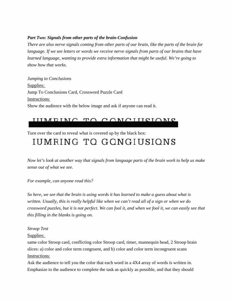
Part Two: Signals from other parts of the brain-Confusion There are also nerve signals coming from other parts of our brain, like the parts of the brain for language. If we see letters or words we receive nerve signals from parts of our brains that have learned language, wanting to provide extra information that might be useful. We’re going to show how that works. Jumping to Conclusions Supplies: Jump To Conclusions Card, Crossword Puzzle Card Instructions: Show the audience with the below image and ask if anyone can read it.
Turn over the card to reveal what is covered up by the black box:
Now let’s look at another way that signals from language parts of the brain work to help us make sense out of what we see. For example, can anyone read this? So here, we see that the brain is using words it has learned to make a guess about what is written. Usually, this is really helpful like when we can’t read all of a sign or when we do crossword puzzles, but it is not perfect. We can fool it, and when we fool it, we can easily see that this filling in the blanks is going on. Stroop Test Supplies: same color Stroop card, conflicting color Stroop card, timer, mannequin head, 2 Stroop brain slices: a) color and color term congruent, and b) color and color term incongruent scans Instructions: Ask the audience to tell you the color that each word in a 4X4 array of words is written in. Emphasize to the audience to complete the task as quickly as possible, and that they should

identify the color not read the word. First use the array with word color matching the written word. Time the audience in this task. Next do the same thing with the 4X4 array in which the color of the word does not match the word written on the array. Time this as well. When done, compare the times of the two tasks, relate this to the audience’s felt perception of greater difficulty and show the fMRI brain scans showing the many, broad areas of greater activation in the mismatched color tasks compared to the one very small area of increased activation when the color and the color term were congruent. First we’re going to see what happens when the brain receives conflicting nerve signals. When you are seeing this sign, your brain has signals both from your eyes, and nerve signals from parts of your brain that are used to read the words you are seeing.
fMRI scan showing areas of increased blood flow during color and word matching condition. (Leung et al. 2000)

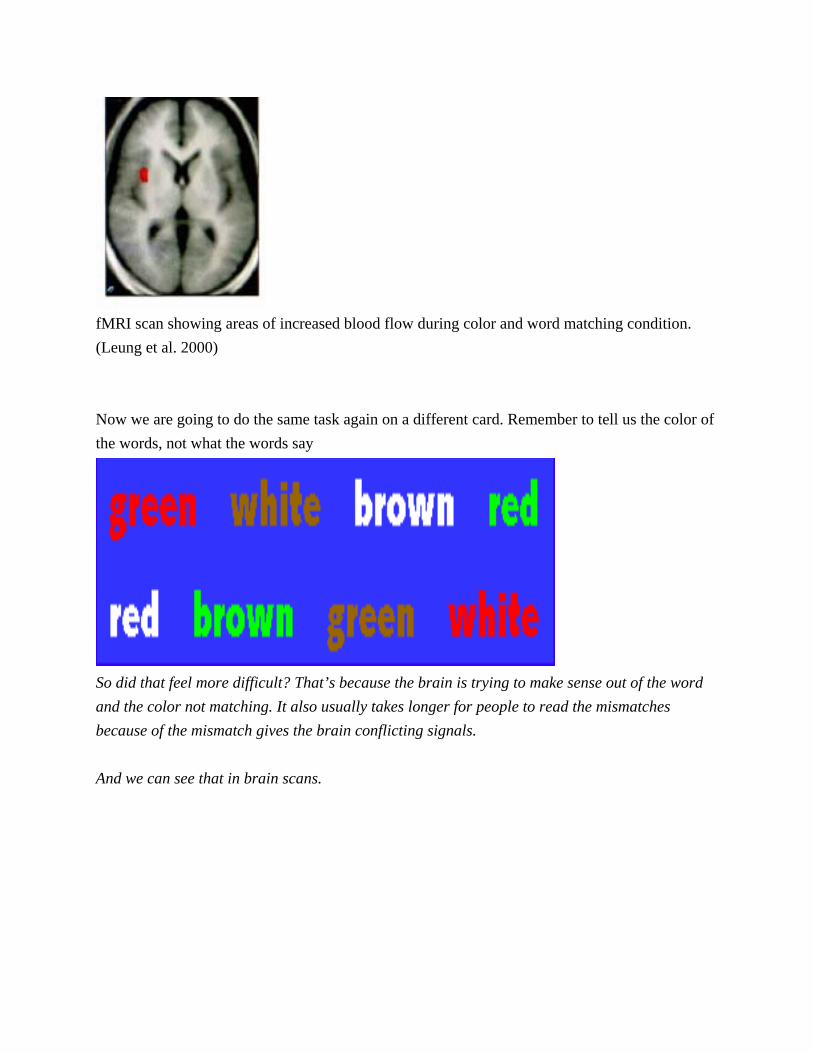
Now we are going to do the same task again on a different card. Remember to tell us the color of the words, not what the words say
So did that feel more difficult? That’s because the brain is trying to make sense out of the word and the color not matching. It also usually takes longer for people to read the mismatches because of the mismatch gives the brain conflicting signals. And we can see that in brain scans.
fMRI of areas of increased blood flow during color and word mismatch condition (Leung et al. 2000)

Comparing fMRI scans of Stroop Test (photo by Mary Leighton) Part Three: Signals from other parts of the brain - Helping us out One really important kind of nerve signals that go from one part of the brain to another are signals from memory. These signals are a big help to us when we try to make sense out of something that is unclear. Degraded Cow (Optional, a visual equivalent of Sine Wave Speech but without brain scans) Supplies: Degraded Cow picture Instructions: Show the audience the degraded image of the cow, and ask them if they can see anything (It is

best not to ask what they see, since some members may have seen this before and could spoil the effect for others). Suggest possibilities like “abstract art.” Then turn over the card to reveal the picture of a cow with the cow’s head outlined. Once everyone sees the cow, show them the original image again, and they will be able to see the cow without the assistance of any outline of the cow’s head. Degraded Image:
©IllusionWorks, L.L.C.1
What do you see in this picture? Maybe it looks like abstract art? How about now? (Showing outlined cow) Doe everyone see the cow? OK, now let’s look at the first picture again. Does everyone see the cow still? Your brain has formed a new memory from the outlined cow, and now nerve signals from that new memory of the outlined cow will send signals that help you see the cow in the first picture I showed you. And you should still be able to see the cow if I shoe you this picture a week or a month from now.
1 Museum will need to obtain permission for use of this image from IllusionWorks, L.L.C.

IT’S A COW!
©IllusionWorks, L.L.C.

Sine Wave Speech Supplies: MP3 Player with Sine Wave Speech Files (http://www.lifesci.sussex.ac.uk/home/Chris_Darwin/SWS/)2 loaded, Speakers, Sine Wave Brain Slice, Mannequin Head Instructions: Play the sine wave speech file to the audience. The audience will not be able to recognize the phrase played, and may not recognize that it is speech at all. Then play the clear human voice file corresponding to the sine wave speech file. Play the sine wave speech file again. After hearing the clear version, the audience will be able to recognize the distorted phrase spoken. Show the audience the fMRI slice showing the additional brain areas that are activated when the sine wave sound file is recognized as speech. In each of these examples, your brain forms a memory of the clearer picture and speech. This signals from this memory then helps us to recognize the original picture and speech that at first we couldn’t make any sense of. And you will probably be able to still see the cow and read the sentence weeks and even months from now. That’s because your brain stores the memory of the clear version, and the nerve signals from this memory help us make sense of the signals coming from our eyes and ears. And we can see that in brain scans.
2 Instructions for creating new Sine Wave Speech files can be found here: http://www.lifesci.sussex.ac.uk/home/Chris_Darwin/Praatscripts/SWS(using this Pratt Program http://www.fon.hum.uva.nl/praat/)

fMRI Scan of a Brain Recognized Sine Wave Speech as Speech (Liebenthal et al. 2003) Part Four: Combining Signals Finally we will look at how the brain combines nerve signals to make sense out of something. Jellybeans Supplies: A bag of Jellybeans Instructions: Give each audience member a jellybean. Ask the audience members to pinch their nose closed when they first place the jelly bean in their mouth. After they have chewed the jellybean with their nose plugged, ask the audience to unplug their noses and notice the difference. They will then be able to perceive much more of the flavor of the jellybean and be much more likely to be able to identify the flavor, At first, you can tell that the Jelly Bean might be sweet or sour, but when you had your nose plugs, but the full taste of the jelly bean only emerges when your brain is also receiving the signals from the nose. It combines the two and BOOM you get the full taste. In the brain nerve signals from your tongue and brain are combined to give us the full flavor of the jelly bean. When you had your nose pinched, you could probably tell a little bit about the jelly bean, like that it was sweet. But you couldn’t tell the full flavor until the receptors in the nose could send

nerve signals to the brain. Next time you are stuffed up with a cold, pay attention to the flavor of things you eat. Because the nose receptors may be blocked, you probably won’t be able to taste as much of your food as usual. Evaluations This demonstration was front and back-end evaluated. Front end evaluations involved using concept mapping, concept boards and short semi-structured interviews to discover if visitors to MSI would be interested in a demonstration about the brain and the nervous system. Visitors questioned were interested in learning more about the senses and the brains role in making sense of the world, but were wary of too 'boring' or intimidating an approach. We also noticed that optical illusions were considered to be interesting by younger visitors, but were already over-familiar to older visitors. We therefore developed a demo that would involve a hands-on approach and experimentation that engaged visitors' own bodies, but avoided the clichéd optical illusions. Back-end evaluations involved participant-observation and brief conversations with visitors who had seen the exhibit in its final form, as well as throughout the development process. Through observing a broad range of visitors (families, individuals, school groups and age ranges from Kindergarten to Adult) as they interacted with the demonstration and demonstrator, the following major points were noted: Children up to older-teenagers enjoy the competitive nature of the Compass experiment and the Stoop test, making these highly popular parts of the demonstration. In fact, the compass test is so popular that young children often want to do this experiment exclusively, over and over again, and at the end of the demonstration request it for all those who didn't have a chance to volunteer before. The demonstrator is able to point out that the 'trick' is natural and not a 'failure' on the part of the volunteer, and in so doing makes it non-intimidating. This part of the demonstration involves a lot of group interaction and discussion in both child and adult groups. All age groups enjoy the jelly bean experiment, and this has even been used successfully to draw people over to the demo! For very young children this experiment is particularly good because the effect is immediate and can be easily repeated at home. Audience members often related the change in taste to previous experiences, for instance commenting on holding their nose when taking medicine, or not being able to taste food with a cold. The degraded cow image has occasionally been seen before, so the demonstrator should be careful to warn people not to shout out if they have already seen it. The reaction to learning that 'a new memory has just been created' was significant to many of the visitors. The experiments involve both individuals and the whole group, which allows many different

people to be involved. The demo should run no longer than 15 minutes to retain engagement, and therefore the demonstrator must select which experiments to conduct. Modularity We have also developed two alternative configurations of the demonstration. For Younger Audiences Here the focus is on just a basic demonstration of nerve signals. The shortened combination of demonstrations is •Hot and Cold Water Compass Jellybeans This combination communicates the message that the brain receives nerve signals from the outside world. Each of these demonstrations was highly successful for younger audiences, keeping their attention and generating considerable excitement. This small number of demonstrations also allows time for each student to try the Compass and Hot and Cold Water demonstrations. Learning and Memory Here the emphasis of the demonstration is changed from nerve signals to learning and memory. This demonstration includes just those components relevant to learning and memory: • Jump to Conclusions • Stroop Test • Degraded Cow • Sine Wave Speech • Jellybeans In this series of demonstrations, automatic recall (in all of this set of demonstrations), new memory formation (Degraded Cow, Sine Wave Speech), and thresholds for recalling memories (Jelly Beans) are discussed. Here, automatic nature of some memory is emphasized. Connection to MSI Exhibits

This demonstration was designed to complement the forthcoming Body Human Exhibit at the Museum of Science and Industry Relevant Illinois State Educational Goals • Goal 12A: Know and apply concepts that explain how living things function, adapt and change. •Goal 12B: Know and apply concepts that describe how living things interact with each other and with their environment. References Leung, Hoi-Chung, Pawel Skudlarski, James Gatenby, Bradley Peterson and John Gore. 2000.
“An Event-related Functional MRI Study of the Stroop Color Word Interference Task” Cerebral Cortex, 10:552-560.
Liebenthal, Einat, Jeffrey Binder, Rebecca Piorkowski, Robert Remez. 2003. “Short-Term Reorganization of Auditory Analysis Induced by Phonetic Experience” Journal of Cognitive Neuroscience 15:4, 549-558. Weinstein S,. 1968 “Intensive and extensive aspects of tactile sensitivity as a function of body part, sex, and laterality.” In D. R. Kenshalo (Ed.), The skin senses. Springfield, Ill.: Thomas, 1968.
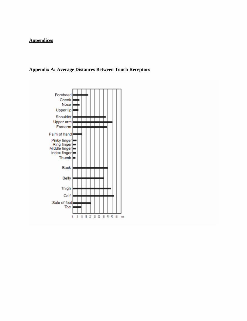
Appendices Appendix A: Average Distances Between Touch Receptors

Appendix B: Stroop Test Images

Appendix C: Demonstration Script Part One: Introduction Signals and the Brain: Hot and Cold Water Supplies: 3 buckets, hot water bottle, ice, hot water, complete nervous system picture Instructions: Ask one or two audience members to place one hand in a bucket of cold water and one in a bucket of hot water. Have them keep their hands in the water for 30 seconds to 1 minute (while you explain the basics of the nervous system receptors, nerve signals, and brain processing of signals–and the ways that perception can be disrupted–damage to receptors (frostbite), interruption of nerve signals (paralysis), and brain damage). After this period, ask the volunteers to place both hands in a bucket of lukewarm or room temperature water. The hand from the cold water will feel hot, the hand from the hot water will feel cold. Welcome to our demonstration on the brain. I want to start off by having two volunteers put each of their hands into two of these buckets of water. While they keep their hands in the water, I’d like for you all to take a look at this diagram. This is the nervous system. Here we can see the brain, which you probably recognize. What you see coming out the brain are the nerves that are connected to the brain. And inside the human brain there are about 50 billion nerves, many more nerves in the brain than in the rest of the body. [NOTE: The brain is not “made of nerves” there are other support cells called glial cells that make up a large part of the brain’s mass (there are 10-50 times more glial cells than nerves in the brain)] Together the brain and the nerves make up what we call the nervous system [insert joke about “nervous” here if you like] So what do these nerves do? These nerves in our body and in our brain send signals. Most of these signals go from one nerve to another. Whenever we move, this is a nerve sending out a signal to a muscle. What we are going focus on today are the signals coming into our brains, from our skin, our eyes, our ears and our mouth and nose. [NOTE: for younger audiences perhaps ask if they can tell us what the 5 senses are]

When we feel or see or hear or taste something, this is because of we have our receptors in our skin, eyes, ears, tongue and nose. Different receptors will detect different things. Right now temperature receptors are detecting this hot and cold water. Receptors in our ears detect sound. Our eyes detect light. When we see, hear, taste, touch or smell things signals are sent from receptors in our eyes, ears, skin, tongue and nose through nerves to our brains. The brain then has to make sense of the nerve signals it gets. If we lose our ability to see or hear or taste, or touch thing, this can happen three ways. We can damage the part of our body that sends the signals to our brains, we can damage the parts of our brain that receive these nerve signals, or we can damage the nerves somewhere in the path from the receptor to the brain. So if someone loses their sight or has problems seeing, the problem could be in their eyes or in this part of the brain that receives nerve signals from our eyes. If we lose our sense of touch, it could be that we have damage to the touch receptors that send signals to the brain, or we could have damaged a nerve between the receptor we could have damage to the part of the brain that receives these nerve signals. One way that people lose touch is very bad frostbite. That’s because some part of our body gets so frozen that the touch receptor dies. So the brain can’t get any more signals from this part of the body. When someone is paralyzed the paralyzed person can’t feel the paralyzed parts of the body because brain isn’t getting any nerve signals from below the point where the nerve signal is broken, just like you can’t get electricity when your power cord that sends electrical signals is broken. In this case, even though the water is the same temperature now, we see that in each hand, the brain is comparing the new signal of temperature with the ones that each had before it, and it is receiving nerve signals from receptors that just detect changes in temperature. Because of these kinds of signals, one hand feels cold because it was in hot water before, while the other hand feels hot because it was in cold water before. Compass (Optional, although highly successful) Supplies: Compass, distances between receptors chart (see appendix A) Instructions: Ask for a volunteer to close eyes and present forearm. Using a compass, asks the volunteer with eyes closed how many points they feel touching their arm. Begin with touching forearm with two

points widely spaced, and then proceed by gradually decreasing the distance (and throwing in a single point occasionally) until the volunteer feels only one point when two points are touching the arm. With the hot and cold water, the brain was receiving nerve signals from temperature receptors. Now we are going to look at another kind of touch receptor: the pressure receptor. This receptor signals when something is pushing on your skin. When some of you couldn’t tell that there were two points, that was because the brain was only getting one signal. Your forearm didn’t have touch receptors close enough to recognize that Part Two: Signals from other parts of the brain-Confusion There are also nerve signals coming from other parts of our brain, like the parts of the brain for language. If we see letters or words we receive nerve signals from parts of our brains that have learned language, wanting to provide extra information that might be useful. We’re going to show how that works. Jumping to Conclusions (Optional, although the simplest demonstration of concept) Supplies: Jump To Conclusions Card, Crossword Puzzle Card Instructions: Show the audience with the below image and ask if anyone can read it.
Turn over the card to reveal what is covered up by the black box:
Now let’s look at another way that signals from language parts of the brain work to help us make sense out of what we see. For example, can anyone read this? So here, we see that the brain is using words it has learned to make a guess about what is

written. Usually, this is really helpful like when we can’t read all of a sign or when we do crossword puzzles, but it is not perfect. We can fool it, and when we fool it, we can easily see that this filling in the blanks is going on. Stroop Test Supplies: same color Stroop card, conflicting color Stroop card, timer, mannequin head, 2 Stroop brain slices: a) color and color term congruent, and b) color and color term incongruent scans Instructions: Ask the audience to tell you the color that each word in a 4X4 array of words is written in. Emphasize to the audience to complete the task as quickly as possible, and that they should identify the color not read the word. First use the array with word color matching the written word. Time the audience in this task . Next do the same thing with the 4X4 array in which the color of the word does not match the word written on the array. Time this as well. When done, compare the times of the two tasks, relate this to the audience’s felt perception of greater difficulty and show the fMRI brain scans showing the many, broad areas of greater activation in the mismatched color tasks compared to the one very small area of increased activation when the color and the color term were congruent. First we’re going to see what happens when the brain receives conflicting nerve signals. When you are seeing this sign, your brain has signals both from your eyes, and nerve signals from parts of your brain that are used to read the words you are seeing.
fMRI scan showing areas of increased blood flow during color and word matching condition. (Leung et al. 2000) Now we are going to do the same task again on a different card. Remember to tell us the color of the words, not what the words say
So did that feel more difficult? That’s because the brain is trying to make sense out of the word and the color not matching. It also usually takes longer for people to read the mismatches because of the mismatch gives the brain conflicting signals. And we can see that in brain scans.

fMRI of areas of increased blood flow during color and word mismatch condition (Leung et al. 2000) Comparing fMRI scans of Stroop Test (photo by Mary Leighton) Part Three: Signals from other parts of the brain - Helping us out One really important kind of nerve signals that go from one part of the brain to another are signals from memory. These signals are a big help to us when we try to make sense out of something that is unclear. Degraded Cow (Optional, a visual equivalent of Sine Wave Speech but without brain scans) Supplies: Degraded Cow picture Instructions: Show the audience the degraded image of the cow, and ask them if they can see anything (It is best not to ask what they see, since some members may have seen this before and could spoil the effect for others). Suggest possibilities like “abstract art.” Then turn over the card to reveal the picture of a cow with the cow’s head outlined. Once everyone sees the cow, show them the original image again, and they will be able to see the cow without the assistance of any outline of the cow’s head. Degraded Image:

©IllusionWorks, L.L.C. What do you see in this picture? Maybe it looks like abstract art? How about now? (Showing outlined cow) Doe everyone see the cow? OK, now let’s look at the first picture again. Does everyone see the cow still? Your brain has formed a new memory from the outlined cow, and now nerve signals from that new memory of the outlined cow will send signals that help you see the cow in the first picture I showed you. And you should still be able to see the cow if I shoe you this picture a week or a month from now. IT’S A COW!
©IllusionWorks, L.L.C.

Sine Wave Speech Supplies: MP3 Player with Sine Wave Speech Files (http://www.lifesci.sussex.ac.uk/home/Chris_Darwin/SWS/) loaded, Speakers, Sine Wave Brain Slice, Mannequin Head Instructions: Play the sine wave speech file to the audience. The audience will not be able to recognize the phrase played, and may not recognize that it is speech at all. Then play the clear human voice file corresponding to the sine wave speech file. Play the sine wave speech file again. After hearing the clear version, the audience will be able to recognize the distorted phrase spoken. Show the audience the fMRI slice showing the additional brain areas that are activated when the sine wave sound file is recognized as speech. In each of these examples, your brain forms a memory of the clearer picture and speech. This signals from this memory then helps us to recognize the original picture and speech that at first we couldn’t make any sense of. And you will probably be able to still see the cow and read the sentence weeks and even months from now. That’s because your brain stores the memory of the clear version, and the nerve signals from this memory help us make sense of the signals coming from our eyes and ears. And we can see that in brain scans.
fMRI Scan of a Brain Recognized Sine Wave Speech as Speech

(Liebenthal et al. 2003) [NOTE: Memory is a very complex neural phenomenon, and different parts of memories are distributed across many different areas of the brain. There is no memory center where memories are stored.] Part Four: Combining Signals Finally we will look at how the brain combines nerve signals to make sense out of something. Jellybeans Supplies: A bag of Jellybeans Instructions: Give each audience member a jellybean. Ask the audience members to pinch their nose closed when they first place the jelly bean in their mouth. After they have chewed the jellybean with their nose plugged, ask the audience to unplug their noses and notice the difference. They will then be able to perceive much more of the flavor of the jellybean and be much more likely to be able to identify the flavor, At first, you can tell that the Jelly Bean might be sweet or sour, but when you had your nose plugs, but the full taste of the jelly bean only emerges when your brain is also receiving the signals from the nose. It combines the two and BOOM you get the full taste. In the brain nerve signals from your tongue and brain are combined to give us the full flavor of the jelly bean. When you had your nose pinched, you could probably tell a little bit about the jelly bean, like that it was sweet. But you couldn’t tell the full flavor until the receptors in the nose could send nerve signals to the brain. Next time you are stuffed up with a cold, pay attention to the flavor of things you eat. Because the nose receptors may be blocked, you probably won’t be able to taste as much of your food as usual.