brad sutton, o.d., f.a.a.o. indiana university school of ... · favorite cases grand rounds brad...
TRANSCRIPT

FAVORITE CASES GRAND ROUNDS
Brad Sutton, O.D., F.A.A.O.
Indiana University School of Optometry
Indianapolis Eye Care Center
Pink sand beach in Bermuda

FINANCIAL DISCLOSURES
None

CASE # 1 Well I’ll be dog gone!
CASE # 1 41 year old white male In for yearly exam, “always had poor vision in left
eye” Diagnosed with coloboma OS many years ago No medical history BVA 20/15 OD, hand motion @ five feet OS

CASE # 1 45 prism diopter constant LXT Other entrance testing and slit lamp findings
unremarkable Fundus as shown OS, unremarkable OD


TOXOCARA CANIS (TOXOCARIASIS)Nematode carried by dogs. Usually
southeastern US. Major problem worldwide
Ingested by humans as the result of eating tainted soil (dog feces) or occasionally undercooked meat
Eggs can remain viable in humans for years then activate into mobile larvae. Entire life cycle can be carried out in dogs, larvae only in humans
Larvae enter the eye via the blood stream, result in formation of a granuloma

TOXOCARA CANIS
Two forms: VLM and OLM Do not typically co-exist together VLM occurs between ages one and four, OLM
later in childhood into adolescence VLM symptoms of fever, weight loss, vomiting,
etc. Vague nature of symptoms often prevents accurate diagnosis

TOXOCARA CANIS
Typical ocular finding is a large granulomatous scar with fibrous bands radiating from the lesion and RPE hyperplasia
Associated vitritis and even iritis are possible
Treatment options include Albendazole 400mg BID in children (800 adults), Thiabendazole, cryotherapy, and photocoagulation.

TOXOCARA CANIS
Steroids can be utilized to curb inflammation Success of treatment is very limited. Death of the
organism leads to a greater inflammatory reaction so treatment is sometimes not indicated

TOXOCARIASIS

TOXOCARIASIS

TRUE COLOBOMA

TOXOPLASMOSIS

“Man…….I am just dying to give somebody Histotoday.”

HISTOPLASMOSIS: FLORIDA STRAIN!

CASE # 2 We’ve got it in the
fold!

CASE # 2 87 year old white female Chief complaint “ blurry vision in right eye” History of cataract extraction and yag
capsulotomy OS HTN Entrance testing non-contributory

CASE # 2 BVA 20/70 OD, 20/20 OS NS cataract OD, centered IOL OS with open
capsule Fundi as shown


CHOROIDAL FOLDS
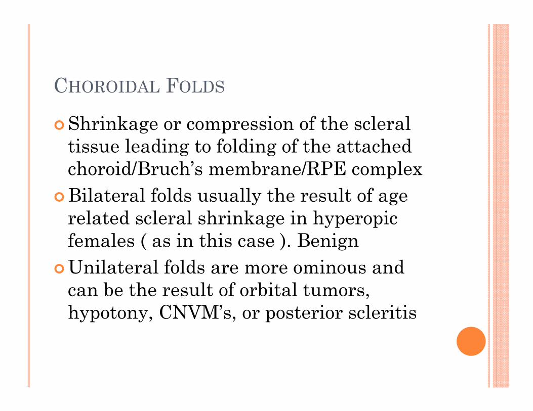
Shrinkage or compression of the scleral tissue leading to folding of the attached choroid/Bruch’s membrane/RPE complex
Bilateral folds usually the result of age related scleral shrinkage in hyperopic females ( as in this case ). Benign
Unilateral folds are more ominous and can be the result of orbital tumors, hypotony, CNVM’s, or posterior scleritis
CHOROIDAL FOLDS
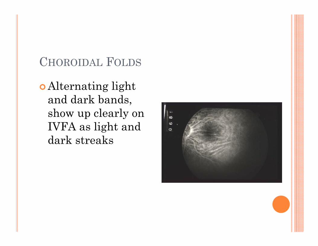
Alternating light and dark bands, show up clearly on IVFA as light and dark streaks

CHOROIDAL VS. RETINAL FOLDS
Choroidal…
Usually roughly horizontal
Usually posterior pole Light and dark
streaks Visible on IVFA
Retinal…
Often stellate alignment
Can be anywhere Similar but finer Visible on IVFA only
if vascular traction

CHOROIDAL FOLDS DUE TO HYPOTONY

WOUND LEAK (POST CATARACT SURGERY) WITHCHOROIDALS: SHALLOW / FLAT CHAMBER
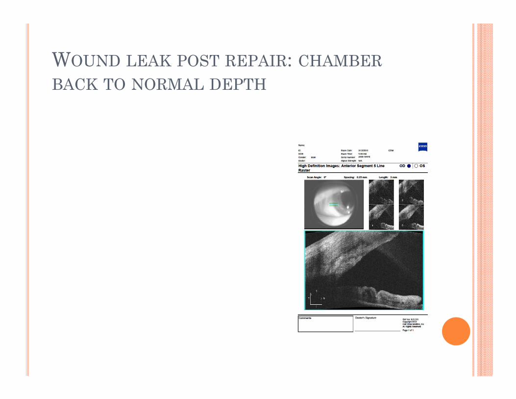
WOUND LEAK POST REPAIR: CHAMBERBACK TO NORMAL DEPTH

Choroidal effusion (different pt.)

Choroidal effusion

Verhoeff’s streaks in choroidal effusion

HYPEROPIC CHOROIDAL FOLDS

PATTON’S FOLDS IN PAPILLEDEMA

CHOROIDAL FOLDS IN POSTERIORSCLERITIS

CASE # 3 Vasculitis? I don’t
think so……….

CASE # 3 23 year old AA female presenting for low vision
consultation BCVA of 20 / 40 -2 OD and 20/800 OS Large, constant LXT, significant VF constriction
OS > OD Other entrance tests and slit lamp findings
unremarkable. Fundus appearance…….

CASE # 3


LEBER’S HEREDITARY OPTICNEUROPATHY
Hereditary mitochondrial disease process affecting the retinal ganglion cells
Maternal inheritance pattern (mitochondrial DNA in embryo comes only from the egg)
Genetic point mutations that have been fully indentified / mapped
Because of inheritance pattern, males can not pass to offspring

LHON Males more commonly afflicted but females can
be Typically strikes in early adulthood, but can
strike later Most commonly affects one eye followed by the
fellow eye within several weeks Clinical trials ongoing with gene therapy; some
encouraging early results

LHON
Reported associations
Can get pseudopapilledemasecondary to peripapillary NFL swelling
Reports of vasculitis (older literature) and pseudovasculitis
What are we looking at here? Pseudovasculitisfrom redundant ILM
Our patient……

LHON
The ganglion cells die so quickly that the redundant internal limiting membrane of the retina collapses and folds on itself, especially around vessels
This appearance lasts several months then tends to go away

CASE # 4
“Looks like drops of water on a windshield”

CASE # 4 60 YO Caucasian
female In for exam to check
on cataracts Arthritis, Asthma,
OSA
BCVA 20/25- OD, 20/20 OS
IOP 13,12 All preliminary
testing normal 1+ NS, trace PSC OU ONH Drusen OU Multiple, small PED’s
OU within the arcades

CASE # 4


MULTIPLE IDIOPATHIC PED SYNDROME
Very rare, with few cases in the literature
Multiple PED’s with little or no neurosensory retinal detachment
Usually females, often related to pregnancy
One theory is a variant of ICSC involving only the RPE
May be related to sleep apnea, which has been linked to ICSC

CASE # 5 “We all have it doc….”

CASE # 5 58 year old Caucasian
male Complaining of flashes
OD for four days. Has floaters, but longstanding with no increase
Told by a retinal specialist 17 years prior that he has a progressive retinal disease but would not go “completely blind”
BCVA of 20 / 20 in each eye
IOP 18 OD, 19 OS Entrance testing
unremarkable Anterior segment
unremarkable OU Posterior segment
reveals significant drusenoid changes OU with pigment mottling OU

CASE # 5 Posterior segment also
reveals a fresh PVD OD with no holes, tears, or breaks
Posterior segment appearance………

CASE # 5

CASE # 5

CASE # 5
OD

CASE # 5 So what have we
here…….? Doyne’s Honeycomb
Dystrophy!
AKA: Mallattia-LeventineseDystrophy

CASE # 5 Doyne’s Honeycomb
Dystrophy Described by Doyne in
England in 1898
Malattia-LeventineseDystrophy described by Alfred Vogt in Switzerland in 1925
Now believed to be phenotypic variants of the same condition
Both caused by genetic mutation in EFEMP1 gene

CASE # 5 The affected gene
encodes a protein that is expressed in the retina and the RPE
Leads to drusenformation early in life
Located all throughout the posterior pole, including nasal to the disc
The drusen coalesce over time leading to radial (MLD), honeycomb (DHD), or mosaic patterns (MLD and DHD)
Symptoms are rare until early middle age
Macula is affected with drusen, so can get vision loss and SRNVM

CASE # 5 Because the condition
is genetic, the role of nutritional supplements is unclear
Manage with regular follow-up, amsler at home, possibly supplements
CASE # 5 Differential diagnoses: 1) AMD : affects
macular area only. More likely to have RPE loss, more likely to have an SRNVM
2) Familial or basil laminar drusen: cluster in groups throughout the posterior pole, especially in arcades, but do not coalesce. No effect on vision.
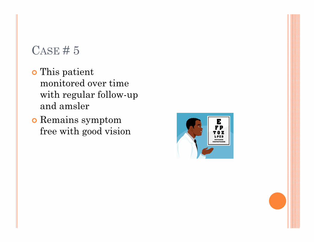
CASE # 5 This patient
monitored over time with regular follow-up and amsler
Remains symptom free with good vision

ANOTHER DOYNE’S EXAMPLE

CASE # 5ANOTHER DOYNE’S EXAMPLE

CASE # 5ANOTHER DOYNE’S EXAMPLE

CASE # 5: ANOTHER DOYNE’S EXAMPLE

CASE # 5FAMILIAL / BASIL LAMINAR DRUSEN EXAMPLE

CASE # 5AMD EXAMPLE

CASE # 6 “Are you sure this
picture is not upside down?”

CASE # 6 43 year old AA male
complaining of poor vision, pain, itching and watering OD
History of RD OD due to trauma and subsequent repair 10 years prior
Taking Naphcon-A and Hydrocodone
BCVA of LP OD, 20 / 30 + OS
IOP 50 OD, 15 OS EOM’s normal VF normal OS Pupil unreactive OD,
but “reverse APD” OS White appearance to
upper half of iris with the naked eye
CASE # 6 Anterior segment
evaluation…….
Eyelid edema OD Aphakic OD Solid, clear bubble in
central AC OD with top half of chamber filled with a white substance
2+ Conj. injection OU
“reverse pseudohypopyon”
Anterior segment OS unremarkable
Posterior segment OS unremarkable. No view of fundus OD
Anterior segment appearance…..

CASE # 6

CASE # 6

CASE # 6: LOOKS LIKE STYROFOAM!

CASE # 6 So what have we
here………? Silicone oil
emulsification!

CASE # 6 Silicone oil tamponade
is routinely used in complicated retinal detachment repair
It must be removed later to avoid potential complications
One complication is emulsification
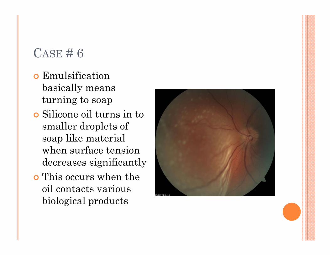
CASE # 6 Emulsification
basically means turning to soap
Silicone oil turns in to smaller droplets of soap like material when surface tension decreases significantly
This occurs when the oil contacts various biological products

CASE # 6 Proteins, lipids, and
phospholipids Particularly HDL Occurs up to 50% of
the time if oil is not removed
Happens in the vitreal cavity, then droplets travel to the AC, especially in an aphakic patient

CASE # 6 “inverse
pseudohypopyon” Leads to corneal
endothelial toxicity and edema, band keratopathy, and increased IOP / glaucoma
In a seeing eye, prompt removal is indicated

CASE # 6 This patient was
given cycloplega for ciliary spasm
Also offered pressure lowering agents for comfort, but deferred
Sent for consideration of surgical removal of emulsified oil
Lost to follow up

ANOTHER EXAMPLE OF EMULSIFICATION

MORE AC OIL WITH AC IOL (SAY THAT 10 TIMES QUICKLY!)


CASE # 7 Maybe it IS a tumor!

CASE # 7 39 year old white male Chief complaint: spot on right eye Reports that spot has been present since high
school but has just recently gotten much larger BCVA: 20/20 OD & OS Entrance testing unremarkable

CASE # 7 Slit lamp exam revealed findings seen as well as
an inferior cortical cataract OD, OS unremarkable
DFE unremarkable OU B-scan ultrasound of posterior segment OD
unremarkable


AMELANOTIC IRIS MELANOMA
Pathology revealed spindle cell morphology (relatively non-aggressive )
Systemic work-up revealed no sign of metastases Iris melanomas account for only a small portion
of uveal tumors ( < 10% ) Seen most frequently in blue irides

IRIS MELANOMA
Inferiorly located due to sun exposure, may distort pupil
Rarely metastasize ( 3-5% ) because visibility typically leads to early detection
Satellite lesions can be seen, increased IOP common with seeding of tumor cells into the TM
Differential diagnoses include iris nevi, Lischnodules, Koeppe and Busacca nodules, etc.
Sector iridectomy performed for complete resection ( see post-op picture next )
Pt. did very well with an opaque contact lens to give him a new pupil


ANOTHER IRIS MELANOMA
