book of abstracts - technische universität dresden · pdf filebook of abstracts. february...
TRANSCRIPT
1
JOINT INDO-GERMAN SYMPOSIUMStrategies for Improved Bone Replacement Materials and Orthopaedic Implants: Design – Manufacturing – Technologies
BOOK OF ABSTRACTS
February 19 – 21, 2014Technische Universität Dresden
Centre for Translational Bone, Joint and Soft Tissue ResearchGermany

2
19th of February, MTZ, Lecture Hall 1
9.00 Registration
9.30 WelcomeandOpeningCeremonyoftheScientificProgramme
10.00 Coffee Break
10.30 SESSION 1: BIOMATERIALS DESIGN AND MANUFACTURING
10.30 Bikramjit Basu (IISc Bangalore): Development of Multifunctional Bioceramics and Polymer-Ceramic Based Hybrid Biocomposites for Orthopedic Applications: a New Paradigm
11.00 Michael Gelinsky (TU Dresden): 3D Plotting of Complex Scaffolds and Tissue Engineering Constructs
11.30 Anindya Deb (IISc Bangalore): Prediction of the Behavior of Total Knee Replacement Implants Using Explicit Finite Element Modeling and an Exploration of the Performance of Alternative Designs
12.00 Christian Hannemann (Fraunhofer IWU Chemnitz): Porous Metal Implant Structures – A Human Bone Copy?
12.30 Lunch,PostersandExhibition
13.30 SESSION2:RAPIDPROTOTYPINGTECHNOLOGIESI
13.30 Alok Kumar (IISc Bangalore): Fabrication of Biomaterial Scaffolds with Gradient Porosity Using 3D Printing
13.50 Bernhard Müller (Fraunhofer IWU Dresden): Multifunctional Implants Realised by Additive Manufacturing
14.10 Rainer Detsch (University Erlangen): Challenges in Biofabrication of Alginate Based Matrices for Vascularized Bone Tissue Regeneration
14.40 Petra Kluger (Fraunhofer IGB Stuttgart): Additive Manufacturing of Bio-Inspired Blood Vessel Systems
15.10 Coffee Break
15.30 SESSION3:DRUGDELIVERYANDRAPIDPROTOTYPING TECHNOLOGIES II
15.30 Kurosch Rezwan (University Bremen): Calcium Phosphate-Based Materials for Advanced Drug Delivery
16.00 Uwe Gbureck (University Würzburg): 3D Powder Printing of Drug Loaded Ceramic Implants
PROGRAMME

3
16.30 Rahul Akkineni (TU Dresden): Design and Fabrication of Core / Shell Structures by 3D Plotting: Applications in Tissue Engineering
16.50 Coffee Break
17.10 SESSION4:CLINICALAPPLICATIONAND COMMERCIALISATION
17.10 Tanvir Momen (Apollo Gleneagles Hospital Kolkata): Hip Replacement: Surgical Techniques and Advancements with Special Emphasis on Metal-on-Metal Hip Replacement and Prognosis
17.40 Maik Stiehler (University Hospital Dresden): Biomaterials in Orthopaedic Surgery: Metallic Implants, Bone Grafts and Bone Substitutes
18.10 Aroop Kumar Dutta (Excel Matrix Biological Devices Pvt. Ltd., Hyderabad): Assembly Line for Tissues Manufacturing
18.40 Gediminas Kostkevicius (Baltic Orthoservice, Kaunas, Lithuania): Mass Customization of Orthopedic Implants and Patient Specific Instruments: the Business Model
19.10 Get Together (MTZ Foyer)
20th of February, MTZ, Lecture Hall 1
8.30 SESSION5:METALLICIMPLANTSI
8.30 Rainer Bader (University Rostock): Evaluation of the Bone Ingrowth of Numerically Optimized and Additive Manufactured Open-Porous Titanium Bone Scaffolds
9.00 Kanyakumari Datta (Data Metallurgical Company, Kolkata): Choice of Materials for Orthopaedic Implants: A Study of the Suitability of Cellular Metals Using Finite Element Modelling
9.30 Annett Gebert (Leibniz IFW Dresden): New Ti-Nb-Based Alloys for Implant Applications
10.00 Christine Schöne (TU Dresden): Individual Contour Adapted Functional Implant Structures in Titanium – From the Theoretical Model to the Practical Application
10.30 Coffee Break
11.00 SESSION6:METALLICIMPLANTSIIANDBIOMECHANICS
11.00 W. Mark Rainforth (Sheffield University, UK): Dynamic Surface Microstructural Changes During Tribological Contact that Determine the Wear Behaviour of Hip Prostheses; Metals and Ceramics

4
11.30 Uta Kremling (IMA GmbH Dresden): Mechanical and Tribological Test Methods for Joint Implants
11.50 Malhar Rao N. Kumar (Hosmat Hospital Bangalore): Clinical and Engineering Assessments of the Effects of Surgical Procedures and Fixations in Spine
12.20 R. Srinivas Gunti (IISc Bangalore): Experimental and Numerical Insights into the Mechanical Behaviour of a Truncated Vertebral Unit under Compressive Static and Impact Dynamic Loads
12.40 Christian Rotsch (Fraunhofer IWU Dresden): Application of Shape Memory Alloys for Active Loosening Protection of Implant Structures
13.00 Lunch,PostersandExhibition
14.30 SESSION 7: CERAMICS
14.30 Manoj Kumar Mitra (Jadavpur Univ. Kolkata): Processing and Characterization of Ceramic Materials in Implants
15.00 Janis Locs (Riga Technical Univ., Latvia): Synthesis and Application of Calcium Phosphates in Maxillofacial and Orthopaedic Surgery
15.20 Hari Krishna Varma (SCTIMST Thiruvananthapuram): Tailor Made Bioactive Ceramics for Specialty Clinical Applications
15.50 Matthias Schumacher (TU Dresden): Modified Calcium Phosphate Bone Cements for the Local Delivery of Therapeutic Ions in Osteoporotic Bone Defects
16.10 Coffee Break
16.40 SESSION8:ELECTRIC/MAGNETICSTIMULATION,POLYMERS
16.40 Debasish Sarkar (NIT Rourkela): Hydroxyapatite Nanoparticles and Nanobiocomposite Scaffold for Protein Adsorption / Release
17.00 Greeshma Thrivikraman Nair (IISc Bangalore): Interplay of Substrate Conductivity and Electric Stimuli in Directing Cell Fate on Implantable Biomaterials
17.20 Sunil Kumar Boda (IISc Bangalore): Differential Response of Prokaryotic and Eukaryotic Cells on Engineered Biomaterials in Magnetic Field Stimulated Culture Conditions
17.40 Ravikumar Krishnamurthy (IISc Bangalore): Bioelectric Stress Induced Cell Deformation and Stability in an Electric Field Stimulated Medium

5
18.00 Yashoda P. Chandorkar (IISc Bangalore): Crosslinking as a Strategy to Design Multifunctional, Tunable Polymer Matrices for Tissue Engineering Applications
18.20 Closing Remarks
21st of February (closed to the public)
8.30 ProjectMeetings
11.00 VisitoftheAmbassadorofIndiatoGermany,HisExcellency Mr.ShriVijayKeshavGokhale
13.30 Lab Demonstration

6

7
We have the great pleasure to welcome you to this Indo-German symposium on “Strategies for improved bone replacement materials and orthopaedic implants: design – manufacturing – technologies”.
The aim is to bring together a number of active researchers from the biomaterials, tissue engineering and medical field to present and discuss state-of-the-art of rapid prototyping technologies for implant design and other emerging manufacturing techniques for novel biomaterials and tissue engineering constructs for regeneration of musculoskeletal tissues. Conceptual contribution and synergistic interaction among academia and industries will strongly influence the direction of translational research, and consequent conversion to applied technology.
The programme covers contributions of experienced scientists and clinicians, as well as of young researchers. Beside the Indian delegation and speakers from the host institutions in Dresden and Chemnitz, colleagues from other German universities and some other European countries will also present their newest research results. Therefore, this symposium is expected to provide a stimulating environment for scientific discussions and to give valuable suggestions concerning translation of research into
clinical application.The financial support of Indo-German Science and Technology Centre (IGSTC), jointly funded by German Ministry for Education and Research (BMBF) and Department of Science and Technology (DST, Government of India) is gratefully acknowledged.
We hope you enjoy your stay in Dresden.
Welcome AddressProf. Dr. Michael Gelinsky, Technische Universität Dresden, GermanyProf. Dr. Bikramjit Basu, Indian Institute of Science, Bangalore, IndiaProf. Dr. Anindya Deb, Indian Institute of Science, Bangalore, IndiaDipl.-Ing. Christian Hannemann, Fraunhofer Institute for Machine Tools and Forming Technology IWU, Chemnitz / Dresden, GermanyDr. Kanyakumari Datta, Data Metallurgical Company, Kolkata, India
8
CONTENT
11
1314
16
17
18
SESSION2:RAPIDPROTOTYPINGTECHNOLOGIESIFabricationofBiomaterialScaffoldswithGradientPorosityUsing3DPrintingDr. Alok Kumar
MultifunctionalImplantsRealisedbyAdditiveManufacturingDr. Bernhard Müller
Challenges in Biofabrication of Alginate Based Matrices for Vascularized Bone Tissue RegenerationDr. Rainer Detsch
AdditiveManufacturingofBio-InspiredBloodVesselSystemsProf. Dr. Petra Kluger
SESSION3:DRUGDELIVERYANDRAPIDPROTOTYPINGTECHNOLOGIES IICalciumPhosphate-BasedMaterialsforAdvancedDrugDeliveryProf. Dr. Kurosch Rezwan
3DPowderPrintingofDrugLoadedCeramicImplantsProf. Dr. Uwe Gbureck
WELCOMEANDOPENINGOFTHESCIENTIFICPROGRAMMEA. ChakrabortyProf. Dr. Stefan R. BornsteinK. Venkatarama SharmaPriv.-Doz. Dr.-Ing. Welf-Guntram DrosselDr. Martin Goller
SESSION 1: BIOMATERIALS DESIGN AND MANUFACTURINGDevelopmentofMultifunctionalBioceramicsandPolymer- Ceramic Based Hybrid Biocomposites for Orthopedic Applications:ANewParadigmProf. Dr. Bikramjit Basu
3DPlottingofComplexScaffoldsandTissueEngineeringConstructsProf. Dr. Michael Gelinsky
PredictionoftheBehaviorofTotalKneeReplacementImplantsUsingExplicitFiniteElementModelingandanExplorationofthePerformanceofAlternativeDesignsProf. Dr. Anindya Deb
PorousMetalImplantStructures–AHumanBoneCopy?Christian Hannemann
1910
22
23
24
27
28
30

9
DesignandFabricationofCore/ShellStructuresby3DPlotting:Applications in Tissue EngineeringAshwini Rahul Akkineni
SESSION4:CLINICALAPPLICATIONAND COMMERCIALISATIONHipReplacement:SurgicalTechniquesandAdvancementswithSpecialEmphasisonMetal-on-MetalHipReplacementandPrognosisDr. Tanvir Momen
Biomaterials in Orthopaedic Surgery: Metallic Implants, Bone Grafts and Bone SubstitutesDr. Maik Stiehler
Assembly Line For Tissue Manufacturing Dr. Aroop Kumar Dutta
MassCustomizationofOrthopedicImplantsandPatientSpecificInstruments:TheBusinessModelDr. Gediminas Kostkevicius
SESSION5:METALLICIMPLANTSIEvaluationoftheBoneIngrowthofNumericallyOptimizedandAdditiveManufacturedOpen-PorousTitaniumBoneScaffoldsProf. Dr. Rainer Bader
32
33
34
35
36
38
4142
44
45
47
4950
51
53
Choice of Materials for Orthopaedic Implants: A Study of the Suitability of Cellular Metals Using Finite Element ModellingDr. Kanyakumari Datta
NewTi-Nb-BasedAlloysforImplantApplicationsDr. Annett Gebert
IndividualContourAdaptedFunctionalImplantStructuresinTitanium–FromtheTheoreticalModeltothePracticalApplicationDr. Christine Schöne
SESSION6:METALLICIMPLANTSIIANDBIOMECHANICSDynamic Surface Microstructural Changes During Tribological ContactthatDeterminetheWearBehaviourofHipProstheses; Metals and CeramicsProf. W. Mark Rainforth
Mechanical and Tribological Test Methods for Joint ImplantsDr. Uta Kremling
Clinical and Engineering Assessments of the Effects of SurgicalProceduresandFixationsinSpineDr. Malhar Rao N. Kumar
10
ExperimentalandNumericalInsightsintotheMechanical BehaviorofaTruncatedVertebralUnit(TVU)under CompressiveStaticandImpactDynamicLoadsGunti Ranga Srinivas
ApplicationofShapeMemoryAlloysforActiveLooseningProtectionofImplantStructuresChristian Rotsch
SESSION 7: CERAMICSProcessingandCharacterizationofCeramicMaterialsinImplantsProf. Dr. Manoj Kumar Mitra
SynthesisandApplicationofCalciumPhosphatesin MaxillofacialandOrthopaedicSurgeryDr. Janis Locs
TailorMadeBioactiveCeramicsforSpecialtyClinical ApplicationsDr. Hari Krishna Varma
ModifiedCalciumPhosphateBoneCementsfortheLocalDeliveryofTherapeuticIonsinOsteoporoticBoneDefectsMatthias Schumacher
SESSION8:ELECTRIC/MAGNETICSIMULATION,POLYMERSHydroxyapatiteNanoparticlesandNanobiocompositeScaffoldforProteinAdsorption/ReleaseProf. Dr. Debasish Sarkar
InterplayofSubstrateConductivityandElectricStimuliinDirecting Cell Fate on Implantable BiomaterialsGreeshma Thrivikraman
DifferentialResponseofProkaryoticandEukaryoticCellsonEngineered Biomaterials in Magnetic Field Stimulated Culture ConditionsB. Sunil Kumar
Bioelectric Stress Induced Cell Deformation and Stability in an Electric Field Stimulated MediumRavikumar Krishnamurthy
Crosslinking as a Strategy to Design Multifunctional, Tunable PolymerMatricesforTissueEngineeringApplicationsYashoda ChandorkarS
54
55
5758
59
60
62
6566
67
69
71
73

11
WELCOME AND OPENING OF THE SCIENTIFIC PROGRAMME
Welcome AddressesA. ChakrabortyDirectorIndo-German Science & Technology Centre (IGSTC)Gurgaon, India
Prof. Dr. Stefan R. BornsteinVice Dean for Development and International AffairsMedical Faculty of Technische Universität DresdenDresden, Germany
K. Venkatarama SharmaCounsellor (Science and Technology)Indian EmbassyBerlin, Germany
Priv.-Doz. Dr.-Ing. Welf-Guntram DrosselDirectorFraunhofer Institute for Machine Tools and Forming Technology IWUChemnitz / Dresden, Germany
Dr. Martin GollerInternational Bureau of the German Federal Ministry of Education and Research (BMBF)Bonn, Germany

12
13
Session 1
BIOMATERIALS DESIGN AND MANUFACTURING
DevelopmentofMultifunctionalBioceramicsand Polymer-CeramicBasedHybridBiocompositesfor OrthopedicApplications:ANewParadigmProf. Dr. Bikramjit BasuLaboratory for Biomaterials, Materials Research Centre and Bio-Engi-neering Program, Indian Institute of Science (IISc), Bangalore, IndiaE-Mail: [email protected]
3DPlottingofComplexScaffoldsandTissue Engineering ConstructsProf. Dr. Michael GelinskyCentre for Translational Bone, Joint and Soft Tissue Research, University Hospital and Medical Faculty, Technische Universität Dresden, GermanyE-Mail: [email protected]
PredictionoftheBehaviorofTotalKneeReplacementImplantsUsingExplicitFiniteElementModelingandan ExplorationofthePerformanceofAlternativeDesignsProf. Dr. Anindya DebCentre for Product Design and Manufacturing, Indian Institute of Science (IISc), Bangalore, IndiaE-Mail: [email protected]
14
16
17
31
PorousMetalImplantStructures–AHumanBoneCopy?Christian HannemannDepartment of Lightweight Structures, Fraunhofer Institute for Machine Tools and Forming Technology IWU, Chemnitz, GermanyE-Mail: [email protected]
18

14 Session 1: Biomaterials Design and Manufacturing
DevelopmentofMultifunctionalBioceramicsandPolymer-CeramicBasedHybrid BiocompositesforOrthopedicApplications:ANewParadigmBikramjit BasuLaboratory for Biomaterials, Materials Research Center and Bio-Engineering Program, Indian Institute of Science (IISc), Bangalore, India
and they are used either as bulk or as coatings for a variety of applications, e.g. dental implants, healing of bone defects, fracture treatment, total HIP joint replacement, and spinal surgery. Despite several years of research, various issues related to poor fracture toughness and lack of antibacterial/electrical conductivity property in CaP-based bioceramics have not been addressed in an integrated manner. In the above perspective, this talk will cover the development of calcium phosphate-based multifunctional composites for hard tissue regeneration. In particular, the challenges in designing the synthetic ceramics to mimic bone-like strength / fracture tough-ness and electrical or piezoelectric properties will be emphasized. A major emphasis will be placed on the development of HA-based multifunctional biomaterial platform to facilitate cell growth in vitro/osseointegration in vivo, while restricting prosthetic infection.In the second part of the presentation, the development of chemically coupled/toughened HDPE-based biocomposites reinforced with bioinert (Al2O3) and bioactive (HA) ceramic fillers using compression molding as well as injection moulding will be discussed. Our experimental results demonstrate that a combina-tion of higher elastic modulus (12 GPa) and improved hardness (260 MPa) and strength of approx. 800 MPa in compression moulded composites is achievable. In an attempt to assess the
Fig. 1: Schematic illustration of the research themes centeredaround various aspects of the cell-material interaction.
One of the important innovations in the field of materials science in last three decades has been the development of biomaterials for the replacement and regeneration of the human tissues. In view of the similarity with the natural bone composition, Calcium phosphate (CaP)-based bioceramics attracted wider attention

15Session 1: Biomaterials Design and Manufacturing
suitability of the developed composites as acetabular sockets in Total Hip Replacement (THR) application, the detailed tribological investigation in vitro of the developed composites established better combination of wear resistance and frictional properties in the hybrid composites than HDPE. In addition, in vitro cell culture study using different cell lines (L929, SaOS2, HOBs) confirms cell adhesion properties of the investigated composites. Importantly, this presentation will also illustrate that it is possible to modulate the cell viability and bone mineralization on HDPE-HA-Al2O3 hybrid composites with the change in substrate modulus/stiffness within a narrow window of the respective parametric values. The progressive healing of cylindrical femoral bone defects in rabbit animal model was assessed by implantation experiments over 1, 4 and 12 weeks. BTaken together, this talk will establish that despite addition of 20 wt.% Al2O3, HDPE-based hybrid biocomposites are as biocompatible in vitro like HA or in vivo like/better than HDPE.
References:1. G. Tripathi, J. E. Dough, A. Dinda and B. Basu; In vitro cyto-
toxicity and in vivo osseointergration property of compression molded HDPE-HA-Al2O3 hybrid biocomposites; Journal of Bio-medical Materials Research Part A 101 [6] (2013) 1539–1549.
2. Garima Tripathi and BikramjitBasu; Injection-Molded High-Density Polyethylene–Hydroxyapatite–Aluminum Oxide Hybrid Composites for Hard-Tissue Replacement: Mechanical, Biological, and Protein Adsorption Behavior; J. Applied Polymer Science,124(2012)2133–2143.
3. Indu Bajpai, Kantesh Balani and Bikramjit Basu, Spark Plasma Sintered HA-Fe3O4 based Multifunctional Magnetic Biocom-posites, J. Am. Cer. Soc. 96 [7] (2013) 2100-2108.
4. A. K. Dubey, Anumol E. A., K. Balani and B. Basu; Multi-functional properties of multi-stage spark plasma sintered HA-BaTiO3 based piezobiocomposites for bone replacement applications; J. Am. Cer. Soc. (in Press, 2013; DOI: 10.1111/jace.12566).

16 Session 1: Biomaterials Design and Manufacturing
3DPlottingofComplexScaffoldsandTissueEngineeringConstructsRahul Akkineni, Yongxiang Luo, Kathleen Schütz, Anja Lode, Michael GelinskyCentre for Translational Bone, Joint and Soft Tissue Research, University Hospital and Medical Faculty, Technische Universität Dresden, Germany
Many rapid prototyping technologies, originally developed for mechanical engineering, have been adapted to scaffold fabrication and manufacturing of tissue engineering constructs. The method of 3D plotting – layer by layer deposition of pasty biomaterials to create 3D objects of pre-defined inner and outer morphology – offers a variety of options for creating complex structures. Using multiple dispensing channels, scaffolds consisting of more than one (bio)material can easily be processed. Due to the mild manufacturing conditions also delicate components like biopolymers, growth factors and even live cells can be included in the 3D plotting process. In addition, this technique easily can be performed under sterile conditions because the necessary instrumentation is small compared to e.g. 3D powder printers or machines for selective laser sintering. The presentation will give an overview of some of our recent developments in the field of 3D plotting, especially concerning manufacturing of calcium phosphate cement (CPC) scaffolds under mild conditions for bone tissue engineering, combination of CPC and biopolymers within one object and direct plotting of blood capillary-like hollow strands. Finally, also the inclusion of living cells in the 3D plotting process will be briefly described.
References:A. Lode, K. Meißner, Y. Luo, F. Sonntag, S. Glorius, B. Nies, C. Vater, F. Despang, Th. Hanke, M. Gelinsky: Fabrication of porous scaffolds by 3D plotting of a pasty calcium phosphate bone cement under mild conditions. J Tissue Eng Reg Med 2012 (in press, DOI 10.1002/term.1563)Y. Luo, A. Lode, F. Sonntag, B. Nies, M. Gelinsky: Well-ordered biphasic calcium phos-phate/alginate scaffolds fabricated by multi-channel 3D plotting under mild conditions. J Mater Chem B 2013, 1, 4088-4098Y. Luo, A. Lode, M. Gelinsky: Direct plotting of three-dimensional hollow fiber scaffolds based on concentrated alginate pastes for tissue engineering. Adv Healthcare Mater 2013, 2, 777-783Y. Luo, Ch. Wu, A. Lode, M. Gelinsky: Hierarchical mesoporous bioactive glass/alginate composite scaffolds fabricated by three-di-mensional plotting for bone tissue engineering. Biofabrication 2013, 5, 015005 (13 pages)

17Session 1: Biomaterials Design and Manufacturing
PredictionoftheBehaviorofTotalKneeReplacementImplantsUsingExplicitFiniteElementModelingandanExplorationofthePerformanceofAlternativeDesignsAnindya DebCenter for Product Design and Manufacturing, Indian Institute of Science, Bangalore, India
In the current study, an advanced finite element model of a total knee arthroplasty (TKR) installed on a representative human knee has been developed. The model includes TKR implant components such as a femoral part, a tibial tray and a HDPE (high density polyethylene) insert, and a knee sub-system comprising parts of femur and tibia as well as the relevant ligaments. The model is initially calibrated using dynamic responses reported in published literature for a mobile bearing TKR evaluated in a knee simulator. It is noted that mobile bearing TKR designs were thought to be associated with lower stresses in the polyethylene (PE) insert as compared to fixed bearing designs. The wear rate of the PE liner was felt to be less in the mobile bearing knees due to the decrease in the stresses. However, a re-evaluation of the biomechanics of the mobile bearing design became necessary due to the recent clinical reports on the long term outcome of mobile bearing knees which have not demonstrated any significant benefit in terms of implant survival and polyethylene wear rate. In the present explicit finite element analysis of mobile bearing and fixed bearing knee designs, no significant differences have been found in the maximal stresses in the superior (articulating) surface of the PE insert in mobile and fixed bearing designs. On the inferior surface of the PE insert, the computed peak stresses were nearly 30% higher in the mobile bearing implant compared to the
fixed bearing design. Thus, contrary to earlier expectations, mobile bearing designs may be associated with higher overall PE stresses and wear as compared to the fixed bearing designs. It appears that further research is necessary to generate a solution that will minimize the wear rate of the non-metallic tibial insert in mobile bearing TKR implants.

18 Session 1: Biomaterials Design and Manufacturing
PorousMetalImplantStructures–AHumanBoneCopy?Christian Hannemann, Steffen ScholzFraunhofer Institute for Machine Tools and Forming Technology IWU, Chemnitz, Germany
In this study the development process of the medical engineering activities of the Department of Lightweight Design of the Fraunhofer IWU is explained and background information given. Coming from research and development for tooling machines the approaches are based upon a completely different way of thinking. The similarity of the cellular structures - currently realized for fast moving slides of milling machines or heat exchangers - to these of cortical or cancellous bone are visible in Figure 1. As these foams are usually made of Aluminum alloys new ideas were needed resulting in extended efforts on Titanium foam. Many research institutions are already working on that topic as the human life span is extending further and further and in many cases bone substitute material is needed.Instead of turning into a “fast follower on open porous structures” the idea of closed or semi-closed cellular Titanium was raised creating something closer to cortical bone cells. By mechanically opening of the outer skin the emerging cavities can give hold of bone cells leading to a form fit between bone and implant. The lecture visualizes the activities, problems and ideas on the way to a closed cellular Titanium structure.
Keywords: Titanium, Metal Foam, Prostheses.
Figure 1: Closed porous Titanium, vertebrae cross section, open porous structure.

19
Session 2
RAPID PROTOTYPING TECHNOLOGIES I
Fabrication of Biomaterial Scaffolds with Gradient PorosityUsing3DPrintingDr. Alok KumarMaterials Research Centre, Indian Institute of Science (IISc), Bangalore, IndiaE-Mail: [email protected]
MultifunctionalImplantsRealisedbyAdditive ManufacturingDr. Bernhard MüllerDepartment of Lightweight Structures, Fraunhofer Institute for Machine Tools and Forming Technology IWU, Chemnitz / Dresden, GermanyE-Mail: [email protected]
Challenges in Biofabrication of Alginate Based Matrices for Vascularized Bone Tissue RegenerationDr. Rainer DetschDepartment of Materials Science and Engineering, Institute for Biomaterials, University of Erlangen-Nuremberg, Erlangen, GermanyE-Mail: [email protected]
20
22
23
31
AdditiveManufacturingofBio-InspiredBloodVesselSystemsProf. Dr. Petra KlugerDepartment of Cell and Tissue Engineering, Fraunhofer Institute for Interfacial Engineering and Biotechnology IGB, Stuttgart and University of Applied Sciences, Reutlingen,Germany E-Mail: [email protected]
24

20 Session 2: Rapid Prototyping Technologies I
FabricationofBiomaterialScaffoldswithGradientPorosityUsing3DPrintingAlok Kumar, Sourav Mandal, Ramakrishna Vasireddi, Bikramjit BasuMaterials Research Centre, Indian Institute of Science, Bangalore, India
With the advancement in biomedical technology, solid freeform fabrication (SFF) has begun to provide biomedical engineers with a number of potential solutions for problems encountered, when trying to create larger and more complex implants. The poor survival of cells were found on conventionally made 3D scaffoldsdue to unavailability of nutrient and growth factors at the central region of scaffold, due to long distance from blood vessels of these regions and absence of interconnected/ organized pores. In this context, one can develop designed porous scaffolds with good vascularization potential due to interconnected pore network using SFF technique. In this perspective, few key design options for the porous scaffold and their relevance for orthopedic applications will be presented in the beginning of talk (see fig. 1). A porous scaffold with a wide range of pore sizes is necessary to meet various functions involved in osseointegration and scaffolds with gradient porosity can mimic the bone structure more closely at microscopic level. For example, highly porous side facing towards the bone defect allows the enhanced osseointegration. While, low porous side will in contact with connecting tissues restrict the growth of fibrous tissue to the wound area(see fig. 2). In order to illustrate the importance of above mentioned issues, the results obtained while fabricating hydroxyapatite as a model system will be presented. As part of our experiment, a spray dried
mixture of hydroxyapatite (HA), maltodextrin (MA), and polyvinyl alcohol (PVA) was used for scaffold fabrication. A powder composition with HA-20 wt. % MA-1 wt. % PVA shows the minimum setting time. The binders were tested for setting time, binding strength, pH and viscosity. Some preliminary experimental results on the efficacy of various scaffolds for three dimensional tissue formationin vitro will be discussed. To this end, the use of 3D culture bioreactor as well as micro-computed tomography (micro-CT) will be highlighted.
Figure 1:Schematic diagrams showing various design options for the orthopedic applications.

21Session 2: Rapid Prototyping Technologies I
Figure 2: Animation showing the healing of bone defect without (a) and with the help of functionally graded biomaterial (b) (adapted from http://www.curasan.de/imgs/usa/bone-defects.gif)
References:1. B.Basu, B., D. S. Katti, A. Kumar. Advanced Biomaterials:
Fundamentals, Processing and Applications. John Wiley & Sons Inc, New Jersey, USA (2009) 746.
2. Simske SJ, Ayers RA, Bateman TA. 1997; Porous Materials for Bone Engineering. Mater Sci Forum 250:151-182.
3. Zhang C, Wang J, Feng H, Lu B, Song Z, Zhang X. 2001; Re-placement of segmental bone defects using porous bioceramic cylinders: a biomechanical and X-ray diffraction study. J Biomed Mater Res 54:407-411.
22 Session 2: Rapid Prototyping Technologies I
MultifunctionalImplantsRealisedbyAdditiveManufacturingBernhard Müller, Thomas TöppelFraunhofer Institute for Machine Tools and Forming Technology IWU, Chemnitz / Dresden, Germany
Keywords: implant design, additive manufacturing; laser beam melting; selective laser melting; functional integration
At the current state of the art, endoprostheses are predominantly manufactured by cutting, forming or casting technologies. Another, rather new way of implant manufacturing is the additive manufacturing process called Beam Melting, using a laser or electron beam. In particular the customized production with no need for any type of tooling, combined with the unique freedom of design spark interest in this technology for implant manufacturing. The use of Beam Melting enables the fabrication of endoprostheses with almost any design of inner and outer geometries. Previous developments and research activities were focussing either on customized, patient specific implant designs with a production batch size of only one piece, or on structured surfaces of specific design for better bone ingrowth and improved stability of the implant-bone bonding. Both approaches have not yet reached a significant breakthrough for additive manufacturing as an adopted technology to produce (metal) implants, besides some niche applications, e. g. for individual cranial or jaw plates or some Electron Beam Melting series production of hip cups.
This presentation presents the results of a completely new approach to trigger this awaited breakthrough of additive implant manufacturing. This approach focuses on the integration of completely new features and functions into endoprostheses which give various added value opportunities to implants that where unthinkable before. The presentation describes how strategies from tooling applications of additive manufacturing were adopted which have proved very successful in giving added value to tools and dies by implementing complex inner cooling channels. The presentation describes the innovative inner design features like functional channels and cavities introduced in standard implants, e. g. hip stems.

23Session 2: Rapid Prototyping Technologies I
Challenges in Biofabrication of Alginate Based Matrices for Vascularized Bone Tissue RegenerationRainer Detsch, Tobias Zehnder, Bapi Sarker, Aldo R. BoccacciniDepartment of Materials Science and Engineering, Institute for Biomaterials, University of Erlangen-Nuremberg, Erlangen, Germany
Biofabrication has been suggested as a promising method for cell-hydrogel-printing in the field of regenerative medicine. These constructs have to be customized to the defect, utilising materials tailored for artificial organs. The challenges involve the fabrication of bone-like tissue combining relevant cells and suitable biomate-rial structures exhibiting an interconnecting and high porosity that facilitates vascularisation. Such tailored 3D-constructs can ideally be realised with rapid-prototyping (RP-) techniques in the context of emerging biofabrication approaches. Both, hydrogels and bone cells can be processed in one step using these methods. For suc-cessful functional tissue, high cell viability and proliferation as well as material degradation are required. Since cells have to be viable, all fabrication processes should be “cell-friendly” and performed under sterile and biocompatible conditions. Several RP-techniques are available for biofabrication, e.g. dispense-plotting, which is the one used in this study. By using lay-down-pattern, the 3D-hydro-gel constructs were produced layer by layer. In this presentation, the following topics will be discussed:– Application of alginate based hydrogels - preparation and
characterisation – Degradation of hydrogels - in vitro and in vivo– Rapid Prototyping of hydrogels for the fabrication of 3D
constructs with tailored pore structures and geometries
– Cell plotting - Cell attachment and cell behaviour - VEGF release of immobilised cells
– Rapid prototyping of support structures and vessels
Fig. 1: Principle of the RP- technique dispense-plotting for biofabrication of cell-loaded scaffolds
Designing materials that can promote cell adhesion and migration starts with the understanding of cell-material interactions in 3D. The results of this study show that the constructs fabricated from alginate based hydrogels with biofabrication methods support and promote the growth of cells and repair of natural tissues promoting neo-vascularisation.

24
AdditiveManufacturingofBio-InspiredBloodVesselSystemsP.J. Kluger1,2, K. Borchers1, Eva Hoch1,2, O. Refle3, S. Engelhard4, W. Meier5, B. Huber2, E. Novosel2, C. Graf3, C. Bierwisch6, N. Notrodt4, M.Wegener5, H. Krüger5, R. Jaeger6, T. Hirth1,2, A. Giller4, G. E.M. Tovar1,2
1 Fraunhofer Institute for Interfacial Engineering and Biotechnology IGB, Germany, 2 Institute for Interfacial Engineering, University of Stuttgart, Germany, 3 Fraunhofer Institute for Manufacturing Engineering and Automation IPA, Germany, 4 Fraunhofer Institute for Laser Technology ILT, Germany, 5 Fraunhofer Institute for Applied Polymer Research IAP, Germany, 6 Fraunhofer Institute for Mechanics of Materials IWM, Germany
The key technology for in 3-D vitro engineered tissues is the establishment of vascular scaffolds. Today it is not yet possible to generate vascular structures resembling the typical organisation of mature blood vessels in vivo. In the past, significant progress has been made mainly in the development of tissue-like engineered products that are not dependent on a significant level of vascular support, such as bioartificial cartilage and skin equivalents. The generation of adequate tissue substitutes of most other types of tissues require a functional vascular network for the supply of nutrients and the disposal of metabolites throughout a functional and growing tissue. Therefore, the generation of vascularised arti-ficial tissues defines a challenging topic for future developments. Natural systems are able to execute complex functions because their forms and materials have been optimized in the course of evolution. In order to develop artificial structures which perform as well as natural ones we need a) fabrication processes that do not set any limits to the genera-
tion of structures and shapes, b) materials that allow for tailoring of their physical, chemical, and
biological properties. A team of five Fraunhofer Institutes is currently developing materials and techniques to fabricate artificial blood vessel system that will be able to supply artificial tissue in future.
Session 2: Rapid Prototyping Technologies I
Fig. Schematic of layer by layer fabrication of bio-inspiered structures, fluid dynamic simulation of wall sheer stress in branched tube, artifical vessel, fabricated by laser-based stereolithography, endothelial cells gro-wing on new flexible biomaterial, inkjet printed chondrocytes after 72 h cultivation (scale bar 1000 µm),non-gelling inkjet-printable bioink based on modified gelatine.

25Session 2: Rapid Prototyping Technologies I
We introduce freeform fabrication for the manufacturing of flexi-ble structures from the micro-meter to the centimeter range. The technology uses manufacturing processes combining 3-D inkjet printing and laser-based polymerization techniques. Based on the essential features of the natural vascular system models were developed to find the optimal branching of the artificial network tree, i.e. the length of individual branches, their branching points and branching angles. Computational fluid dynamics calculations which take into account the complex blood rheology and the elasticity of the walls are used to find the optimal geometry of bifurcations.In order to characterize the artificial structures and to validate the model predictions, an experimental set-up was established for studying pulsatile flows and mechanical responses in artificial vascular systems. Resins for 3-D rapid prototyping of biomaterials have been developed that fulfill a wide range of requirements such as photo cross-linkability, viscosity, flexibility, tensile strength and biocompatibility of the post-cured materials. Cytocompatible synthetic materials as well as biomolecules from the natural extra cellular matrix are used for resin formulation. Photo-cured synthetic materials are covalently coated with biologically active substances e.g. heparin-RGD in order to create biofunctional materials that promote the interaction between material and cells.
Endothelial cells are seeded onto the biological coating at the in-ner vessel wall. The cells are cultured in a bioreactor system which provides a pulsatile flow of culture media mimicking the natural blood flow. Biobased inks are used for direct cell printing of e.g. cartilage cells and optimization of the matrix has been performed for stabilization of the native chondrocyte morphology.

26 Session 2: Rapid Prototyping Technologies I

27
Session 3
DRUG DELIVERY AND RAPID PROTOTYPING TECHNOLOGIES II
CalciumPhosphate-BasedMaterialsforAdvancedDrugDeliveryProf. Dr. Kurosch RezwanAdvanced Ceramics, University of Bremen, GermanyE-Mail: [email protected]
3DPowderPrintingofDrugLoadedCeramicImplantsProf. Dr. Uwe GbureckDepartment of Functional Materials in Medicine and Dentistry, University of Würzburg, GermanyE-Mail: [email protected]
Design and Fabrication of Core / Shell Structures by 3D Plotting:ApplicationsinTissueEngineeringAshwini Rahul AkkineniCentre for Translational Bone, Joint and Soft Tissue Research, University Hospital and Medical Faculty, Technische Universität Dresden, GermanyE-Mail: [email protected]
28
30
32

28 Session 3: Drug Delivery and Rapid Prototyping Technologies II
CalciumPhosphate-BasedMaterialsforAdvancedDrugDeliveryKurosch RezwanAdvanced Ceramics, University of Bremen, Germany
Controlled multi-staged drug release: Challenges and Opportuni-ties. A typical open bone injury, e. g. compound fracture or loss of a tooth, undergoes a local inflammation and generally needs bone grafting for defect sizes larger than 0.5 cm as well as local appli-cation of e. g. antibiotics. Even though scaffolds for bone healing incorporating several types of drugs are heavily investigated and sought-after, a highly desirable controlled multi-stage release of combined drugs such as antibiotics and bone growth factors is still unavailable and most challenging. While a short-term release within hours and days is desirable for antibiotics, a subsequent release of bone growth factors over days and weeks would be ideal. Degradable polymer-based systems providing a sequential release are commonly not suitable for bone tissue engineering as their acidic dissolution attacks and decomposes bone [1]. Calcium phosphate is an ideal substrate material for drugs as it is next to collagen the major constituent of bone while being available in different crystal phases. Hydroxyapatite and beta-tricalciumphos-phate are the most prominent ones featuring varied degradation rates. The main challenge is to obtain these biphasic material substrates without sintering while introducing the drugs during colloidal processing.Aim and Approach. The aim of our studies is to provide a ceramic material substrate for a controlled multi-staged drug release.
After synthesizing tailored calcium phosphate microbeads [2,3] containing growth factors, embedment into a freeze-gelation [4] obtained porous CaP-matrix incorporating antibiotics is envisaged. The hypothesized multi-staged drug release is investigated in vitro for short- (days) and mid-term (weeks) use. By tailoring the bipha-sic material microstructure and composition while incorporating two types of CaP crystal phases the dissolution rates become adjustable.
References:1. Rezwan, K.; Chen, Q. Z.; Blaker, J. J.; Boccaccini, A. R., Biode-
gradable and bioactive porous polymer/inorganic composite scaffolds for bone tissue engineering. Biomaterials 2006, 27, (18), 3413-3431.
2. Klein, T. Y.; Treccani, L.; Rezwan, K., Ceramic Microbeads as Adsorbents for Purification Technologies with High Specific Sur-face Area, Adjustable Pore Size, and Morphology Obtained by Ionotropic Gelation. J. Am. Ceram. Soc. 2012, 95, (3), 907-914.
3. Schumacher, T. C.; Klein, T. Y.; Treccani, L.; Rezwan, K., Rapid sintering of porous monoliths assembled from microbeads with high specific surface area and multimodal porosity. Advanced Engineering Materials 2013 - in print, DOI 10.1002/adem.201300220

29Session 3: Drug Delivery and Rapid Prototyping Technologies II
4. Blindow, S.; Pulkin, M.; Koch, D.; Grathwohl, G.; Rezwan, K., Hydroxylapatite / SiO2 composites via freeze casting for bone tissue engineering. Advanced Engineering Materials (Cover Page) 2009, 11, (11), 875 - 884.

30 Session 3: Drug Delivery and Rapid Prototyping Technologies II
3DPowderPrintingofDrugLoadedCeramicImplantsElke Vorndran, Uwe GbureckDepartment of Functional Materials in Medicine and Dentistry, University of Würzburg, Germany
3D powder printing is an attractive rapid prototyping technology for the fabrication of porous scaffolds with pore sizes spanning from micro to macro scale, accompanied with an anisotropic alignment of the pores and a spatial control over structure geom-etry and composition. Although primarily invented for technical applications, the method has gained increasing attention for the fabrication of scaffolds for bone tissue engineering and custom made implants for cranio-maxillofacial surgery. The method is based on the localised deposition of a reactive binder liquid onto thin ceramic powder layers. Hardening of samples is achieved either by using swellable polymeric additives to the ceramic powder or by a chemical reaction between the powder and the binding liquid. The latter has the advantage that a bioceramic implant is formed at room temperature without the need of a further heat treatment. Powders used for 3D printing must fulfil two crucial criteria, firstly they must allow the formation of thin powder layers 100-200 µm in thickness with a smooth surface (to obtain high printing quality) and secondly they must rapidly harden with the binder solution during printing. The first criterion is associated with the particle size distribution of the powder, it has been demonstrated that ideal particle sizes are in the range of 20-50 µm with the absence of small particle fractions < 5 µm. Rapid hardening is necessary to avoid spreading of the binder
liquid in the porous powder bed by capillary forces (this would be detrimental to printing accuracy); suitable material systems must have a fast crystallisation rate in case of hydraulic setting. Material systems processed by 3D printing at room temperature can be either non-degradable hydroxyapatite or more chemically degradable brushite or struvite cements.
Recent developments concern the use of microporous CaP-scaf-folds as drug carriers. Drug modification is possible by immersion of the porous ceramic scaffolds in an aqueous drug solution. Al-though this procedure is working for many types of drugs, such as antibiotics, bone growth factors, VEGF, dexamethasone etc., the major disadvantage is the homogeneous distribution of the drug within the scaffold structure. A more sophisticated approach is the use of multicolour-printers, whereas bioactive compounds can be added at desired locations in the 3D scaffolds for a spatial control of tissue response and drug release kinetics. Localised delivery of therapeutic substances can also reduce the dose required to achieve a biological response compared with systemic delivery. In this way, both the risk of side-effects and cost of treatment can be significantly reduced. Spatial control of scaffold modification with various drugs was recently demonstrated by using a commercial multi-colour printer for sample preparation [1], in which the black

31Session 3: Drug Delivery and Rapid Prototyping Technologies II
channel was used for applying the phosphoric acid binder, while the other three channels were filled with drug solutions (BMP-2, vancomycin, heparin) and polymer solution (chitosan hydrochlo-ride). A spatial resolution of approximately 300 µm of the drugs within the matrix was achieved by using a cellulose modified tricalcium phosphate powder. Drug release kinetics were shown to depend on the drug localisation (homogeneous, depot or graded) within the scaffolds; while homogeneously loaded scaffolds pro-vided first order release kinetics, drug depots or gradients resulted in zero order release over a period of 3-4 days with release rates in the range 0.68-0.96 %/h.
Reference:[1] Vorndran E, Klammert U, Ewald A, Barralet JE, Gbureck U.
Similtaneous bioactive immobilisation during 3D powder printing of bioceramic drug release matrices, Adv Funct Mater 2010; 20: 1585-1591.

32 Session 3: Drug Delivery and Rapid Prototyping Technologies II
DesignandFabricationofCore/ShellStructuresby3DPlotting:ApplicationsinTissue EngineeringAshwini Rahul Akkineni, Tilman Ahlfeld, Anja Lode, Michael GelinskyCentre for Translational Bone, Joint and Soft Tissue Research, University Hospital and Medical Faculty, Technische Universität Dresden, Germany
3D scaffolds consisting of hollow fibers made of highly concen-trated alginate pastes can be potentially used for engineering of vascularized tissue substitutes [1]. In the present work, we could simultaneously extrude two different materials to form a core/shell strand using the plotting technique with a coaxial needle. Fabrication of scaffolds made of such core/shell structures with two different materials further expands the potential of 3D plotting for tissue engineering applications. Higher mechanical strengths were observed for such scaffolds when compared to hollow fiber and conventional scaffolds. Moreover, the advantage of 3D plotting, i.e. the ability to fabricate scaffolds at physiological conditions, enabled us to incorporate sensitive components such as drugs, growth factors and living cells into the scaffolds. BSA as a model protein was loaded into the core of the strands and 3D scaffolds were fabricated. Release kinetics of BSA from these scaffolds was measured over an extended period of time. Different material combinations (highly concentrated (16%) alginate pastes as shell material with low concentrated (3%) alginate, gellan gum, chitosan and gelatin hydrogels as core material, the latter loaded with BSA) showed varying release kinetics. Further, core/shell structures can potentially be developed in vitro as a functional tissue substitute with formation of vascular structures in the core. To assess this possibility, human endothelial cells (HUVEC) and
human mesenchymal stem cells (hMSC) were loaded in the core material and plotted core/shell structures were cultured for 3-4 weeks and analysed.
(A) Core/shell scaffolds of 1%chitosan (core) and 16% alginate (shell) ; (B) Core/shell scaffolds of 3% gelatin (blue, core) and 16% alginate (shell) (C) Cumulative release of BSA from core/shell scaffolds.(C/S=core/shell; A-A=alginate-16% & 3%; A-C= alginate 16% and 1% chitosan
References: 1. Y. Luo, A. Lode, M. Gelinsky: Direct plotting of three-dimen-
sional hollow fiber scaffolds based on concentrated alginate pastes for tissue engineering. Adv Healthcare Mater 2013, 2, 777-783

33
Session 4
CLINICAL APPLICATION AND COMMERCIALISATION
Hip Replacement: Surgical Techniques and AdvancementswithSpecialEmphasison Metal-on-MetalHipReplacementandPrognosisDr. Tanvir MomenFRCS (Edin) Consultant Orthopaedic Surgeon, Kolkata, IndiaE-Mail: [email protected]
Biomaterials in Orthopaedic Surgery: Metallic Implants, Bone Grafts and Bone SubstitutesDr. Maik StiehlerUniversityCentre for Orthopaedics and Trauma Surgery, University Hospital Carl Gustav Carus at Technische Universität Dresden, GermanyE-Mail: [email protected]
Assembly Line For Tissue Manufacturing Dr. Aroop Kumar DuttaExCel Matrix Biological Devices Pvt. Ltd., Hyderabad, IndiaE-Mail: [email protected]
34
35
36
Mass Customization of Orthopedic Implants and PatientSpecificInstruments:TheBusinessModelDr. Gediminas KostkeviciusBaltic Orthoservice, Kaunas, LithuaniaE-Mail: [email protected]
38

34 Session 4: Clinical Application and Commercialisation
HipReplacement:SurgicalTechniquesandAdvancementswithSpecialEmphasison Metal-on-MetalHipReplacementandPrognosisTanvir MomenFRCS (Edin) Consultant Orthopaedic Surgeon, Kolkata, India
Hip Replacement Surgery is the most commonly performed arthroplasty surgery in the world. We have come a long way from the original Charnley Hips using PTFE (Poly Tetra Fluoro Ethylene) in 1950s. Unfortunately the Teflon-on-Teflon articulation was as-sociated with high failure rate. From cemented hip replacements, we have progressed to uncemented Hip with Hydroxy-Apatite coating which helps in bone ingrowth. With younger patients, Metal-on-Metal Hip Resurfacing has revolutionized survival rate as it is associated with less wear and tear and preservation of bone stock. Other option for younger patients with Hip Arthritis is cementless Alumina Ceramic-on-Ceramic bearing surfaces. But both these implants are still on the developing stages and we are going to discuss about the failures and survival rates and complications in Primary Hip Arthroplasty.
Keywords: Arthroplasty, Surgery, Hip Replacement, Hip Resurfac-ing.

35Session 4: Clinical Application and Commercialisation
Biomaterials in Orthopaedic Surgery: Metallic Implants, Bone Grafts and Bone SubstitutesMaik StiehlerUniversity Centre for Orthopaedics and Trauma Surgery, University Hospital Carl Gustav Carus at Technische Universität Dresden, Germany
Surgical treatment of degenerative joint diseases and critical bone defects is challenging and causes tremendous annual health care costs. With more than one million total joint replacements (TJR) performed worldwide each year the frequency of surgically challenging revision procedures accompanied with extended periprosthetic bone loss is expected to rise dramatically within the near future. [1]Currently established strategies comprise the use of metallic os-teosynthetic and endoprosthetic devices. Failure of these is usually multifactorial and can be attributed to insufficient primary fixation and osteointegration, periprosthetic infection, compromised bone stock, as well as comorbidities systemically affecting the bone quality, e.g. diabetes and osteoporosis. Notably, periprosthetic tissue adverse reactions to metal debris (ARMD) appear to be an increasingly alarming issue in patients with metal-on-metal (MoM) articular bearings affected by both material-, design-, and surgeon-specific factors. [2] Bone grafting is one of the most commonly performed procedures in the field of Orthopaedic Surgery. In the light of an aging pop-ulation in most western countries accompanied by an increasing prevalence of patients suffering from musculoskeletal diseases, the demand for bone grafts is expected to rise significantly in the near future. Bone autografts as the gold standard-type
biomaterial supplying both growth factors, cellular components, and a structural scaffold are, however, associated with drawbacks, e.g. donor site morbidity and limited graft availability. Allogeneic bone grafts, usually derived from femoral heads during total hip joint surgery, denote an alternative void filling biomaterial. To avoid transmission of infectious agents the process of bone graft sterilization is key. However, this step may impede successful oste-ointegration of the bone graft which in turn may have an adverse impact on the long-term clinical outcome. In an effort to improve the clinical performance of inactivated bone allograft and bone substitute materials, innovative strategies include growth-factor and stem cell-based tissue-engineering concepts aiming at the biofunctionalization of bone allograft material. [3]The talk will give an overview on the requirements and limitations of currently available biomaterials in the context of clinical bone regeneration.
References:1. Kurtz SM et al. Clin Orthop Relat Res. 467(10): 2606-12, 2009.2. Stiehler et al. Orthopade. 43(1): 79-91, 2014.3. Rauh et al. Tissue Eng Part B Rev. 17(4): 263-80, 2011.

36 Session 4: Clinical Application and Commercialisation
Assembly Line For Tissue Manufacturing A.K. Dutta, R. DuttaExCel Matrix Biological Devices P. Ltd. Hyderabad, India
In order to engineer human tissues as alternative to ever widening gap between demand and supply for surgical transplantations, we must resort to a faster and reliable tissue engineering method. A method for tissues manufacturing is needed in near future similar to the current manufacturing practices for FMCS products. Attempts are made worldwide to create scaffolds of biocompati-ble polymers and grow cells upon them as tissues. This approach is the current paradigm for building a tissue or an organ. How-ever, this approach is slow, capital intensive, costly, skill intensive and very risky due to long-term cell culture steps involved in man-ufacturing. Such limitations make cell culture based therapeutic approaches unviable for most of world population. We present here, concept of tissue assembly line for rapid-artificial tissue manufacturing. System requirement for such assembly line are disused in the presentation. We begin with fresh outlook towards our goal “the artificial tissue”. How do we define a tissue? Tissues are a combination of live cells and extracellular matrix in a simple most manner of description to us without involving complexities of biology. This definition is instrumental in conceptualizing an artificial tissue with appropriate type of cells and extracellular matrix to maintain cell function similar to its natural counterpart. This could be done in principle by manipulating cells and extracellular matrix.
Further, it needs to be scalable for an assembly line adaptation to create an affordable tissue unlike handcrafted versions of tissues currently available at an enormous price.
Mainly two technologies are required for assembly line manufac-turing of tissues. First, we need to assemble specific cell types and extracellular matrix composition at a micron level resolution to create functional tissues. Secondly, we need to render this cell and matrix jigsaw assembly as three-dimensional object, solid enough to construed tissues and possibly an organ. Solution to the first requirement exists at two levels. At the pri-mary level, cells in appropriate composition of extracellular matrix reorganize themselves gradually and spontaneously to create
37Session 4: Clinical Application and Commercialisation
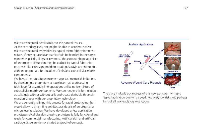
micro-architectural detail similar to the natural tissues. At the secondary level, one might be able to accelerate these micro-architectural assemblies by typical micro-fabrication tech-niques, if only extracellular matrix could be handled in the same manner as plastic, alloys or ceramics. The external shape and size of an organ or tissue can then be crafted by typical fabrication processes like extrusion, molding, coating, spraying, printing etc. with an appropriate formulation of cells and extracellular matrix components. We have attempted to overcome major technological limitations by developing a proprietary extracellular matrix processing technique for assembly line operations unlike native mixture of extracellular matrix components. We can render this formulation as solid gels with or without cells and create desirable three-di-mension shapes with our proprietary technology. We are currently refining this process for rapid prototyping that would allow to attain fine architectural details of an organ at a micron level resolution. We have developed a few application prototypes. Acellular skin dressing prototype is fully functional and ready for commercial manufacturing. Artificial skin and artificial cartilage tissue are demonstrated as proof-of-concept.
There are multiple advantages of this new paradigm for rapid tissue fabrication due to its speed, low cost, low risks and perhaps best of all, no regulatory restrictions.

38 Session 4: Clinical Application and Commercialisation
MassCustomizationofOrthopedicImplantsandPatientSpecificInstruments: The Business ModelGediminas KostkeviciusBaltic Orthoservice, Kaunas, Lithuania
Mass production of standard orthopedic implants and the system of their distribution result in a dilemma to be tackled by orthope-dists on a daily basis: how to apply standard implants to a specific clinical case? How to make do with what is available, with what a healthcare institution can afford?However, radiological diagnostics (digitization of internal human organs), image engineering technologies, object design in a virtual environment, 3D printing and modern non-destructive testing and quality control solutions make for technical means enabling design and production of custom-made implants and Patient Specific Instruments (PSI) based on virtual skeleton modules of a patient obtained by digitizing patient’s organs with a x-ray CT. This is a big step forward in improvement of treatment quality.This new business model of orthopedic implant design and production precipitates changes in the surgical thought paradigm. Instead of letting orthopedists continue to twist and turn trying to solve the problem of “how to make do with what we have” we can now ask them to describe the individual structural and func-tional properties to be implemented in an implant that is going to be used in treatment of a specific patient, in a specific clinical case. This in turn increases orthopedist’s personal responsibility for treatment results.
Technologies, personnel, and processes are the basis for national healthcare systems. They are the three resting pillars of these systems. Although available technologies already enable produc-tion of custom-made implants and PSI their wider use is impeded by growing treatment costs. These products are expensive. At the same time national healthcare systems are deeply engaged in funds saving policy as the needs of the ageing public for healthcare services grow faster that the funding assigned to meet them. This is why they are careful in accepting all innovations that increase treatment expenses.New technological innovations in medicine can pay off in two following ways:– by themselves if they help to save on other treatment costs
by reducing the number of repeat operations and length of hospitalization and disability as well as ensuring a higher degree of recovery from disability, etc.
– by creating conditions for redesign of activity processes. Rearrangement of processes will not only help reduce activity expenses but also allow for improved service accessibility and faster rendering of treatment services.

39Session 4: Clinical Application and Commercialisation
The third basis of national healthcare systems, along with tech-nologies and processes, is personnel. Success in implementation of both new technologies and new activity processes depends on how friendly these technologies and processes are for doctors and paramedics, how they will help solve issues that have to be han-dled by healthcare institution administrators on a daily basis. New technologies and processes require that all personnel engaged in the healthcare system should obtain additional competencies and change the limits of personnel responsibility.The presentation concerns changes in the processes of surgical treatment of patients using orthopedic implants occurring with transition from the model of mass production and distribution of standard implants towards mass customization of orthopedic implants. Production of custom made implants and PSI is not the challenge. Singular production of those products is already in place. The challenge is mass customization of orthopedic implants and special surgery instruments to make them affordable to national health care systems in terms of price and delivery terms, which encompasses a fundamental change in the surgical treatment planning paradigm.

40 Session 4: Clinical Application and Commercialisation
41
Session 5
METALLIC IMPLANTS I
EvaluationoftheBoneIngrowthofNumerically OptimizedandAdditiveManufacturedOpen-PorousTitanium Bone ScaffoldsProf. Dr. Rainer BaderDepartment of Orthopaedics, Biomechanics and Implant Technology Research Laboratory, University Medicine Rostock, GermanyE-Mail: [email protected]
Choice of Materials for Orthopaedic Implants: A Study of the Suitability of Cellular Metals Using Finite Element ModellingDr. Kanyakumari DattaData Metallurgical Company, Consulting Engineers, Kolkata, IndiaE-Mail: [email protected]
NewTi-Nb-BasedAlloysforImplantApplicationsDr. Annett GebertInstitute for Complex Materials, IFW (Leibniz Institute for Solid Stateand Materials Research) Dresden, GermanyE-Mail: [email protected]
42
44
45
1
IndividualContourAdaptedFunctionalImplant StructuresinTitanium–FromtheTheoreticalModel tothePracticalApplicationDr. Christine SchöneFaculty of Mechanical Science and Engineering, Chair of EngineeringDesign and CAD, Technische Universität Dresden, GermanyE-Mail: [email protected]
47
42 Session 5: Metallic Implants I
EvaluationoftheBoneIngrowthofNumericallyOptimizedandAdditiveManufacturedOpen-PorousTitaniumBoneScaffoldsRainer Bader, Jan WiedingDepartment of Orthopaedics, Biomechanics and Implant Technology Research Laboratory, University Medicine Rostock, Germany
Restoration of large segmental defects caused by fractures, tumors or infections is still a biomechanical challenge in ortho-paedic surgery. Autologous bone is still the ‘Gold Standard’ but has several disadvantages like limited availability and donor site morbidity [1]. Furthermore, artificial materials, such as calcium phosphate and titanium, have to fulfill biological and mechanical requirements in order to support bone regeneration. Interconnect-ing pores with a suitable pore size as well as sufficient mechanical stability are of great importance to enable bone and endothelial cell ingrowth [2]. To cope with all these requirements, additive manufacturing (AM) techniques like e.g. selective laser melting (SLM) offers the possibility to gain control about the architecture of the pore geometry and subsequent on the mechanical prop-erties [3]. The objective of this in-vivo study was the investigation of the biomechanical capability of custom made open-porous titanium implants for segmental defects of the metatarsal bone of sheep as well as the determination of the bone ingrowth into the open-porous structure. Cylindrical open-porous titanium scaffolds (20 mm in height and 17 mm in diameter, pore size of 700 x 700 µm) were generated by means of SLM fabrication, based on the CAD data of a biomechanically optimized scaffold design [4]. For this experi-mental study segmental defects of 20 mm were created in the
mid-diaphysis of the right metatarsal bone of fourteen three year old female sheep. The open-porous scaffolds were placed into the defect within the metatarsal bone, stabilized with additional osteosynthesis plate. After 12 (n=10) and 24 weeks (n=4) the animals were sacrificed. Both metatarsal bone were harvested in order to compare the treated and the non-treated contralateral side under torsional load. Maximum torsional moment, torsional angle, torsional stiffness and fracture energy have been calculat-ed. Furthermore, data from CT scans were used to determine the amount of newly formed bone around the implant.Metatarsal bones treated with the titanium scaffold showed differ-ent stages of new bone formation around the implant. 499 ± 461 and 667 ± 595 mm³ of new formed bone after 12 and 24 weeks, respectively, could be determined from CT data. In comparison to the non-treated side, maximum torsional moment was 49 ± 19 % after 12 weeks and increased to 69 ± 17 % after 24 weeks. For the torsional angle 51 ± 22 % and 80 ± 40 % could be achieved after 12 and 24 weeks, respectively. In contrast, torsional stiffness did not change significantly and was 107 ± 37 % after 12 weeks and 96 ± 40 % after 24 weeks. The ability of energy absorption was 29 ± 21 % for the treated bones after 12 weeks increasing up to nearly 60 ± 42 % after 24 weeks.

43Session 5: Metallic Implants I
In summary, our results showed the ability of open-porous titani-um scaffolds acting as bone scaffolds for large segmental defects. After the removal of the osteosynthesis system the treated bones showed lower mechanical loading capability of approximately 50 % after 12 weeks and 20 % after 24 weeks compared to the non-treated bones. A further increase in the biomechanical stability can be assumed for later follow-up periods. Therefore, open-porous titanium scaffolds represent a good alternative for autologous or allogenic material. Nevertheless, the ingrowth of bone tissue into the open-porous structure of the scaffolds has to be determined by further histomorphometric analyses.
References[1] Wippermann BW et al. Chirurg 1997; 68(12): 1286-91.[2] Karageorgiou V et al. Biomaterials 2005; 26(27): 5474-91.[3] Wieding J et al. Materials 2012; 5(8): 1336-47.[4] Wieding J et al. MED ENG PHYS 2013; 35(4): 422-432.

44 Session 5: Metallic Implants I
Choice of Materials for Orthopaedic Implants: A Study of the Suitability of Cellular Metals Using Finite Element ModellingK. Datta1, T. Momen2, F. Gagliardi3, D. Umbrello3, G. L. Manco3, C. Hannemann4, W. M. Rainforth5
1 Data Metallurgical Company, Consulting Engineers, Kolkata, India I 2 Consultant Orthopaedic Surgeon, Kolkata, India I 3 Department of Mechanical Engineering, University of Calabria, Rende (CS), Italy I 4 Fraunhofer Institute for Machine Tools and Forming Technology IWU, Chemnitz, Germany I 5 Department of Materials Science & Engineering, University of Sheffield, UK
In this study, the advantages of cellular structures like metal foam in the design of orthopaedic implants are discussed with respect to existing materials for implant. A finite element method (FEM) analysis of the forging process of a simple-shaped metallic foam billet is carried out using mathematical models in order to obtain a 3D geometry of a complex form. In particular, the deforming behaviour of a metallic foam and the development of density gradients are investigated in order to produce a selected portion of a hip prosthesis, for replacement of human bone. A commercial FEM code is used and the results are compared with previously done experiments. The complex 3D structure is successfully modelled. The constitutive behaviour of the porous material model was used. The model was validated by the comparison of the predicted results with the experimental evidence. Fig. 1 shows a contour plot of the variation in the void volume fraction across a central cross section of the forged foam billet. Forged metallic foam stands out in its ability to control the size and distribution of the pores during forging to manipulate the strength of the finished implant. When appropriately coated, it also offers better fixation in cementless joint replacement by triggering bone ingrowth.
Keywords: FEM, Metal Foam, Prostheses.
Figure 1. A contour plot showing the variation in the void volume frac-tion across a central cross section of the forged billet with initial foam density of 0.82 g/cc.

45Session 5: Metallic Implants I
NewTi-Nb-BasedAlloysforImplantApplicationsJ. Eckert1,2, A. Gebert1, M. Bönisch1,3, A. Helth1,2, K. Zhuravleva1,2, P.F. Gostin1, U. Hempel4, M. Calin1
1 IFW Dresden, Institute of Complex Materials, Germany I 2 TU Dresden, Institute of Materials Science, Faculty of Mechanical Science & Engineering, Germany I 3 TU Dresden, Institute of Structural Physics, Faculty of Science, Germany I 4 TU Dresden, Institute of Physiological Chemistry, Carl Gustav Carus Faculty of Medicine, Germany
Current research efforts are dedicated to the development of new Ti-based alloys with improved mechanical performance and biological compatibility expressed in low rigidity, high strength, composition of non-toxic elements and optimum surface condi-tions for osseointegration [1,2]. Alloys in the Ti-Nb based system show a promising spectrum of relevant properties. Young’s modu-lus, yield strength and microhardness typically follow a W-shaped curve as a function of the Nb content. The two minima in Young’s modulus correspond to the stabilization of α’’-martensite at 14-16.5 wt.-% Nb and the ß-Ti phase at >40 wt.-% Nb. These distinct compositions are promising starting points for further increase of the strength-to-Young’s modulus ratio [3,4].
Ti-40Nb shows the lowest Young’s modulus of 60 GPa and a compression strength up to 1200 MPa. A new casting technology was developed for additions of In as alloying element. Already a small amount of In leads to an evident decrease of the Young’s modulus below 50 GPa. The adaptation of thermo-mechanical post-treatments to those alloys is subject of current research. Interactions at the bone tissue/implant interface determine the biocompatibility. We demonstrated that acid etchings established for Ti and Ti6Al4V are not suitable for Ti-(40-45)Nb. The effective use of a H2SO4/H2O2 treatment for nano-roughening the
alloy surface was revealed [5,6]. This treatment accelerates the adhesion and spreading of human mesenchymal stem cells, increases the metabolic activity and the enzyme activity of tissue non-specific alkaline phosphatase (TNAP).
Porous metallic implants provide several advantages over bulk ones, e.g. very low stiffness and better ingrowth of bone tissue. Porous ß-type Ti-40Nb samples were prepared by a powder metallurgical approach starting with mechanical alloying [7]. The alloy powder was compacted with space holder material (Mg, NaCl) and sintered under strict temperature and time control. Samples with a porosity of 60-80% were tested in compression. The Young´s modulus reached very low values in the range of 1.5–2.5 GPa and the strength decreased with increasing porosity from 40-10 MPa. Surface modifications by electrodeposition of hydroxyapatite are demonstrated [8].
Acknowledgements: S. Oswald, H. Wendrock, A. Teresiak, S. Scudino, M. Uhlemann; for funding from the DFG - SFB/Transregio 79 and from the EC within the framework of FP7/2007-13 grant agreement no. 264635 (BioTiNet-ITN)
46 Session 5: Metallic Implants I
References:[1] M. Niinomi, J. Mech. Behav. Biomed. 1 (2008) 30[2] M. Calin et al., Mater. Sci. Eng. C 33 (2013) 875 [3] S. Hanada et al., Int. Congr. Series 1284 (2005) 239 [4] M. Bönisch et al., Sci. Technol. Adv. Mater. 14 (2013) 055004[5] P.F. Gostin et al., J. Biomed. Mater. Res. B 101B (2013) 269[6] A. Helth et al., J. Biomed. Mater. Res. B (DOI: 10.1002/
jbm.b.32976)[7] K. Zhuravleva, et al., Adv. Eng. Mater. 15 (2013) 257 [8] K. Zhuravleva et al., Mater. Sci. Eng. C 33 (2013) 2280

47Session 5: Metallic Implants I
IndividualContourAdaptedFunctionalImplantStructuresinTitanium–FromtheTheoreticalModeltothePracticalApplicationRalph Stelzer, Christine Schöne, Philipp SembdnerFaculty of Mechanical Science and Engineering, Chair of Engineering Design and CAD, Technische Universität Dresden, Germany
In the medical domain, the use of biocompatible materials, such as titanium or titanium alloys is essential to produce individual implants. As a result of this development, it is now possible to generate new patient-specific geometries fitted to the contour. This presentation elucidates the process chain to derive individual design variants and to produce patient-specific bone replacement implants for the lower jaw-bone regions by using innovative reverse engineering, design and direct manufacturing methods based on CT-data. The design of individualized lower jaw implants made of pure titanium was the subject of two projects, financed by the Saxon Bank for Reconstruction and Development (SAB). The authors’ partners in this interdisciplinary endeavor are doctors from the University Clinical Center Dresden and designing engineers from a product development firm. New technologies from medical image processing, Direct Manufacturing (generative manufacturing) and Reverse Engineering are thus brought together. Below, we list the principal steps needed to create individualized implants:1) Recording of the lower jaw region by means of computer
tomography (CT) and generation of a surface model by means of the “marching cubes“ algorithm
2) Alignment of the lower jaw model in a defined co-ordinate sys-tem and definition of cutting planes (marking of the damaged
area), fixing screws and dental implants3) Surface representation of the lower jaw contour with follow-up
design of implant and cutting templates (marking of the cutting position during operation)
4) Preparation for generative production by means of LaserCUS-ING® with subsequent manufacture of the implant and the cutting templates
5) Preparation for generative production by means of LaserCUS-ING® with subsequent manufacture of the implant and the cutting templates
In the presentation the authors reports about the different steps in the project, the difficulties and the results.

48 Session 5: Metallic Implants I

49
Session 6
METALLIC IMPLANTS II AND BIOMECHANICS
Dynamic Surface Microstructural Changes During Tribological Contact that Determine the Wear BehaviourofHipProstheses;MetalsandCeramicsProf. W. Mark RainforthDepartment of Materials Science and Engineering, The University of Sheffield, UKE-Mail: [email protected]
Mechanical and Tribological Test Methods for Joint ImplantsDr. Uta KremlingLaboratory for Medical Product Testing, IMA Dresden GmbH,GermanyE-Mail: [email protected]
Clinical and Engineering Assessments of the Effects of SurgicalProceduresandFixationsinSpineDr. Malhar Rao N. KumarConsultant Orthopaedic Surgeon, HOSMAT Hospital, Bangalore, IndiaE-Mail: [email protected]
50
51
53
31
ExperimentalandNumericalInsightsintothe MechanicalBehaviorofaTruncatedVertebralUnit(TVU)underCompressiveStaticandImpactDynamicLoadsGunti Ranga SrinivasCenter for Product Design and Manufacturing, Indian Institute of Science (IISc), Bangalore, IndiaE-Mail: [email protected]
ApplicationofShapeMemoryAlloysforActive LooseningProtectionofImplantStructuresChristian RotschDepartment of Lightweight Structures, Fraunhofer Institute for Machine Tools and Forming Technology IWU, Chemnitz / Dresden, GermanyE-Mail: [email protected]
54
55

50 Session 6: Metallic Implants II and Biomechanics
Dynamic Surface Microstructural Changes During Tribological Contact that Determine the WearBehaviourofHipProstheses;MetalsandCeramicsW. Mark Rainforth, Peng Zeng, Le Ma, Akemi Nogiwa ValdezDepartment of Materials Science and Engineering, The University of Sheffield, UK
It is often the dynamic microstructural changes induced by tribological contact that determine whether or not a material provides good wear resistance. It is well known that the mechan-ical properties of a surface are significantly different from the bulk even for the starting surface, but particularly as a result of tribological contact, which induces surface deformation (often to high strain) and interaction with the environment and/or the counterface and electrochemical effects. Despite the importance of these dynamic microstructural changes, there remains little quantitative understanding of how the surface microstructure changes during tribo-contact, and how this modifies the surface mechanical properties and chemical activity. This contribution will focus on key hip prosthetic materials, including CoCrMo alloys, third and fourth generation alumina/ zirconia toughened alumina and zirconia ceramics. High resolution techniques have been used to characterise the wear induced microstructural changes for both in vivo and in vitro samples, which has provided new insight into the wear mechanisms. The results are discussed in detail, in particular, how they inform future materials development for this important application.

51Session 6: Metallic Implants II and Biomechanics
Mechanical and Tribological Test Methods for Joint ImplantsUta KremlingLaboratory for Medical Product Testing, IMA Dresden GmbH
The presentation aims to give a survey about services that our lab offers to manufacturers of joint implants and implants for osteosynthesis. It will give some details to better understand the tests, like the standards, the test configuration and the obtained results. Our laboratory works as an accredited lab for mechanical and tribological tests on these implants since 1995.The first step of investigation should be the confirmation of suitability of the used materials. That includes metallic materials, ceramics and polyethylene. Investigations on the implant materials have the aim to validate the conformity in terms of chemical composition, mechanical strength and microstructure. Special attention should be paid to the investigation of polyethylene regarding the influence of aging, sterilization and crosslinking on the mechanical and wear behavior. The tests of coatings are another important part of material testing. On this field it is necessary to do for instance such important tests like tensile and shear static and fatigue tests or wear tests using the taber abraser. Some demanded limit values for the test results are contained in ASTM F 2068. Investigations on implants for osteosynthesis include tests of bone screws, bone plates and intramedullary nails. They will be done according ASTM standards. A special field is the investigation of the spinal implants like fixation devices and intervertebral cages.
Static and dynamic compression, torsion and shear tests are described in ASTM F 1717, 1798 und 2193 for fixation devices and in ASTM F 2077 and 2267 for cages. For joint implants a lot of ISO and ASTM standards are available to secure the safety in vivo. Hip implants have to fulfill the requirements of all parts of the ISO 7206 regarding geometrical and mechanical requests. Tests according to ISO 7206-4 and ISO 7206-6 are the most common ones. If the implants did not fulfill the demands it is necessary to explore the nature and the origin of the failures. The problems often result from the notch sensitivity of Titanium alloys or from problems in the process of casting in the case of CoCrMo alloys. The increasing modularity makes great demands on the corrosion resistance of the connections. A test according to ASTM F 1875 could be useful to get information about the corrosion resistance of cone connections. Thereby for example the appearance of the contact areas, the metallic content in the test medium and the morphology of the created particles are investigated.Tests regarding the wear behavior of the joint implants are more complicated and time-consuming than mechanical tests. But they are very important because the produced wear particles play an important role in the process of loosening of the implants. They can also influence the health of the patients. That is why

52 Session 6: Metallic Implants II and Biomechanics
standardized wear tests on hip and knee implants like ISO 14242 and ISO 14243 were developed. For shoulder implants our lab created own in-house standards. Beside the determination of the amount of wear the characterization of the wear particles and the determination of the metallic content in the test medium are of special interest. Standard tests are not always sufficient. In future these tests have to be extended by tests that are more relevant for practical usage of the endoprostheses. Problems that have to be solved are for instance the prevention of damages cause by rim loading, securing the unlimited functionality by activities with higher angle movements and higher loads and the safety of modular connections.

53Session 6: Metallic Implants II and Biomechanics
ClinicalandEngineeringAssessmentsoftheEffectsofSurgicalProceduresandFixationsinSpineMalhar Rao N. KumarConsultant Orthopaedic Surgeon, HOSMAT Hospital, Bangalore, India
Adjacent segment degeneration is a clinically observed compli-cation of segmental spinal fusion in the lumbar spine. In order to throw light into this clinical experience, an engineering tool in the form of finite element analysis is used to assess the stresses in the discs adjacent to a fused segment of a vertebral column under various loading conditions. A detailed finite element model of the lumbar spine (L1 to L5) is made followed by numerical analysis using LS-DYNA, a commercially available package for nonlinear dynamic analysis with powerful contact algorithms. Using this model, the stresses in the adjacent lumbar discs have been predicted for the following situations: disc adjacent to a degenerative disc without any fusion; disc adjacent to posterior decompression without fusion; disc adjacent to non-instrumented posterior, anterior and circumferential fusion (with and without decompression); and, disc adjacent to instrumented posterior, anterior and circumferential fusion (with and without decompres-sion). It was found that there was no significant difference in the adjacent level disc stresses with anterior fusion alone. There was a mild increase in the adjacent level disc stresses with posterior fusion without decompression. There was a 30% increase in the adjacent level stresses following posteriror fusion with decompres-sion. It is hoped that the information will be useful in providing a comprehensive insight into the parameters influencing the
outcome of segmental lumbar spinal fusion. It is hoped that the information generated will be useful in providing a comprehensive insight into the parameters influencing the outcome of segmental lumbar spinal fusion, and the finite element modeling technique discussed can be a powerful means for developing improved surgical interventions for spine as well as for other orthopedic applications.

54 Session 6: Metallic Implants II and Biomechanics
ExperimentalandNumericalInsightsintotheMechanicalBehaviorofaTruncatedVertebralUnit(TVU)underCompressiveStaticandImpactDynamicLoadsGunti Ranga SrinivasCenter for Product Design and Manufacturing, Indian Institute of Science, Bangalore, India
Human lumbar spine may experience considerable loads during daily activities and severe loads in events such as automotive accidents, blasts, falls and assaults. An understanding of the mechanical response of spinal vertebrae, especially under com-pressive loads, is essential for the assessment of injury tolerance and orthopedic interventions. In the present work, experimental and numerical investigations into the load-displacement responses of a human lumbar truncated vertebral unit (TVU) under qua-si-static and impact loading conditions have been carried out. TVU samples obtained from the lumbar spinal column of an adult human male cadaver were subject to quasi-static compressive tests in a UTM. Impact tests were then conducted on a similar TVU sample in a drop-weight testing device instrumented with a load cell and a high-speed data acquisition system. From the recorded dynamic responses, impact performance characteristics such as mean crush load and energy absorbed were obtained. An explicit finite element model of the TVU was developed for predicting the experimental quasi-static and impact dynamic responses. SEM images of vertebral cancellous and cortical bone specimens revealed their cellular microstructures similar to that of open cell foam. Thus crushable foam plasticity material models were used to simulate the cancellous and cortical bone responses. It was found that the results obtained from the computational
model correlated well with the experimental results. The enhanced understanding of the inelastic behaviour of a fundamental unit of a human vertebral bone complex gained in this study can aid in simulation-driven predictions of impact injuries such as fractures, and biomechanical assessments of orthopedic applications such as implants.
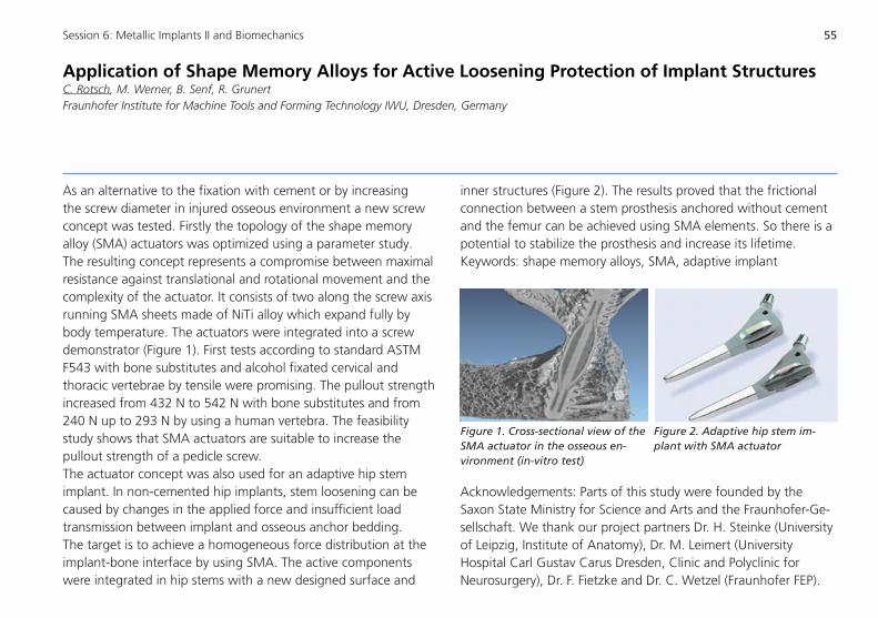
55Session 6: Metallic Implants II and Biomechanics
ApplicationofShapeMemoryAlloysforActiveLooseningProtectionofImplantStructuresC. Rotsch, M. Werner, B. Senf, R. GrunertFraunhofer Institute for Machine Tools and Forming Technology IWU, Dresden, Germany
As an alternative to the fixation with cement or by increasing the screw diameter in injured osseous environment a new screw concept was tested. Firstly the topology of the shape memory alloy (SMA) actuators was optimized using a parameter study. The resulting concept represents a compromise between maximal resistance against translational and rotational movement and the complexity of the actuator. It consists of two along the screw axis running SMA sheets made of NiTi alloy which expand fully by body temperature. The actuators were integrated into a screw demonstrator (Figure 1). First tests according to standard ASTM F543 with bone substitutes and alcohol fixated cervical and thoracic vertebrae by tensile were promising. The pullout strength increased from 432 N to 542 N with bone substitutes and from 240 N up to 293 N by using a human vertebra. The feasibility study shows that SMA actuators are suitable to increase the pullout strength of a pedicle screw. The actuator concept was also used for an adaptive hip stem implant. In non-cemented hip implants, stem loosening can be caused by changes in the applied force and insufficient load transmission between implant and osseous anchor bedding. The target is to achieve a homogeneous force distribution at the implant-bone interface by using SMA. The active components were integrated in hip stems with a new designed surface and
inner structures (Figure 2). The results proved that the frictional connection between a stem prosthesis anchored without cement and the femur can be achieved using SMA elements. So there is a potential to stabilize the prosthesis and increase its lifetime.Keywords: shape memory alloys, SMA, adaptive implant
Figure 1. Cross-sectional view of the SMA actuator in the osseous en-vironment (in-vitro test)
Figure 2. Adaptive hip stem im-plant with SMA actuator
Acknowledgements: Parts of this study were founded by the Saxon State Ministry for Science and Arts and the Fraunhofer-Ge-sellschaft. We thank our project partners Dr. H. Steinke (University of Leipzig, Institute of Anatomy), Dr. M. Leimert (University Hospital Carl Gustav Carus Dresden, Clinic and Polyclinic for Neurosurgery), Dr. F. Fietzke and Dr. C. Wetzel (Fraunhofer FEP).

56 Session 6: Metallic Implants II and Biomechanics

57
Session 7
CERAMICS
ProcessingandCharacterizationofCeramicMaterialsin ImplantsProf. Dr. Manoj Kumar MitraDepartment of Metallurgical and Material Engineering, Jadavpur University, Kolkata, IndiaE-Mail: [email protected]
SynthesisandApplicationofCalciumPhosphatesinMaxillofacialandOrthopaedicSurgeryDr. Janis LocsInstitute of General Chemical Engineering, Rudolfs Cimdins Riga Biomaterials Innovations and Development Centre, Riga Technical University, LatviaE-Mail: [email protected]
TailorMadeBioactiveCeramicsforSpecialtyClinicalApplicationsDr. Hari Krishna VarmaBioceramic Laboratory, Biomedical Technology Wing, Sree Chitra Tirunal Institute for Medical Sciences and Technology (SCTIMST), Trivandrum, IndiaE-Mail: [email protected]
58
59
60
3
1
ModifiedCalciumPhosphateBoneCementsforthe LocalDeliveryofTherapeuticIonsinOsteoporoticBone DefectsMatthias SchumacherCentre for Translational Bone, Joint and Soft Tissue Research, University Hospital and Medical Faculty, Technische Universität Dresden, GermanyE-Mail: [email protected]
62

58 Session 7: Ceramics
ProcessingandCharacterizationofCeramicMaterialsinImplantsManoj Kumar MitraDepartment of Metallurgical & Material Engineering, Jadavpur University, Kolkata, India
The paper describes experiences in relation to development of two implants – high purity alumina based hip joint prosthesis and porous hydroxyapatite based optical implant. Effect of initial grain size of alumina on fracture, wear and impact fatigue char-acteristics of the sintered product have been assessed. Detailed studies on general as well as sliding wear behavior under different medium have been described. Submicron grained alumina exhibits much improved performance under wear condition but results in a poorer impact fatigue resistance. In case of optical implant control of porosity made it possible to limit the weight of the implant to below 2gm. As a result, in addition to the expected performance of the implant in service a sympathetic motion of the eyeball could also be achieved. Detailed characterization of the material and in vivo evaluation of implant will be presented.
References:1. Role of apparent contact pressure on sliding wear of polycrys-
talline alumina in conformal contacts, Inter Ceram, Vol 55, No 2, 2006, pp 94 – 98
2. Sliding wear behaviour of submicron grained alumina in biolog-ical environment, Jl Biomed Mater Res – Part A, 2007,83(2), pp 257-262
3. Improved sliding wear resistance of submicron grained alumina : a comparison with coarser grained material, Jl Eur. Ceram. Soc, 2007, 27(16), pp4737-4743
4. Distinct wear characteristics of submicron grained alumina in air and distilled water – A brief analysis on experimental observa-tion, Jl Am. Ceram. Soc., 2007, 90(9), pp 2987-2991
5. Impact Fatigue Behaviour of fully dense Alumina Ceramics with Different Grain Sizes – Int Jl Sc Engg Res, 2011, Vol 2, Issue 4, pp 1-5
6. Fabrication and characterization of porous hydroxyapatite ocular implant followed by an in vivo study in dogs, Bull. Mater. Sc.,Vol. 27, No. 2 (2004), pp 133-140

59Session 7: Ceramics
SynthesisandApplicationofCalciumPhosphatesinMaxillofacialandOrthopaedicSurgeryLiga Berzina-Cimdina1, Janis Locs1, Girts Salms2, Sandris Petronis3, Simonas Grybauskas4
1 Riga Technical University, Institute of General Chemical Engineering, Rudolfs Cimdins Riga Biomaterials Innovations and Development Centre, Latvia I 2 Riga Stradins University, Institute of Stomatology, Latvia I 3 Riga 2nd Hospital and Riga Stradins University, Riga, Latvia I 4 Department of Maxillofacial Surgery, Vilnius University Zalgirio Hospital, Lithuania
The recent development in synthesis of calcium phosphate materials and preparation of implant materials in Riga Technical University will be described. The advantages of wet precipitation technology for the preparation of calcium phosphate ceramics with tailored phase composition will be discussed. The application examples of clinical trials in maxilla facial (see Fig.1) and ortho-paedic surgery (see Fig.2) will be demonstrated. The variations of calcium phosphate phase composition and corresponding applications will be discussed.
Fig.2 X-ray image of fracture of the proximal humerus: a) before surgery and b) postoperative follow up at 5 months
Fig.1 X-ray image before and after regulation of facial symmetry
60 Session 7: Ceramics
TailorMadeBioactiveCeramicsforSpecialtyClinicalApplicationsHari Krishna VarmaBioceramic laboratory, Biomedical Technology Wing, SreeChitraTirunal Institute for Medical Sciences and Technology, Trivandrum, India
Synthetic biomaterials have been used as bone grafts and devices over the last two decades. While autogenic bone is the gold standard, their limited availability and donor site morbidity lead to the search for alternate sources such as allografts and synthetic biomaterials. Synthetic biomaterials, mainly, bioactive ceramics are widely tried as bone substitutes owing to their biocompatibility. They elicit very little inflammation and have the ability to integrate with the surrounding bone. In clinical point of view the ideal ceramic bone graft substitute should be the one that rapidly inte-grates with the host bone, attains optimum mechanical stability very quickly and disappears rapidly after its function has been achieved with minimum toxic byproducts, allowing replacement by host bone. Bioactive ceramics, glasses and cements have al-ready made their mark in bone graft substitution. The Bioceramics Laboratory at SCTIMST had been working on these materials over the last decade dealing from the synthesis to clinical applications. Various technologies for manufacturing calcium phosphates based ceramics (dense and porous), bioactive ceramic composites (cal-cium-phosphate-silica systems) and calcium phosphate cements have been developed. A number of such products are used in India by orthopaedic surgeons as well as dental doctors. Bone tissue engineering is emerging as a solution for problems such as large bone segmental defects. Search for new generation bioac-
tive materials and devices is a never ending process and essentially to meet the demand from clinicians for better and faster solutions to unmet clinical problems.
References: Manitha B. Nair, Varma HK, S.Shenoy and Annie John, Treatment of Goat Femur Segmental Defects with Silica-Coated Hydroxyap-atite – One Year Follow-Up, Tissue Engineering Part A. 2010;16: 385. Acharya NK, Mahajan CV, Kumar RJ, Varma HK and Menon KV Can Iliac Crest Reconstruction Reduce Donor Site Morbidity? A Study Using Degradable Hydroxyapatite-bioactive Glass Ceramic Composite, J Spinal Disord Tech. 2010; 23: 266-271.JB Rajesh, K Nandakumar, HK Varma and Manoj Calcium Phos-phate Cement as a “Barrier-Graft” for the Treatment of Human Periodontal Intraosseous Defects Komath Indian J Dent Res, 2009; 20: 471-479 Acharya N K, Kumar R J, Varma H K, Menon KV, Hydroxyapa-tite-Bioactive Glass Ceramic Composite as Stand-alone Graft Substitute for Posterolateral Fusion of Lumbar Spine: A Prospec-tive, Matched, and Controlled Study. Journal of Spinal Disorders & Techniques 2008; 21(2):106-111

61Session 7: Ceramics
Easwer HV, Rajeev A, Varma HK, Vijayan S, Bhattacharya RN, Cosmetic and radiological outcome following the use of synthetic hydroxyapatite porous-dense bilayer burr-hole buttons. ActaNeu-rochir, 2007;149(5):481-5 Menon K V and Varma HK Radiological outcome of tibial plateau fractures treated with percutaneously introduced synthetic porous Hydroxyapatite granule, European Journal of Orthopaedic Surgery & Traumatology, 15, 205-213 (2005).

62 Session 7: Ceramics
ModifiedCalciumPhosphateBoneCementsfortheLocalDeliveryofTherapeuticIonsin Osteoporotic Bone DefectsMatthias Schumacher, Anja Lode, Anne Bernhardt, Michael GelinskyCentre for Translational Bone, Joint and Soft Tissue Research, University Hospital and Medical Faculty, Technische Universität Dresden, Germany
In the last years divalent strontium ions have gained considerable attention in osteoporosis therapy due to their dual mode of action on bone cells: on the one hand, they stimulate bone formation by osteoblasts and on the other hand inhibit the osteoclastic bone resorption. Thus, Sr2+ could help to regulate the impaired balance of osteoblast and osteoclast activity that is characteristic for osteoporosis [1].To ensure the local availability at a specific defect site and to overcome drawbacks of systemic Sr-based drug administration, strontium-containing biomaterials have recently been subject of intense research. In particular calcium phosphates (CaP) are of high interest in this field. Their easy applicability in the form of cal-cium phosphate cements (CPC), their intrinsic degradability as well as the possibility to substitute Sr2+ into their bulk structure offer the possibility to locally release therapeutic doses of strontium into a specific defect site. However, most SrCPC formulations imply elaborate synthesis of Sr-substituted CaP species. In contrast to that, we developed a SrCPC that can easily be doped with Sr2+ in varying concentrations up to 8.3 wt-%. The cement was shown to have good applicability and enhanced mechanical properties compared to standard CPC [2]. Depending on the Sr-content, a sustained release of Sr2+ was shown. The released Sr-doses had significant impact on both osteoblasts and
osteoclasts in vitro: human mesenchymal stem cells were shown to have higher proliferation rates and more advanced osteogenic differentiation on SrCPC [3], whilst the activity of characteristic enzymes in human osteoclast like cell cultures as well as the formation of resorption pits were reduced compared to Sr-free control samples. After 6 weeks in a critical-size bone defect in ovariectomized rats, histomorphometric analysis showed increased bone formation at the bone-implant interface of SrCPC compared to CPC [4]. Thus, SrCPC can be regarded as promising new material for the treatment of osteoporotic bone defects.
Fig. 1: Effect of Sr-modification on the mechanical properties (A) and osteoclastic material resorption (B).

63Session 7: Ceramics
References:[1] Pierre J. Marie. Bone, 40:5–8, 2007.[2] M. Schumacher, A. Henß, M. Rohnke, and M. Gelinsky. Acta
Biomater, 9:7536–7544, 2013.[3] M. Schumacher, A. Lode, A. Helth, and M. Gelinsky. Acta
Biomater, 9:9547–9557, 2013.[4] U. Thormann et al. Biomaterials, 34:8589–8598, 2013.

64 Session 7: Ceramics
65
Session 8
ELECTRIC / MAGNETIC STIMULATION, POLYMERS
HydroxyapatiteNanoparticlesandNanobiocompositeScaffoldforProteinAdsorption/ReleaseProf. Dr. Debasish SarkarDepartment of Ceramic Engineering, National Institute of Technology, Rourkela, IndiaE-Mail: [email protected]
InterplayofSubstrateConductivityandElectricStimuliin Directing Cell Fate on Implantable BiomaterialsGreeshma Thrivikraman Laboratory for Biomaterials, Materials Research Centre and Centre for Nanoscience and Engineering, Indian Institute of Science (IISc), Bangalore, IndiaE-Mail: [email protected]
DifferentialResponseofProkaryoticandEukaryoticCells on Engineered Biomaterials in Magnetic Field Stimulated Culture ConditionsB. Sunil KumarMaterials Research Centre, Indian Institute of Science (IISc), Bangalore, IndiaE-Mail: [email protected]
66
67
69
3
1
Bioelectric Stress Induced Cell Deformation and Stability in an Electric Field Stimulated MediumRavikumar KrishnamurthyMaterials Research Centre, Indian Institute of Science (IISc), Bangalore, IndiaE-Mail: [email protected]
Crosslinking as a Strategy to Design Multifunctional, TunablePolymerMatricesforTissueEngineering ApplicationsYashoda ChandorkarLaboratory for Biomaterials, Materials Research Centre, Indian Institute of Science (IISc), Bangalore, INDIAE-Mail: [email protected]
71
73

66 Session 8: Electric / Magnetic Stimulation, Polymers
HydroxyapatiteNanoparticlesandNanobiocompositeScaffoldforProteinAdsorption/ ReleaseDebasish SarkarDepartment of Ceramic Engineering, National Institute of Technology, Rourkela, India
Spherical, rod and fibrous hydroxyapatite (HA) nanoparticles synthesize through a common co-precipitation technique and fabricate macroporous HA-gelatin nanobiocomposite scaffolds for protein adsorption/release study. Three fundamental processing parameters such as solution pH, temperature and Ca/P ratio synchronize the morphology and crystallinity of nano HA from identical precursors Ca(CH3COO)2 and KH2PO4. Dispersion study illustrates the HA nanoparticle suspension stability phenomenon in aqueous media. Rod shaped HA exhibits relatively better bovine serum albumin (BSA) protein adsorption efficacy with compare to other two morphologies. In aqueous media, one gram nanorod HA particle adsorb 28 mg BSA within a time frame of 48 h and subsequently 75 wt.% release after 96h in phosphate buffer solution. Low temperature freeze casting of homogenous aqueous slurry of HA nanoparticles, gelatin and biocompatible polyvinyl alcohol binder develops nano HA – gelatin nanobiocomposite macroporous scaffolds. Freeze casted nanorod HA-gelatin macro-porous (70 vol.%) scaffold demonstrate highest yield compressive strength of ~2 MPa compare to other scaffolds prepared from spherical and fibrous HA because of high surface area and the effective anchoring. An optimum cryogenic treatment time at 77K promotes the mechanical response of this low strength scaffold and designates as cryo-treated hydroxyapatite–gelatin
macroporous scaffold (CHAMPS). CHAMPS has a high degree of interconnected pores of 50-200 µm in size, compressive strength up to 5.6 MPa and larger strain failure up to 25%. L929 mouse fibroblast cell interaction supports the cytotoxicity and cell adherence behavior with CHAMPS. Porous scaffold exhibits bioactivity in simulated body fluid (SBF) solution through preferable deposition of carbonated apatite layer around the pores. Biodegradation of scaffold in tris-HCl solution reveals a slow but systematic decrease in weight over incubation up to 7 days. Importantly, the excellent adsorption (upto 50 wt.%) and release (upto 60 wt.% of adsorbed protein) of BSA within 48h has been uniquely attributed to the inherent porous microstructure of the CHAMPS. Protein adsorption behavior for both of the particles and scaffolds follow the classical Langmuir isotherm. The extensive micro-computed tomography (micro-CT) analysis establishes cancellous bone-like highly interconnected and complex porous architecture of the protein loaded and original CHAMPS. Overall, the present study provides an assessment of the interaction of protein with HA nanoparticles and their cryotreated HA-gelatin scaffold in vitro to support as drug delivery media and tissue engineering, respectively.

67Session 8: Electric / Magnetic Stimulation, Polymers
InterplayofSubstrateConductivityandElectricStimuliinDirectingCellFateonImplantableBiomaterialsGreeshma Thrivikraman1,2, Giridhar Madras2, Bikramjit Basu1
1 Laboratory for Biomaterials, Materials Research Centre, Indian Institute of Science, Bangalore, India2 Centre for Nanoscience and Engineering, Indian Institute of Science, Bangalore, India
Most current strategies in biomaterials science revolve around altering the physical attributes of the substrates to modulate the cell functionality for desired therapeutic outcomes. Amongst those external cues, the response of cells towards underlying substrate conductivity, when stimulated by exogenous electric field, remains unexplored. In this light, the first part of the talk will address the ability of electroactive C2C12 muscle cells to sense the differences in electrical conductivity of the substrate. The cell morphology under fluorescence microscope after 72 hrs of culture revealed parallel arrangement of myoblast cells with increase in substrate conductivity. Furthermore, enhanced myoblast assembly and myotube formation (see Fig. 1) was also observed in a conductivity dependent manner.Knowing its biological significance, the study was further extend-ed to understand and determine the effect of electroconductivity and electric field stimuli in switching the lineage commitment of stem cells to desired phenotype. We used polyaniline (PANI) film as the model system to demonstrate this effect, wherein the substrate conductivity has been systematically varied over a wide range, by protonating it by dopant addition. Using flow cytometry based approach, the intracellular ROS level as well as cell cycle analysis was performed to determine the cellular response after electric field treatment. After 7 days of electric field treatment,
hMSCs adopted neural-like morphology from fibroblast-like mor-phology, with elongated actin rich unidirectional protrusions (see Fig. 2). Cytoskeletal reorganization led to the increase in length of cellular outgrowth, as evidenced with rising conductivity. More-over, with increasing substrate conductivity, electrically stimulated cells showed immunoreactivity towards neural lineage markers such as nestin and ßIII tubulin. These findings establish the key role of substrate conductivity in combination with electric stimuli as novel guidance cues in promoting neural-like differentiation of hMSCs.Overall, the results altogether will point towards the usefulness of tailoring the substrate conductivity and external electric field stimulation, which synergistically can modulate the in vitro cell fate processes. References[1] Greeshma Thrivikraman, Mallik PK, Basu B: Substrate
Conductivity Dependent Modulation of Cell Proliferation and Differentiation in vitro. Biomaterials, 2013 ;34:7073-7085.
[2] Gupta S, Greeshma T, Basu B, Goswami S, Sinha A: Stiffness- and wettability-dependent myoblast cell compati-bility of transparent poly(vinyl alcohol) hydrogels. Journal of Biomedical Materials Research Part B: Applied Biomaterials, 2012;101B(2):346-354.

68 Session 8: Electric / Magnetic Stimulation, Polymers
Fig. 1: Immunofluorescence image showing myogenin expression on myotube formed on conducting ceramic substrate. Scale bar: 100 µm
Fig. 2: Morphology of hMSCs grown on conducting polyaniline substrate after 7 days of electric field stimulated culture, exhibiting elongated filopodial structures. Scale bar: 50 µm
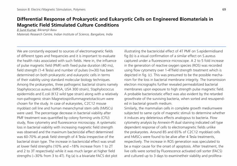
69Session 8: Electric / Magnetic Stimulation, Polymers
DifferentialResponseofProkaryoticandEukaryoticCellsonEngineeredBiomaterialsinMagnetic Field Stimulated Culture ConditionsB Sunil Kumar, Bikramjit BasuMaterials Research Centre, Indian Institute of Science, Bangalore, India
We are constantly exposed to sources of electromagnetic fields of different types and frequencies and it is important to evaluate the health risks associated with such fields. Here in, the influence of pulse magnetic field (PMF) with fixed pulse duration (40 ms), field strength (1-4 Tesla) and number of pulses (n=30) has been determined on both prokaryotic and eukaryotic cells in terms of their viability using standard molecular biology techniques. Among the prokaryotes, three pathogenic bacterial strains namely Staphylococcus aureus (MRSA, USA 300 strain), Staphylococcus epidermidis and E.coli (K12 wild type strain) along with a relatively non-pathogenic strain Magnetospirillummagnetotacticum were chosen for the study. In case of eukaryotes, C2C12 mouse myoblast cell line and human mesenchymal stem cells (hMSCs) were used. The percentage decrease in bacterial viability after PMF treatment was quantified by colony forming units (CFU) study, flow cytometry and fluorescence microscopy. A systematic loss in bacterial viability with increasing magnetic field strength was observed and the maximum bactericidal effect determined was 60-70% at peak field strength of 4 Tesla irrespective of the bacterial strain type. The increase in bactericidal effect was small at lower field strengths (10% and ~18% increase from 1 to 2T and 2 to 3T respectively) while it was much larger at higher field strengths (~30% from 3 to 4T). Fig (a) is a bivariate FACS dot plot
illustrating the bactericidal effect of 4T PMF on S.epidermidisand fig (b) is a visual confirmation of a similar effect on S.aureus captured under a fluorescence microscope. A 2 to 5 fold increase in the generation of reactive oxygen species (ROS) was recorded using flow cytometry over 1-4Tfield strength treatment which is depicted in fig. (c). This was presumed to be the possible mecha-nism for the loss in bacterial membrane integrity. The transmission electron micrographs further revealed permeabilized bacterial membranes upon exposure to high strength pulse magnetic field. A probable bacteriostatic effect was also evident by the retarded growthrate of the surviving bacteria, when sorted and resuspend-ed in bacterial growth medium.Similarly, the mammalian cells in complete growth mediumwere subjected to same cycle of magnetic stimuli to determine whether it induces any deleterious effects analogous to bacteria. Flow cytometry analysis by Annexin-PI dual staining indicated cell type dependent response of cells to electromagnetic fields unlike the prokaryotes. Around 85 and 65% of C2C12 myoblast cells and hMSCs were found to be alive after 4 Tesla treatments, respectively. The increase in ROS generation was speculated to be a major cause for the onset of apoptosis. After treatment, the live cells were sorted by Fluorescence activated cell sorter (FACS) and cultured up to 3 days to examinetheir viability and prolifera-
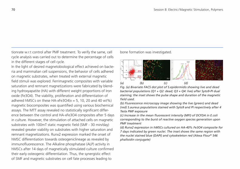
70 Session 8: Electric / Magnetic Stimulation, Polymers
tionrate w.r.t control after PMF treatment. To verify the same, cell cycle analysis was carried out to determine the percentage of cells in the different stages of cell cycle. In the light of desired magnetobiological effect achieved on bacte-ria and mammalian cell suspensions, the behavior of cells adhered on magnetic substrates, when treated with external magnetic field stimuli was explored. Ferrimagnetic composites with variable saturation and remnant magnetizations were fabricated by blend-ing hydroxyapatite (HA) with different weight proportions of Iron oxide (Fe3O4). The viability, proliferation and differentiation of adhered hMSCs on these HA-xFe3O4(x = 5, 10, 20 and 40 wt%) magnetic biocomposites was quantified using various biochemical assays. The MTT assay revealed no statistically significant differ-ence between the control and HA-xFe3O4 composites after 5 days in culture. However, the stimulation of attached cells on magnetic substrates with 100mT static magnetic field (SMF - 30 min/day) revealed greater viability on substrates with higher saturation and remnant magnetizations. Runx2 expression marked the onset of hMSC differentiation towards osteogeniclineage as revealed by immunofluorescence. The Alkaline phosphatase (ALP) activity in hMSCs after 14 days of magnetically stimulated culture confirmed their early osteogenic differentiation. Thus, the synergistic effect of SMF and magnetic substrates on cell fate processes leading to
bone formation was investigated.
(a) (b) (c) (d)Fig. (a) Bivariate FACS dot plot of S.epidermidis showing live and dead bacterial populations (Q1 + Q2: dead; Q3 + Q4: live) after Syto9-PI dual staining; the inset shows the pulse shape and duration of the magnetic field used. (b) Fluorescence microscopy image showing the live (green) and dead (red) S.aureus populations stained with Syto9 and PI respectively after 4 Tesla PMF exposure(c) Increase in the mean fluorescent intensity (MFI) of DCFDA in E.coli corresponding to the burst of reactive oxygen species generation upon PMF treatment (d) Runx2 expression in hMSCs cultured on HA-40% Fe3O4 composite for 7 days indicated by green nuclei. The inset shows the same region with the nuclei stained blue (DAPI) and cytoskeleton red (Alexa Flour® 546 phalloidin conjugate)

71Session 8: Electric / Magnetic Stimulation, Polymers
Bioelectric Stress Induced Cell Deformation and Stability in an Electric Field Stimulated MediumRavikumar Krishnamurthy1, V. Kumaran2, Bikramjit Basu1
1 Materials Research Centre, Indian Institute of Science, Bangalore, India2 Department of Chemical Engineering, Indian Institute of Science, Bangalore, India
Electric field has been proved to be a versatile stimulus which is capable of altering cell functionality both in vitro and in vivo. Experiments on both conducting and insulating substrates have shown that by altering stimulation parameters (field strength, pulse duration), a host of responses like apoptosis, necrosis, cell deformation, migration and in some cases, enhanced proliferation and differentiation can be observed. It has been reported that depending on the cell type and the substrate, only a narrow win-dow ofstimulation parameters can enhance the cell proliferation on biomaterial substrates [1].While the theoretical framework for some of the observed effects like electrophoresis [2] and electro-poration [3] are well developed, these predictive/analytical models are unable to explain the recent experimental results. In the above perspective, in an effort to rationalise such experimental observations, we provide here a theoretical model proposing the development of bioelectric stress field around a single cell. A single cell is modelled as a double layered membrane separating the cell culture medium and the cytoplasm with differ-ent dielectric properties. This well-known system is linearized by invoking Debye-Huckel approximation of the Poisson-Boltzmann equation. With appropriate boundary conditions, the system is solved to obtain bioelectric stress (Maxwell stress) in the presence of an external electric in terms of parameters like cell size,
permittivity of the medium and the cell, electric field strength, orientation and radial distance. The cell is found to be under both normal and shear stresses in the presence of external electric field. Under representative conditions of electric field stimulation (100 V/m, DC), a membrane without surface charges experiences normal and shear stresses of the order of 0.1 µPa. In the presence of surface charges on the cell membrane, the stresses are much higher at the membrane with normal stress of 10 mPa (5 orders higher) and shear stress of 0.1 mPa (3 orders higher). Based on the moments of the normal stresses, we predict shape changes of the viscoelastic cell membrane under the influence of electric field. In addition, the effect of shear stress on the membrane is quanti-fied by determining the electric field required to cause membrane rupture by overcoming the cohesive forces between the lipid molecules (membrane strength). It is found that this electric field is much higher for a charged membrane (30 kV/mm) as compared to a membrane without surface charges (0.6 kV/mm), highlighting the significance of membrane surface charge density.

72 Session 8: Electric / Magnetic Stimulation, Polymers
Fig.1: Deformation of a spherical cell into prolate shaped cell due to pre-sence of an external electric field.
References:[1] Dubey AK, Gupta SD, Basu B. Optimization of electrical
stimulation parameters for enhanced cell proliferation on biomaterial surfaces. Journal of Biomedical Materials Research Part B: Applied Biomaterials. 2011;98:18-29.
[2] Bier M. Electrophoresis: theory, methods, and applications. Electrophoresis: theory, methods, and applications. 1959.
[3] Weaver JC, Chizmadzhev YA. Theory of electroporation: a re-view. Bioelectrochemistry and bioenergetics. 1996;41:135-60.
E

73Session 8: Electric / Magnetic Stimulation, Polymers
CrosslinkingasaStrategytoDesignMultifunctional,TunablePolymerMatricesforTissue Engineering ApplicationsYashoda Chandorkar1, Giridhar Madras2, Bikramjit Basu1
1 Laboratory for Biomaterials, Materials Research Centre, Indian Institute of Science, Bangalore, India2 Department of Chemical Engineering, Indian Institute of Science, Bangalore, India
In our attempt to effectively mimic natural soft tissue, we realized that there is an increasing need to generate matrices for soft tissue engineering with varying mechanical properties. The achievement of tunable mechanical properties without sacrificing other essential properties like biodegradability and cytocompatibil-ity has been a major challenge.
In the context of biodegradability, the commonly used biopoly-mers e.g. poly lactic-co-glycolic acid (PLGA) releases chemicals, which can potentially cause inflammation. In order to address these issues, we have incorporated ricinoleic acid, which is known to have analgesic and anti-inflammatory effects in a crosslinked polymer. The crosslinking allows variation in mechanical and physical properties. The other monomers included the crosslinkers, citric acid and mannitol and the chain extender, sebacic acid.Although crosslinking generally causes polymers to form thermosets, we have followed a unique sequence of synthesis of a pre-polymer, which is soluble in solvents and then can be crosslinked (cured) to different degrees (Figure 1a). Hence, inspite of forming thermosets, the fabrication requirements are not so rigid, and the alterations to the polyester structure are possible.Consequently, we were able to generate a family of biodegrad-able, cytocompatible polyesters with tunable elastic modulus,
surface wettability and degradation properties. An analysis of the curing results led us to develop a predictive model to assess the degree of curing and control the properties (Figure 1 b).
Importantly, the tensile modulus range of 22-327 MPa, which matches that of human ligament and human tendon was ob-tained.Another important highlight is the variation in the erosion mechanismwith curing, from bulk to surface eroding. Another part of the present talk will focus on the development of salicylic acid (SA)-based biodegradable polymers. The in situ delivery of non-steroidal anti-inflammatory drugs (NSAIDs) like
Figure 1: (a) Central theme depicting variation in elastic properties and other mechanical properties achieved due to different curing conditions; (b) A theoretical model proposed for curing, when applied in the sense of condensation polymerization
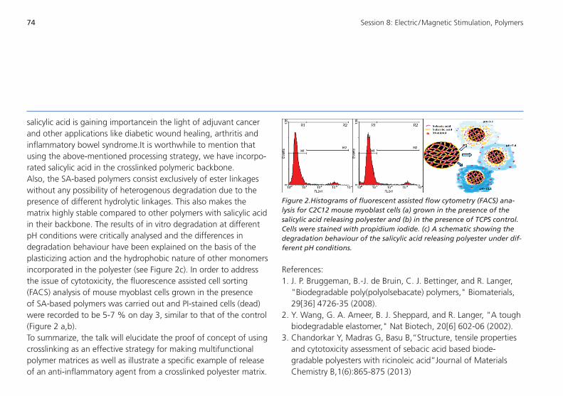
74
References:1. J. P. Bruggeman, B.-J. de Bruin, C. J. Bettinger, and R. Langer,
"Biodegradable poly(polyolsebacate) polymers," Biomaterials, 29[36] 4726-35 (2008).
2. Y. Wang, G. A. Ameer, B. J. Sheppard, and R. Langer, "A tough biodegradable elastomer," Nat Biotech, 20[6] 602-06 (2002).
3. Chandorkar Y, Madras G, Basu B,“Structure, tensile properties and cytotoxicity assessment of sebacic acid based biode-gradable polyesters with ricinoleic acid”Journal of Materials Chemistry B,1(6):865-875 (2013)
salicylic acid is gaining importancein the light of adjuvant cancer and other applications like diabetic wound healing, arthritis and inflammatory bowel syndrome.It is worthwhile to mention that using the above-mentioned processing strategy, we have incorpo-rated salicylic acid in the crosslinked polymeric backbone.Also, the SA-based polymers consist exclusively of ester linkages without any possibility of heterogenous degradation due to the presence of different hydrolytic linkages. This also makes the matrix highly stable compared to other polymers with salicylic acid in their backbone. The results of in vitro degradation at different pH conditions were critically analysed and the differences in degradation behaviour have been explained on the basis of the plasticizing action and the hydrophobic nature of other monomers incorporated in the polyester (see Figure 2c). In order to address the issue of cytotoxicity, the fluorescence assisted cell sorting (FACS) analysis of mouse myoblast cells grown in the presence of SA-based polymers was carried out and PI-stained cells (dead) were recorded to be 5-7 % on day 3, similar to that of the control (Figure 2 a,b). To summarize, the talk will elucidate the proof of concept of using crosslinking as an effective strategy for making multifunctional polymer matrices as well as illustrate a specific example of release of an anti-inflammatory agent from a crosslinked polyester matrix.
Figure 2.Histograms of fluorescent assisted flow cytometry (FACS) ana-lysis for C2C12 mouse myoblast cells (a) grown in the presence of the salicylic acid releasing polyester and (b) in the presence of TCPS control. Cells were stained with propidium iodide. (c) A schematic showing the degradation behaviour of the salicylic acid releasing polyester under dif-ferent pH conditions.
Session 8: Electric / Magnetic Stimulation, Polymers

75
February 21, 2014
VISIT OF THE AMBASSADOR OF INDIA TO GERMANY
11:00 – 12.30 amHis Excellency Mr. Shri Vijay Keshav Gokhale
(event closed to the public)

76
published by
Prof. Dr. Michael Gelinsky
Centre for Translational Bone, Joint and Soft Tissue Research
Technische Universität Dresden
Fetscherstrasse 74
01307 Dresden, Germany
and
Christian Hannemann
Fraunhofer Institute for Machine Tools
and Forming Technology IWU
Reichenhainer Strasse 88
09126 Chemnitz, Germany
February 2014