bone-repair properties of biodegradable hydroxyapatite
TRANSCRIPT

Nanoscale
PAPER
Cite this: Nanoscale, 2015, 7, 18751
Received 20th July 2015,Accepted 10th October 2015
DOI: 10.1039/c5nr04850h
www.rsc.org/nanoscale
Bone-repair properties of biodegradablehydroxyapatite nano-rod superstructures†
Noelia L. D’Elía,a Colleen Mathieu,b Caroline D. Hoemann,b,c,d Juan A. Laiuppa,e
Graciela E. Santilláne and Paula V. Messina*a
Nano-hydroxyapatite (nano-HAp) materials show an analogous chemical composition to the biogenic
mineral components of calcified tissues and depending on their topography they may mimic the specific
arrangement of the crystals in bone. In this work, we have evaluated the potential of four synthesized
nano-HAp superstructures for the in vitro conditions of bone-repair. Experiments are underway to investi-
gate the effects of the material microstructure, surface roughness and hydrophilicity on their osseo-inte-
gration, osteo-conduction and osteo-induction abilities. Materials were tested in the presence of both, rat
primary osteoblasts and rabbit mesenchymal stem cells. The following aspects are discussed: (i) cytotoxi-
city and material degradation; (ii) rat osteoblast spreading, proliferation and differentiation; and (iii) rabbit
mesenchymal stem cell adhesion on nano-HAp and nano-HAp/collagen type I coatings. We effectively
prepared a material based on biomimetic HAp nano-rods displaying the appropriate surface topography,
hydrophilicity and degradation properties to induce the in vitro desired cellular responses for bone bonding
and healing. Cells seeded on the selected material readily attached, proliferated and differentiated, as
confirmed by cell viability, mitochondrial metabolic activity, alkaline phosphatase (ALP) activity and cyto-
skeletal integrity analysis by immunofluorescence localization of alpha-smooth muscle actin (α-SMA)
protein. These results highlight the influence of material’s surface characteristics to determine their tissue
regeneration potential and their future use in engineering osteogenic scaffolds for orthopedic implants.
1. Introduction
The current options related to bone injury therapies such asauto-grafts, allo-grafts and a variety of synthetic or biomimeticdevices are unsatisfactory. The evidence base is the existenceof significant limitations including the need for additional sur-geries, morbidity associated with the donor site, limitedmaterial supply and inadequate size or shape of the implanta-ble donor tissue.1,2 All these limitations lead to the develop-ment of advanced approaches to assist the skeletal tissue’s
reparation and reconstruction; avoiding the requirement of apermanent prosthesis.3–5 Overcoming these challengesrequires not only that the engineered substitute has bothstructural resemblance with the native tissue and ability toperform similar biological functions, but also avoid problemsof donor site scarcity, immune rejection and pathogentransference.6–9 In addition, a bone substitute should ideallydegrade at an equivalent rate to tissue regrowth.7 In thisregard, nanosized hydroxyapatite (nano-HAp) materials havebeen widely investigated for many biomedical applicationsrelated to calcified tissues, i.e., bone and dental compositefillers,10,11 maxillofacial reconstruction and augmentation,12,13
tissue engineering scaffolding14–16 and coatings for metallicorthopedic prosthesis.17 The main reason is that nano-HApmimics the natural chemical composition and dimensions ofthe mineralized phase found in the extracellular matrix (ECM)of bone.18 The most significant advantages of nano-HAppowders compared to coarse synthetic crystals are theirenhanced sinterability and densification, better osteo-conduc-tive and osteo-inductive capabilities, and improved fracturetoughness as a consequence of their greater surface area.19
However, despite these valuable properties, the effect of nano-HAp properties on tissue response has not yet been fullyunderstood.20 When a biomaterial is introduced into a living
†Electronic supplementary information (ESI) available: Calculation of roughnessparameters Rz, Rz,max, and Rz, prom. Nano-HAp powder degradation after immer-sion in phosphate buffer (pH = 7.4). Optical phase contrast microphotographsof MSC adhesion on nano-HAp and nano-HAp/Co I coatings at different concen-trations. Laser scanning confocal microphotographs of MSCs’ α-SMA expressionspreading on large amounts of nano-HAp (MI) coatings. Immunofluorescencemicrophotograph analysis by image software. See DOI: 10.1039/c5nr04850h
aDepartment of Chemistry, Universidad Nacional del Sur, INQUISUR-CONICET,
8000 Bahía Blanca, Argentina. E-mail: [email protected]; Fax: +54 291 4595160;
Tel: +54 291 4595159bInstitute of Biomedical Engineering, École Polytechnique, Montréal, QC, CanadacGroupe de Recherche en Sciences et Technologies Biomédicales (GRSTB), CanadadDepartment of Chemical Engineering, École Polytechnique, Montréal, QC, CanadaeDepartment of Biology, Biochemistry and Pharmacy, Universidad Nacional del Sur,
CONICET, 8000 Bahía Blanca, Argentina
This journal is © The Royal Society of Chemistry 2015 Nanoscale, 2015, 7, 18751–18762 | 18751
Publ
ishe
d on
12
Oct
ober
201
5. D
ownl
oade
d by
Eco
le P
olyt
echn
ique
de
Mon
trea
l on
23/0
5/20
18 1
6:25
:00.
View Article OnlineView Journal | View Issue

tissue, the ensuing biocompatibility and the host responsesdepend on their physical and chemical properties thatpromote the initial reactions of cells upon the materialsurface.20,21 In such terms, it was determined that thematerial’s surface roughness is first closely related to theextent and strength of cell’s adhesion, proliferation and differ-entiation; subsequently to the material’s osteo-induction,osteo-conduction and osseo-integration, thereby the bonerepair abilities. The microstructure and topology of nanostruc-tured materials depend on their synthesis method, as well ason their processing conditions; a minor change in the powdercompaction or in the sintering conditions can result in avariety of different porosity characteristics.22 Consequently, itis extremely important to select the most appropriate tech-nique when formulating nano-materials with desired pro-perties and their combinations. In a previous work, weprepared four hydroxyapatite materials templated by differentmicelle–polymer structured networks.23 The synergistic inter-action of each polymer in contact with hexadecyl-trimethylammonium bromide (CTAB) rod-like micelles results in crystal-line HAp nano-rods of 25–50 nm length organized in hierarchi-cal structures with different roughnesses and porosities. Therole of the material’s surface texture was revealed as a keyfactor in the nucleation and growth of a bone-like apatitecoating in contact with simulated body fluid (SBF); an essen-tial requirement to estimate the materials’ bone-bonding andosseo-integration potential.24,25 The growth of the differentstructures is attributed to a dissimilar matching of crystalplanes in the material and in the apatite layer formed. Underspecific synthesis conditions, a biocompatible material with aCa/P ratio close to that existing in the trabecular bone and, amorphology that is considered essential for bone-bonding wasobtained.23 Since the roughness, morphology and micro-patterning of the four investigated nano-HAp superstructures arecrucial points for their optimal osseo-integration capabilities,the aim of this study was to carry out an in vitro evaluation ofthe effect of these parameters to induce the desired bonerepair cellular responses, i.e. osteo-induction and osteo-conduction abilities. The effects of the synthesized materials’surface characteristics on the following factors are discussed:(i) nano-HAp material degradation and cytotoxicity; (ii) ratosteoblast spreading, proliferation and differentiation; and(iii) rabbit mesenchymal stem cell adhesion, mitochondrialmetabolic activity, and spreading when cultured on surfacescoated with HAp nanoparticles and fibrillar collagen type I.
The results of this study were expected to serve as a refer-ence in the selection and/or in the manufacturing of theproper nano-HAp materials’ surface characteristics to maxi-mize their bone-repair properties.
2. Materials and methods2.1 Materials
Hexadecyl-trimethyl ammonium bromide (CTAB, MW =364.48 g mol−1, 99%, Sigma-Aldrich); poly(ethylene glycol) 400
(PEG 400, Sigma-Aldrich, MW = 380–420 g mol−1, δ = 1.126 g mL−1
at 25 °C); poly(propylene glycol) (PPG, Sigma-Aldrich,MW = 425 g mol−1, δ = 1.004 g mL−1 at 25 °C); poly(ethyleneglycol)-block-poly(propylene glycol)-block-poly(ethylene glycol)(PEG-PPG-PEG, PEG 10 wt%, Sigma-Aldrich, MW = 2800 g mol−1,δ = 1.018 g mL−1); octylphenyl-polyethylene glycol (IGEPAL®CA630, (C2H4O)nC14H22O, Sigma-Aldrich, MW = 603 g mol−1,δ = 1.06 g mL−1 at 25 °C); sodium phosphate (Na3PO4, MW =148 g mol−1, 96%, Sigma-Aldrich); calcium chloride (CaCl2,MW = 91 g mol−1, 99%, Sigma-Aldrich) and sodium nitrite(NaNO2, MW = 69 g mol−1, 97%, Sigma-Aldrich) were usedwithout further purification. Alpha-Minimum EssentialMedium supplemented with 15% fetal bovine serum(α-MEM-15% FBS, Sigma-Aldrich); ALP activity kit (WienerLab., Rosario, Argentina), xylazine (Rompun, CDMV, 20 mg mL−1);ketamine (Rogarsetic, CDMV, 100 mg mL−1); sodiumpentobarbital (Euthanyl) solution IV (CDMV, 240 mg mL−1).Collagen type I from rat tail solution (3.9 mg mL−1, Sigma-Aldrich); Dulbecco’s Modified Eagle Medium: NutrientMixture F-12 (DMEM/F12, Gibco) with supplemented sodiumbicarbonate (2.438 g, Sigma-Aldrich); phosphate bufferedsaline (PBS without Ca/Mg, 1X, Sigma-Aldrich); paraformalde-hyde (HO(CH2O)nH, Sigma-Aldrich); normal Goat Serum(Sigma-Aldrich); Triton X-100 (Sigma-Aldrich); Bovine SerumAlbumin (BSA, Sigma-Aldrich); Alexa Fluor® 488 goat anti-mouse IgG (H + L) (Molecular Probes, 2 mg mL−1); MouseMonoclonal Anti-Actin, α-Smooth Muscle antibody (clone 1A4,Isotype IgG2a, Sigma-Aldrich, 1 μg mL−1); Hoechst 33342(Gibco, 2 μg mL−1); Fluoromount-G (Southern biotech); 3-(4,5-dimethylthiazol-2-yl)-2,5-diphenyltetrazolium bromide (MTT,Sigma-Aldrich, 5 mg mL−1). For solution preparation, onlytriple-distilled water was used. For all experiments passage twoto four (P2–P4) cells were used.
2.2 Bone-like HAp synthesis
Four HAp nanoparticle (7–9 nm diameter and 25–50 nmlength) materials exhibiting different topographies and mor-phologies were synthesized by a previously described method-ology.23 First, 350 mL of a 3.13 mM CTAB aqueous solutionwas mixed with 20 mL of PPG (MI), PEG (MII), PEG-PPG-PEG(MIII) or IGEPAL (MIV)23 and stirred at 500 rpm for10 minutes. Second, 200 mL of 2 M sodium nitrite aqueoussolution and 2.2 g calcium chloride were incorporated insequence. Finally, 200 mL of 0.14 M Na3PO4 aqueous solutionwas added to the above mixed solution drop by drop at roomtemperature (RT) under magnetic stirring at 500 rpm. After theintegration of all reactants, the solution was magneticallystirred for 1 h. The resulting gels were left for 24 h in an auto-clave at 100 °C. The obtained materials were filtered andwashed with triplet-distilled water to remove impurities.Finally, the surfactant was completely removed by calcinationat 400 °C for 3 h under air flux.
2.3 Near infrared spectroscopy
A Nicolet iS50 FTIR-NIR spectrophotometer (Thermo Scienti-fic, Waltham, MA, USA) along with a diffuse reflectance
Paper Nanoscale
18752 | Nanoscale, 2015, 7, 18751–18762 This journal is © The Royal Society of Chemistry 2015
Publ
ishe
d on
12
Oct
ober
201
5. D
ownl
oade
d by
Eco
le P
olyt
echn
ique
de
Mon
trea
l on
23/0
5/20
18 1
6:25
:00.
View Article Online

accessory (DRA, also called an integrating sphere) was usedto measure the reflectance properties of the powders. Thespectra were obtained under an air atmosphere and at RT.In the present study the integrating sphere was operated inthe reflectance mode in the region of 1000–2500 nm. AGold NIR Diffuse Reflection Standard (99.9% Reflective) wasused as a reference to calibrate the baseline. The powdersamples were supported inside flat bottom glass vialsto form pellets of 10 mm diameter and 5 mm thick formeasurements.
2.4 In vitro biodegradability under acidic conditions
Assuming that the local pH around the ruffled border ofosteoclasts is from 4.0 to 5.0 during the bone remodelingperiod,26 in vitro degradation behavior of HAp particlesunder acidic conditions was evaluated by soaking them inan acetic acid/sodium acetate buffer solution (AcOH buffer) witha pH of 4.24 according to the method of Matsumoto et al.27
HAp particles (200 mg) were soaked in 100 ml of theAcOH buffer solution at 25 °C and 36 °C for 3, 12, 16and 25 days. The degradability of nano-HAp materials wasestimated from the rate of weight loss (WL) accordingly toTampieri et al.:16
%WL ¼ ðW0 �WtÞW0
� 100
where W0 and Wt are the dry weights of the initial and thedegraded specimens at different immersion times, t, respect-ively. The same procedure was followed at pH 7.4 (PBS) as acontrol.
2.5 Rat osteoblast isolation, culture and treatment
Calvarial osteoblasts were obtained from 5-day-old neonatalrats as previously described23 and the cells were frozen inliquid nitrogen until their use. For the experiment, cells werecultured at 37 °C in α-MEM-15% FBS, 1% penicillin and strep-tomycin (P–S) under humidified air (5.5% CO2). After 24 h, themedium was replaced by α-MEM supplemented with 10%(FBS) and 1% (P–S), and the cells were cultured until ∼80% ofconfluence (2–3 days). Calvarial osteoblasts were seeded in 48-well plates in the presence or the absence of the nano-particles(71.42 μg ml−1) and cultured for 4 and 7 days in α-MEM-10%(FBS)-1% (P–S), under a humidified atmosphere (5.5% CO2) at37 °C. It is known that the presence of calcium in the culturemedia stimulates osteoblast differentiation,28 therefore thecells were cultured in the presence of 4 mM of CaCl2 as a posi-tive control.
2.6 Alkaline phosphatase activity in rat osteoblast
After cell treatments, the ALP activity was measured in lysedprimary rat osteoblasts as described by Ayala-Peña et al.29 TheALP activity was estimated using a commercially available kitand indirectly measured by the amount of phenol released;and the results were based on the increase in absorbance atoptical density (OD) 520 nm. A blank (B) and a standard (S)(200 IU l−1 phenol) were also processed. Optical density of the
samples (D) was measured and ALP activity was calculated asfollows:
ALPIUl
� �¼ 200 IU l�1 � ðD� BÞ
ðS� BÞ
2.7 Rabbit mesenchymal stem cell (MSC) isolation
All protocols involving animals were approved by institutionalethics committees. MSCs were obtained from 9–11 month oldNew Zealand White rabbits which were sacrificed underxylazine–ketamine induced anesthesia using 150 mg kg−1 sodiumpentobarbital (Euthanyl) solution IV. Bone fragments from thesubchondral bone of the distal femur were subjected to col-lagenase digestion to release cells from marrow stroma thatwere pelleted, and deposited in petri dishes in completeculture media, DMEM/F12 + 10% FBS + 1% P–S, in a humidi-fied 5% CO2 incubator. The obtained cells were cultured for5 days during which the medium was changed every 2 days toremove non-adherent cells and to select adherent cells. Theprimary MSCs were cryopreserved at P0 and at P1, then thawedand passaged for the cell assays.
2.8 Adhesion assay
2.8.1. Rabbit MSC metabolic assay on nano-HAp coatings.Viable MSCs with active metabolism reduce MTT into a purplecolored formazan product that can be solubilized and quanti-fied by spectrophotometric means. When cells die, they losethe ability to convert MTT into formazan, thus color formationserves as a marker of only the metabolically active cells.30,31
Therefore, this assay was performed with the objective toevaluate the MSC metabolic activity in wells coated with thefour different nano-HAp materials (MI, MII, MIII and MIV)and indirectly their viability.31 For sterilization, nano-HApmaterials were autoclaved for 30 min at 120 °C. Then a watermaterial dispersion was prepared by placing the componentson a rotating mixer for 5 min following which, a 96-well plate(0.32 cm2 per well) was filled with increasing amounts ofnano-HAp material dispersions to have a final coating of15–3100 µg HAp per cm2. Finally, the material-coatings wereallowed to dry overnight on a shaker in a biological safetycabinet to obtain a homogeneous dry-coat surface on thebottom of the well. MSCs were seeded at a density of 10 000cells per well and were cultured in a pre-coated culture platefor 24 h and 48 h in complete culture media (DMEM/F12 +10% FBS + 1% P–S). The homogeneous distribution andattachment of HAp nanoparticles on the bottom of the wells,before and after MSC seeding, was checked by opticalmicroscopy; see Fig. 6, ESI.† Pre-coated wells with mediumand no cells were used as negative controls (C−) and uncoatedwells were used as positive controls (C+). The MTT solutionwas added in each well at 0.5 mg mL−1 final concentration andthe plate was incubated for 3 h under humidified air (5%CO2). The wells were aspirated and 100% DMSO (200 µL) wasadded and the resulting formazan was quantified using anELISA plate reader (OD 570). The absorbance of coated wellswithout cells was used as blanks.
Nanoscale Paper
This journal is © The Royal Society of Chemistry 2015 Nanoscale, 2015, 7, 18751–18762 | 18753
Publ
ishe
d on
12
Oct
ober
201
5. D
ownl
oade
d by
Eco
le P
olyt
echn
ique
de
Mon
trea
l on
23/0
5/20
18 1
6:25
:00.
View Article Online

2.8.2 Rabbit MSC metabolic assay on nano-HAp (MI)/collagen type I (Co I) coatings. Rabbit MSC adhesion and meta-bolic activity under four different MI/Co I weight ratio coatingconditions were studied (MI/Co I: 0/1; 0.5/1; 1/1 and 2/1, withCo I = 31 µg cm−2); and for this a MTT assay was done in a 96-well plate as described above. To prepare MI/Co I coatings, thecommercial rat tail collagen-acetic acid (20 mM) solution wasneutralized with a 20 mM NaOH solution. Then, threedifferent concentrations of MI water dispersions were preparedand mixed with a suitable amount of Co I neutral solution toobtain different MI/Co I weight ratio coatings. Finally, the solu-tions were allowed to bind to the well plate by a dry-coatingprocess (see section 2.8.1). Adhesion of MSCs on non-coatedwells was used as a control.
2.8.3. MSC actin-based spreading on collagen type Iwith and without nano-HAp: immunofluorescence confocalmicroscopy. The cytoskeleton is known to play a key role inthe mechano-transduction steps, and it can also contribute toMSC differentiation.32 Multipotential mesenchymal stem cellsalso express α-smooth muscle actin (αSMA), and actin stressfibers can be used to demonstrate cell attachment responses.This experiment tested the hypothesis that MSCs attach andspread on collagen type I incorporated with nano-HAp. 10 000rabbit MSCs per well were seeded in individual Culture Slide(Falcon®, Corning) glass chambers dry-coated with Co I orMI-Co I, as described above. Non-coated glass slide chamberswere used as a C+. Cells were incubated for 29 h in a humidi-fied cell culture incubator with 5% CO2, then fixed with 4%paraformaldehyde for 5 min and then washed three times for5 min with PBS. They were blocked and permeabilized with20% v/v Goat serum/PBS/0.1% Triton X-100 for 15 min at RT.After washing with 1% BSA/PBS/0.1% Triton X-100 for 5 minthe cells were incubated with primary antibody anti α-SMA(clone 1A4, Isotype IgG2a, Sigma-Aldrich) diluted to 1 μg mL−1
in 1% BSA/PBS/0.1% Triton X-100 for 30 minutes at RT. Sub-sequent to three washes with 1% BSA/PBS/0.1% Triton X-100for 10 min the glass slides were incubated with secondary anti-mouse antibody conjugated with Alexa 488 diluted to1.3 µg mL−1 in 1% BSA/PBS/0.1% Triton X-100 for 30 minutes atRT. After two 10 min PBS washes a counter stain was done withHoechst 33342 diluted to 2 µg mL−1 in PBS for 1 min at RT.The slides were washed two times with PBS for 10 min. Finally,the slides were separated from the chamber and Fluoromount-G was used to mount the slides. They were covered with alumi-num foil and allowed to dry for 24 h. Images were acquiredusing an Olympus FluoView™ FV1000 laser scanning confocalmicroscope with laser operational wavelength set at 405 nmfor the blue signal with Hoechst counterstain in cell nucleusand at 488 nm for the green signal for α-SMA expression. Allimages were analyzed with the help of the software ImageJ(National Institutes of Health, Bethesda, MD). The cellnumber was determined by a count of nucleus positive forHoechst stain in a histology field per sample, and α-SMAexpression was quantified from total green pixels per imagewith the data calculated as a percentage of (green pixels perimage)/(total pixels). In addition, α-SMA expression was
characterized by the mean brightness value of green pixelsdetermined from the intensity of all green pixels per fieldin monochromatic images of the laser set at 488 nm (seeFig. 10, ESI†).
2.9 Statistical analysis
All quantitative tests were carried out at least in triplicate, andthen mean values with standard deviations were calculated.Statistical analysis of data was accomplished by one factor ana-lysis of variance (ANOVA). Student’s t-test and probabilityvalues below 0.05 (p < 0.05) were considered significantlydifferent. Quantitative data are expressed as mean ± standarddeviation (SD) from the indicated set of experiments.
3. Results and discussion
Osteo-induction, osteo-conduction and osseo-integration areinterrelated and are the required abilities of a bone-healingmaterial.33 Osteo-induction implies the recruitment of imma-ture cells and the stimulation of these cells to develop intomature osteoblasts; this process is part of normal bonehealing and is responsible for the majority of newly formedbone after a fracture or an implant insertion. Osteo-conductionmeans that bone grows on a surface; is a term usually used inconjunction with implants. Osseo-integration is the stableanchorage of an implant achieved by a direct bone-to-implantcontact. Osteo-conduction and osseo-integration both dependnot only on biological factors, but also on their response to thepresence of a foreign material.33 The effect of nano-HAppowder’s surface topography, hydrophobicity and degradationon their in vitro osteo-induction, osteo-conduction and osseo-integration capacity is analyzed in the following sections.
3.1 Nano-HAp powder’s surface topographyand hydrophilicity
Surface roughness degree, determined by the roughness arith-metical average deviation (Ra), can be distinguished accordingto the scale of the irregularities of the material surfaces: (i)surface macro-roughness, ≥100 μm; (ii) microscale surfaceroughness, from 1 to 100 μm; (iii) submicron surface rough-ness, from 100 nm to 1 μm; and (iv) nano-roughness,≤100 nm.34 Each type of surface roughness has a specific influ-ence on the behavior of the implant and also on the adheringcells.34 In a previous work,23 by means of digitalized scanningelectron microscopy images and different software packages,we computed Ra; the root mean square roughness (Rq); themaximum height of peaks (Rp); the maximum depth of valleys(Rv); the maximum height of the profile (Rt); kurtosis (Rku) andskewness (Rsk) coefficients for the studied nano-HAp materials.The results of this study indicate that controlling surfaceroughness is an efficient platform to manipulate the biomi-metic bone-like apatite layer deposition, which is essential forthe material osseo-integration.23 From MI to MIV, thematerials showed a decrease in the surface roughness degree.According to the obtained Ra values, MI and MII are associated
Paper Nanoscale
18754 | Nanoscale, 2015, 7, 18751–18762 This journal is © The Royal Society of Chemistry 2015
Publ
ishe
d on
12
Oct
ober
201
5. D
ownl
oade
d by
Eco
le P
olyt
echn
ique
de
Mon
trea
l on
23/0
5/20
18 1
6:25
:00.
View Article Online
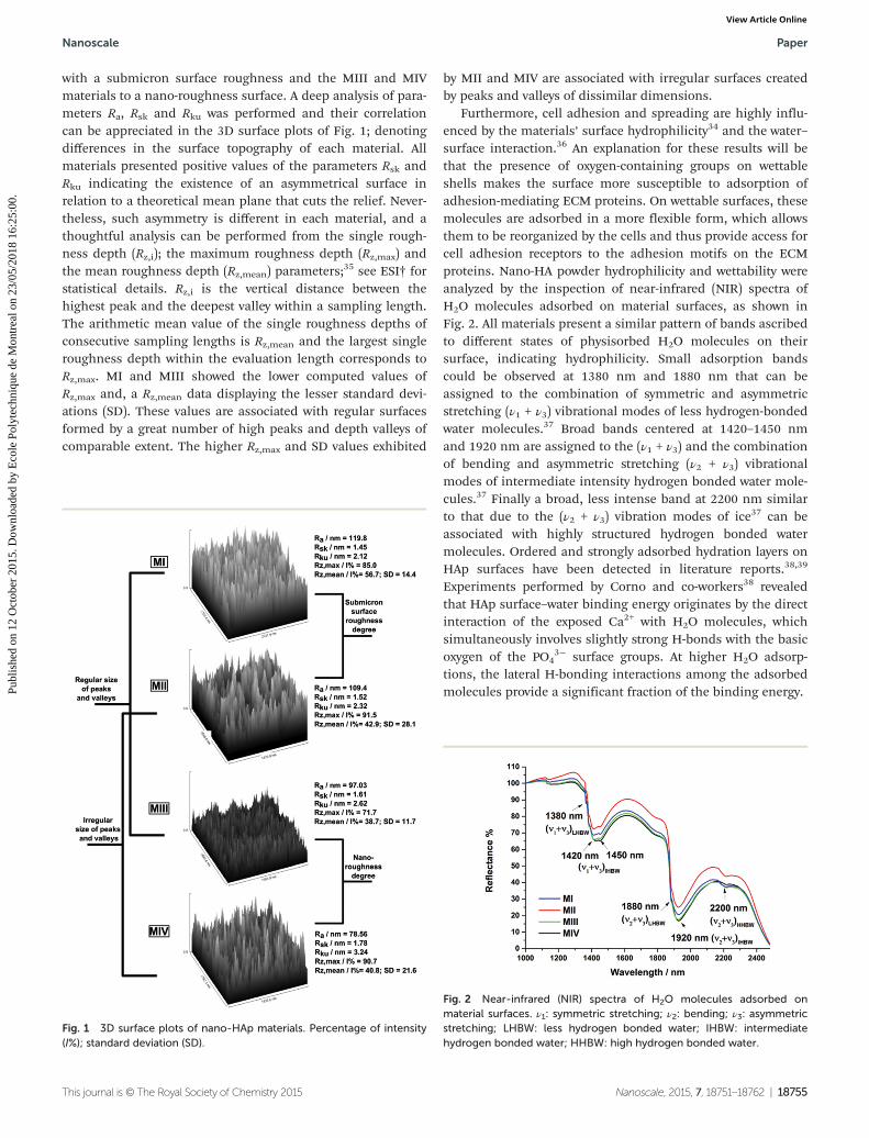
with a submicron surface roughness and the MIII and MIVmaterials to a nano-roughness surface. A deep analysis of para-meters Ra, Rsk and Rku was performed and their correlationcan be appreciated in the 3D surface plots of Fig. 1; denotingdifferences in the surface topography of each material. Allmaterials presented positive values of the parameters Rsk andRku indicating the existence of an asymmetrical surface inrelation to a theoretical mean plane that cuts the relief. Never-theless, such asymmetry is different in each material, and athoughtful analysis can be performed from the single rough-ness depth (Rz,i); the maximum roughness depth (Rz,max) andthe mean roughness depth (Rz,mean) parameters;35 see ESI† forstatistical details. Rz,i is the vertical distance between thehighest peak and the deepest valley within a sampling length.The arithmetic mean value of the single roughness depths ofconsecutive sampling lengths is Rz,mean and the largest singleroughness depth within the evaluation length corresponds toRz,max. MI and MIII showed the lower computed values ofRz,max and, a Rz,mean data displaying the lesser standard devi-ations (SD). These values are associated with regular surfacesformed by a great number of high peaks and depth valleys ofcomparable extent. The higher Rz,max and SD values exhibited
by MII and MIV are associated with irregular surfaces createdby peaks and valleys of dissimilar dimensions.
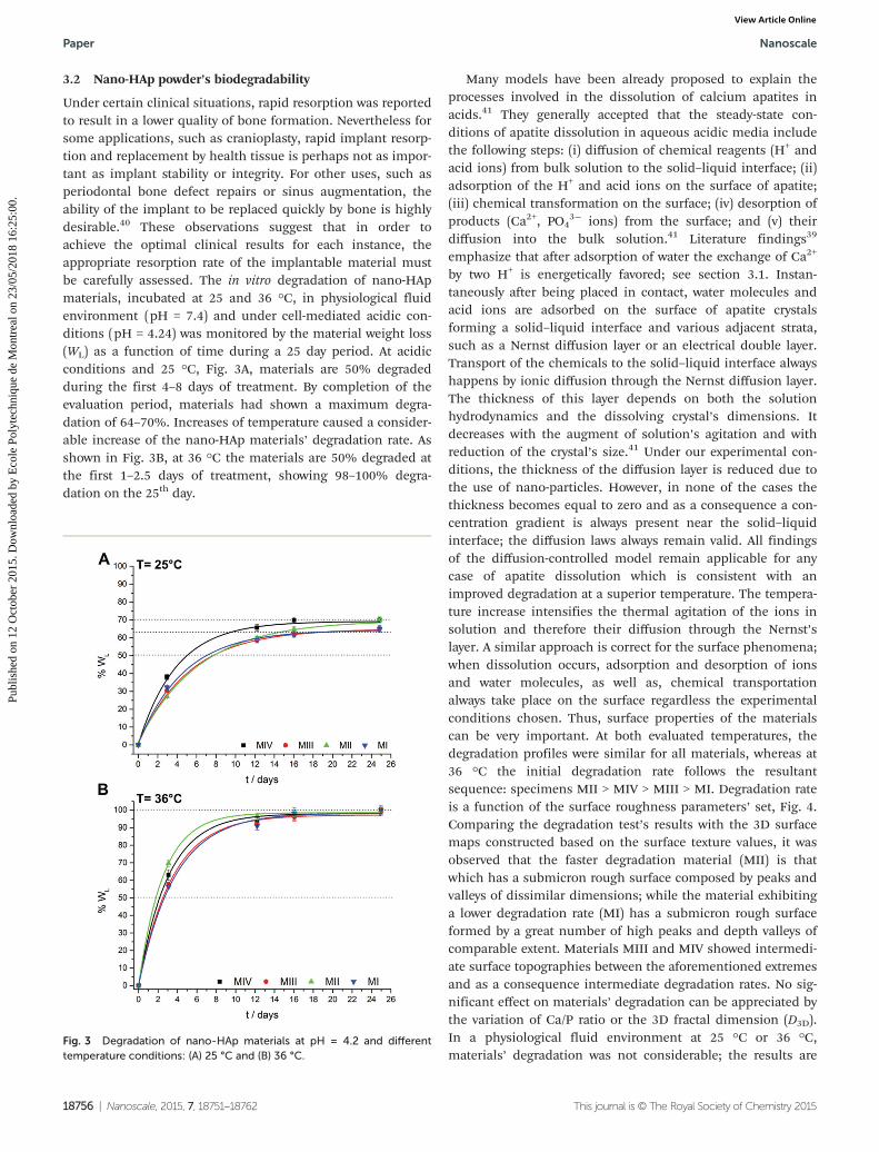
Furthermore, cell adhesion and spreading are highly influ-enced by the materials’ surface hydrophilicity34 and the water–surface interaction.36 An explanation for these results will bethat the presence of oxygen-containing groups on wettableshells makes the surface more susceptible to adsorption ofadhesion-mediating ECM proteins. On wettable surfaces, thesemolecules are adsorbed in a more flexible form, which allowsthem to be reorganized by the cells and thus provide access forcell adhesion receptors to the adhesion motifs on the ECMproteins. Nano-HA powder hydrophilicity and wettability wereanalyzed by the inspection of near-infrared (NIR) spectra ofH2O molecules adsorbed on material surfaces, as shown inFig. 2. All materials present a similar pattern of bands ascribedto different states of physisorbed H2O molecules on theirsurface, indicating hydrophilicity. Small adsorption bandscould be observed at 1380 nm and 1880 nm that can beassigned to the combination of symmetric and asymmetricstretching (ν1 + ν3) vibrational modes of less hydrogen-bondedwater molecules.37 Broad bands centered at 1420–1450 nmand 1920 nm are assigned to the (ν1 + ν3) and the combinationof bending and asymmetric stretching (ν2 + ν3) vibrationalmodes of intermediate intensity hydrogen bonded water mole-cules.37 Finally a broad, less intense band at 2200 nm similarto that due to the (ν2 + ν3) vibration modes of ice37 can beassociated with highly structured hydrogen bonded watermolecules. Ordered and strongly adsorbed hydration layers onHAp surfaces have been detected in literature reports.38,39
Experiments performed by Corno and co-workers38 revealedthat HAp surface–water binding energy originates by the directinteraction of the exposed Ca2+ with H2O molecules, whichsimultaneously involves slightly strong H-bonds with the basicoxygen of the PO4
3− surface groups. At higher H2O adsorp-tions, the lateral H-bonding interactions among the adsorbedmolecules provide a significant fraction of the binding energy.
Fig. 1 3D surface plots of nano-HAp materials. Percentage of intensity(I%); standard deviation (SD).
Fig. 2 Near-infrared (NIR) spectra of H2O molecules adsorbed onmaterial surfaces. ν1: symmetric stretching; ν2: bending; ν3: asymmetricstretching; LHBW: less hydrogen bonded water; IHBW: intermediatehydrogen bonded water; HHBW: high hydrogen bonded water.
Nanoscale Paper
This journal is © The Royal Society of Chemistry 2015 Nanoscale, 2015, 7, 18751–18762 | 18755
Publ
ishe
d on
12
Oct
ober
201
5. D
ownl
oade
d by
Eco
le P
olyt
echn
ique
de
Mon
trea
l on
23/0
5/20
18 1
6:25
:00.
View Article Online
3.2 Nano-HAp powder’s biodegradability
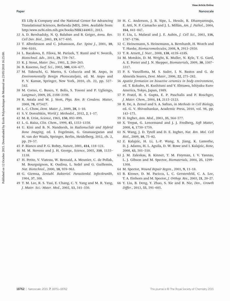
Under certain clinical situations, rapid resorption was reportedto result in a lower quality of bone formation. Nevertheless forsome applications, such as cranioplasty, rapid implant resorp-tion and replacement by health tissue is perhaps not as impor-tant as implant stability or integrity. For other uses, such asperiodontal bone defect repairs or sinus augmentation, theability of the implant to be replaced quickly by bone is highlydesirable.40 These observations suggest that in order toachieve the optimal clinical results for each instance, theappropriate resorption rate of the implantable material mustbe carefully assessed. The in vitro degradation of nano-HApmaterials, incubated at 25 and 36 °C, in physiological fluidenvironment (pH = 7.4) and under cell-mediated acidic con-ditions (pH = 4.24) was monitored by the material weight loss(WL) as a function of time during a 25 day period. At acidicconditions and 25 °C, Fig. 3A, materials are 50% degradedduring the first 4–8 days of treatment. By completion of theevaluation period, materials had shown a maximum degra-dation of 64–70%. Increases of temperature caused a consider-able increase of the nano-HAp materials’ degradation rate. Asshown in Fig. 3B, at 36 °C the materials are 50% degraded atthe first 1–2.5 days of treatment, showing 98–100% degra-dation on the 25th day.
Many models have been already proposed to explain theprocesses involved in the dissolution of calcium apatites inacids.41 They generally accepted that the steady-state con-ditions of apatite dissolution in aqueous acidic media includethe following steps: (i) diffusion of chemical reagents (H+ andacid ions) from bulk solution to the solid–liquid interface; (ii)adsorption of the H+ and acid ions on the surface of apatite;(iii) chemical transformation on the surface; (iv) desorption ofproducts (Ca2+, PO4
3− ions) from the surface; and (v) theirdiffusion into the bulk solution.41 Literature findings39
emphasize that after adsorption of water the exchange of Ca2+
by two H+ is energetically favored; see section 3.1. Instan-taneously after being placed in contact, water molecules andacid ions are adsorbed on the surface of apatite crystalsforming a solid–liquid interface and various adjacent strata,such as a Nernst diffusion layer or an electrical double layer.Transport of the chemicals to the solid–liquid interface alwayshappens by ionic diffusion through the Nernst diffusion layer.The thickness of this layer depends on both the solutionhydrodynamics and the dissolving crystal’s dimensions. Itdecreases with the augment of solution’s agitation and withreduction of the crystal’s size.41 Under our experimental con-ditions, the thickness of the diffusion layer is reduced due tothe use of nano-particles. However, in none of the cases thethickness becomes equal to zero and as a consequence a con-centration gradient is always present near the solid–liquidinterface; the diffusion laws always remain valid. All findingsof the diffusion-controlled model remain applicable for anycase of apatite dissolution which is consistent with animproved degradation at a superior temperature. The tempera-ture increase intensifies the thermal agitation of the ions insolution and therefore their diffusion through the Nernst’slayer. A similar approach is correct for the surface phenomena;when dissolution occurs, adsorption and desorption of ionsand water molecules, as well as, chemical transportationalways take place on the surface regardless the experimentalconditions chosen. Thus, surface properties of the materialscan be very important. At both evaluated temperatures, thedegradation profiles were similar for all materials, whereas at36 °C the initial degradation rate follows the resultantsequence: specimens MII > MIV > MIII > MI. Degradation rateis a function of the surface roughness parameters’ set, Fig. 4.Comparing the degradation test’s results with the 3D surfacemaps constructed based on the surface texture values, it wasobserved that the faster degradation material (MII) is thatwhich has a submicron rough surface composed by peaks andvalleys of dissimilar dimensions; while the material exhibitinga lower degradation rate (MI) has a submicron rough surfaceformed by a great number of high peaks and depth valleys ofcomparable extent. Materials MIII and MIV showed intermedi-ate surface topographies between the aforementioned extremesand as a consequence intermediate degradation rates. No sig-nificant effect on materials’ degradation can be appreciated bythe variation of Ca/P ratio or the 3D fractal dimension (D3D).In a physiological fluid environment at 25 °C or 36 °C,materials’ degradation was not considerable; the results are
Fig. 3 Degradation of nano-HAp materials at pH = 4.2 and differenttemperature conditions: (A) 25 °C and (B) 36 °C.
Paper Nanoscale
18756 | Nanoscale, 2015, 7, 18751–18762 This journal is © The Royal Society of Chemistry 2015
Publ
ishe
d on
12
Oct
ober
201
5. D
ownl
oade
d by
Eco
le P
olyt
echn
ique
de
Mon
trea
l on
23/0
5/20
18 1
6:25
:00.
View Article Online

shown in Fig. 5, ESI.† This is a desirable characteristic becausethe biodegradation rate must be comparable to the formationof bone tissue, which is between 8 and 16 weeks.42
This degradation study was carried out in the absence ofenzymes and cells, and therefore cannot be predictive of theresorption times in vivo; however an important in vitro result ofthe degradation analysis is that nano-HAp materials do notdissolve under pH physiological fluid conditions, yet areresorbable under cell-mediated acidic conditions26 similar tothose that are set in vivo during new bone formation43,44 andbone remodeling. The goal of this in vitro biodegradationstudy is to ascertain that nano-HAp materials can act as tem-porary templates, providing structural and mechanical supportinitially and regenerative properties over time and finally,under resorption conditions, its complete degradation.
3.3 nano-HAp powder’s osteo-induction, osteo-conductionand osseo-integration properties
Bone repair properties of nano-HAp powders were tested in thepresence of two different cell types that could be potentially beused in orthopedic or tissue engineering applications:45,46
primary mature osteoblast and mesenchymal stem cells thatare known to form bone tissues when implanted in vivo ordifferentiated into bone cells.47
3.3.1 Osteoblast spreading, proliferation and differen-tiation. The variation of nano-HAp powders’ surface topo-graphy and degradation on neonatal rat calvarial osteoblasts’viability, spreading and morphology were preliminarily ana-lyzed by simple microscopic observation of cultured cellsthrough two different treatments for 24, 48 and 72 h, accord-ing to a previous work.23 During a first procedure (T1), thenano-HAp powder was extended in the culture media and cellswere seeded in the presence of the material from the begin-ning of the test. Then, a second treatment (T2) was applied toevaluate the effect of material on cells’ spreading perturbation;at this time osteoblasts were plated in culture media andnano-HAp were added 24 h later. The cells’ behavior wassimilar to control after 72 h for both treatments, Fig. 5A. To
evaluate the cell’s morphology, the samples were thensmeared on a microscope slide, air dried, fixed with absoluteethanol and stained by Giemsa dye.48 In the presence of alltested nano-HAp samples, rat osteoblasts displayed normalmorphological features49 for both applied culture methods;and they showed their typical polygonal or widespread formswith fine filopodia and abundant surface folds, Fig. 5B. Theobtained results exclude any significant difference due to thenano-HAp powder surface properties or particle degradationon the process of adhesion, spreading and morphology of thetested cells. There are no apparent differences in the cell via-bility after they are cultured in the presence of the preparedmaterials. Further inspection of cell–material interactions, wasanalyzed by differentiation assays.
The induction of APL activity is used as a predictive para-meter for in vivo osteoblast maturation. A successful bonescaffold must demonstrate support for enhanced bone for-mation, including organic and inorganic components ofnatural tissue. ALP is a key component of bone matrix vesiclesbecause of its role in the formation of apatite calcium phos-phate,50 and it is an early indicator of immature osteoblastactivity. Although cells in several tissues (liver, kidney,placenta, etc.) generate the enzyme, elevated levels of ALP in bonetissue typically are observed several days prior to neo-mineral-ization and during the initial phase of bone matrix depo-sition.51,52 Fig. 6 displays the influence of different materialproperties on the ALP activity of rat primary calvarial osteo-blast after 4 and 7 days of culture in the synthetic serum sub-stitute with and without the addition of 4 mM Ca2+ ionconcentration.
The material exhibiting a submicron rough surface com-posed by peaks and valleys of comparable extent (MI), showsan ALP activity statistically superior to the control during the7 days of treatment (p < 0.05); while those materials having asurface composed by peaks and valleys of dissimilar dimen-sions (MII), nano-surface roughness degree (MIII) or both(MIV) present a considerable decrease in ALP activity thatintensified with time. Investigations performed by Maenoet al.28 revealed that low concentrations of Ca2+ (2–6 mM) canprovide suitable conditions for osteoblast proliferation anddifferentiation in both monolayer and 3D cultures. Our experi-ments showed a clear decrease in rat calvarial osteoblast differ-entiation after the addition of 4 mM Ca2+ in culture media inthe presence of all tested nano-HAp materials, while in theabsence of the materials the ALP activity increases with respectto the control and in agreement with Maeno et al. results.28
Statistically considerable and equivalent ALP activity reductionwas noticed in the presence of materials MII, MIII and MIV.The obtained results may be associated with an increase ofCa2+ concentration due to nano-HAp degradation above theacceptable limit to promote cell differentiation. The pH of theinterstitial fluid bathing cell tissues will generally be slightlyacidic and subject to complex gradients, depending on themetabolic activity of the cells.53 Being MI the less degradedmaterial under acidic conditions, its presence generates asmaller reduction in ALP activity.
Fig. 4 Variation of degradation rate versus the material’s roughnessparameters.
Nanoscale Paper
This journal is © The Royal Society of Chemistry 2015 Nanoscale, 2015, 7, 18751–18762 | 18757
Publ
ishe
d on
12
Oct
ober
201
5. D
ownl
oade
d by
Eco
le P
olyt
echn
ique
de
Mon
trea
l on
23/0
5/20
18 1
6:25
:00.
View Article Online

3.3.2 Effects of nano-HAp powders on MSC adhesion. RabbitMSC spreading was evaluated on coatings of collagen type I,the principle organic component of bone, with and withoutincorporated nano-HAp. The experiments were performed inan attempt to mimic a remodeling bone environment in whichthe MSCs attach and differentiate. First, a cytocompatible levelof nano-HAp to use in the coating was identified. Rabbit MSCswere treated with different amounts of nano-HAp particles(15–3100 μg HAp per cm2 coating), and their mitochondrialmetabolic activity was measured after culture for 24 and 48 h.The obtained data collected after the first 24 hours were incon-clusive, while by the completion of 48 hours the results showthat the inhibition effect produced by the particles was dose-and material type-dependent, Fig. 7. Mitochondrial metabolicactivity was correlated with the cells viability; proliferation
status was confirmed by microscopic observation, Fig. 7, inset;for more details see, Fig. 6, ESI.† A measurement of MSC via-bility after adhesion is imperative to evaluate the capacity for amaterial to support initial cell spreading and proliferation. Inorder to be successful in providing a scaffold with therapeuticpotential, the cells must demonstrate viability properties statis-tically comparable to the control. As expected, our resultssuggest that cell viability is related to the materials surface pro-perties. Materials having comparable dimensions of peaks andvalleys on their surfaces (MI and MIII) exhibited a 50% cell
Fig. 6 ALP activity of rat primary osteoblast after culture in the pres-ence of different nano-HAp materials. [Ca2+] = 4 mM. Statistical analysiswas performed between different materials and control, *p < 0.05; sig-nificant differences between the samples are indicated with brackets.
Fig. 7 Mitochondrial metabolic activity of rabbit mesenchymal stemcells adhered on different concentrations of nano-HAp material coat-ings. Experimental results were fitted to Boltzmann sigmoidal curve(continuous line); non-coated well was used as a control. Statistical ana-lysis was performed among different materials, *p < 0.05; significantdifferences between the samples are indicated with brackets. Inset:optical microphotographs of adhered cells on (I) 15 μg cm2, (II) 300 μg cm2
and (III) 3100 μg cm2 MI coating.
Fig. 5 (A) Rat primary osteoblast viability after cultured through to two different treatments (T1 and T2) during 72 h in the presence of differentnano-HAp materials. (B) Giemsa stains of rat primary osteoblast after culture in the presence MI sample.
Paper Nanoscale
18758 | Nanoscale, 2015, 7, 18751–18762 This journal is © The Royal Society of Chemistry 2015
Publ
ishe
d on
12
Oct
ober
201
5. D
ownl
oade
d by
Eco
le P
olyt
echn
ique
de
Mon
trea
l on
23/0
5/20
18 1
6:25
:00.
View Article Online

proliferation inhibition at 300 μg HAp per cm2 coating; while itrequired 162 and 124 μg HAp per cm2 to reach similar cellmortality in the presence of materials with more asymmetricalsurfaces (MII and MIV). The sample MIV, which has a combi-nation of nano-surface roughness degree and an asymmetricaltopography, causes the greatest toxic effect on initial MSC via-bility. As the concentration of particles exceeded the 300 μgHAp per cm2 coating, the inhibition effect is similar for alltested materials. Literature findings54 inform that the degreeof toxicity of nano-HAp powder correlated well with the degreeof cellular uptake. This strongly suggests that cellular particleload is the main cause of cytotoxicity, probably due to therelease of calcium.54 This fact is consistent with our resultssince the less degraded material, MI, causes the lowest cyto-toxicity of cells of mesenchymal origin.
3.3.3 MSC adhesion on a mimic bone matrix. Regardingthe cell–material interactions, the information discussed inthe preceding sections indicate a clear difference amongsample MI and the rest of the studied nano-HAp superstruc-tures. MI provides an adequate environment to permit ratprimary osteoblast differentiation, and higher ALP activity rela-tive to the control compared to the attained values in the pres-ence of the other three materials. Even though there are nodifferences among rat primary osteoblast spreading and pro-liferation in the presence of the four tested nano-HAppowders, rabbit MSC adhesion and viability on the MI coatingis slightly favored in comparison to control at low materialconcentration. Furthermore, a greater amount of MI materialis required to reach the 50% of rabbit MSC mortality. Resultsobtained in a previous work23 showed that the mineral HApcoating formed on the surface of MI material after it wasimmersed in simulated body fluid (SBF) giving an estimate ofthe material’s bone-bonding potential,24,25 exhibited a globu-lar morphology and a Ca/P weight ratio of 1.56 close to whatexists in trabecular bone.55 Both characteristics are consideredessential for bone-bonding.55,56 Accordingly, the specimen MIwas selected as the material with the best osseo-integration,osseo-induction and osteo-conduction characteristics to becombined with collagen type I in MI/Co I coatings and toindirectly evaluate their potential use in bone tissue repair. Itis considered that the average mineral content of bone tissueis within the range of 30 to 55 vol% or 50 to 75 wt%, and col-lagen is the principal organic matrix in bone,57 thus the MI/CoI coatings were prepared in compliance of these proportionsand taking into account that higher amounts of MI (>300 μg cm−2)tested as coatings caused the inhibition of MSCs’ adhesionand metabolic activity, see Fig. 6–8, ESI.† Fig. 8 showsthe rabbit MSC mitochondrial metabolic activities afterculture in the presence of different MI/Co I weight ratio coat-ings. No significant statistical difference with respect tocontrol was observed among the MSC viability after adhesionon the different MI/Co I coatings. Current discoveries haverecognized that mechanical properties of the cellular environ-ment such as its rigidity, geometry, and external stresses playimperative roles in the definition of the cellular function andfate.58 It has been shown that mechanical properties influence
cell shape and orientation, regulate cell proliferation anddifferentiation and even govern the development and organiz-ation of tissues.58 It is widely known that for most tissue cellsthe traditional picture of the cell as a lipid membrane that sur-rounds a liquid-like cytosol containing localized organelles isnot relevant.59 Adherent cells such as fibroblasts, neurons,endothelial cells, and bone cells, contain an actively regulatedgel-like elastic cytoskeleton.60 The elastic nature of the cyto-skeleton is important not only for the mechanical stability ofthe cell, but also for transmitting mechanical signals in theform of elastic stress and strain fields, from the environment,through the cytoskeleton and to the nucleus.61
The cytoskeleton is a complex dynamical network of actin,intermediate filaments, and microtubules that interact with amyriad of molecular motors and cross-linking proteins. Manyof these proteins are collectively responsible for cellularmechano-sensing. Recent studies have identified a contractilemuscle actin isoform, α-SMA, in osteoblasts62 as well as in anumber of other musculoskeletal connective tissue cells.Associated studies demonstrated the ability of α-SMA-expres-sing osteoblastic cells to contract a collagen-glycosamino-glycan analog of extracellular matrix in vitro.63 It was proposedthat α-SMA enabled contraction might be responsible for theretraction of osteoblasts on the bone surface at the initiationof the remodeling cycle. Moreover, α-SMA-enabled contractionmay allow for the generation of the higher forces required forthe cellular modeling of the newly synthesized extracellularmatrix, to impart the tissue specific architecture.64 Thus, theelevation of α-SMA expression might be expected to be associ-ated with an osteogenic process such as bone transport.65 Fur-thermore, the α-SMA expression is a relatively reliableindicator that the MSC cells maintain their mesenchymal stemcells phenotype.66 MSCs’ α-SMA-based spreading on MI/Co Icoatings was evaluated by confocal fluorescence microscopyand histomorphometry.
The results indicate that the rabbit MSCs spread, adhere,and proliferate on 0.5/1, 1/1 and 2/1 MI/Co I coatings cultured
Fig. 8 Mitochondrial metabolic activity of rabbit MSCs adhered ondifferent MI/Co I weight ratio coatings. Non-coated wells were used aspositive control (C+).
Nanoscale Paper
This journal is © The Royal Society of Chemistry 2015 Nanoscale, 2015, 7, 18751–18762 | 18759
Publ
ishe
d on
12
Oct
ober
201
5. D
ownl
oade
d by
Eco
le P
olyt
echn
ique
de
Mon
trea
l on
23/0
5/20
18 1
6:25
:00.
View Article Online

Fig. 10 (A) Relative percentage of MSCs on cultured surface. (B) Relative percentage of positive stain for α-SMA. (C) Mean brightness value per celland (D) confocal microphotographs in order to show the α-SMA expression of MSCs adhered on different MI/Co I weight ratio coatings per cell.Non-coated glass slide was used as positive control (C+).
Fig. 9 Immunofluorescence assay: (A–E) 40× magnification laser scanning confocal microphotographs showing cell nucleus and α-SMAexpression, blue and green stains respectively, scale bar 50 µm; (F–J) 400× magnification laser scanning confocal microphotographs showingα-SMA filaments’ size and morphology, scale bar 5 µm. (K–O) Optical phase contrast images showing MSCs’ morphology and adherence to surface.Non-coated glass slide was used as positive control (C+).
Paper Nanoscale
18760 | Nanoscale, 2015, 7, 18751–18762 This journal is © The Royal Society of Chemistry 2015
Publ
ishe
d on
12
Oct
ober
201
5. D
ownl
oade
d by
Eco
le P
olyt
echn
ique
de
Mon
trea
l on
23/0
5/20
18 1
6:25
:00.
View Article Online

in proliferation medium for 29 h in the same manner as onbiocompatible glass control surfaces exhibiting α-SMA fila-ments of similar size and morphology, Fig. 9. The time ana-lyzed, the visualization of the cell number and therearrangement of the α-SMA’s cytoskeleton do not show anysignificant difference with respect to control, Fig. 10A–C. Thevisual inspection of proliferation, as well as a measurement ofcellular metabolism and spreading provided information onthe ability of cells to populate MI/Co I coatings. Altogether,these data demonstrate that, under the conditions tested,in vitro culture MI/Co I materials are cytocompatible, and thatthe HAp nanoparticles do not interfere with mesenchymalstem cell character or spreading.
4. Conclusion
We successfully fabricated a biocompatible nano-HAp super-structure that possesses the precise controlled surface topo-graphy and hydrophilicity allowing normal growth anddifferentiation of bone cells in vitro. The material has a regularsub-micron rough surface and the optima degradation rateunder cell-mediated acidic conditions, which resulted in auniform cell seeding, spreading, adhesion and differentiation.
The obtained results highlight the importance of micro-patterning and surface properties of nano-HAp superstructuresand provide new insight for the design of scaffolds based onHAp nanoparticles for effective bone repair and regeneration.
Acknowledgements
The authors acknowledge Universidad Nacional del Sur (PGI 24/Q064 and PGI 24/B211), Concejo Nacional de InvestigacionesCientíficas de la República Argentina (CONICET, PIP11220130100100CO) and the Canadian Institutes of HealthResearch (CIHR, MOP 303615 to CDH). Also they acknowledgeSylvie Taillon for technical contributions. NLD and JL have doc-toral fellowships of CONICET; GES and PVM are independentresearchers of CONICET. NLD thanks the Emerging Leaders inthe Americas Program (ELAP).
References
1 A. J. Salgado, O. P. Coutinho and R. L. Reis, Macromol.Biosci., 2004, 4, 743–765.
2 W. L. Fodor, Reprod. Biol. Endocrinol., 2003, 1, 102–102.3 W.-J. Li, R. Tuli, X. Huang, P. Laquerriere and R. S. Tuan,
Biomaterials, 2005, 26, 5158–5166.4 M. D. Kofron, X. Li and C. T. Laurencin, Curr. Opin. Biotech-
nol., 2004, 15, 399–405.5 Y. Shandalov, D. Egozi, J. Koffler, D. Dado-Rosenfeld,
D. Ben-Shimol, A. Freiman, E. Shor, A. Kabala andS. Levenberg, Proc. Natl. Acad. Sci. U. S. A., 2014, 111, 6010–6015.
6 L. Zhang and T. J. Webster, Nano Today, 2009, 4, 66–80.
7 V. J. Shirtliff and L. L. Hench, J. Mater. Sci., 2003, 38, 4697–4707.
8 C. J. Koh and A. Atala, J. Am. Soc. Nephrol., 2004, 15, 1113–1125.
9 L. L. Hench and J. M. Polak, Science, 2002, 295, 1014–1017.10 M. Sadat-Shojai, M. Atai, A. Nodehi and L. N. Khanlar,
Dent. Mater., 2010, 26, 471–482.11 H.-W. Kim, H.-H. Lee and J. C. Knowles, J. Biomed. Mater.
Res., Part A, 2006, 79A, 643–649.12 E. Neovius and T. Engstrand, J. Plast. Reconstr. Aesthet.
Surg., 2010, 63, 1615–1623.13 D. W. Lee, J. Y. Kim and D. H. Lew, J. Craniofac. Surg., 2010,
21, 1084–1088.14 H. Wang, Y. Li, Y. Zuo, J. Li, S. Ma and L. Cheng, Bio-
materials, 2007, 28, 3338–3348.15 S. J. Hollister, Nat. Mater., 2005, 4, 518–524.16 A. Tampieri, M. Iafisco, M. Sandri, S. Panseri, C. Cunha,
S. Sprio, E. Savini, M. Uhlarz and T. Herrmannsdörfer, ACSAppl. Mater. Interfaces, 2014, 6, 15697–15707.
17 A. K. Lilikakis, S. L. Vowler and R. N. Villar, Orthop. Clin.North Am., 2005, 36, 215–222.
18 C. Liang, M. M. Joseph, C. M. L. James and L. Hao, Nat.Nanotechnol., 2011, 22, 105708.
19 S. V. Dorozhkin, Acta Biomater., 2010, 6, 715–734.20 D. D. Deligianni, N. D. Katsala, P. G. Koutsoukos and
Y. F. Missirlis, Biomaterials, 2001, 22, 87–96.21 H.-H. Huang, C.-T. Ho, T.-H. Lee, T.-L. Lee, K.-K. Liao and
F.-L. Chen, Biomol. Eng., 2004, 21, 93–97.22 H. Gleiter, Acta Mater., 2000, 48, 1–29.23 N. L. D’Elía, A. N. Gravina, J. M. Ruso, J. A. Laiuppa,
G. E. Santillán and P. V. Messina, Biochim. Biophys. Acta,Gen. Subj., 2013, 1830, 5014–5026.
24 T. Kokubo, H. Kushitani, S. Sakka, T. Kisugi andT. Yamamuro, J. Biomed. Mater. Res., Part B, 1990, 24, 721–734.
25 M. Bohner and J. Lemaitre, Biomaterials, 2009, 30, 2175–2179.
26 R. Baron, L. Neff, D. Louvard and P. J. Courtoy, J. Cell Biol.,1985, 101, 2210–2222.
27 T. Matsumoto, M. Okazaki, M. Inoue, S. Yamaguchi,T. Kusunose, T. Toyonaga, Y. Hamada and J. Takahashi,Biomaterials, 2004, 25, 3807–3812.
28 S. Maeno, Y. Niki, H. Matsumoto, H. Morioka, T. Yatabe,A. Funayama, Y. Toyama, T. Taguchi and J. Tanaka, Bio-materials, 2005, 26, 4847–4855.
29 V. B. Ayala-Peña, L. A. Scolaro and G. E. Santillán, Exp. CellRes., 2013, 319, 2028–2036.
30 N. J. Marshall, C. J. Goodwin and S. J. Holt, Growth. Regul.,1995, 5, 69–84.
31 T. L. Riss, R. A. Moravec, A. L. Niles, H. A. Benink,T. J. Worzella, L. Minor, D. Storts and Y. Reid, in AssayGuidance Manual [Internet], ed. G. S. Sittampalam,N. P. Coussens, H. Nelson, M. Arkin, D. Auld, C. Austin,B. Bejcek, M. Glicksman, J. Inglese, P. W. Iversen,Z. Li, J. McGee, O. McManus, L. Minor, A. Napper,J. M. Peltier, T. Riss, O. J. Trask Jr. and J. Weidner,
Nanoscale Paper
This journal is © The Royal Society of Chemistry 2015 Nanoscale, 2015, 7, 18751–18762 | 18761
Publ
ishe
d on
12
Oct
ober
201
5. D
ownl
oade
d by
Eco
le P
olyt
echn
ique
de
Mon
trea
l on
23/0
5/20
18 1
6:25
:00.
View Article Online
Eli Lilly & Company and the National Center for AdvancingTranslational Sciences, Bethesda (MD), 2004. Available from:http:/www.ncbi.nlm.nih.gov/books/NBK144065/, 2013.
32 A. D. Bershadsky, N. Q. Balaban and B. Geiger, Annu. Rev.Cell Dev. Biol., 2003, 19, 677–695.
33 T. Albrektsson and C. Johansson, Eur. Spine J., 2001, 10,S96–S101.
34 L. Bacakova, E. Filova, M. Parizek, T. Ruml and V. Svorcik,Biotechnol. Adv., 2011, 29, 739–767.
35 K. J. Stout, Mater. Des., 1981, 2, 260–265.36 B. Kasemo, Surf. Sci., 2002, 500, 656–677.37 M. Takeuchi, G. Martra, S. Coluccia and M. Anpo, in
Environmentally Benign Photocatalysts, ed. M. Anpo andP. V. Kamat, Springer, New York, 2010, ch. 22, pp. 527–542.
38 M. Corno, C. Busco, V. Bolis, S. Tosoni and P. Ugliengo,Langmuir, 2009, 25, 2188–2198.
39 R. Astala and M. J. Stott, Phys. Rev. B: Condens. Matter,2008, 78, 075427.
40 L. C. Chow, Dent. Mater. J., 2009, 28, 1–10.41 S. V. Dorozhkin, World J. Methodol., 2012, 2, 1–17.42 M. R. Urist, Science, 1965, 150, 893–899.43 L. G. Raisz, Clin. Chem., 1999, 45, 1353–1358.44 U. Kini and B. N. Nandeesh, in Radionuclide and Hybrid
Bone Imaging, ed. I. Fogelman, G. Gnanasegaran andH. van der Waals, Springer, Berlin, Heidelberg, 2012, ch. 2,pp. 29–57.
45 P. Bianco and P. G. Robey, Nature, 2001, 414, 118–121.46 M. M. Stevens and J. H. George, Science, 2005, 310, 1135–
1138.47 H. Petite, V. Viateau, W. Bensaid, A. Meunier, C. de Pollak,
M. Bourguignon, K. Oudina, L. Sedel and G. Guillemin,Nat. Biotechnol., 2000, 18, 959–963.
48 G. Giemsa, Zentabl. Bakteriol. Parasitenkd. Infectkrankh,1904, 37, 308.
49 T. M. Lee, R. S. Tsai, E. Chang, C. Y. Yang and M. R. Yang,J. Mater. Sci.: Mater. Med., 2002, 13, 341–350.
50 H. C. Anderson, J. B. Sipe, L. Hessle, R. Dhamyamraju,E. Atti, N. P. Camacho and J. L. Millán, Am. J. Pathol., 2004,164, 841–847.
51 F. Liu, L. Malaval and J. E. Aubin, J. Cell Sci., 2003, 116,1787–1796.
52 C. Heinemann, S. Heinemann, A. Bernhardt, H. Worch andT. Hanke, Biomacromolecules, 2008, 9, 2913–2920.
53 T. R. Arnett, J. Nutr., 2008, 138, 415S–418S.54 M. Motskin, D. M. Wright, K. Muller, N. Kyle, T. G. Gard,
A. E. Porter and J. N. Skepper, Biomaterials, 2009, 30, 3307–3317.
55 P. S. Vanzillotta, M. S. Sader, I. N. Bastos and G. deAlmeida Soares, Dent. Mater., 2006, 22, 275–282.
56 Apatite formation on bioactive ceramics in body environment,ed. T. Kokubo, H. Kushitani and Y. Ebisawa, Ishiyaku Euro-America, Tokyo, Japan, 1989.
57 P. Fratzl, H. S. Gupta, E. P. Paschalis and P. Roschger,J. Mater. Chem., 2004, 14, 2115–2123.
58 R. De, A. Zemel and S. A. Safran, in Methods in Cell Biology,ed. G. V. Shivashankar, Academic Press, 2010, vol. 98, pp.143–175.
59 D. Ingber, Ann. Med., 2003, 35, 564–577.60 X. Trepat, G. Lenormand and J. J. Fredberg, Soft Matter,
2008, 4, 1750–1759.61 N. Wang, J. D. Tytell and D. E. Ingber, Nat. Rev. Mol. Cell
Biol., 2009, 10, 75–82.62 Z. Kalajzic, H. Li, L.-P. Wang, X. Jiang, K. Lamothe,
D. J. Adams, H. L. Aguila, D. W. Rowe and I. Kalajzic, Bone,2008, 43, 501–510.
63 J. M. Zaleskas, B. Kinner, T. M. Freyman, I. V. Yannas,L. J. Gibson and M. Spector, Biomaterials, 2004, 25, 1299–1308.
64 M. Spector, Wound Repair Regen., 2001, 9, 11–18.65 B. Kinner, D. M. Pacicca, L. C. Gerstenfeld, C. A. Lee,
T. A. Einhorn and M. Spector, J. Orthop. Res., 2003, 21, 20–27.66 Y. Liu, B. Deng, Y. Zhao, S. Xie and R. Nie, Dev., Growth
Differ., 2013, 55, 591–605.
Paper Nanoscale
18762 | Nanoscale, 2015, 7, 18751–18762 This journal is © The Royal Society of Chemistry 2015
Publ
ishe
d on
12
Oct
ober
201
5. D
ownl
oade
d by
Eco
le P
olyt
echn
ique
de
Mon
trea
l on
23/0
5/20
18 1
6:25
:00.
View Article Online