bone infections
TRANSCRIPT
BONE INFECTIONS
OSTEOMYELITISSEPTIC ARTHRITIS
PREPARED BY:
PRABITA SHRESTHA
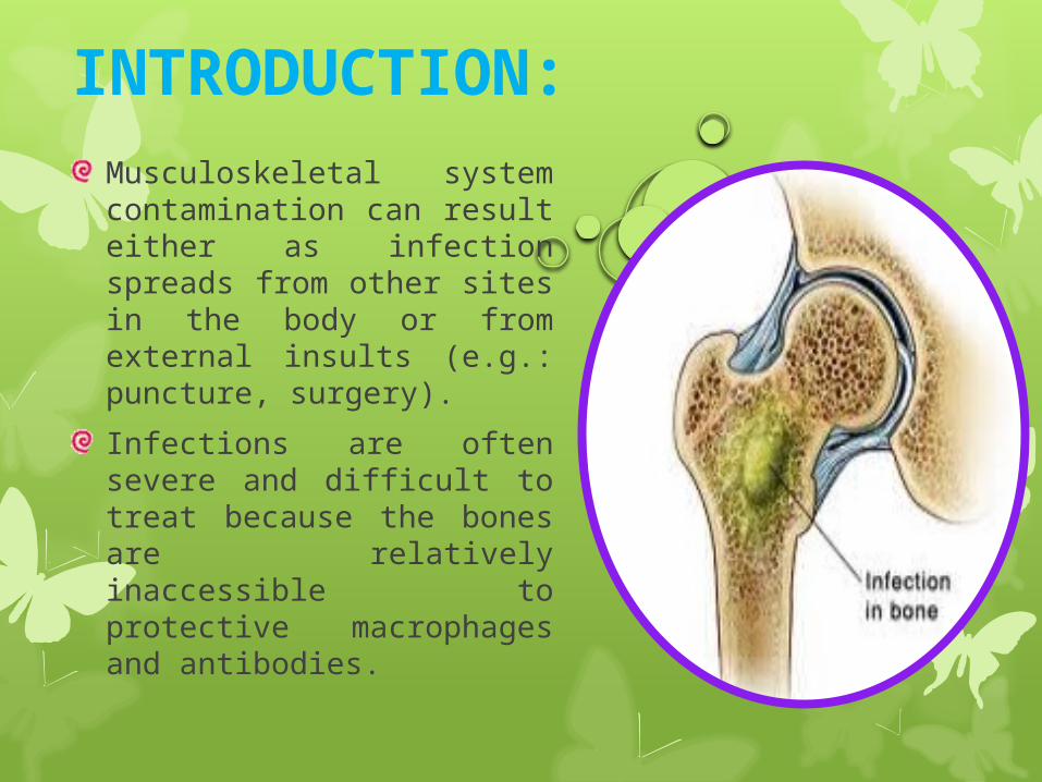
INTRODUCTION:Musculoskeletal system contamination can result either as infection spreads from other sites in the body or from external insults (e.g.: puncture, surgery).
Infections are often severe and difficult to treat because the bones are relatively inaccessible to protective macrophages and antibodies.
Contd…
Even a small number of microorganisms can be enough to establish a serious infection that can lead to loss of function or even death.
For this reason, nurses should be diligent in wound care and alert to any manifestations that suggest infection.
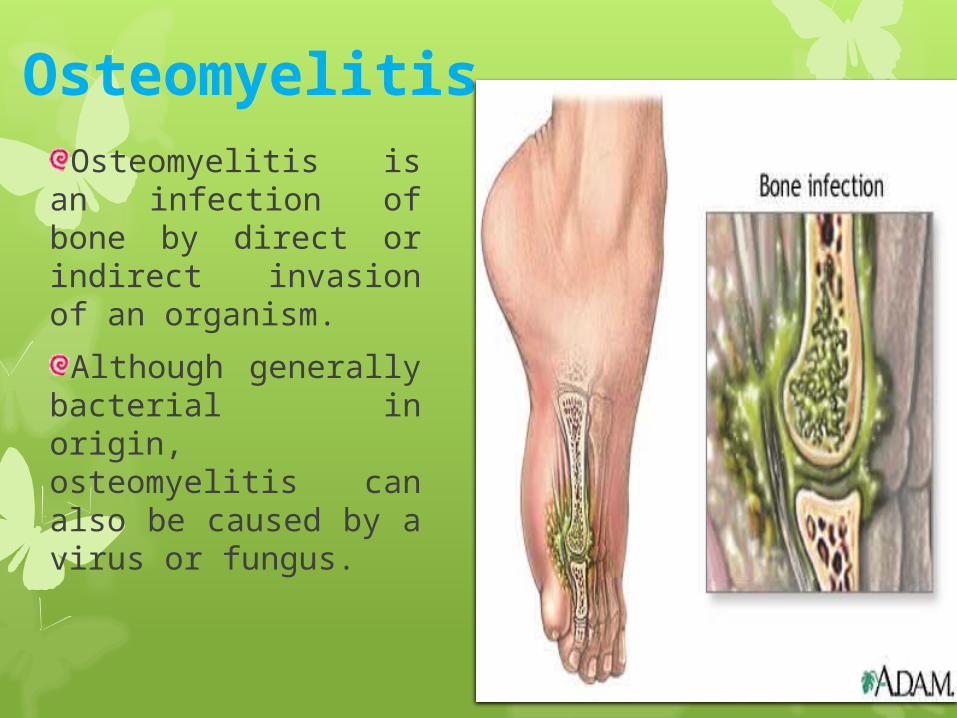
Osteomyelitis Osteomyelitis is an
infection of bone by direct or indirect invasion of an organism.
Although generally bacterial in origin, osteomyelitis can also be caused by a virus or fungus.
Types of osteomyelitis:
On the basis of mode of entry of the pathogen; it is divided into two types:
1. Exogenous osteomyelitis
2. Haematogenous osteomyelitis
1. Exogenous osteomyelitis Exogenous osteomyelitis is secondary to a
contagious source of infection, is caused by a pathogen from outside the body.
E.g. pathogens from an open fracture or surgical procedure, involving instrumentation.
This spreads from soft tissues to bone.
2.Haematogenous osteomyelitis
It is caused by blood-borne pathogen originating from infectious site within the body.
E.g. sinus, ear, dental, respiratory and genitourinary infections.
It spreads from bone to soft tissues and can even break through the skin, becoming a draining fistula.
Causative agents: Staphylococcus aureus (most common)
Pseudomonas
Klebsiella
Salmonella
Escherichia coli
Pneumococcus species
Proteus vulgaris
Pasteurella multicida
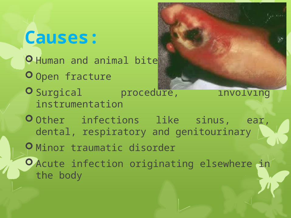
Causes: Human and animal bites
Open fracture
Surgical procedure, involving instrumentation
Other infections like sinus, ear, dental, respiratory and genitourinary
Minor traumatic disorder
Acute infection originating elsewhere in the body
Risk factors: Chronic illness
Diabetes mellitus
Alcohol/drug abuse
Immunosuppression
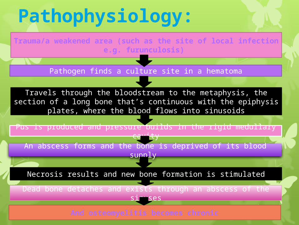
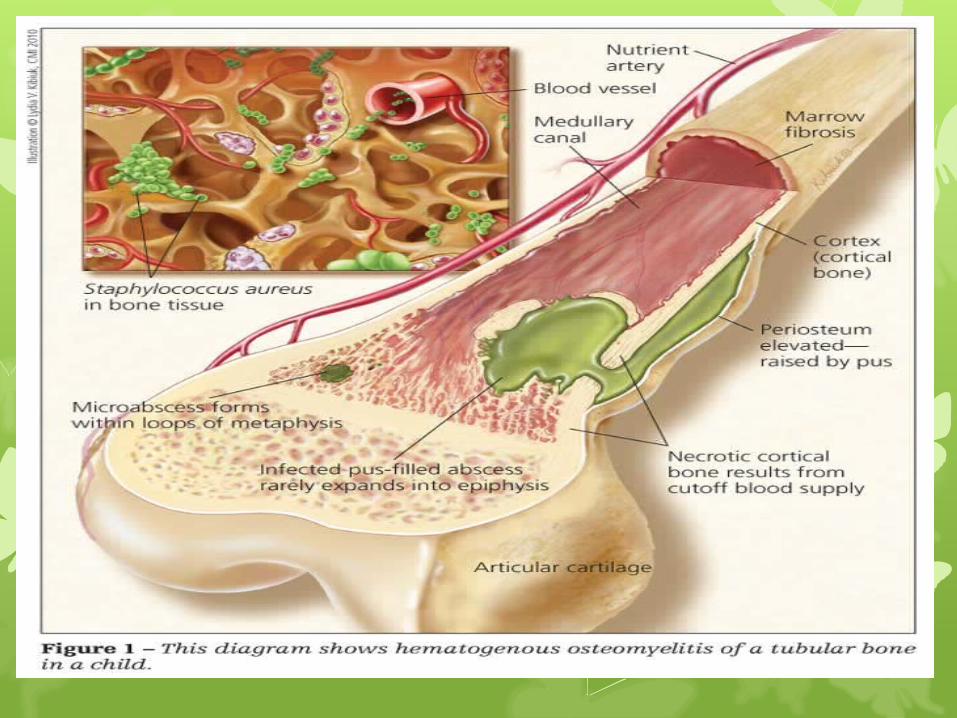
Pathophysiology:Trauma/a weakened area (such as the site of local infection
e.g. furunculosis)
And osteomyelitis becomes chronic
Dead bone detaches and exists through an abscess of the sinuses
Necrosis results and new bone formation is stimulated
An abscess forms and the bone is deprived of its blood supply
Pus is produced and pressure builds in the rigid medullary cavity
Travels through the bloodstream to the metaphysis, the section of a long bone that’s continuous with the epiphysis plates, where the
blood flows into sinusoids
Pathogen finds a culture site in a hematoma
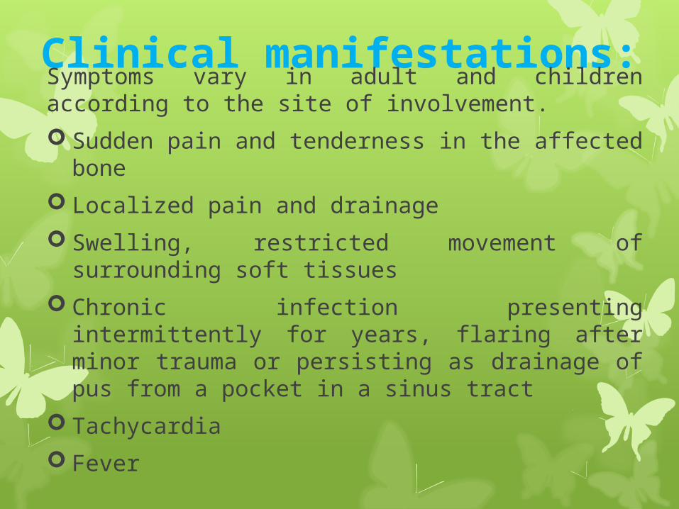
Clinical manifestations:Symptoms vary in adult and children according to the site of involvement.
Sudden pain and tenderness in the affected bone
Localized pain and drainage
Swelling, restricted movement of surrounding soft tissues
Chronic infection presenting intermittently for years, flaring after minor trauma or persisting as drainage of pus from a pocket in a sinus tract
Tachycardia
Fever
Contd…
Chills
Nausea
Malaise
Dysphagia
Dyspnea
Decreased oesophageal motility
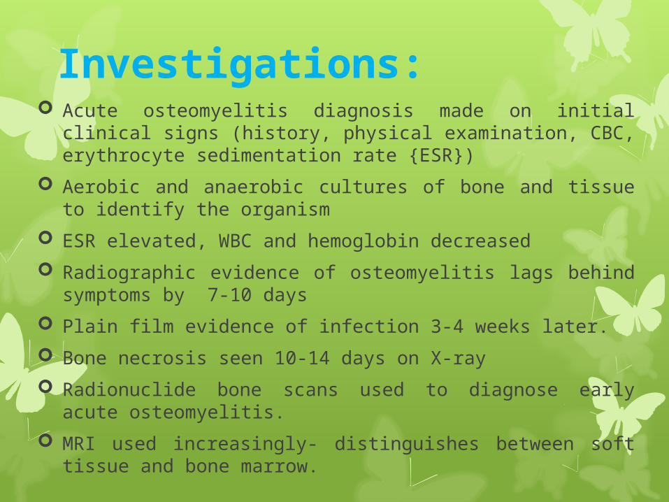
Investigations: Acute osteomyelitis diagnosis made on initial clinical signs
(history, physical examination, CBC, erythrocyte sedimentation rate {ESR})
Aerobic and anaerobic cultures of bone and tissue to identify the organism
ESR elevated, WBC and hemoglobin decreased
Radiographic evidence of osteomyelitis lags behind symptoms by 7-10 days
Plain film evidence of infection 3-4 weeks later.
Bone necrosis seen 10-14 days on X-ray
Radionuclide bone scans used to diagnose early acute osteomyelitis.
MRI used increasingly- distinguishes between soft tissue and bone marrow.
Treatment:
Use of treatment modality is used depends on the area of bone involved.
Antibiotic therapy: IV antibiotics may be prescribed for up to 6 weeks and oral antibiotics therapy may continue for up to 6 months. E.g. : ciprofloxacin and ofloxacin.
Analgesics and antipyretics as necessary.
Hyperbaric O2 therapy may be used as an adjunctive therapy.
Irrigation and drainage systems : This involves a surgical procedure in which holes are drilled into the cortex of bone, allowing continuous infusion of antibiotic solution and drainage of inflammatory exudate. Drains are usually removed after a few days to prevent secondary infection.
Complications:
Non-healing wound
Sepsis
Immobility
amputation
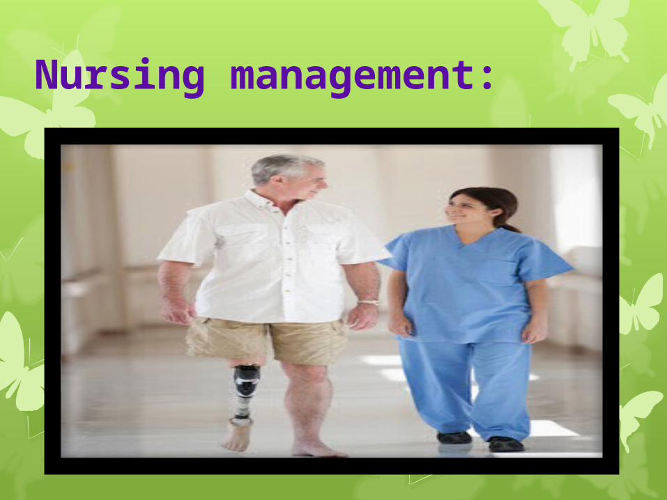
Nursing management:
Nursing assessment
Obtain detailed history of injury
Assess pain and functional deficits
Be aware that systemic symptoms are acute in children but vary in intensity with adults
Perform general systemic assessment because adults with long bone involvement generally have more systemic septic symptoms.
Nursing diagnosis:
Acute/chronic pain related to inflammatory process.
Impaired physical mobility related to rest of affected part.
Risk for extension of infection: bone abscess formation.
Nursing interventions:
Relieving pain:
1. Administer opiods for acute pain; non-narcotics for chronic pain.
2. Administer medications around the clock versus as necessary to establish a consistent blood level.
3. Report any decreased in pain that may indicate worsening infection.
Contd….
Increasing physical mobility:
1. Treatment regimens restrict activity. The bone is weakened by the infective process and must be protected by immobilization devices and by avoidance of stress on the bone.
2. The patient must understand the rationale for the activity restrictions. The joints above and below the affected part should be gently moved through their range of motion. The nurse encourages full participation in ADLs within the physical limitations to promote general well being.
Increasing knowledge:
1. Describe the infectious process and rationale for prolonged treatment with osteomyelitis.
2. Explain IV antibiotic therapy, potential adverse effects, and reactions.
3. Explain strict adherence to infection control practices ( sterile technique, hand washing) to prevent spread of infection in some cases.
Contd….
Promoting rest without complication:
1. Support the affected extremity to minimize pain.
2. If patient is on bed rest, prevent hazards of immobility (passive ROM, position change, coughing and deep breathing exercises)
3. Encourage distraction activities.
Contd….
Patient education and health maintenance:
1. Advise patient to adhere to infection control principles- proper hand washing, disposal of wound drainage, dressings to prevent reinfection/transmission of infection at home.
2. Stress adherence to medication regimen, which may be prolonged, with frequent follow up visits.
3. Teach care of indwelling device for medication delivery (such as Hickman catheter)
Contd….
Expected outcomes:
Pain managed with non-narcotic analgesics.
Infectious process minimized.
Functional status of affected joint intact.