bode index in copd final
TRANSCRIPT

1
DISSERTATION ON
A STUDY ON BODE INDEX AS A PREDICTOR OF SEVERITY AND
SYSTEMIC INVOLVEMENT IN PATIENTS WITH CHRONIC
OBSTRUCTIVE PULMONARY DISEASE
Submitted in partial fulfillment of the requirements for
M.D.DEGREE BRANCH -1 GENERAL MEDICINE
of
THE TAMILNADU DR. M.G.R. MEDICAL UNIVERISTY,
MADRAS MEDICAL COLLEGE
CHENNAI – 600 003
MARCH – 2009.

2
CERTIFICATE This is to certify that this dissertation entitled “A STUDY ON
BODE INDEX AS A PREDICTOR OF SEVERITY AND
SYSTEMIC INVOLVEMENT IN PATIENTS WITH CHRONIC
OBSTRUCTIVE PULMONARY DISEASE” submitted by Dr.
SOJAN GEORGE K. appearing for Part II M.D. Branch I General
Medicine Degree examination in March 2009 is a bonafide record of
work done by him under my direct guidance and supervision in partial
fulfillment of regulations of the Tamil Nadu Dr. M.G.R. Medical
University, Chennai. I forward this to the Tamil Nadu Dr.M.G.R.
Medical University, Chennai, Tamil Nadu, India.
Director,
Institute of Internal Medicine,
Government General Hospital,
Chennai – 600 003.
The Dean,
Madras Medical College,
Government General Hospital,
Chennai – 600 003.
Additional Professor,
Institute of internal medicine,
Madras Medical College,
Government General Hospital,

3
DECLARATION
I solemnly declare that the dissertation entitled “A STUDY ON
BODE INDEX AS A PREDICTOR OF SEVERITY AND
SYSTEMIC INVOLVEMENT IN PATIENTS WITH CHRONIC
OBSTRUCTIVE PULMONARY DISEASE” is done by me at t he
madras medical college and Government General Hospital, Chennai during 2007-
2008 under the guidance and supervision of Prof .D. Rajasekaran, M.D.
This dissertation is submitted to The Tamilnadu Dr. M.G.R Medical
University, towards partial fulfillment of regulation for the award of M.D.
DEGREE IN GENERAL MEDICINE (BRANCH–I).
Place:
Date:
Dr. SOJAN GEORGE K.
M.D. post graduate,
Institute of Internal Medicine,
Madras Medical College,
Government General Hospital,
Chennai-600003.

4
ACKNOWLEDGEMENT
I would like to express my sincere gratitude to Prof. T. P.
KALANITI, M.D., The Dean, Madras Medical College, for having
permitted me to use the hospital material in this study.
I am immensely grateful to Prof. C. Rajendran, M.D.,
Director, Institute of Internal Medicine, for his suggestions and
encouragement.
I am greatly indebted to my beloved unit chief and teacher
Prof. D. Rajasekaran, M.D., Additional professor, Institute of Internal
Medicine, who encouraged, helped and guided me throughout this study.
I express my sincere thanks to my unit Assistant Professors,
Dr. G. Subburagavalu, M.D., Dr. A. Aravind, M.D., Dr. S. Tito,
M.D. for their thoughtful guidance throughout the work.
I thank all the professors, assistant professors and post
graduates of the department of biochemistry for their valuable support in
the biochemical analysis.

5
I thank all my colleagues and friends for their constant
encouragement and valuable criticism.
I express my gratitude for the generosity shown by all the
patients who participated in the study. If at all this study contributes a
little to relieve their suffering, I feel that I have repaid a part of my debt.
I am extremely thankful to my family members for their
continuous support. Above all I thank my God Almighty for His
immense blessings.

6
CONTENTS
S.NO
TITLE
PAGE NO.
1 INTRODUCTION
1
2 AIMS AND OBJECTIVES
4
3 REVIEW OF LITERATURE
5
4 MATERIALS AND METHODS
24
5 STUDY PROTOCOL
26
6 OBSERVATION AND RESULTS
33
7 DISCUSSION
45
8 CONCLUSION AND SUMMARY
53
9 PROFORMA
10 MASTER CHART
11 ETHICAL COMMITTEE CERTIFICATE
12 ABBREVIATIONS
13 BIBLIOGRAPHY

7
INTRODUCTION
Chronic obstructive pulmonary disease (COPD) is a major cause of
morbidity and mortality throughout the world. The prevalence and burden of
COPD are projected to increase in the coming decades due to continued exposure
to COPD risk factors and the changing age structure of the world’s population. It is
projected to rank fifth in 2020 in burden of disease caused worldwide, according to
a study published by the World Bank/World Health Organization1. The disease
causes a heavy burden on the global health care resources. The costs involved in
the treatment and evaluation is directly proportional to the pulmonary and the extra
pulmonary components of the disease2.
‘Chronic obstructive pulmonary disease (COPD) is defined as a
preventable and treatable disease with some significant extra pulmonary effects
that may contribute to the severity in individual patients. Its pulmonary component
is characterized by airflow limitation that is not fully reversible. The airflow
limitation is usually progressive and associated with an abnormal inflammatory
response of the lung to noxious particles or gases’ 3.
The pathogenesis and clinical manifestations of COPD are not
restricted to pulmonary inflammation and structural remodeling. Rather, this

8
disorder is associated with clinically significant systemic alterations in
biochemistry and organ function. The systemic aspects of COPD include oxidative
stress and altered circulating levels of inflammatory mediators and acute-phase
proteins. As in other chronic inflammatory conditions, weight loss, muscle
wasting, hypo proteinemia and tissue depletion are commonly seen in COPD
patients4. Selective wasting of fat-free mass coupled with impaired respiratory and
peripheral muscle function and a reduced capacity for exercise occur in COPD
patients. Indeed, weight loss may directly impact poor prognosis in COPD patients.
The severity of COPD is usually assessed on the basis of a single
parameter – forced expiratory volume in one second (FEV1). However the
patients with COPD have systemic manifestations that are not reflected by the
FEV1. Hence a multidimensional grading system that assessed the respiratory and
systemic expressions of COPD was designed to predict outcome in these patients5.
The four factors that predicted the severity most were the body-mass index (B), the
degree of airflow obstruction (O) and dyspnea (D), and exercise capacity (E),
measured by the six-minute–walk test. These variables were used to construct the
BODE index, a multidimensional 10-point scale in which higher scores indicate a
higher risk of death.

9
The process of allocating scarce medical resources to the most needed
patients can be extremely difficult in diseases which affect a large number of
patients. Decision makers need a rational and consistent scoring system that is
designed to identify those who are maximally in need of a diagnostic or a
therapeutic intervention under a health-care budget constraint. BODE index has
been proposed to serve this purpose in patients with chronic obstructive pulmonary
disease (COPD)6.
In our study we analyzed the BODE index as a predictor of
hospitalization and severity of systemic involvement.

10
AIMS AND OBJECTIVES
1. To determine whether higher BODE index in chronic obstructive pulmonary
disease correlates with more years of cigarette smoking.
2. To determine whether higher BODE index is associated with more days of
hospitalisation.
3. To determine whether higher BODE index is associated with more severe
cardiac involvement.
4. To determine whether higher BODE index correlates with poor nutritional
status.
5. To determine the correlation between BODE index and the level of systemic
inflammation in patients with COPD.

11
REVIEW OF LITERATURE
Chronic obstructive pulmonary disease (COPD) is a lung disease
characterized by chronic obstruction of lung airflow that interferes with normal
breathing and is not fully reversible. COPD is one of the leading causes of
morbidity and mortality worldwide and imparts a substantial economic burden on
individuals and society.
DEFINITION
Chronic obstructive pulmonary disease (COPD) was initially defined as
‘a disease state characterized by chronic airflow limitation due to chronic
bronchitis and emphysema’. Chronic bronchitis has been defined in clinical terms
as ‘the presence of chronic productive cough for at least 3 consecutive months in 2
consecutive years. Other causes of chronic productive cough must be ruled out’.
Emphysema, on the other hand, has been defined by its pathologic description: ‘an
abnormal enlargement of the air spaces distal to the terminal bronchioles
accompanied by destruction of their walls and without obvious fibrosis’.
However the definition of COPD has undergone major revision. The
new GOLD guidelines3 and the ATS/ERS definition7 reflect these scientific

12
advances: “Chronic obstructive pulmonary disease (COPD) is defined as a
preventable and treatable disease with some significant extra pulmonary effects
that may contribute to the severity in individual patients. Its pulmonary component
is characterized by airflow limitation that is not fully reversible. The airflow
limitation is usually progressive and associated with an abnormal inflammatory
response of the lung to noxious particles or gases” 3. While the new guidelines do
not specifically include chronic bronchitis and emphysema in the definition of
COPD, it is made clear that they are considered the predominant causes of COPD.
The airflow limitation is caused by mixture of small airway disease (obstructive
bronchiolitis) and parenchymal destruction (emphysema) the relative contribution
of each varies from person to person3.
EPIDEMIOLOGY
Using the World Health Organization/World Bank Global Burden of
Disease Study data, the worldwide prevalence of COPD in 1990 was estimated at
9.34/1000 in men and 7.33/1000 in women8,9. This was an underestimation of true
prevalence of COPD since the estimates included all age groups. In a large
epidemiologic study from Korea involving 9,243 subjects, Kim and his colleagues
reported that the prevalence of COPD was 17.2 % among subjects older than 45

13
years10. There is plenty of information on the prevalence and burden of COPD
from the developed countries. Such an assessment is rather scarce from most of the
developing world.
The prevalence of COPD reported in different population based
studies from India is highly variable11. The prevalence rates in male subjects of
2.12% to 9.4% in studies reported from the North are generally higher than 1.4% to
4.08% reported from South India. The respective ranges for female subjects vary
from 1.33% to 4.9% in the North and from 2.55% to 2.7% in South India. For
epidemiological assessment, the rounded-off median prevalence rates were
assessed as 5 percent for male and 2.7 percent for female subjects of over 30 years
of age11. The disease is distinctly more common in males.
The prevalence was found to increase with increasing age, especially n
the males, in those with more than 20 pack–yrs of smoking and in low income
subjects. The male to female ratio had varied from 1.32:1 to 2.6:1 in different
studies with a median ratio of 1.6:111.

14
RISK FACTORS
A. Genetic risk factor:
a rare recessive severe hereditary deficiency of alpha-1 antitrypsin14, a
major circulating inhibitor of serine proteases, most commonly seen in
individuals of Northern European origin15.
B. Exposure to environmental particles:
• Tobacco smoke:
Cigarette smokers have a greater annual rate of decline in FEV1 and a
greater COPD mortality rate than non smokers16,17. Not all smokers develop
clinically significant COPD, which suggests that genetic factors must
modify each individual’s risk18. Passive exposure to cigarette smoke may
also contribute to respiratory symptoms19 and COPD20 by increasing the
lungs’ total burden of inhaled particles and gases21,22.
• Occupational dusts organic and inorganic Chemicals:
A statement published by the American Thoracic Society concluded
that occupational exposures account for 10-20% of either symptoms or
functional impairment consistent with COPD23.

15
• Indoor and Outdoor Air Pollution:
The evidence that indoor pollution from biomass cooking and heating
in poorly ventilated dwellings and high levels of urban pollution is an
important risk factor for COPD continues to grow24-30. This has been proved
by many case-control studies29.30 and other robustly designed studies .
C. Gender:
Studies from developed countries31 show that the prevalence of the
disease is now almost equal in men and women. Some studies have
suggested that women are more susceptible to the effects of tobacco smoke
than men32-34.
D. Infection:
A history of severe childhood respiratory infection has been
associated with COPD35-37.
E. Low birth weight:
F. Socioeconomic Status:
The risk of developing COPD is inversely related to socioeconomic
status 38-40.
16
G. Poor nutritional status
H. Co morbidities
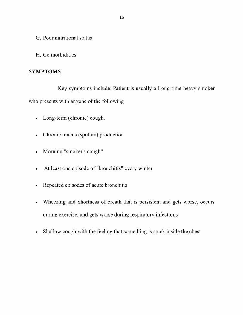
SYMPTOMS
Key symptoms include: Patient is usually a Long-time heavy smoker
who presents with anyone of the following
• Long-term (chronic) cough.
• Chronic mucus (sputum) production
• Morning "smoker's cough"
• At least one episode of "bronchitis" every winter
• Repeated episodes of acute bronchitis
• Wheezing and Shortness of breath that is persistent and gets worse, occurs
during exercise, and gets worse during respiratory infections
• Shallow cough with the feeling that something is stuck inside the chest

17
Patients may have a rapid, sometimes sudden, and prolonged increase
in symptoms (cough, amount of mucus, and/or shortness of breath), especially if
the COPD is mainly chronic bronchitis. This is called a COPD exacerbation.
PATHOPHYSIOLOGY OF COPD
Quantitative evidence of increased expiratory flow resistance in
emphysema was first obtained in one patient by Neergard and Wirz in 192741. In
1934, Christie described elastic properties or distensibility of the lung in
emphysema42. The “golden age” of pulmonary macrophysiology, extending from
about the 1960s to the 1980s provided new insights regarding the determinants of
flow limitation at the levels of the airway and parenchyma. Corbin and coworkers
showed that smoking was associated with the loss of lung recoil pressure and with
increased static lung volumes (RV, FRC, and TLC), even among individuals who
had relatively normal FEV143. Up to a point, these changes appeared reversible
with smoking cessation.
COPD, or chronic obstructive pulmonary disease, is a progressive
inflammatory disease connecting the airways, lung parenchyma, and vasculature. It
causes the damage and remodeling of the airways and lung tissue. The
inflammatory process is a driving aspect in the pathophysiology of COPD. Recent
verification suggests that the inflammatory response results in a number of effects,

18
including an arrival of inflammatory cells such as macrophages, neutrophils and
lymphocytes. Thickened airways and structural changes such as increased smooth
muscle and fibrosis may also be manifested.
Cigarette smoking causes an inflammatory response in the lungs. This
response does not cease with the removal of the stimulus, but progresses for an
unlimited period of time. These processes result in emphysema, chronic bronchitis,
or both. Emphysema begins with a small airway disease and progresses to alveolar
destruction, with a predominance of small airway narrowing and mucous gland
hyperplasia.
The basic pathophysiologic process in COPD consists of increased
resistance to airflow, loss of elastic recoil and decreased expiratory flow rate. The
alveolar walls frequently break because of the increased resistance of air flows.
The hyper inflated lungs flatten the curvature of the diaphragm and enlarge the rib
cage. The altered configuration of the chest cavity places the respiratory muscles,
including the diaphragm, at a mechanical disadvantage and impairs their force-
generating capacity. Consequently, the metabolic work of breathing increases and
the sensation of dyspnea heightens.
Hogg has focused on the importance of small airway obstruction, most
recently showing that airway remodeling and wall thickening, presence of

19
inflammatory mucous exudates, and B cell and CD8 T cell inflammation are all
associated with severity of COPD and progression of the disease44. Christie,
Thurlbeck, and others have championed emphysema as the dominant pathology
accounting for abnormal physiology, and have shown, for example, that in a
subgroup of patients, the relationship between flow and recoil pressure is indeed
normal. While many important insights came from this line of investigation, to this
day we still do not understand the relative contribution of small airway obstruction
versus emphysema in an individual patient, nor the potential relationship between
these two lesions. This line of investigation is still fruitful, particularly with the
ongoing revolution in imaging.
THE ELASTASE:ANTIELASTASE HYPOTHESIS
Just over 40 years ago, two lines of evidence, one experimental and
one clinical, suggested that emphysema is caused by destruction of elastic fibers by
elastases. The first was by Laurell and Eriksson who, in 1963, described five
patients with deficiency of α-1AT, the primary inhibitor of the neutral serine
proteinase neutrophil elastase (NE). Three of these five patients had emphysema45.
The second came in 1965 when Gross and coworkers instilled papain into the lungs
of rodents in an attempt to produce granulomas. Instead they found emphysema46.

20
Subsequently, investigators have instilled a variety of proteinases into
animal lungs. Kuhn and colleagues47, Senior and coworkers48, Janoff and
associates49, and Snider and colleagues50 were among the group of investigators
who subsequently demonstrated that only elastolytic proteinases—including
pancreatic elastase and the more relevant human neutrophil elastase (HNE)—
caused emphysema. Hoidal’s group showed that proteinase 351 also caused
destructive lung disease. These seminal experiments formed the basis for the
elastase : antielastase hypothesis, which states that the relative balance between
elastases and their inhibitors determines the susceptibility of the lung to the
destruction characteristic of emphysema.
INFLAMMATION–EXTRACELLULAR MATRIX TURNOVER
A classic study by Damiano and coworkers correlated the presence of
HNE with COPD using immunogold staining52. However, other studies actually
showed a negative correlation between emphysema and HNE or neutrophil
number53, 54. As discussed above, macrophages are abundant in COPD, yet the
capacity of the macrophage to degrade elastin and hence contribute to disease
pathogenesis was unproven until Senior and colleagues demonstrated that
macrophages produce elastolytic matrix metalloproteinases55,56, and Chapman and
coworkers found elastolytic cysteine proteinases57.

21
Retamales and colleagues58 found that even in end-stage lung disease,
long after smoking cessation, there remains an exuberant inflammatory response.
This suggests that the mechanisms of cigarette smoke–induced inflammation that
initiate the disease differ from mechanisms that sustain inflammation after
smoking cessation. Moreover, this study suggests that multiple inflammatory (and
likely structural) cells interact to cause COPD, and that focusing on single cells
and proteinases in isolation will not provide a comprehensive understanding of the
disease process.
Cigarette smoke causes constitutive macrophages to produce MMP-
12, which, in turn, cleaves elastin into fragments chemotactic for monocytes. This
positive feedback loop perpetuates macrophage accumulation and lung destruction.
The concept that proteolytically generated elastin fragments mediate monocyte
chemotaxis was proven by Senior and coworkers59 and Hunninghake and
colleagues60.At the very least, this study demonstrates a critical role for
macrophages in the development of emphysema and unmasks a proteinase-
dependent mechanism of inflammatory cell recruitment. Of note, last year
Grumelli and coworkers found that human CD8+ T cells derived from patients
with COPD generate interferon (IFN)-γ–inducible chemokines that also function to
upregulate expression of human macrophage MMP-1261. Studies by Churg and

22
coworkers demonstrate that acute neutrophil inflammation secondary to smoking is
related to MMP-12–dependent tumor necrosis factor (TNF) shedding 62.
OXIDANT–ANTIOXIDANT BALANCE
Cigarette smoke and inflammatory cells have the capacity to produce
reactive oxygen species, and they have been postulated to play a variety of roles in
the pathogenesis of emphysema. One intriguing finding was that cigarette smoke
can oxidize a methionine residue in the reactive center of A1PI, inactivating A1PI
and thus altering the elastase:antielastase balance. Oxidants cannot degrade
extracellular matrix but might modify elastin, making it more susceptible to
proteolytic cleavage.
Recently, Barnes and colleagues have found that cigarette smoke
oxidizes and inactivates histone deacetylase 2 (HDAC2), which acts to counter
histone acetylase (HAT)63. Acetylation of histone unwinds chromatin, allowing
transcriptional complexes to bind to DNA. Thus, in the absence of HDAC2, RNA
polymerase II and NF-kβ form a proinflammatory transcription complex. Finally,
reactive oxygen species may also promote apoptosis of structural cells, a recent
concept for initiation of emphysema.

23
APOPTOSIS
Kasahara and colleagues found that exposure to agents that initiate
endothelial cell death (via VEGFRII inhibition) leads to non inflammatory airspace
enlargement64. Nagai and coworkers then found that epithelial cell death (via
caspase 3 delivery) also causes emphysema65. As mentioned above, the loss of an
acinar unit results from the destruction of both the extracellular matrix (ECM) and
the structural cells. These models suggest that death of structural cells may be an
initiating event, with subsequent release of matrix-degrading proteinases. Whether
this occurs in human COPD as a primary event is uncertain,
INEFFECTIVE REPAIR
The ability of the adult lung to repair damaged alveoli appears
limited. In fact, as we define genetic predisposition to COPD, we speculate that
smoking routinely leads to inflammation and lung damage, and those at risk lack
the capacity to repair this damage. In emphysema, aberrant alveolar and
extracellular matrix repair results in coalesced and enlarged airspaces with
depleted and disordered parenchymal elastic fibers, and excess and abnormally
arranged collagen.

24
SYSTEMIC INVOLVEMENT IN COPD
The pathogenesis and clinical manifestations of COPD are not
restricted to pulmonary inflammation and structural remodeling. Rather, this
disorder is associated with clinically significant systemic alterations in
biochemistry and organ function. The systemic aspects of COPD include oxidative
stress and altered circulating levels of inflammatory mediators and acute-phase
proteins. Indeed, an impaired endogenous oxidant-antioxidant balance66-68 has been
reported in patients experiencing exacerbations of COPD, and others have observed
altered circulating levels of several cytokines and adhesion molecules in patients
with stable disease. As in other chronic inflammatory conditions, weight loss,
muscle wasting, and tissue depletion are commonly seen in COPD patients.
Wasting is a generally occurring manifestation in a wide variety of
different chronic conditions and can be considered to be an important systemic
manifestation as a loss of > 40% of actively metabolizing tissue is incompatible
with life69-70. The body cell mass (BCM) represents the actively metabolizing
(organs) and contracting (muscles) tissue. This BCM cannot be measured directly.
Changes in BCM can be clinically recognized by decrease in body mass index
(BMI) in general and by loss in fat-free mass (FFM) in particular. In a
retrospective study of 400 patients with COPD, Schols et al71 demonstrated that

25
low body mass index (BMI), age, and low PaO2 were significant independent
predictors of increased mortality rates. After stratification of the group into BMI
quintiles, a threshold value of 21 kg/m2 was identified below which the mortality
risk was clearly increased.
SPIROMETRIC CLASSIFICATION OF COPD SEVERITY
The present widely accepted classification of COPD is mainly based
on the FEV1 values1. It is as follows
Stage I: Mild COPD –
Characterized by mild airflow limitation (FEV1/FVC < 0.70; FEV1 >/= 80%
predicted). Symptoms of chronic cough and sputum production may be present.
Patients are usually unaware of the illness.
Stage II: Moderate COPD –
Characterized by worsening airflow limitation (FEV1/FVC < 0.70; 50% </=
FEV1 < 80% predicted), with shortness of breath typically developing on exertion
with or without cough and sputum production. This is the stage at which patients
typically seek medical attention.

26
Stage III: Severe COPD –
Characterized by further worsening of airflow limitation (FEV1/FVC < 0.70;
30% </= FEV1 < 50% predicted), greater shortness of breath, reduced exercise
capacity, fatigue, and repeated exacerbations that almost always have an impact on
patients’ quality of life.
Stage IV: Very Severe COPD –
Characterized by severe airflow limitation (FEV1/FVC < 0.70; FEV1 < 30%
predicted or FEV1 < 50% predicted plus the presence of chronic respiratory
failure). Respiratory failure is defined as arterial partial pressure of oxygen (PaO2)
less than 8.0 kPa (60 mm Hg) with or without arterial partial pressure of CO2
(PaCO2) greater than 6.7 kPa (50 mm Hg) while breathing air at sea level.
LIMITATIONS OF SPIROMETRIC CLASSIFICATION
The spirometric classification, though good in many ways is not full
proof for the assessment of severity of COPD. The FEV1 is essential for the
diagnosis and quantification of the respiratory impairment resulting from COPD72-
74. In addition, the rate of decline in FEV1 is a good marker of disease progression
and mortality75,76. However, the FEV1 does not adequately reflect all the systemic
manifestations of the disease. For example, the FEV1 correlates weakly with the

27
degree of dyspnea77, and the change in FEV1 does not reflect the rate of decline in
patients' health78. More important, prospective observational studies of patients
with COPD have found that the degree of dyspnea79 and health-status scores80 are
more accurate predictors of the risk of death than is the FEV1. Thus, although the
FEV1 is important to obtain and essential in the staging of disease in any patient
with COPD, it alone as the sole parameter of severity does not throw light on the
systemic involvement and progession of the disease.
BODE INDEX
Due to reasons above stated, researchers described a new index –
BODE index, for the comprehensive evaluation of patients with COPD. This
multisystem grading index has four variables.
• Body mass index
• Obstruction to airflow (FEV1)
• Dyspnea (MMRC dyspnea scale)
• Effort tolerance (6 minute walk test)

28
Each variable in the index correlates independently with the prognosis
of COPD, is easily measurable, and serves as a surrogate for other potentially
important variables. In the BODE index, two descriptors of systemic involvement
were included: the body-mass index and the distance walked in six minutes. Both
are simply obtained and independently predict the risk of death81-83. It is likely that
they share some common underlying physiological determinants, but the distance
walked in six minutes contains a degree of sensitivity not provided by the body-
mass index. The six-minute walk test is simple to perform and has been
standardized84. Its use as a clinical tool has gained acceptance, since it is a good
predictor of the risk of death among patients with other chronic diseases, including
congestive heart failure84 and pulmonary hypertension. Indeed, the distance walked
in six minutes has been accepted as a good outcome measure after interventions
such as pulmonary rehabilitation.
The body-mass index was also an independent predictor of the risk of
death and was therefore included in the BODE index. We evaluated the
independent prognostic power of body-mass index in our cohort using different
thresholds and found that values below 21 were associated with an increased risk of
death, an observation similar to that reported by Landbo and coworkers in a large
population study79.

29
The Global Initiative for Chronic Obstructive Lung Disease and the
American Thoracic Society recommend that a patient's perception of dyspnea be
included in any new staging system for COPD. Dyspnea represents the most
disabling symptom of COPD; the degree of dyspnea provides information
regarding the patient's perception of illness and can be measured. The MMRC
dyspnea scale is simple to administer and correlates with other dyspnea scales and
with scores of health status80. Furthermore, in a large cohort of prospectively
followed patients with COPD, which used the threshold values included in the
BODE index, the score on the MMRC dyspnea scale was a better predictor of the
risk of death than was the FEV1.
The BODE index combines the four variables by means of a simple
scale. Weighting the variables included in the index did not improve the predictive
power of the BODE index. Most likely, it failed to do so, because each variable
included has already proved to be a good predictor of the outcome of COPD.

30
MATERIALS AND METHODS
SETTING
Institute of internal medicine
Madras Medical college and Government General hospital
Chennai – 600 003
INSTITUTIONAL ETHICS COMMITTEE APPROVAL
Obtained
STUDY DESIGN
To evaluate the BODE index as a predictor of hospitalization and
severity of systemic involvement in patients with Chronic Obstructive Pulmonary
Disease, a cross sectional study design was chosen.
PERIOD OF STUDY
May 2007 to July 2008
SAMPLE SIZE
Cases : 90 ; controls : 30

31
INCLUSION CRITERIA
• Male patients with symptoms suggestive of COPD as cases
• Male patients who came for master health check up as controls
EXCLUSION CRITERIA
• Spirometry proved bronchial asthma defined as an increase in the FEV1 of
more than 15 percent above the base-line value or of 200 ml after the
administration of a bronchodilator
• recent myocardial infarction < 4months
• unstable angina
• congestive heart failure (NYHA class III or IV)
• inability to perform spirometry or 6 minute walk test
• Unrelated life threatening major illness
• liver disease
• patients with acute exacerbation

32
STUDY PROTOCOL
A total of 120 patients who attended our outpatient clinic at the
Madras Medical College and Government General Hospital, Chennai were
enrolled into the study. Of these, 90 patients with symptoms suggestive of COPD
were selected as cases and 30 patients who came for Master health checkup were
selected as controls.
The patients with the following diagnostic criteria (according to the
GOLD guidelines) were defined as having COPD
1. the presence of cough and sputum production for at least 3 months in each of
the two consecutive years
2. exertional dyspnoea
3. physical examination showing
(a) Signs of airflow limitation like prolonged expiration and expiratory
wheeze which is not fully reversible;
(b) Signs of hyperinflation
4. Spirometry showing post bronchodilator FEV1/FVC ratio < 0.70

33
The present analysis was restricted to male patients only, who met the
acceptability and reliability criteria of the American Thoracic Society to improve
the diagnostic accuracy as sex may be a confounding factor in many of the
parameters assessed.
For each enrolled subject, detailed history of smoking, personal and
family medical histories were obtained. On the day of enrollment, height and
weight were measured twice during the examination. Weight was measured to the
nearest 100 grams with bare foot. Height was measured to the nearest mm with the
stadometer. Body mass index (BMI) was calculated by the formula.
BMI = Weight in Kgs / (Height in Ms)2
Spirometry was performed with an equipment that met the American
thoracic society performance criteria, in each of the cases on enrollment into the
study and 20 minutes following the administration of salbutamol nebulisation. To
adjust for the height, sex, age and sex published prediction equations for forced
expiratory volume in 1 second (FEV1) and forced vital capacity (FVC) were used.
FEV1 and FVC were calculated. The procedure was repeated on 2 occasions and
the average value was taken.

34
A detailed history of the dyspnea experienced by the patient was
taken. MMRC dyspnea scale was used to score the patients dyspnea. Six minute
walk test was performed twice with a gap of 30 minutes rest in between and the
average was taken. Patients were asked to walk on a level ground for maximum
possible distance within a duration of 6 minutes. Periods of rest taken, was also
included in the 6 minutes test period.
The BODE index was calculated for each patient using the body mass
index, the threshold value of FEV1, the distance walked in 6 min, and the score on
the modified Medical Research Council (MMRC) dyspnea scale. The patients
received points ranging from 0 (lowest value) to 3 (maximal value). For body mass
index the values were 0 (>21) or 1 (<21). The scores for FEV1 were 0 (more than
or equal to 65%), 1 (50 – 64%), 2 (36 – 49%) and 3 (less than or equal to 35%).
The 6 minute walk test scores were 0 (> 350 ms), 1 (250 – 350 ms), 2 (150 – 249
ms) and 3 (< 150 ms). The MMRC dyspnea class 0 and I were given 0 points, class
II – 1 point, class III – 2 points and class IV – 3 points. The points for each
variable were added, so that the BODE index ranged from 0 to 10 points in each
patient. The BODE score of 0 – 2 was taken as mild COPD. Scores between 3 – 5
was considered as moderate disease and those more than or equal to 6 was
considered as severe COPD.

35
MMRC dyspnea scale
Grade 0 – no dyspnea / only on severe exertion
Grade 1 – dyspnea on hurrying / walking up a hill
Grade 2 – walks slower than normal at level/ pause while walking on level ground
Grade 3 – stops for breath after walking 100 yards/few mins on level ground
Grade 4 – too breathless to leave the house/ dyspnea on dressing
BODE INDEX:
BODE score 0 1 2 3
FEV1 >/= 65 % 50 – 64% 36 – 49% < /= 35%
6 min walk test
>350 ms 250 – 349ms 150 – 249 ms <149 ms
Dyspnea scale
0 – 1 2 3 4
BMI > 21 kg/m2 <21 kg/m2
Mild COPD 0 – 2
Moderate COPD 3 – 5
Severe COPD >/= 6

36
A detailed history regarding number of days of hospital admission in
the last two years was obtained from the patients response to the question “how
many days have you been admitted in hospital in the past 2 years due to reasons
related to COPD ?” Patient’s discharge cards were also reviewed.
A standard 12 lead ECG was taken for each of the individual patients.
QRS axis was determined by plotting the QRS potentials on a graph with lead I as
X axis and aVF as Y axis. – 30 to + 90 was considered as normal axis, – 30º to –
90º as left axis , +90º to +180º as right axis and – 90º to + 180º was considered as
north west axis. Echo cardiography was performed using 2D echo in the institute
of cardiology, Madras Medical College. Ejection fraction and pulmonary pressure
gradient was assessed. Pulmonary artery hypertension was graded as mild,
moderate and severe.
Hemoglobin estimation was performed according to the routine
standards using an automated analyzer at the biochemistry lab attached to the
institute of biochemistry, Madras Medical College and Government General
Hospital. CRP was estimated in the lab using the latex agglutination method.
Agglutination at a dilution of 6 or less was considered as negative result. Positive
result was expressed as the dilution at which agglutination occurred.

37
FINANCIAL SUPPORT: nil
CONFLICT OF INTEREST: nil

38
STATISTICAL ANALYSIS
Statistical analysis was carried out in all the 120 subjects (including 90
COPD patients and 30 controls) after categorizing the variable. Baseline data was
collected from patients without and with mild, moderate and severe COPD. Ages,
body mass index, days of hospitalization, mean hemoglobin concentration, QRS
axis, ejection fraction, pulmonary hypertension, serum albumin concentration, and
CRP of all subjects were the parameters analyzed.
The significance of difference in means between two groups was
analyzed using the one way ANOVA F-test and the significance of difference in
proportions by the Chi square test. Multiple comparisons were done by fishers least
significant difference (LSD) t-test. Statistical significance was taken when the p
value was less than 0.05.
Statistical analysis was carried out using the standard formula.
Microsoft excel 2007 and SPSS (statistical package for social sciences)version 13
software was used for data entry and analysis.

39
RESULTS AND OBSERVATIONS
A total of 120 patients including 90 patients with COPD as cases and
30 healthy individuals as controls were enrolled in the study. All the cases and
controls were males. Among patients with COPD, there were 32 (35.56%) patients
who had mild COPD with a BODE score between 0 – 2. Moderate (BODE score of
3 – 5) and severe COPD (BODE score more than or equal to 6) groups had 29
patients (32.22%) each.
Table 1: Age wise distribution in years
Group N Mean
(yrs)
Std. deviation One way ANOVA F-
test
Control 30 54.70 5.603
Mild (0-2) 32 53.47 7.362
Moderate (3-5) 29 55.00 8.627
Severe (>/=6) 29 59.93 7.606
Total 120 55.71 7.679
F=4.440
P=0.005
significant

40
The average age of participants in the study was 55.71 years. Among
the COPD patients, BODE index was found to increase with age with the mild
group having a mean age of 53.47 years, moderate group 55.00 years and the
severe group with 59.93 years as the mean age. The difference was statistically
significant with a P value of 0.005.

41
Table 2: Smoking status
Smoker
Yes No
Groups
N % N %
Total N
Pearson
chi square
test
Control 12 40.0 18 60 30
Mild 14 43.7 18 56.3 32
Moderate 19 65.5 10 34.5 29
Severe 26 89.7 3 10.3 29
Total 71 59.2 49 40.8 120
X2 -19.352
P =0.000
significant
The proportion of smokers was higher in the higher BODE index
group compared to the lower index group. There was no significant difference
between the control group and the lower score group. Thus smoking status had a
positive risk correlation with higher BODE index (P = 0.000).

42
Table 3: Smoking in pack years
Group N Mean
(pack yrs)
Std. deviation Oneway ANOVA F test
Control 30 4.55 5.603
Mild 32 7.42 7.362
Moderate 29 15.07 8.627
Severe 29 26.90 7.606
Total 120 13.26 7.679
F = 26.936
P = 0.000
Significant
The study revealed that the BODE score was significantly associated
with the number of pack years of smoking. It was 4.55 pack yrs in controls, 7.42
pack yrs in mild cases, 15.07 in moderate and 26.90 in severe cases. On multiple
comparison by LSD the difference between control group and mild group was not
statistically significant but that of the other 2 groups were highly significant (P =
0.000).

43
Table 4: Body Mass index
Group N Mean
(kg/m2)
Std. deviation
Oneway ANOVA F
test
Multiple comparison
(LSD)
Control 30 24.294 2.544
Mild 32 22.476 2.455
Moderate 29 21.711 2.552
Severe 29 20.260 3.212
Total 120 22.210 3.035
F = 11.431
P = 0.000
Significant
1Vs2,3,4
2Vs1,4
3Vs1,4
4Vs1,2,3
P = 0.05
The average BMI of the patients in our study was 22.21 kg/m2. The
control group had a BMI of 24.294 kg/m2 with a standard deviation of 2.544. The
BMI was found to be significantly lower in patients with COPD. It was 22.476
kg/m2 (standard deviation –2.455) in the mild group, 21.711 (std. deviation –
2.552) in the moderate group and 20.260 (std. deviation – 3.212) in the severe
group. On multiple comparisons the significance between mild and moderate
groups was not found to be significant. All other comparisons showed significant
difference.

44
Table 5: Duration of hospital stay over last 2 years (days)
Group N Mean
(days)
Std. deviation
Oneway ANOVA F
test
Multiple comparison
(LSD)
Control 30 0.07 0.365
Mild 32 0.13 0.492
Moderate 29 3.17 2.929
Severe 29 16.00 9.177
Total 120 4.68 8.041
F = 75.340
P = 0.000
Significant
1Vs3,4
2Vs3,4
3Vs1,2,4
4Vs1,2,3
P = 0.05
The study results showed that a higher BODE score was associated
with a higher incidence of hospital stay due to reasons related to COPD, over the
past 2 years. The control group and the mild COPD group did not have any
significant hospital admission during the past 2 years. The average duration of stay
in the moderate study group was 3.17 days while it was 16 days in the group with
severe COPD according to the BODE score. Both these values were found to be
significant on multiple comparisons to other groups.

45
Table 6: Hemoglobin concentration in gm/ dL
Group N Mean
(gm/dL)
Std. deviation
Oneway ANOVA F
test
Multiple comparison
(LSD)
Control 30 11.040 1.305
Mild 32 10.713 1.439
Moderate 29 12.176 1.566
Severe 29 14.869 1.460
Total 120 12.153 2.168
F = 50.733
P = 0.000
Significant
1Vs3,4
2Vs3,4
3Vs1,2,4
4Vs1,2,3
P = 0.05
Comparing the hemoglobin values in various groups of the study it
was found that the mean hemoglobin concentration was lower (10.713 gm/dL) in
those patients with COPD compared to controls (11.04 gm/dL). However this
correlation was not found to be significant on multiple comparisons trial. The
values in the other 2 groups were significantly higher (moderate – 12.176 gm/dL
and severe 14.869 gm/dL). This was found to be statistically significant at a P
value of 0.05.

46
Table 7: QRS axis in ECG and BODE score
ECG axis
Normal RAD LAD North West axis
Group
N % N % N % N %
Pearson chi square
test
Control 26 86.7% 0 0 % 4 13.3% 0 0 %
Mild 27 84.4% 0 0 % 5 15.6% 0 0 %
Moderate 20 69.0% 9 31.0% 0 0 % 0 0 %
Severe 1 3.4 % 25 86.2% 0 0 % 3 10.3%
Total 74 61.7% 34 28.3% 9 7.5 % 3 2.5 %
df= 9.0
P = 0.000 significant
The QRS axis was found to vary among the different groups studied.
The control group had 26 patients with normal axis and 4 with left axis. Mild
COPD group had 27 patients with normal axis and 5 patients with right axis
deviation. Out of 29 patients in moderately severe COPD group 20 had normal axis
and 9 had right axis deviation. In patients with the highest BODE score 1 patient
had normal axis, 34 had right axis deviation, 9 with left axis deviation and 3 had
North West axis.

47
Table 8: Ejection fraction Vs BODE score
Group N Mean
(%)
Std. deviation
Oneway ANOVA F
test
Multiple comparison
(LSD)
Control 30 73.43 8.597
Mild 32 65.06 3.089
Moderate 29 56.45 6.500
Severe 29 47.31 7.177
Total 120 60.78 11.689
F = 85.476
P = 0.000
Significant
1Vs2,3,4
2Vs1,3,4
3Vs1,2,4
4Vs1,2,3
P = 0.000
The Ejection fraction varied considerably among various groups in the
study. For the control group, mean EF was 73.43% (std. deviation 8.597). The
mean ejection fraction for the other groups were mild COPD group 65.06% (std.
deviation 3.089), the moderate COPD group 56.45% (std. deviation 6.500) and the
severe group 47.31% (std. deviation 7.177). The difference of mean ejection
fractions was significant between the various groups was statistically significant
with a P value of 0.000.

48
Table 9: Pulmonary hypertension and BODE score
pulmonary hypertension
Normal mild moderate severe
Group
N % N % N % N %
Pearson chi square
test
Control 30 100% 0 0 % 0 0 % 0 0 %
Mild 32 100% 0 0 % 0 0 % 0 0 %
Moderate 19 65.5% 8 27.6% 2 6.9 % 0 0 %
Severe 0 0 % 5 17.3% 17 58.6% 7 24.1%
Total 81 67.5% 13 10.8% 19 15.8% 7 5.8 %
df= 9.0
P = 0.000 significant
This study showed that there was no incidence of pulmonary
hypertension in the controls and the group with mild COPD according to BODE
scores. In the moderate COPD group 19 patients did not have pulmonary
hypertension while 8 showed mild and 2 patients had severe PHT. However in the
severe COPD group all patients had PHT with 13 patients having mild PHT, 19
having moderate and 7 patients having severe PHT.

49
Table 10: Albumin concentration Vs BODE score
Group N Mean
(gm/dL)
Std. deviation
Oneway ANOVA F test
Multiple comparison
(LSD)
Control 30 3.900 .2901
Mild 32 3.850 .2664
Moderate 29 3.462 .4057
Severe 29 2.997 .4579
Total 120 3.563 .5084
F = 40.010
P = 0.000
Significant
1Vs3,4
2Vs3,4
3Vs1,2,4
4Vs1,2,3
P = 0.05
Albumin concentration was found to progressively decrease with
increase in BODE score. The mean albumin concentrations were 3.900 gm/dL (std.
deviation .2901), in the control group, 3.850 gm/dL (std. deviation .4507) in the
mild group, 3.462 gm/dL (std. deviation 0.4057) in the moderate group and 3.563
gm/dL (std. deviation 0.5084) in the severe COPD group. The difference was not
significant between the controls and patients with mild COPD. However the
difference of the moderate and severe COPD groups with the other groups were
highly significant (P = 0.000).

50
Table 11: C reactive protein Vs BODE score
Group N Mean Std. deviation
Oneway ANOVA F test
Multiple comparison
(LSD)
Control 30 2.60 3.410
Mild 32 7.31 4.993
Moderate 29 33.72 18.452
Severe 29 105.93 53.480
Total 120 36.35 49.577
F = 85.476
P = 0.000
Significant
1Vs3,4
2Vs3,4
3Vs1,2,4
4Vs1,2,3
P = 0.05
The marker of systemic inflammation the C reactive protein was found
to be highest in the group with the highest BODE scores 105.93 (std. deviation
53.48). it was not significantly different between the control(2.60) and the mild
COPD (7.13) groups and in the moderate group the titer was 33.72. The difference
was statistically significant with a P value of 0.05.

51
fig 2: Age distribution
20
40
60
80
control mild moderate severe

52

53
Fig 5: Duration of hospital stay in the last 2 yrs (days)

54
Fig 7: QRS AXIS IN ECG

55

56

57
DISCUSSION
COPD is predicted to be one among the most common killer diseases
affecting a large number of individuals by the year 2020. In the recent past, more
stress has been given to formulate a simple but effective index for assessing the
severity of COPD. Researchers have found that BODE index would fulfill this
necessity. But most of the research has been limited to finding the usefulness of the
index in predicting the mortality and hospitalization in patients with COPD. In our
study we tried to evaluate its usefulness in predicating the severity of COPD in
terms of hospitalization, systemic involvement and the level of systemic
inflammation. Our research has brought out many results which would have a
significant impact in the management of COPD in the future.
We included only male patients in our research, since COPD is more
common among male patients. This was aimed at making the study group as
uniform as possible. Such a selection would negate the differences in the BODE
index among various patients studied, by removing the gender related differences
in FEV1, BMI and patient perception of dyspnea.
Studies by Celli etal5 and Kian-Chung etal2 has proven that grouping
COPD patients into three groups with BODE scores 0 – 2 as first group, 3 – 5 as
second and 6 or more as the third group correlates well with severity in terms of

58
hospitalization and mortality. Hence we have accepted the same classification and
grouped the above groups as mild, moderate and severe COPD. Our study
individuals were almost equally distributed in the various groups. 30 controls were
also selected.
Kian-chung etal2 and Celli etal5 has shown in their respective studies
that BODE score increases with age. This study also shows a significant increase
in the severe and moderately severe group compared to controls. This could be due
to the progression of COPD with age. However results from a few other studies 76,
77 do not significant progression with age. This difference is mainly due to the fact
that duration of smoking was not proportional to age in those groups unlike in our
study.
Results from this study go along with most other studies, in the
relationship of smoking to BODE index. Studies by Kian-chung etal2, Celli etal5,
and Karoli etal85 have all proven beyond doubt that higher duration of smoking is
associated with higher BODE index. The study revealed that there was significant
increase in the BODE index in patients with a higher duration of smoking. The
difference was not statistically significant among the control group and those in the
mild COPD group. This probably means that the disease could still be reversed
with the cessation of smoking.

59
A multiple component staging system combining FEV1, 6-min walking
distance, dyspnea scored with the MMRC scale, and PaO2 was reported to better
describe health-care resources utilization among COPD patients in different
geographic areas when compared to international COPD classifications (ATS,
British Thoracic Society, and GOLD)86. The BODE index was also reported to be a
much better predictor of the severity in COPD acute exacerbations than FEV12. Our
findings of the usefulness of the BODE index in predicting hospitalization for
COPD are also supported by the findings of a prospective study29 of risk factors of
hospital readmissions for COPD exacerbation. In that study, a strong association
between the usual physical activity and reduced risk of COPD readmission was
demonstrated. Moreover, the association did not change when adjusted for FEV1 or
nutritional status. These results are in agreement with the increased risk of COPD
hospital admission associated with a limited 6-min walking test reported by another
group of investigators23. Therefore, it may be speculated that the superior value of
the BODE index compared to FEV1 in predicting hospital admissions for COPD
that we have observed, is accounted for by the evaluation of physical performance
status among the individual components of the BODE scoring system.
Admission to the hospital and heavy use of health-care resources is a
common feature of COPD. A clinical implication of the present study is that the

60
BODE scoring system may prove to be helpful in health-care resource allocation
and in guiding therapy for individual patients in the future. This multistage scoring
system, which incorporates variables that can be evaluated easily in any office
setting, should not be difficult or costly to implement routinely. As the BODE
index can provide useful prognostic information of survival and hospitalization, the
findings of the present study are in support of the utility of the BODE index as an
assessment tool for COPD patients.
To our knowledge, this is the first study to show that the BODE
staging system predicts the severity of systemic involvement in patients with
COPD. The parameters that we assessed to this regard were the body mass index,
hemoglobin and albumin concentration, ECG axis, ejection fraction and pulmonary
hypertension in ECHO and systemic inflammation as assessed by the CRP value.
While considering BMI as a criteria for BODE index scoring,
significance is only given to whether it is more, or less than 21. In our study we
found that the BMI progressively declines with severity among the patients with
COPD. Emil etal4 had described the depletion of free fat mass and thereby a
reduction in BMI in patients with COPD. Our finding is further supported by
various studies69, 70 that evaluated the systemic effects of COPD. An imbalance in

61
the continuously ongoing process of protein degradation and replacement can be
hypothesized as a mechanism contributing to this wasting condition.
Polycythemia has frequently been reported in patients with COPD,
owing to the increased erythropoietin production induced by chronic hypoxia.
However in our study we found that in the group with mild disease according to
BODE scores, the mean hemoglobin concentration was lower than the control
group. But as severity increases the mean hemoglobin concentration was found to
increase. Though the initial decrease was statistically insignificant, it could be
attributed to the nutritional deficiencies that occur due to the disease state. More
studies are required to prove this.
Burch et al65 and Caird et al69 have shown that most of the cases (80%)
of severe COPD are associated with right axis deviation. We could replicate this in
our study population. In our study 61.7 % of individuals had normal axis, 28.3 %
had right axis deviation, 7.5 % LAD and 2.5 % had northwest axis. However in
the severe COPD group 86.3 % individuals had right axis and 10.3 % Northwest
axis which was significantly higher compared to other groups. This could be
attributed to the higher level of deterioration in lung function and pulmonary
hypertension in these individuals.

62
The echocardiography findings in our study were generally in
agreement with other studies conducted. However studies have detected only mild
reduction in ejection fraction among patients with COPD. In our study the
reduction in ejection fraction was very significant especially in the group with
severe COPD. This could probably be because of higher incidence of other causes
of dilated cardiomyopathy in our group like ischemia, as smoking is a common
risk factor. The LV dysfunction in COPD is believed to be due to Bernheim’s
effect due to paradoxical movement of the interventricular septum in patients with
COPD.
Arcasoy et al87 has demonstrated an incidence of pulmonary
hypertension of around 16 % in patients with COPD. Stevens et al88 showed that
the proportion of patients with pulmonary hypertension is higher among patients
with severe COPD. Our study revealed a total incidence of 32.5 % of PAH among
patients with COPD. The proportion was higher in the severe group with 58.6 %
having moderate PAH and 24.1 % having severe PAH. The pulmonary
hypertension in patients with COPD occurs due to a variety of factors including
pulmonary vasoconstriction due to alveolar hypoxia, acidemia and hypercarbia;
compression of pulmonary vessels due to increased lung volume; loss of small

63
vessels due to lung destruction, and increased blood viscosity and cardiac output
due to polycythemia secondary to hypoxia.
Li et al80 have reported direct effects of TNF α and demonstrated
time-dependent and concentration-dependent reductions in total protein content in
patients with COPD. Wouters et al81 also demonstrated hypoalbuminemia in
patients with COPD. In our study we found significant reduction in serum albumin
concentrations with increase in severity of COPD as assessed by the BODE score.
Among the markers of systemic inflammation, we concentrated on C
reactive protein since it has been shown to upregulate the production of
proinflammatory cytokines and tissue factors by monocytes, increase the uptake of
LDL by macrophages and directly induce expression of adhesion molecules by the
human endothelial cells. Additionally CRP may deposit directly into the arterial
wall during atherogenesis, interacting with other inflammatory mediators to create
foam cells, which serve as building blocks to atherosclerotic plaques. Cirillo et al 90
showed an increasing CRP value with worsening airflow obstruction. Our study
has shown that moderate and severe, but not mild COPD is associated with
significant levels of low grade systemic inflammation. Study done by Sin et al89,
also revealed similar findings.

64
LIMITATIONS OF THE STUDY
1. We would like to acknowledge that a relatively small number of patients
were evaluated and that the number of patients required to detect a
significant difference in the predictive power of the BODE index and FEV1
were not been prospectively determined.
2. The study is a hospital based study and may not be representative of the
general population.
3. Caution is required while using the results in populations outside India
because there have been no systematic comparisons of the regional
manifestations of COPD.
4. Only males were inducted into the study. Hence the results of the study
cannot be used in female patients with COPD.
5. As a cross sectional study, the present analysis is limited in its ability to
elucidate, whether improving the BODE index reverses the various
parameters analyzed.
6. Alternate causes and medication effects influencing the parameters analyzed
should also be considered.

65
CONCLUSIONS AND SUMMARY
1. BODE index can be used as a reliable index to assess the severity of
chronic obstructive pulmonary disease.
2. BODE index is directly correlated with the duration and intensity of
smoking.
3. BODE index predicts hospitalization due to causes related to COPD.
4. Polycythemia is associated with more severe disease.
5. Cardiac effects of the disease increases with the severity of disease as
assessed by BODE index.
6. BODE index directly correlates with nutritional derangement in
patients with COPD as evidenced by the changes in BMI and serum
albumin.
7. Intensity of systemic inflammation increases with increase in the
severity of disease.
Thus our study concludes that BODE index is reliable method to
predict hopitalisation and the severity of systemic involvement in patients
with COPD. Since the assessment of BODE index requires only a
spirometer, which is relatively inexpensive and can easily be made available,
this index could be of great practical value in a primary health care setup to

66
identify individuals who are at need for further evaluation in a higher center.
Thus the BODE index can be used for judicious referral of patients with
COPD thereby preventing the wastage of the limited resources available.

67
SCOPE FOR FUTURE STUDIES
The study conducted in our population has many significant
observations and potential implications. Our study concludes that BODE index is
reliable method to predict hospitalization and the severity of systemic involvement
in patients with COPD. Future studies are needed to assess whether it can be used
as a reliable index to monitor the progress of disease. Studies are also needed to
assess whether reduction in BODE index improves the disease status. The
incidental finding whether hemoglobin concentration decreases initially in patients
with COPD could also be subjected to further research.
Future research should also aim at finding the intervention measures
which have the greatest impact on BODE index and thereby the severity of the
disease. We do not know whether it will be a useful indicator of the outcome in
clinical trials, the degree of utilization of health care resources, or the clinical
response to therapy. More studies are needed in this regard.
To summarize, the BODE scoring system is reliable index to predict
hospitalizations and the severity of systemic involvement in patients with COPD.
Besides its excellent predictive power with regard to outcome, the BODE index is
simple to calculate and requires no special equipment. This makes it a practical tool
of potentially widespread applicability.

68
REFERENCES
1. National Heart, Lung and Blood institute. Morbidity and mortality chart
book on cardiovascular, lung and blood diseases. Bethesda, Maryland: Us
department of health and human services, public health service, national
institute of health accessed at http://www.nhlbi.nih.gov/resources/docs/cht-
book.htm; 2004.
2. Kian Chung Ong, FRCP; Arul Earnest, MSc; and Suat-Jin Lu, MBBS: A
multidimensional grading system (BODE INDEX) as a predictor of
hospitalization for COPD. Chest 2005; 128:3810-3816
3. Global initiative for chronic obstructive lung disease. Global strategy for the
diagnosis, management and prevention of chronic obstructive pulmonary
disease. 2007; available from www.goldcopd.com: accessed Oct 5, 2008.
4. Emil F. M. Wouters, MD, PhD, FCCP; Eva C. Creutzberg, PhD and
Annemie M. W. J. Schols, PhD. Systemic effects in COPD. Chest. 2002
May;121(5 Suppl):127S-130S.

69
5. Celli BR, Cote CG, Marin JM, et al. The body-mass index, airflow
obstruction, dyspnoea, and exercise capacity index in chronic obstructive
pulmonary disease. N Engl J Med 2004;350:1005–12
6. Sean D. Sullivan, PhD; Scott D. Ramsey, MD, PhD and Todd A. Lee, Pharm
D. The Economic Burden of COPD. Chest. 2000;117:5S-9S
7. Sidney S. Braman, MD, FACP, FCCP. Update on the ATS Guidelines for
COPD. Medscape Pulmonary Medicine. 2005; 9(1) ©2005 Medscape.
8. Murray CJL, Lopez AD, evidence based health policy lessons from the
global burden of diseases study. Science 1996; 274: 740-3
9. Murray CJL, Lopez AD, global burden of diseases; a comprehensive
assessment of morbidity and mortality from diseases, injuries and risk
factors in 1990 and projected to 2020 Cambridge MA; Harvard university
press 1996

70
10. Kim DS, Kim YS, Jung KS, Chang JH, Lim CM, Lee JH, etal. Prevalence of
chronic obstructive pulmonary disease in Korea : a population based
spirometry survey . Am J Respir care Med 2005;172:842-7
11. Jindal SK, Gupta D, Aggarwal AN. Guidelines for management of chronic
obstructive pulmonary disease in India: a guide for physicians (2003). Indian
J Chest Dis Allied Sci 2004; 46: 137-93.
12. Stoller JK, Aboussouan LS. Alpha1-antitrypsin deficiency.
Lancet 2005;365(9478):2225-36.
13. Blanco I, de Serres FJ, Fernandez-Bustillo E, Lara B, Miravitlles M.
Estimated numbers and prevalence of PI*S and PI*Z alleles of alpha1-
antitrypsin deficiency in European countries. Eur Respir J 2006;27(1):77-84.
14. Stoller JK, Aboussouan LS. Alpha1-antitrypsin deficiency. Lancet
2005;365(9478):2225-36.
15. Blanco I, de Serres FJ, Fernandez-Bustillo E, Lara B, Miravitlles M.
Estimated numbers and prevalence of PI*S and PI*Z alleles of alpha1-

71
antitrypsin deficiency in European countries. Eur Respir J 2006; 27(1):77-
84.
16. Jindal SK, Aggarwal AN, Chaudhry K, Chhabra SK, D'Souza GA, Gupta D,
et al. A multicentric study on epidemiology of chronic obstructive
pulmonary disease and its relationship with tobacco smoking and
environmental tobacco smoke exposure. Indian J Chest Dis Allied Sci
2006;48(1):23-9.
17. Al-Fayez SF, Salleh M, Ardawi M, AZahran FM. Effects of sheesha and
cigarette smoking on pulmonary function of Saudi males and females. Trop
Geogr Med 1988;40(2):115-23.
18. Smith CA, Harrison DJ. Association between polymorphism in gene for
microsomal epoxide hydrolase and susceptibility to emphysema. Lancet
1997;350(9078):630-3.

72
19. The Health Consequences of Involuntary Exposure to Tobacco Smoke: A
Report of the Surgeon General, Department of Health and Human Services.
Washington, DC, US; 2006.
20. Eisner MD, Balmes J, Katz BP, Trupin L, Yelin E, Blanc P. Lifetime
environmental tobacco smoke exposure and the risk of chronic obstructive
pulmonary disease. Environ Health Perspect 2005;4:7-15.
21. Leuenberger P, Schwartz J, Ackermann-Liebrich U, Blaser K, Bolognini G,
Bongard JP, et al. Passive smoking exposure in adults and chronic
respiratory symptoms (SAPALDIA Study). Swiss Study on Air Pollution
and Lung Diseases in Adults, SAPALDIA Team. Am J Respir Crit Care
Med 1994;150(5 Pt 1):1222-8.
22. Dayal HH, Khuder S, Sharrar R, Trieff N. Passive smoking in obstructive
respiratory disease in an industrialized urban population. Environ Res
1994;65(2):161-71.

73
23. Balmes J, Becklake M, Blanc P, Henneberger P, Kreiss K, Mapp C, et al.
American Thoracic Society Statement: Occupational contribution to the
burden of airway disease. Am J Respir Crit Care Med 2003;167(5):787-97
24. Warwick H, Doig A. Smoke the killer in the kitchen: Indoor air pollution in
developing countries. ITDG Publishing, 103-105 Southampton Row,
London WC1B HLD, UK 2004: URL: http://www.itdgpublishing.org.uk.
25. Ezzati M. Indoor air pollution and health in developing countries. Lancet
2005;366(9480):104-6.
26. Smith KR, Mehta S, Maeusezahl-Feuz M. Indoor air-pollution from
household solid fuel use. In: Ezzati M., Lopez, AD, Rodgers M., Murray CJ,
eds. Comparative quantification of health risks: global and regional burden
of disease attributable to selected major risk factors. Geneva: World Health
Organization; 2004.
27. Mishra V, Dai X, Smith KR, Mika L. Maternal exposure to biomass smoke
and reduced birth weight in Zimbabwe. Ann Epidemiol 2004;14(10):740-7.

74
28. Boman C, Forsberg B, Sandstrom T. Shedding new light on wood smoke: a
risk factor for respiratory health. Eur Respir J 2006;27(3):446-7.
29. Oroczo-Levi M, Garcia -Aymerich J, Villar J, Ramirez-Sarmiento A, Anto
JM, Gea J. Wood smoke exposure and risk of chronic obstructive pulmonary
disease. Eur Respir J 2006;27:542-6.
30. Sezer H, Akkurt I, Guler N, Marakoglu K, Berk S. A case-control study on
the effect of exposure to different substances on the development of COPD.
Ann Epidemiol 2006;16(1):59-62.
31. Mannino DM, Homa DM, Akinbami LJ, Ford ES, Redd SC. Chronic
obstructive pulmonary disease surveillance– United States, 1971-2000.
MMWR Surveill Summ 2002;51(6):1-16.
32. Xu X, Weiss ST, Rijcken B, Schouten JP. Smoking, changes in smoking
habits, and rate of decline in FEV1: new insight into gender differences. Eur
Respir J 1994;7(6):1056-61.

75
33. Anthonisen NR, Connett JE, Kiley JP, Altose MD, Bailey WC, Buist AS, et
al. Effects of smoking intervention and the use of an inhaled anticholinergic
bronchodilator on the rate of decline of FEV1. The Lung Health Study.
JAMA 1994;272(19):1497-505.
.
34. Silverman EK, Weiss ST, Drazen JM, Chapman HA, Carey V, Campbell EJ,
et al. Gender-related differences in severe, early-onset chronic obstructive
pulmonary disease. Am J Respir Crit Care Med 2000;162(6):2152-8.
35. Tager IB, Segal MR, Speizer FE, Weiss ST. The natural history of forced
expiratory volumes. Effect of cigarette smoking and respiratory symptoms.
Am Rev Respir Dis 1988;138(4):837-49.
36. Barker DJ, Godfrey KM, Fall C, Osmond C, Winter PD, Shaheen SO.
Relation of birth weight and childhood respiratory infection to adult lung
function and death from chronic obstructive airways disease. BMJ
1991;303(6804):671-5.
37. Shaheen SO, Barker DJ, Shiell AW, Crocker FJ, Wield GA, Holgate ST.
The relationship between pneumonia in early childhood and impaired lung

76
function in late adult life. Am J Respir Crit Care Med 1994;149(3 Pt 1):
616-9.
38. Prescott E, Lange P, Vestbo J. Socioeconomic status, lung function and
admission to hospital for COPD: results from the Copenhagen City Heart
Study. Eur Respir J 1999;13(5):1109-14.
39. Tao X, Hong CJ, Yu S, Chen B, Zhu H, Yang M. Priority among air
pollution factors for preventing chronic obstructive pulmonary disease in
Shanghai. Sci Total Environ 1992;127(1-2):57-67.
40. US Centers for Disease Control and Prevention. Criteria for a recommended
standard: occupational exposure to respirable coal mine dust: National
Institute of Occupational Safety and Health; 1995.
41. Neergaard Kv, Wirz K. U¨ ber eine Methode zur Messung der
Lungenelastizita¨ t am lebenden Menschen, insbesondere beim Emphysem.
Z Klin Med 1927;105:35–48.

77
42. Christie R. The elastic properties of the emphysematous lung and their
clinical significance. J Clin Invest 1934;13:295.
43. Corbin RP, Loveland M, Martin RR, Macklem PT. A four-year followup
study of lung mechanics in smokers. Am Rev Respir Dis 1979;120: 293–
304.
44. Hogg J, Chu F, Utokaparch S, Woods R, Elliott W, Buzatu L, Cherniack R,
Rogers R, Sciurba F, Coxson H, et al. The nature of small-airway
obstruction in chronic obstructive pulmonary disease. N Engl J Med
2004;350:2645–2653.
45. Laurell CB, Eriksson S. The electrophoretic alpha-globulin pattern of serum
in alpha-antitrypsin deficiency. Scand J Clin Invest 1963;15:132–140.
46. Gross P, Pfitzer E, Tolker E, Babyak M, Kaschak M. Experimental
emphysema: its production with papain in normal and silicotic rats.Arch
Environ Health 1965;11:50–58.

78
47. Kuhn C, Yu S, Chraplyvy M, Linder H, Senior RM. The induction of
emphysema with elastase: changes in connective tissue. Laboratory
Investigation 1976;34:372–380.
48. Senior RM, Tegner H, Kuhn C, Ohlsson K, Starcher BC, Pierce JA. The
induction of pulmonary emphysema induced with human leukocyte
elastase. Am Rev Respir Dis 1977;116:469.
49. Janoff A, Sloan B, Weinbaum G, Damiano V, Sandhaus RA, Elias J,
Kimbel P. Experimental emphysema induced with purified human
neutrophil elastase: tissue localization of the instilled protease. Am Rev
Respir Dis 1977;115:461.
50. Snider G, Lucey EC, Christensen TG, Stone PJ, Calore JD, Catanese A,
Franzblau C. Emphysema and bronchial secretory cell metaplasia induced in
hamsters by human neutrophil products. Am Rev Respir Dis 1984;129:155–
160.

79
51. Kao RC, Wehner NG, Skubitz KM, Gray BH, Hoidal JR. Proteinase 3: A
distinct human polymorphonuclear leukocyte proteinase that produces
emphysema in hamsters. J Clin Invest 1988;82:1963–1973.
52. Eidelman D, Saetta MP, Ghezzo H, Wang N-S, Hoidal JR, King M, Cosio
MG. Cellularity of the alveolar walls in smokers and its relation to lung
destruction: functional implications. Am Rev Respir Dis 1990;141:1547–
1552.
53. Finkelstein R, Fraser RS, Ghezzo H, Cosio MG. Alveolar inflammation and
its relation to emphysema in smokers. Am J Respir Crit Care Med
1995;152:1666–1672.
54. Senior RM, Griffin GL, Fliszar CJ, Shapiro SD, Goldberg GI, Welgus HG.
Human 92- and 72-kilodalton type IV collagenases are elastases. J Biol
Chem 1991;266:7870–7875.
55. Shapiro SD, Kobayashi DK, Ley TJ. Cloning and characterization of a
unique elastolytic metalloproteinase produced by human alveolar
macrophages. J Biol Chem 1993;268:23824–23829.

80
56. Chapman HAJ, Stone OL, Vavrin Z. Degradation of fibrin and elastin by
intact human alveolar macrophages in vitro. Characterization of a
plasminogen acivator and its role in matrix degradation. J Clin Invest
1984;73:806–815.
57. Campbell E, Senior R, Welgus H. Extracellular matrix injury during lung
inflammation. Chest 1987;92:161–167.
58. Retamales I, Elliott W, Meshi B, Coxson H, Pare P, Sciurba F, Rogers R,
Hayashi S, Hogg J. Amplification of inflammation in emphysema and its
association with latent adenoviral infection. Am J Respir Crit Care Med
2001;164:469–473.
59. Senior RM, Griffin GL, Mecham RP. Chemotactic activity of elastinderived
peptides. J Clin Invest 1980;66:859–862.
60. Huninghake GW, Davidson JM, Rennard S, Szapiel S, Gadek JE, Crystal
RG. Elastin fragments attract macrophage precursors to diseased sites in
pulmonary emphysema. Science 1981;212:925–927.

81
61. Grumelli S, Corry D, Song L, Green L, Huh J, Hacken J, Espada R, Bag R,
Lewis D, Kheradmand F. An immune basis for lung parenchymal
destruction in chronic obstructive pulmonary disease and emphysema. PLoS
Med. 2004;1:e8.
62. Churg A, Wang R, Tai H, Wang X, Xie C, Dai J, Shapiro S, Wright J.
Macrophage metalloelastase mediates acute cigarette smoke-induced
inflammation via tumor necrosis factor-alpha release. Am J Respir Crit Care
Med 2003;167:1083–1089.
63. Ito K, Ito M, Elliott M, Cosio MG, Caramori G, Kon O, Barczyk A,
Hayashi S, Adcock I, Hogg J, et al. Decreased histone deacetylase activity in
chronic obstructive pulmonary disease: Relationship to severity. N Engl J
Med
64. Kasahara Y, Tuder RM, Cool CD, Lynch DA, Flores SC, Voelkel NF.
Inhibition of VEGF receptors causes lung cell apoptosis and emphysema. J
Clin Invest 2000;106:1311–1319.

82
65. Aoshiba K, Yokohori N, Nagai A. Alveolar wall apoptosis causes lung
destruction and emphysematous changes. Am J Respir Cell Mol Biol
2003;28:552–562.
66. Rahman, I, Morrison, D, Donaldson, K, et al (1996) Systemic oxidative
stress in asthma, COPD, and smokers. Am J Respir Crit Care Med
154,1055-1060
67. Schols, AM, Buurman, WA, Staal van den Brekel, AJ, et al (1996) Evidence
for a relation between metabolic derangements and increased levels of
inflammatory mediators in a subgroup of patients with chronic obstructive
pulmonary disease. Thorax 51,819-824
68. Takabatake, N, Nakamura, H, Abe, S, et al (1999) Circulating leptin in
patients with chronic obstructive pulmonary disease. Am J Respir Crit Care
Med 159,1215-1219

83
69. Engelen, MP, Schols, AM, Baken, WC, et al (1994) Nutritional depletion in
relation to respiratory and peripheral skeletal muscle function in out-patients
with COPD. Eur Respir J 7,1793-1797
70. Schols, AM, Soeters, PB, Dingemans, AM, et al (1993) Prevalence and
characteristics of nutritional depletion in patients with stable COPD eligible
for pulmonary rehabilitation. Am Rev Respir Dis 147,1151-1156
71. Baarends, EM, Schols, AM, Mostert, R, et al (1997) Peak exercise response
in relation to tissue depletion in patients with chronic obstructive pulmonary
disease. Eur Respir J 10,2807-2813
72. Pauwels RA, Buist AS, Calverley PM, Jenkins CR, Hurd SS. Global strategy
for the diagnosis, management, and prevention of chronic obstructive
pulmonary disease: NHLBI/WHO Global Initiative for Chronic Obstructive
Lung Disease (GOLD) Workshop summary. Am J Respir Crit Care Med
2001;163:1256-1276.
73. Definitions, epidemiology, pathophysiology, diagnosis, and staging. Am J
Respir Crit Care Med 1995;152:Suppl: S78-S83.

84
74. Siafakas NM, Vermeire P, Pride NB, et al. Optimal assessment and
management of chronic obstructive pulmonary disease (COPD). Eur Respir
J 1995;8:1398-1420.
75. Anthonisen NR, Wright EC, Hodgkin JE. Prognosis in chronic obstructive
pulmonary disease. Am Rev Respir Dis 1986;133:14-20.
76. Burrows B. Predictors of loss of lung function and mortality in obstructive
lung diseases. Eur Respir Rev 1991;1:340-5.
77. Domingo-Salvany A, Lamarca R, Ferrer M, et al. Health-related quality of
life and mortality in male patients with chronic obstructive pulmonary
disease. Am J Respir Crit Care Med 2002;166:680-685.
78. Nishimura K, Izumi T, Tsukino M, Oga T. Dyspnea is a better predictor of
5-year survival than airway obstruction in patients with COPD. Chest
2002;121:1434-1440.

85
79. Pulmonary rehabilitation -- 1999. Am J Respir Crit Care Med
1999;159:1666-1682.
80. Li, Benoit-Connors ML, Reardon JZ, ZuWallack RL. Variables related to
increased mortality following out-patient pulmonary rehabilitation. Eur
Respir J 1996;9:431-435.
81. Schols AM, Slangen J, Volovics L, Wouters EF. Weight loss is a reversible
factor in the prognosis of chronic obstructive pulmonary disease. Am J
Respir Crit Care Med 1998;157:1791-1797.
82. Landbo C, Prescott E, Lange P, Vestbo J, Almdal TP. Prognostic value of
nutritional status in chronic obstructive pulmonary disease. Am J Respir Crit
Care Med 1999;160:1856-1861.
83. ATS Committee on Proficiency Standards for Clinical Pulmonary Function
Laboratories. ATS statement: guidelines for the six-minute walk test. Am J
Respir Crit Care Med 2002;166:111-117.

86
84. Miyamoto S, Nagaya N, Satoh T, et al. Clinical correlates and prognostic
significance of six-minute walk test in patients with primary pulmonary
hypertension: comparison with cardiopulmonary exercise testing. Am J
Respir Crit Care Med 2000;161:487-492.
85. Karoli NA, Rebrov AP. The BODE index as a predictor of unfavourable
prognosis in chronic obstructive pulmonary disease. Ter Arkh.
2007;79(3):11-4.
86. Marin, JM, Alonso, J, Sanchez, A, et al Value of current COPD
classification versus a multiple component staging system (SCORE) as
predictor of health care resource utilization [abstract]. Am J Respir Crit Care
Med 2002;165,A43
87. Arcasoy SM, Christie DJ, et al; Echocardiographic assessment of pulmonary
hypertension in patients with advanced lung disease; Am J RespiR Crit Care
Med 167;735, 2003
88. Stevens D, Sharma K, Rich S; severe pulmonary hypertension associated
with COPD; Am J Respir Crit Care Med;159:A155;1999

87
89. Sin DD, Man SF; why are patients with COPD at increased risk of
cardiovascular diseases? The potential role of systemic inflammation on
COPD; circulation2003;107:1514-9
90. Cirillo DJ, Agarwal Y, Cassano PA; lipids and pulmonary function in third
national health and nutrition examination survey; Am J Epidemiol;2002:155:
842-848

88
PROFORMA
Name: age: sex: serial no:
Occupation: address:
Symptoms:
Smoker : Y/N
Cough : Y/N
Sputum – nil / scanty / moderate / copious
Breathlessness – nil / exertional / orthopnea / PND
MMRC scale : 0 1 2 3 4
Seasonal variation : Y/N
Nocturnal symptoms: Y/N
Exclusion history:
H/o Unrelated life threatening major illness (If yes specify):
H/o Spirometry proved bronchial asthma :
H/o Myocardial infarction in the past 4 months: Y/N
H/o unstable angina : Y/N
H/o congestive heart failure : Y/N
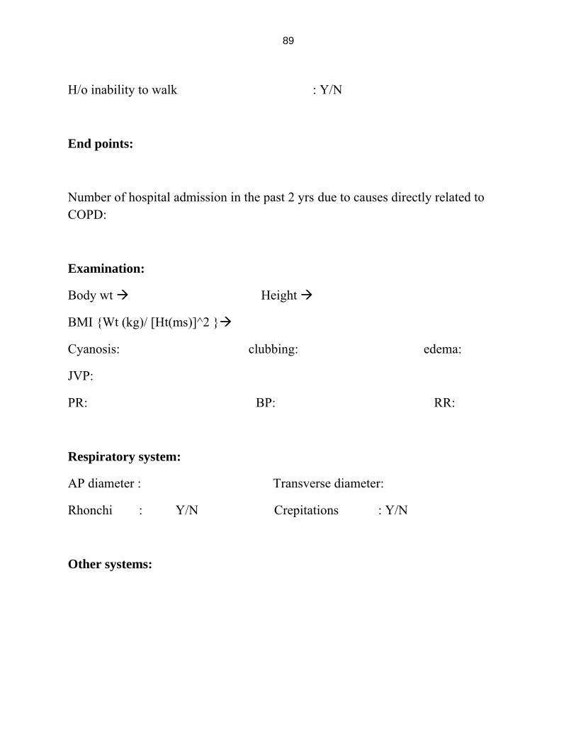
89
H/o inability to walk : Y/N
End points:
Number of hospital admission in the past 2 yrs due to causes directly related to COPD:
Examination:
Body wt Height
BMI {Wt (kg)/ [Ht(ms)]^2 }
Cyanosis: clubbing: edema:
JVP:
PR: BP: RR:
Respiratory system:
AP diameter : Transverse diameter:
Rhonchi : Y/N Crepitations : Y/N
Other systems:

90
Investigations:
Hb – TC DC
ECG QRS AXIS – P pulmonale
Echo EF – PAH – RV diameter –
6 min walk test >350 ms / 250 – 349 / 150 – 249 / <149
FEV1 >/= 65 / 50 – 64 / 36 – 49 / < /= 35
FEV1/FVC
Post bronchodialator FEV1
CRP
LFT total prot : albumin :
Estimated BODE score score
• FEV1 • Distance walked in 6 mins • MMRC dyspnea scale • BMI
------------
* Total BODE score

91
MMRC dyspnea scale
Grade 0 – no dyspnea / only on severe exertion
Grade 1 – dyspnea on hurrying / walking up a hill
Grade 2 – walks slower than normal at level/ pause while walking on level ground
Grade 3 – stops for breath after walking 100 yards/few mins on level ground
Grade 4 – too breathless to leave the house/ dyspnea on dressing
BODE INDEX:
BODE score 0 1 2 3
FEV1 >/= 65 % 50 – 64% 36 – 49% < /= 35%
6 min walk test
>350 ms 250 – 349ms 150 – 249 ms <149 ms
Dyspnea scale
0 – 1 2 3 4
BMI > 21 kg/m2 <21 kg/m2
Mild COPD 0 – 2
Moderate COPD 3 – 5
Severe COPD >/= 6

92
A study on BODE index as a predictor of severity and systemic involvement in patients with chronic obstructive pulmonary disease Dr Sojan George K
Institute of Internal Medicine, MMC, Chennai 3

93
ABBREVIATIONS
COPD Chronic obstructive pulmonary disease
FEV1 forced expiratory volume in one second
BODE body-mass index (B), airflow obstruction (O), dyspnea(D), and exercise
capacity (E)
GOLD Global initiative for obstructive lung diseases
ATS American Thoracic Society
ERS European Respiratory Society
RV Residual Volume
FRC Forced Respiratory Capacity
TV Tidal Volume
α 1 AT Alpha 1 antitrypsin
HNE Neutrophil elastase
BCM Body cell mass
FFM Fat free mass
BMI Body mass index
IFN Interferon
CRP C reactive Protein