bmc oral health article
TRANSCRIPT

RESEARCH ARTICLE Open Access
Three/four-dimensional (3D/4D)microscopic imaging and processingin clinical dental researchPing Ye1,2*, Hong Yu3 and Mojgan Houshmandi1
Abstract
Background: Confocal laser scanning microscope (CLSM) has been widely employed in our laboratory for structuraland functional analysis of clinical dental specimens and live cell imaging of cultured oral epithelial cells.
Methods: In this vitro study, a Fluoview 1000 (Olympus) confocal system was utilised to study thick sections ofcarious lesions (40–100 μm) and periodontal disease tissue samples (20–40 μm) by 2D Z stacking imaging and3-dimentional (3D) reconstruction. Four-dimensional (4D) imaging when including time or position points wasused for live cells to assess penetration/localisation/co-localization of oral pathogen proteins and therapeutic drugs.
Results: Three-dimensional (3D) reconstruction revealed latent features of carious hard tissues (strongly expressedamelogenin proteins in dentin tubules), and soft tissues (increased glial markers GFAP and S100B in pulpcomponents). We also found the oral microbial specific pathogens, Porphyromonas gingivalis to be widely localisedinside the periodontal pocket epithelial tissues as detected by 3D reconstruction from a series of 2D sections fromperiodontal disease tissue samples. 4D live cell imaging showed the diffusion patterns of fluorescent molecules inresponse to a bacterial virulence factor, the pathogen (gingipain haemagglutinin) domain that attacked epithelialintegrity. This technology also showed uptake of a novel porphyrin-linked metronidazole antibiotic into epithelialcells to kill intracellular oral pathogen, P. gingivalis.
Conclusions: Three/four-dimensional (3D/4D) imaging and processing in confocal microscopy is of great interestand benefit to clinical dental researchers.
Keywords: 3D reconstruction, 4D imaging, Live cell imaging, Carious lesion, Periodontitis, Porphyromonas gingivalis
Abbreviations: 3D, Three dimension (XYZ); 4D, Four dimension (XYZT or XYZP); CLSM, Confocal laser scanningmicroscope; EDTA, Ethylenediaminetetraacetic acid; GFAP, Glial fibrillary acidic protein; P. gingivalis, Porphyromonasgingivalis; PBS, Phosphate-buffered saline; S100B, S100 calcium-binding protein B; XYZP, 3D (XYZ) plus positionpoints; XYZT, 3D (XYZ) plus time lapse
BackgroundConfocal laser scanning microscope (CLSM) has beendeveloped and improved enormously over the past10 years. This powerful technology has several advan-tages over conventional epi-fluorescence microscopy,including improved performance in contrast and freeout-of-focus blur for thin or thick specimens [1],
excellent high resolution and analysis of fluorescent la-belled thick specimens without physical sectioning [1, 2],capacity for complex three-dimensional (XYZ) architec-ture called 3D reconstruction [2], and 4D imaging andprocessing on live cells when including time (XYZT) orposition (XYZP) points [3], and even 5D imaging whenincluding numerous channels [3]. These images havevolumetric and texture details and it is impossible to ob-tain such details with conventional microscopes.The technology enables to capture thin optical sections
from thick specimens with controllable depth of field toproduce 2D z-stack images through a three-dimensional
* Correspondence: [email protected] of Dental Research, Oral Health, Westmead Hospital, Westmead,Australia2Affiliation of Faculty of Dentistry, the University of Sydney, Sydney, AustraliaFull list of author information is available at the end of the article
© 2016 The Author(s). Open Access This article is distributed under the terms of the Creative Commons Attribution 4.0International License (http://creativecommons.org/licenses/by/4.0/), which permits unrestricted use, distribution, andreproduction in any medium, provided you give appropriate credit to the original author(s) and the source, provide a link tothe Creative Commons license, and indicate if changes were made. The Creative Commons Public Domain Dedication waiver(http://creativecommons.org/publicdomain/zero/1.0/) applies to the data made available in this article, unless otherwise stated.
Ye et al. BMC Oral Health (2016) 16:84 DOI 10.1186/s12903-016-0282-0

(3D) object with accurate information, allowing 3Dreconstructions to be generated with the digital datafrom 2D z-stack images. Processing turns 2D imagesinto a 3D image [1, 2], revealing latent features ofspecimens from carious lesions and inflamed gingivaltissues.Confocal microscopy is a widely applied tool for
studying the functions and activities of live cellsthrough a time-change, dye diffusion, and concentrationof fluorescent-labeled substances, and also studying forthe cellular functions of drug applicants. For live cell im-aging, confocal optics provides a major improvement indimensional resolution and real time [4].Most microscopic samples are essentially transparent,
and the depths of fields of focused samples are excep-tionally narrow. Therefore, another advantage of con-focal laser scanning microscope (CLSM) is the capacityto distinguish between different depths of a sample,which is free of out-of-focus blur for thin or thick speci-mens [1]. This process can provide good quality highresolution images for publishing.In this vitro study, overviews of four applications
for 3D/4D microscopic imaging and processing inclinical dental research were introduced. We utilisedthe Fluoview 1000 Olympus confocal system to study3D reconstruction for thick sections (up to 100 μm)of dental pulp, 3D reconstruction for oral pathogensin periodontitis tissues, 4D imaging and processing oflive cells including time (XYZT) or position (XYZP)to assess proteins of oral pathogen, and confocal mi-croscopy application in drug development.
MethodsCarious teethThe detailed processing of teeth has been describedin the previous paper [5]. Briefly, healthy (n = 15)and carious teeth (n = 37) were obtained with ap-proval of the Ethics Committee of Sydney WestLocal Health Service and informed consent frompatients, aged from 20 to 45 years. The half toothcontained the pulp was fixed in 1 % paraformalde-hyde in PBS (phosphate-buffered saline) for 3 h,then washed in PBS and EDTA (ethylenediaminetet-raacetic acid, pH 7.0) for 5 or 6 days, changedEDTA each day and equilibrated in 30 % sucrose(w/v) for 3 to 5 days at 4 °C. A diamond disc(Thin-Flex, Abrasive Technology, Chicago, IL) wasused to trim enamel and the majority of dentin.Samples were cooled down with ample water. Specimenswere in cryo-embedding matrix -Tissue Freezing Medium(Triangle Biomedical Sciences, Durham, NC) for quickchill (5 min), and frozen in liquid nitrogen. Sections of40–100 μm were prepared and stored at −80 °C untilrequired.
Gingival tissuesThe detailed processing of gingival tissue specimens hasbeen described in the previous paper [6]. Briefly, ob-tained gingival tissues were approved by the EthicsCommittee of Sydney Dental Hospital and informedconsent from adult participants (n = 26) in the periodon-tal clinics. All patients had detailed clinical records andradiographs with no systemic disease and no periodontaltherapy for the past 3 years. Tissues were grouped byclinical and histological criteria as clinically healthy gin-gival sites and paired periodontitis sites. Gingival tissueswere snap-frozen in isopentane, cooled in liquid nitro-gen, and 20–40 μm sections prepared for study.
Oral epithelial cell culture for live/fixed cell imagingThe H413 epithelial cell line original from a human oralsquamous cell carcinoma [7] exhibits stratified epithelialcell morphology and high CD24 marker expression inculture. Cell clonal lines of H413 were constructed usinga limit dilution method as described previously [8].H413 colone-1 cells were cultured in Joklik modifica-tion’s minimum essential medium (Sigma-Aldrich), sup-plemented with penicillin/streptomycin (100 IU/ml,Sigma) and 10 % fetal calf serum (FCS, CSL Limited,Victoria, Australia) at 37 °C in 5 % CO2 [9]. Cultureswere collected with trypsin replacement - triple express(Invitrogen, Australia) in PBS and sub-cultured every3 days.To further confirm biological function, live cell im-
aging was performed to determine intracellular versusparacellular pathways of movement of labeled dextran.Cloned H413-1 cells (2 × 10⁵/cm2) were cultured in 8-wellslide chambers (ibidi, cat 80826, Germany), 300 μl perwell. Confluence was achieved within 48 h. A haemolyticgingipain adhesin domain K2 (100 pM) from Porphyromo-nas gingivalis [10] or a gingipain haemagglutinin sub-domain named Ka 100 pM (constructed in our laboratory,unpublished) was added and each well was simultaneouslysupplemented with a low molecular weight dextran AlexaFluor 647 (10 kDa, Invitrogen, Australia) at 1:50 dilutionfrom a stock solution of 1 mg/ml in medium.For drug development assay, oral pathogen P.gingivalis
strain ATCC 33277 at MOI (multiplicity of infection)100 cells per one epithelial cell [11] was added toconfluent H413 clone-1 epithelial monolayers for1.5 h at 37 °C in 5 % CO2. Monolayers were washedtwice with Dulbecco’s phosphate-buffered saline (DPBS)[12]. Gentamycin (300 μg/ml, Sigma) and metronidazole(200 μg/ml, Sigma) were added to kill adherent P.gingiva-lis on the cell surface (extracellular bacteria, [12]). Afteran additional 1 h incubation and washing off antibiotics,we added a porphyrin-linked metronidazole adduct(40 μM) developed in our laboratory (auto-fluorescence inred, [13]) to cell culture up to additional 1.5 h. Then P.
Ye et al. BMC Oral Health (2016) 16:84 Page 2 of 9

gingivalis was targeted by mouse monoclonal antibodyIIB2 (5 μm/ml) [14] at various time points. For livecells, nuclei were stained with NucBlue (Invitrogen,Australia). For fixed cells, the secondary antibody wasgoat anti-mouse conjugated with Alexa Fluor 488(Life Technologies, USA).
Immunostaining for frozen sectionsFrozen sections including teeth and gingival tissues werefixed in 4 % paraformaldehyde in PBS for 30 min atroom temperature. Slides were washed with PBS andplaced in glycine-PBS for 10 min, then washed in PBSand incubated in PBS containing 0.2 % Tween-20 and10 % goat serum for 1 h at room temperature. Sectionswere washed in PBS and incubated with primary anti-bodies: polyclonal rabbit anti-human GFAP (5 μg/ml,Dako), polyclonal rabbit anti-human S100 (4 μg/ml, reactsstrongly with human S100B, Dako), polyclonal rabbit anti-human amelogenin (5 mg/ml, Abcam, UK), and mousemonoclonal antibody IIB2 (5 μm/ml) [14] to P. gingivalisbacteria, for 1 h at room temperature. For controls, rabbitor mouse IgG (Dakocytomation) was served as the pri-mary antibody. Sections were washed 3 times in PBS andthen incubated with secondary antibodies: goat anti-rabbitor mouse IgG conjugated with Alexa Fluor 488 or 594(Life Technologies, USA) for 1 h at room temperature.Slides were mounted in Prolong gold anti-fade reagentwith DAPI (Molecular Probes, Invitrogen).
Confocal laser scanning microscopyAn Olympus Fluoview (FV) 1000 was used to captureconfocal images under multi lasers (405 nm, 473 nm,633 nm) and NTT electronic Optiλ (559 nm). Visual ob-servation under the objective lens [Olympus 40X/1.30/0.20 (WD) Oil UPLSAPO] was performed. The cellswere selected at random and adjusted focus beforeimage acquisition.For 3D reconstruction imaging on thick teeth and gin-
gival tissue sections, the fundamental step was in stain-ing of the preparations. An appropriate stained sectionshowed the same intensity from the top to the bottom ofthe stack. Over-stained or under-stained sections wouldgreatly reduce imaging clarity of thick teeth and gingivaltissues. Z-stack imaging was performed with a 40X ob-jective using up to three corresponding lasers (405 nm,473 nm and 559 nm). Briefly, the procedure was to setup step size between 0.5 and 1.5 μm depending on thethickness of samples, and then consecutive cross-sectionimages (XYZ) were acquired from the top to the bottom.After acquisition, projection of z-stack images was dis-played using the click Z mode. 3D reconstruction anddisplay of cubic imaging were built up by 3D OlympusFluoview software. It was essential for 3D reconstruc-tions achieved identical to any z-stack preparation [15].
For 4D (3D time lapse) imaging on live cells, chamberslides with stained samples were operated in the micro-scope stage incubation chamber (with temperature con-troller at 37 °C and CO2 controller at 5 % of CO2) [16].Time-lapse imaging was performed over a period ofhours with a 40X objective using the time-lapse functionof confocal microscopy Auto Imaging System. Two cor-responding lasers (405 nm and 633 nm) were selected.Combined z-stack and time-lapse functions were used inthe present study to swiftly capture the images [16]. Ser-ial images were taken at regular time points to capturethe dynamics.All fluorescence images captured with confocal acqui-
sition software (FV10-ASW 1.7) were stored as OlympusImage Format (OIF) for signal analysis and exported im-ages as TIF files.
Imaging analysisThree-dimensional (3D) reconstruction images were proc-essed using Olympus Fluoview (FV) software (4.2 viewer,Japan) or Image J software (version 1.50). Co-localizationimage analysis was as described in reference papers [17,18] or by using recently developed advanced software:Huygens Professional (https://svi.nl/HuygensProfessional).
ResultsThree-dimensional reconstruction for thick carious lesionsSpecimens used in this study were soft pulp tissues con-nected with hard dentin tissues (Fig. 1a). 2D images withan overlay of the differential interference contrast (DIC)channel image of a healthy control tooth were displayedscant expression of GFAP and S100B (Fig. 1b). For com-parison, a matched set of 2D images from a carioustooth was shown in Fig. 1c. Figure 1d showed projectionof 2D stack images from a carious tooth (Fig. 1c) usingfrozen (40–100 μm) sections, 40–50 images of 0.8–1.5 μm step size acquired as z-stacks by confocal laserscanning microscopy. Three-dimensional (3D) imagingfrom 2D z-stack images (Fig. 1d) was reconstructed andviewed using confocal software (4.2 viewer, Japan) (Fig. 1e).A 3D movie in the Additional file 1: Movie 1 revealed la-tent features of glial cell markers GFAP (green) and S100B(red) from thick specimens.Another example was distribution of amelogenin pro-
tein (green) expressed in carious adult human teeth al-though not detected in healthy adult human teeth.Figure 2a showed 2D images of a healthy control toothdisplaying trace expression of amelogenin protein con-sidered as background staining compared to isotype con-trol antibody staining (data not shown). An overlayimage using the DIC (differential interference contrast)channel showed anatomical structure and orientation ofthe tooth. For comparison, a matched set of 2D imagesfrom a tooth with a carious lesion was shown (Fig. 2b).
Ye et al. BMC Oral Health (2016) 16:84 Page 3 of 9
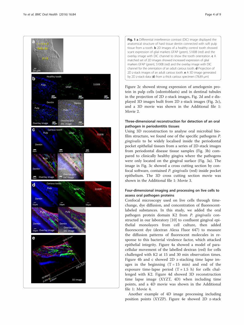
Figure 2c showed strong expression of amelogenin pro-tein in pulp cells (odontoblasts) and in dentinal tubulesin the projection of 2D z-stack images, Fig. 2d and e dis-played 3D images built from 2D z-stack images (Fig. 2c),and a 3D movie was shown in the Additional file 1:Movie 2.
Three-dimensional reconstruction for detection of an oralpathogen in periodontitis tissuesUsing 3D reconstruction to analyse oral microbial bio-film structure, we found one of the specific pathogens P.gingivalis to be widely localised inside the periodontalpocket epithelial tissues from a series of 2D-stack imagesfrom periodontal disease tissue samples (Fig. 3b) com-pared to clinically healthy gingiva where the pathogenswere only located on the gingival surface (Fig. 3a). Theimage in Fig. 3c showed a cross cutting section by con-focal software, contained P. gingivalis (red) inside pocketepithelium. The 3D cross cutting section movie wasshown in the Additional file 1: Movie 3.
Four-dimensional imaging and processing on live cells toassess oral pathogen proteinsConfocal microscopy used on live cells through time-change, dye diffusion, and concentration of fluorescent-labeled substances. In this study, we added the oralpathogen protein domain K2 from P. gingivalis con-structed in our laboratory [10] to confluent gingival epi-thelial monolayers from cell culture, then addedfluorescent dye (dextran Alexa Fluor 647) to measurethe diffusion patterns of fluorescent molecules in re-sponse to this bacterial virulence factor, which attackedepithelial integrity. Figure 4a showed a model of para-cellular movement of the labelled dextran (red) for cellschallenged with K2 at 15 and 30 min observation times.Figure 4b and c showed 2D z-stacking time lapse im-ages in the beginning (T = 15 min) and end of theexposure time-lapse period (T = 1.5 h) for cells chal-lenged with K2. Figure 4d showed 3D reconstructiontime lapse image (XYZT, 4D) when including timepoints, and a 4D movie was shown in the Additionalfile 1: Movie 4.Another example of 4D image processing including
position points (XYZP). Figure 4e showed 2D z-stack
a
b
c
d
e
Fig. 1 a Differential interference contrast (DIC) image displayed theanatomical structure of hard tissue dentin connected with soft pulptissue from a tooth. b 2D images of a healthy control tooth showedscant expression of glial markers GFAP (green), S100B (red) and theoverlay image with DIC channel to show the tooth orientation. c Amatched set of 2D images showed increased expression of glialmarkers GFAP (green), S100B (red) and the overlay image with DICchannel for the orientation of an adult carious tooth. d Projection of2D z-stack images of an adult carious tooth. e A 3D image generatedby 2D z-stack data (d) from a thick carious specimen (78.89 μm)
Ye et al. BMC Oral Health (2016) 16:84 Page 4 of 9

image in which P. gingivalis proteins Ka (green) pene-trated into nuclei (red) after 30 min on live cells.One of the cells was picked from 2D z-stack imagesto build up a 3D image (XYZ Plus position point,Fig. 4f ). A 4D movie was shown in the Additional file 1:Movie 5.
Confocal laser scanning microscopy in drug developmentUsing confocal microscopy, it was possible to identifythe penetration/localization/co-localisation of aporphyrin-linked metronidazole antibiotic and P. gingi-valis bacteria. The porphyrin adducts exhibiting auto-fluorescence in the red range of wavelengths (600–
a
b
c
e
d
Fig. 2 Another example of amelogenin protein strongly re-expressed in newly differentiated pulp cells (odontoblasts) and distributed in dentinaltubules under the lesion site. a 2D images of a healthy control tooth, b a matched set of 2D images was from an adult carious lesion. c Projectionof 2D z-stack images from a carious tooth, d a 3D reconstruction image from (c), e a 3D image plus x and z axes projection
Ye et al. BMC Oral Health (2016) 16:84 Page 5 of 9

650 nm), penetrated into the cells and localized withinthe cytoplasm (Fig. 5a). Immuno-stained P. gingivalispotent gingipain-RgpA was shown in Fig. 5b–d in greenfluorescence, also localized within cytoplasm. This modi-fied drug could kill oral intracellular pathogens P. gingi-valis with a time course at 30 min (Fig. 5b), 1 h (Fig. 5c)and 1.5 h (Fig. 5d).
DiscussionWhy do we need confocal microscopy as a visual toolfor clinical dental research? As noted previously, con-focal microscopy has several advantages particularly for3D visualization on thick specimens to reveal latent fea-tures of cell and tissue structures. The present studydemonstrates that latent patterns of the increased glialcell markers GFAP and S100B on carious teeth lesionscan be revealed through 3D reconstruction from thickspecimens. The increased abundance of these markers incarious teeth lesions indicates a response to initial mi-crobial invasion of dentin [5]. In the current study, 3Dreconstruction also reveals strong expression of amelo-genin protein in pulp cells (odontoblasts) and in dentinaltubules from adult human teeth with carious lesions. Ina normal condition, this amelogenin protein is onlyappeared during tooth embryonic development includ-ing expression in tooth enamel, dentin, and pulp cells
(odontoblasts) [19]. When tooth becomes mature, ame-logenin is absent in dentin and pulp cells (odontoblasts).However, in injured or carious teeth amelogenin proteinis highly re-presented in newly differentiated pulp cells(odontoblasts) and allotted in the dentinal tubules underthe adult carious teeth [19] corresponding to ourfindings.Dental researchers face major issues when cutting thin
sections of soft pulp tissues connected with hard dentintissues. Therefore, we utilise the advantage of analysis offluorescent labeled thick specimens without physical sec-tioning for carious teeth and thick gingival tissues.Confocal laser scanning microscopy (CLSM) is a use-
ful method to study the interface of bacteria within bio-films and inflamed gingival tissues. Images obtainedfrom CLSM are free of out-of-focus blur. CLSM-basedimaging platform has been widely used for analysis oforal bacterial pathogens or biofilm structures on gingivaltissues [20–23]. The images shown in the present studyillustrate that 3D visualization is a promising method forrepresentative analysis of oral bacterial pathogens or bio-film components [20].In the present confocal microscopy study, using 4D
(3D time lapse) imaging and processing on live cells wedemonstrate that time-lapse imaging is a powerful tech-nique for analysis of dynamic cellular events and
a
c
b
Fig. 3 Example of 3D reconstruction to analyse oral microbial biofilm structure. One of the specific pathogens P. gingivalis (green colour) wasfound to be widely localised on the surface of clinically healthy gingiva (a), but inside the periodontal pocket epithelial tissue from a series of 2Dz-stack images (b) and one of 3D cross cutting sections, P. gingivalis (red colour) (c)
Ye et al. BMC Oral Health (2016) 16:84 Page 6 of 9

morphology in real time and real space. There are twoissues that researchers need to be aware of while per-forming experiments on live cells. Firstly, how to choosethe right dye? The excitation (absorption, short wave-length) and emission (long wavelength) spectra of theAlexa Fluor series cover the visible spectrum and extendto the invisible infrared [24]. The closer to infraredwavelengths, the less damage to live cells [25], therefore,the far-red dye of Alexa Fluor 647 was chosen in thecurrent study.
Secondly, illumination by fluorophores can causephoto bleaching and cell damage, hence there is a needto raise the speed of image acquisition by confocal mi-croscopy. However the confocal microscopy has a limita-tion of raising an acquisition speed, which can result ina low resolution of image [26]. This image resolutioncan be improved by the use of DeltaVision super-resolution microscope (GE Healthcare, Japan). DeltaVi-sion microscopy can offer better performance at highmagnification (100 times), absolutely high resolution and
d fe
a
Control cells Cells challenged with bacterial proteins
c
b
T = 1.5 hT = 1.5 h, overlay
T = 15 min, overlay T = 15 min
3D time lapse (1.0 1.5 h)
T = 15 min T = 30 min T = 15 min T = 30 min
Fig. 4 a A model of adding oral pathogen P. gingivalis proteins K2 and fluorescent dye (Alexa Fluor dextran 647, red colour) to confluent gingivalepithelial monolayers, to track pathological changes of epithelial integrity at a time course. Paracellular pathway of movement of the labelleddextran (red) was shown at 15 and 30 min observation times. b and c 2D z-stacking images in the beginning (T = 15 min) and end of theexposure time-lapse period (T = 1.5 h): left panels were the overlay images of cells (green nuclei) with far-red dye channel; right panels werefar-red dye channel only. d 3D reconstruction time lapse images from (b-c) on live cells challenged with bacterial protein (K2) (green colour: cellnuclei; red colour: dye). e Another example of projection of 2D z-stack imaging plus a position point. f A 3D reconstruction image with a positionpoint (e) on live cells challenged with P. gingivalis bacterial protein (Ka) at 30 min (green colour: Ka protein; red colour: nuclei)
Ye et al. BMC Oral Health (2016) 16:84 Page 7 of 9

contrast through de-convolution of images, especially onlive cell imaging where faster image acquisition, lowerexcitation power, and less photo bleaching and cell dam-age [27, 28]. This is a key consideration which can beimproved by DeltaVision microscopy in the future studyon live cells [27, 28].Using confocal microscopy in the present drug devel-
opment study, has determined that a novel porphyrin-linked metronidazole antibiotic can penetrate into epi-thelial cells to kill intracellular oral pathogens. However,researchers need to be aware of that an overlap in fluor-escence does not necessarily demonstrate co-localizationof two colours (probes) in the same cellular structure[17]. Therefore the co-distribution of two colours (probes)in fluorescence microscope images can be evaluated quan-titatively and statistically on either co-localisation or two
colours’ ratio change using Huygens professional software(https://svi.nl/HuygensProfessional). For the images shownin Fig. 5, we could explain it as the ratio change of two col-ours which bacterial spots in green were decreasing with atime lapse comparing to drugs in red colour.
ConclusionsConfocal laser scanning microscope is a valuable tool indental research, particularly for probing thick specimensof carious teeth, formation of microbial biofilms ongingival tissues, tracing pathological changes in live cellsand studying the cellular effects of drug candidates. Fu-ture developments of powerful microscopy will be ofgreat interest and benefit in clinical dental research.
Additional file
Additional file 1: Movie 1–5 (PPTX 2493 kb)
AcknowledgementsWe would like to thank Professor Neil Hunter and Ms Mary Simonian fortheir technical and laboratory supports.
FundingThere was no finding obtained for this study.
Availability of data and materialsAll data and materials have been supported by our previous studies andcited in this manuscript where necessary. The images and videos processingof 3D/4D reconstruction, including the additional file used licensed OlympusFluoview (FV) software (4.2 viewer, Japan). Findings reported in thismanuscript can be shared to public.
Authors’ contributionsPY participated in the experimental design and data analysis, andcontributed to the draft of manuscript. HY provided confocal microscopysupport for the study. MH performed immunohistology work and dataanalysis. The authors reviewed and approved the final manuscript.
Competing interestsThe authors declare that they have no competing interests.
Consent for publicationNot applicable, as this manuscript does not contain any individual persons’identifying information.
Ethics approval and consent to participateEthical approvals for data and sample collection from the patients have beenobtained by Ethics Committees of the Sydney Dental Hospital and theSydney West Area Health Service. All data and samples from the patientswere collected following informed written consent. All the informationincluding personal details, images and videos of patients’ teeth and gingivaltissues that have been used for this study were de-identified.
Author details1Institute of Dental Research, Oral Health, Westmead Hospital, Westmead,Australia. 2Affiliation of Faculty of Dentistry, the University of Sydney, Sydney,Australia. 3Microscopy Laboratory, Westmead Institute for Medical Research,Westmead, Australia.
Received: 26 April 2016 Accepted: 20 August 2016
c
20µm
d
20µm
20µm
a
b
20µm
Fig. 5 a A pathogen-specific antimicrobial compound (a porphyrin-linkedmetronidazole) developed in our laboratory [13] which can targetintracellular oral pathogens P.gingivalis displayed in red auto-fluorescence.Left: nuclei staining with DAPI (blue channel), middle: an overlay image ofdrug (red channel) with nuclei (blue channel), and right image was drugonly (red channel). Images showed that this modified drug penetrated intothe cells and localized within the cytoplasm (red) to kill oral intracellularpathogens P. gingivalis (green) with a time course at 30 min (b), 1 h (c) and1.5 h (d). From (b–d), left panels showed green colour bacteria P. gingivalis;middle panels showed the overlay images of cells' nuclei (blue channel),P.gingivalis (green channel) and drug (red channel); right panels showed theoverlay images of P.gingivalis (green channel) and drug (red channel) only
Ye et al. BMC Oral Health (2016) 16:84 Page 8 of 9

References1. Shotton D, White N. Confocal scanning microscopy: three-dimensional
biological imaging. Trends Biochem Sci. 1989;14:435–9.2. Wright SJ, Schatten G. Confocal fluorescence microscopy and three-
dimensional reconstruction. J Electron Microsc Tech. 1991;18:2–10.3. Roux P, Münter S, Frischknecht F, Herbomel P, Shorte SL. Focusing light on
infection in four dimensions. Cell Microbiol. 2004;6:333–43.4. Zemanová L, Schenk A, Valler MJ, Nienhaus GU, Heilker R. Confocal optics
microscopy for biochemical and cellular high-throughput screening. DrugDiscov Today. 2003;8:1085–93.
5. Houshmandi M, Ye P, Hunter N. Glial network responses to polymicrobialinvasion of dentin. Caries Res. 2014;48:534–48.
6. Guo W, Ye P, Yu H, Liu Z, Yang P, Hunter N. CD24 activates the NLRP3inflammasome through c-Src kinase activity in a model of the liningepithelium of inflamed periodontal tissues. Immun Inflamm Dis. 2014;2:239–53.
7. Prime SS, Nixon SV, Crane IJ, Stone A, Matthews JB, Maitland NJ, Remnant L,Powell SK, et al. The behaviour of human oral squamous cell carcinoma incell culture. J Pathol. 1990;160:259–69.
8. Ye P, Nadkarni MA, Hunter N. Regulation of Ecadherin and TGF-beta3expression by CD24 in cultured oral epithelial cells. Biochem Biophys ResCommun. 2006;349:229–35.
9. Swierenga SH, MacManus JP. Preparation of low calcium growth mediumsuitable for determination of tumorigenicity of cultured cells. J TissueCulture Methods. 1982;7:1–3.
10. Li N, Yun P, Nadkarni MA, Ghadikolaee NB, Nguyen KA, Lee M, Hunter N, CollyerCA. Structure determination and analysis of a haemolytic gingipain adhesindomain from Porphyromonas gingivalis. Mol Microbiol. 2010;76:861–73.
11. Ye P, Harty D, Commandeur Z, Hunter N. Binding of Streptococcus gordoniito oral epithelial monolayers increases paracellular barrier function. MicrobPathog. 2013;56:53–9.
12. Nisapakultorn K, Ross KF, Herzberg MC. Calprotectin expression in vitro byoral epithelial cells confers resistance to infection by Porphyromonasgingivalis. Infect Immun. 2001;69:4242–7.
13. Yap BC, Simpkins GL, Collyer CA, Hunter N, Crossley MJ. Porphyrin-linkednitroimidazole antibiotics targeting Porphyromonas gingivalis. Org BiomolChem. 2009;7:2855–63.
14. DeCarlo AA, Paramaesvaran M, Yun PL, Collyer C, Hunter N. Porphyrin-mediated binding to hemoglobin by the HA2 domain of cysteineproteinases (gingipains) and hemagglutinins from the periodontalpathogen Porphyromonas gingivalis. J Bacteriol. 1999;181:3784–91.
15. Kopecky BJ, Duncan JS, Elliott KL, Fritzsch B. Three-dimensionalreconstructions from optical sections of thick mouse inner ears usingconfocal microscopy. J Microsc. 2012;248:292–8.
16. Attik GN, Gritsch K, Colon P, Grosgogeat B. Confocal time lapse imaging asan efficient method for the cytocompatibility evaluation of dentalcomposites. J Vis Exp. 2014;93:e51949.
17. Dunn KW, Kamocka MM, McDonald JH. A practical guide to evaluatingcolocalization in biological microscopy. Am J Physiol Cell Physiol.2011;300:C723–42.
18. Costes SV, Daelemans D, Cho EH, Dobbin Z, Pavlakis G, Lockett S. Automaticand Quantitative Measurement of Protein-Protein Colocalization in LiveCells. Biophys J. 2004;86:3993–4003.
19. Mitsiadis TA, Filatova A, Papaccio G, Goldberg M, About I, Papagerakis P.Distribution of the amelogenin protein in developing, injured and carioushuman teeth. Front Physiol. 2014;5:477.
20. Karygianni L, Follo M, Hellwig E, Burghardt D, Wolkewitz M, Anderson A, Al-Ahmad A. Microscope-Based Imaging Platform for Large-Scale Analysis ofOral Biofilms. Appl Environ Microbiol. 2012;78:8703–11.
21. Thurnheer T, Belibasakis GN, Bostanci N. Colonisation of gingival epitheliaby subgingival biofilms in vitro: Role of “red complex” bacteria. Arch OralBiol. 2014;59:977–86.
22. Rudney JD, Chen R, Sedgewick GJ. Intracellular Actinobacillusactinomycetemcomitans and Porphyromonas gingivalis in Buccal EpithelialCells Collected from Human Subjects. Infect Immun. 2001;69:2700–7.
23. Zhang B, Elmabsout AA, Khalaf H, Basic VT, Jayaprakash K, Kruse R, Bengtsson T,Sirsjö A. The periodontal pathogen Porphyromonas gingivalis changes the geneexpression in vascular smooth muscle cells involving the TGFbeta/Notchsignalling pathway and increased cell proliferation. BMC Genomics. 2013;14:770.
24. Sowell J, Strekowski L, Patonay G. DNA and protein applications of near-infrared dyes. J Biomed Opt. 2002;7:571–5.
25. Dempsey GT. A user’s guide to localization-based super-resolutionfluorescence imaging. Methods Cell Biol. 2013;114:561–92.
26. Laurent M, Johannin G, Gilbert N, Lucas L, Cassio D, Petit PX, Fleury A.Power and limits of laser scanning confocal microscopy. Biol Cell.1994;80:229–40.
27. Schermelleh L, Heintzmann R, Leonhardt H. A guide to super-resolutionfluorescence microscopy. J Cell Biol. 2010;190:165–75.
28. Ball G, Parton RM, Hamilton RS, Davis I. A cell biologist’s guide to highresolution imaging. Methods Enzymol. 2012;504:29–55.
• We accept pre-submission inquiries
• Our selector tool helps you to find the most relevant journal
• We provide round the clock customer support
• Convenient online submission
• Thorough peer review
• Inclusion in PubMed and all major indexing services
• Maximum visibility for your research
Submit your manuscript atwww.biomedcentral.com/submit
Submit your next manuscript to BioMed Central and we will help you at every step:
Ye et al. BMC Oral Health (2016) 16:84 Page 9 of 9