bmc ophthalmology biomed central - … · and 0.03% triton x-100 (tx-100). cultures were rinsed in...
TRANSCRIPT

BioMed CentralBMC Ophthalmology
ss
Open AcceResearch articlePalm is expressed in both developing and adult mouse lens and retinaMeryl Castellini1,4, Louise V Wolf2, Bharesh K Chauhan2,5, Deni S Galileo1, Manfred W Kilimann3, Ales Cvekl2 and Melinda K Duncan*1Address: 1Department of Biological Sciences, University of Delaware, Newark, DE 19716 USA, 2Depts. of Ophthalmology and Visual Sciences and Molecular Genetics, Albert Einstein College of Medicine, Bronx, NY 10461 USA, 3Department of Cell and Molecular Biology, Uppsala University, S-75124 Uppsala Sweden, 4Department of Pathology, Anatomy, and Cell Biology, Thomas Jefferson University, Philadelphia, PA 19107 USA and 5Developmental Biology Division and Department of Ophthalmology, Children's Hospital Research Foundation, 3333 Burnet Avenue, Cincinnati, OH 45229 USA
Email: Meryl Castellini - [email protected]; Louise V Wolf - [email protected]; Bharesh K Chauhan - [email protected]; Deni S Galileo - [email protected]; Manfred W Kilimann - [email protected]; Ales Cvekl - [email protected]; Melinda K Duncan* - [email protected]
* Corresponding author
AbstractBackground: Paralemmin (Palm) is a prenyl-palmitoyl anchored membrane protein that can drivemembrane and process formation in neurons. Earlier studies have shown brain preferred Palmexpression, although this protein is a major water insoluble protein in chicken lens fiber cells andthe Palm gene may be regulated by Pax6.
Methods: The expression profile of Palm protein in the embryonic, newborn and adult mouse eyeas well as dissociated retinal neurons was determined by confocal immunofluorescence. Therelative mRNA levels of Palm, Palmdelphin (PalmD) and paralemmin2 (Palm2) in the lens and retinawere determined by real time rt-PCR.
Results: In the lens, Palm is already expressed at 9.5 dpc in the lens placode, and this expressionis maintained in the lens vesicle throughout the formation of the adult lens. Palm is largely absentfrom the optic vesicle but is detectable at 10.5 dpc in the optic cup. In the developing retina, Palmexpression transiently upregulates during the formation of optic nerve as well as in the formationof both the inner and outer plexiform layers. In short term dissociated chick retinal cultures, Palmprotein is easily detectable, but the levels appear to reduce sharply as the cultures age. Palm mRNAwas found at much higher levels relative to Palm2 or PalmD in both the retina and lens.
Conclusion: Palm is the major paralemmin family member expressed in the retina and lens and itsexpression in the retina transiently upregulates during active neurite outgrowth. The expressionpattern of Palm in the eye is consistent with it being a Pax6 responsive gene. Since Palm is knownto be able to drive membrane formation in brain neurons, it is possible that this molecule is crucialfor the increase in membrane formation during lens fiber cell differentiation.
Published: 21 June 2005
BMC Ophthalmology 2005, 5:14 doi:10.1186/1471-2415-5-14
Received: 01 March 2005Accepted: 21 June 2005
This article is available from: http://www.biomedcentral.com/1471-2415/5/14
© 2005 Castellini et al; licensee BioMed Central Ltd. This is an Open Access article distributed under the terms of the Creative Commons Attribution License (http://creativecommons.org/licenses/by/2.0), which permits unrestricted use, distribution, and reproduction in any medium, provided the original work is properly cited.
Page 1 of 11(page number not for citation purposes)

BMC Ophthalmology 2005, 5:14 http://www.biomedcentral.com/1471-2415/5/14
BackgroundThe retina and lens form from the neural tube and headectoderm respectively. Despite these different origins, thedevelopment of the mature eye requires mutually induc-tive interactions between these two cell layers [1]. Further,in many cases, the lens and retina express the same devel-opmentally important transcription factors [2-6]. In addi-tion, a number of studies have identified the expression ofproteins with known roles in neuronal function in thelens [7-12] and proteins important in lens function in theretina [13,14]. This may partially be due to the need ofboth retinal neurons and lens fiber cells to develop elabo-rated plasma membranes for their function [15-17].
Pax6 is a paired and homeodomain containing transcrip-tion factor that is required for the formation of the lensplacode from the head ectoderm [18]. Specific loss of Pax6expression from retinal progenitor cells results in the con-version of all retinal cell types to amacrine interneurons[19] and lens epithelial cells heterozygous for a Pax6mutation preferentially differentiate into lens fiber cells[20]. Overexpression of the canonical form of Pax6 in lensfiber cells (Pax6 con transgenics) results in cataracts typi-fied by incomplete lens fiber cell elongation and denucle-ation, instability of the transcription factor c-Maf and adrastic downregulation of βB1-crystallin expression [21]while overexpression of the Pax6 (5a) splice form alsoresults in cataracts without the changes in cMaf stability[22]. Microarray analysis was previously performed onlenses from both Pax6 (con) transgenics and mice hetero-zygous for a Pax6 null allele and 13 genes were found tobe upregulated in the transgenics and downregulated inthe heterozygous knockout mice [23].
One of these genes, paralemmin (Palm), encodes a proteinpresent at the plasma membrane in axons, dendrites andperikarya of differentiating neuronal cell lines, and at highlevels in the processes of the cerebellar molecular layer[24]. Further, this gene is downregulated in lenses overex-pressing the Pax6(5a) splice variant [25] and the proteinis detected in lens cells from both mice and chickens[25,26]. Overexpression of Palm in both neuronal andnon-neuronal cell lines initiates the expansion of theplasma membrane and the development of extendedprocesses and microspikes which is dependent on Palmtargeting to the cytoplasmic face of the plasma membranevia a palmitoyl group covalently linked near the protein'sC-terminus [24,27].
Here we investigate the distribution of Palm in the devel-oping lens and retina, and compare its mRNA levels withtwo other members of the paralemmin family, paralem-min-2 (Palm-2) and palmdelphin/paralemmin-like (PalmD)[28,29].
MethodsAnimalsAll experiments using animals were approved by the boththe University of Delaware and Albert Einstein College ofMedicine Institutional Animal Care Committees and con-form to the ARVO statement for the Use of Animals inOphthalmic and Vision Research. C57Bl/6 mice were gen-erated in-house from breeding stock obtained from Har-lan Sprague Dawley (Indianapolis, IN). CD-1 mice wereobtained directly from Charles River Laboratories (Wilm-ington, MA). Embryonic mice were staged by designatingnoon of the day on which a semen plug was observed inthe dam as 0.5 days post-coitum (dpc). Postnatal micewere staged by designating the day of birth as 1 day post-natal (DPN). All mice were maintained in a 12-hour light/dark cycle at 21–24°C and were given food and water adlibitum.
Immunofluorescent detection of Palm in tissue sectionsPalm was detected by indirect immunofluorescence fol-lowing the protocol previously described [30]. Briefly, tis-sue or embryos were excised from C57Bl/6 mice,embedded in tissue freezing media (TFM, Triangle Bio-medical Sciences, Durham, NC) and sectioned at 16 µMon a Leica CM 3050 S Cryostat (Leica, Deerfield, IL). Sec-tions were mounted on Colorfrost-plus™ slides (FisherScientific; Pittsburgh, PA), fixed in ice-cold acetone:meth-anol (1:1 vol/vol) for 15 minutes, dried and blocked with1% BSA in phosphate buffered saline (PBS), pH 7.4. Theblocking solution was removed and the sections incu-bated with a 1:150 dilution of rabbit polyclonal anti-Palmantibody [24] in 1% BSA-PBS for one hour at room tem-perature. The bound primary antibody was detected withAlexaFluor 568 goat anti-rabbit IgG (Molecular Probes,Inc. Eugene, OR) and cell nuclei were detected by counter-staining with TO-PRO-3 (1:3000 dilution in 1% BSA-PBS;Molecular Probes, Inc). Negative controls consisted ofparallel staining experiments that omitted the primaryantibody. Images were captured on a Zeiss LSM 510 Con-focal Microscope configured with an Argon/Krypton laser(488 nm and 568 nm excitation lines) and Helium Neonlaser (633 nm excitation line)(Carl Zeiss Inc, Göttingen,Germany).
Transfections and reporter assaysFour copies of the Pax6-binding site previously identifiedin the human PALM promoter [25] were cloned intoE4TATA-pGL3 [31] using a synthetic double stranded oli-gonucleotide 5'-ctagGGCTACTTTCACTCTGCGATGGCAGAGCAG-GGCTACTTTCACTCTGCGATGGCAGAGCA-3'. Nucle-otides containing Pax6-binding sites are in bold andnucleotides used for subcloning are indicated by lowercase letters. Transient transfection assays were performed
Page 2 of 11(page number not for citation purposes)

BMC Ophthalmology 2005, 5:14 http://www.biomedcentral.com/1471-2415/5/14
in 293T cells, which do not express endogenous Pax6 pro-teins, as described earlier [32].
Immunofluorescent detection of Palm in cultured chick retinaFertile White Leghorn eggs were obtained from theDepartment of Animal and Food Sciences at the Univer-sity of Delaware and kept in a humidified, forced-draftincubator until embryonic day (E) 7. Retinas were dis-sected in calcium and magnesium-free saline solution(CMF). The neural retina was separated from the pig-mented epithelium with fine forceps. The neural retinawas minced with fine scissors and incubated in 0.25%trypsin in CMF for 20 minutes at 37°C. Retinas were dis-sociated into single cells by trituration with a Pasteur pipetin a 0.3 mg/ml soybean trypsin inhibitor/ 0.03 mg/mlDNaseI in Medium 199 (Cellgro, Herndon, Virginia).Cells were plated at a density of 5 × 10⁵ cells / 12 mmdiameter round glass coverslip in wells of a 24-well platein one milliliter of Medium 199 (Cellgro) supplementedwith 10% fetal bovine serum. Retina cultures were kept ina standard humidified culture incubator with 5% CO2.
Two days or one week after plating, cultures were fixed in1% paraformaldehyde in PBS pH 7.4 for 30 minutes andthen rinsed in PBS. Cells were then incubated for approx-imately 1 hour in a mixture of 1:200 rabbit polyclonalanti-chicken Palm [26] and 1:2 mouse monoclonal anti-neurofilament (RT-97) hybridoma supernatant (Develop-mental Studies Hybridoma Bank, Iowa City, IA; [33,34])in PBS supplemented with 5% normal goat serum (NGS)and 0.03% Triton X-100 (TX-100). Cultures were rinsed inPBS and then incubated for approximately 1 hour in amixture of 1:200 Alexa 488-goat anti-rabbit and 1:200Alexa 594-goat anti-mouse secondary antibodies (bothfrom Molecular Probes, Inc., Eugene OR) in the PBS/NGS/TX-100 mixture. Cultures were then rinsed in PBS andcoverslips were mounted on glass slides in a buffered glyc-erol mounting medium containing ρ-phenylenediamineto retard photo-bleaching. Cultures were observed andphotographed using a Nikon Microphot FX epifluores-cence microscope equipped with a Nikon DXM-1200CCD camera. Red and green channel images were mergedusing Adobe Photoshop.
Real Time RT-PCRTissue microdissected from the lens, cerebellum and tel-encephalon was stored in RNA later (Qiagen, Valencia,California). Total RNA from the lens, cerebellum andforebrain of newborn CD-1 mice was isolated using theRNeasy Protect Mini Kit (Qiagen). Retinal P0, P4 and P22RNA was kindly provided by Drs. Mike Dorrel and Ken-neth Mitton, respectively. DNaseI digestion was per-formed during RNA isolation with RNase-Free DNase Set(Qiagen). The RNA was quantified with an Agilent 2100
Bioanalyzer and first strand cDNA was then synthesizedusing 5 µg of RNA, Oligo(dT)12–18 primer and SuperscriptII RT (Invitrogen, Carlsbad, California) as per manufac-turer's instructions. The cDNA was diluted 1:10 and PCRreactions were conducted using 2 µl of cDNA, 50 nm offorward and reverse primers, and 2X SYBR Green PCRMaster Mix (Applied Biosystems, Foster City, California).Amplification of the cDNA was performed using a 7900HP Applied Biosystems Real Time PCR machine. ThecDNA was initially denatured at 94°C for 5 minutes, fol-lowed by 45 cycles of 94°C for 10 seconds, annealing at60°C for 20 seconds, and extension at 72°C for 30 sec-onds. A final extension at 72°C for 5 minutes was thenconducted. Each gene was amplified nine times (threetimes as triplicate experiments). The primers used withEnsembl or NCBI accession numbers follow: Palm(ENSMUSG00000035863) (5' -AGCAGGCAGAGATTGA-GAGC-3' and 5' -AGCCAGCGTTCCCTCAGT-3'); Palm2(NM 172868) (5' -CGCAGGCAGTCTGAAGAAG-3' and 5'-TTTCGAGCGCTTGTATTTCC-3'); PalmD(ENSMUSG00000033377) (5' -AGTAGCTGGAGACG-GGACTG-3' and 5' -CACGGCTCTCAGATCACCTT-3').The housekeeping genes β2-microglobulin, B2M(ENSMUSG00000033376) (5' -TGGTGCTTGTCTCACT-GACC-3' and 5' -TATGTTCGGCTTCCCATTCT-3'); Hypox-anthine-guanine phosphoribosyltransferase, HPRT(ENSMUSG00000025630) (5' -GTTGTTGGATAT-GCCCTTGA-3' and 5' -GGCTTTGTATTTGGCTTTTCC-3'):and succinate dehydrogenase, SDHA(ENSMUSG00000021577) (5'-GAGGAAGCACAC-CCTCTCATA-3' and 5' -GCACAGTCAGCCTCATTCAA-3')were used for normalization of gene expression levels.Each primer set was designed using Primer3 [35] and spe-cificity verified by NCBI Blast [36]. Standard PCR was thenperformed to verify amplification of a single PCR productbearing the correct size. The dissociation curve of eachPCR amplicon was analyzed using ABI PRISM SDS 2.0and revealed a single peak, indicating specific PCR ampli-fication [37].
The mRNA levels were normalized to the internal house-keeping gene, B2M and the change in Ct values for eachgene (∆Ct) were determined according to the standardmethod [38,39]. The standard deviation calculated foreach sample was less than 5% and was therefore notshown in Figure 5. The primers used had similar efficien-cies for amplification as determined by serial dilutionexperiments [38].
Results and discussionPreviously, we determined that Palm gene expression isdownregulated in lenses from mice lacking one copy ofthe Pax6 gene [25] and upregulated in lenses overexpress-ing Pax6 [23]. Since potential Pax6 binding sites wereidentified upstream of the transcriptional start site of Palm
Page 3 of 11(page number not for citation purposes)

BMC Ophthalmology 2005, 5:14 http://www.biomedcentral.com/1471-2415/5/14
Localization of Palm protein during early mouse eye developmentFigure 1Localization of Palm protein during early mouse eye development. A-C, 9.5 dpc; D-F, 10.5 dpc; G-I, 12.5 dpc; A,D,G Palm; B,E,H cell nuclei stained with ToPro3, C,F,I, merge; Abbreviations- lp, lens placode; ov, optic vesicle; he, head ectoderm; di- lumen of the diencephalon; pce- presumptive corneal epithelium; lv- lens vesicle; pnr- presumptive neural retina; ppe- presump-tive retinal pigmented epithelium; L- lens; nr- neural retina; ce- corneal epithelium; os- optic stalk. Arrowheads denote staining in developing neuronal processes that will grow through optic stalk to form the optic nerve. All scale bars are 77 µm. red- Palm; blue-ToPro3 DNA stain.
Page 4 of 11(page number not for citation purposes)
BMC Ophthalmology 2005, 5:14 http://www.biomedcentral.com/1471-2415/5/14
[25], Palm may be a direct Pax6 target gene. Thus, weundertook a developmental expression study of Palm inthe eye to assess the extent that its expression overlaps thatof Pax6.
At 9.5 dpc, Palm immunoreactivity is prominent in thehead ectoderm overlying the optic vesicle that is fated togive rise to the lens and corneal epithelium with muchlower, but detectable, levels of expression in the optic ves-icle (Figure 1A,B,C). At 10.5 dpc, Palm protein is detectedat relatively similar levels in the presumptive neural ret-ina, retinal pigmented epithelium (RPE), corneal epithe-lium and lens vesicle (Figure 1D,E,F). This overlaps wellwith Pax6 expression in both the optic vesicle and devel-oping lens placode/ vesicle in mice [18,40]. By 11.5 dpc,relative Palm levels have decreased in the presumptiveRPE although staining is still detected in both the periocu-lar mesenchyme and presumptive neural retina (data notshown). At 12.5 dpc, intense Palm immunoreactivity isdetected at the vitreal surface of the neural retina (Figure1G, H, I), corresponding to the formation of the ganglioncell processes that will migrate down the optic stalk toform the neural component of the optic nerve [41,42].
This only partially corresponds with Pax6 expression atthis stage, since Pax6 expression has been reported in thedeveloping RPE of 13 dpc embryos [43], although the RPEcan produce pigment without Pax6 [4]. The presence ofPalm in ganglion cell processes at this stage is interestingsince Pax6 expression is noted in mature ganglion cells ofthe adult retina although Pax6 is detected in only a subsetof 13 dpc neural retinal precursors [43].
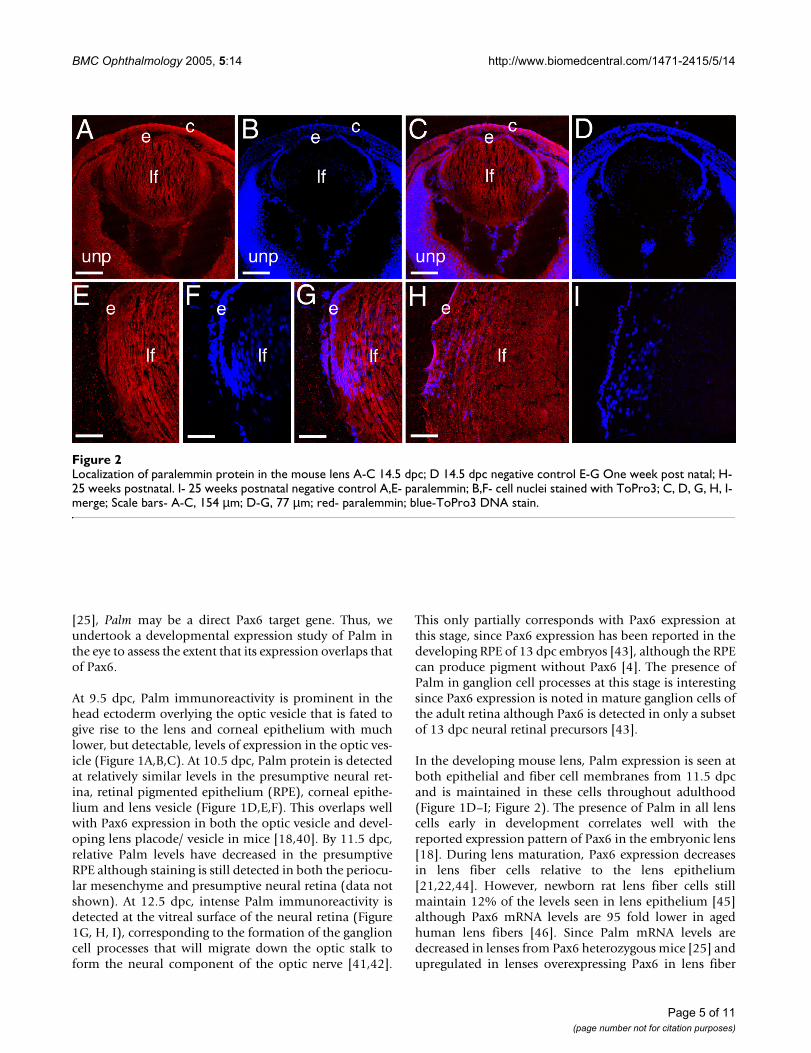
In the developing mouse lens, Palm expression is seen atboth epithelial and fiber cell membranes from 11.5 dpcand is maintained in these cells throughout adulthood(Figure 1D–I; Figure 2). The presence of Palm in all lenscells early in development correlates well with thereported expression pattern of Pax6 in the embryonic lens[18]. During lens maturation, Pax6 expression decreasesin lens fiber cells relative to the lens epithelium[21,22,44]. However, newborn rat lens fiber cells stillmaintain 12% of the levels seen in lens epithelium [45]although Pax6 mRNA levels are 95 fold lower in agedhuman lens fibers [46]. Since Palm mRNA levels aredecreased in lenses from Pax6 heterozygous mice [25] andupregulated in lenses overexpressing Pax6 in lens fiber
Localization of paralemmin protein in the mouse lens A-C 14.5 dpc; D 14.5 dpc negative control E-G One week post natal; H- 25 weeks postnatalFigure 2Localization of paralemmin protein in the mouse lens A-C 14.5 dpc; D 14.5 dpc negative control E-G One week post natal; H- 25 weeks postnatal. I- 25 weeks postnatal negative control A,E- paralemmin; B,F- cell nuclei stained with ToPro3; C, D, G, H, I- merge; Scale bars- A-C, 154 µm; D-G, 77 µm; red- paralemmin; blue-ToPro3 DNA stain.
Page 5 of 11(page number not for citation purposes)

BMC Ophthalmology 2005, 5:14 http://www.biomedcentral.com/1471-2415/5/14
cells [23], it is plausible that Palm expression is eitherdirectly responsive to Pax6 or controlled by genes in thesame pathway.
In order to test this proposition functionally, we clonedthe Pax6 binding site previously identified in the 5'-flank-ing region of human PALM [25] in front of a basal pro-moter and performed transient transfections in 293T cellswhich lack endogenous Pax6 proteins [32]. Co-transfec-tion of this reporter construct with Pax6 and Pax6(5a)expression vectors activated this artificial promoter 3.4-and 2.1-fold, respectively while addition of both expres-sion vectors simultaneously yielded a reporter activationsimilar to that of the Pax6 expression vector alone (Figure3). These levels of Pax6 mediated activation are compara-ble to those typically obtained in transient transfectionswith Pax6 responsive promoters [44,47,48]. From thesedata, it appears likely that the human PALM promotercontains a Pax6-binding site functionally able to interactwith both Pax6 and Pax6(5a) consistent with the upregu-lation of PALM expression in transgenic mice overexpress-ing Pax6 in the lens and reduced expression of Palm inPax6 heterozygous lenses [23,25,49]. However, the func-tional significance of this Pax6 site in the context of thePALM gene is more difficult to ascertain since neither thetranscriptional start site nor the functional minimal pro-moter of PALM have been experimentally investigated.Further studies of PALM/Palm promoters are necessary tofully establish their direct regulation by Pax6 proteins.
In the developing retina, the intense Palm staining seen inelongating ganglion cell axons at 12.5 dpc downregulatedmarkedly by 14.5 dpc as the development of these proc-esses completes [41] (data not shown). At 16.5 dpc, Palmimmunoreactivity is maintained at moderate levels in thecell bodies of both differentiating ganglion cells andundifferentiated neural precursors, but appears slightlystronger in the first morphologically distinguishableaxons of the developing inner plexiform layer (ipl) whichis composed of cell processes of the neurons of the innernuclear layer and ganglion cells [50](Figure 4A,B,C). Atbirth, Palm levels are upregulated in the developing innerplexiform layer (inl) which is in the process of rapidexpansion (Figure 4D,E,F). As the development of ipl pro-ceeds, the intensity of Palm staining in this layer drops tothat seen in the cell bodies of the ganglion cell and inl(Figure 4G–L). While not as dramatic, localized expres-sion is seen in the developing outer plexiform layer (opl)processes at 1 week pn (Figure 4G–I), although both atthat time and in the adult (Figure 4J–L), much less Palmstaining is seen on the photoreceptor cell bodies of theouter nuclear layer then in any other retinal layer. Nota-ble, Pax6 expression persists in both retinal ganglion cellsand the inner nuclear layer into adulthood, correlating
well with the expression pattern of Palm in this tissue[43].
In neuronal cell lines, Palm was previously detected at thecell membrane of the cell body and developing axons aswell as in a granular localization intracellularly. In vivo,Palm co-purifies with chick brain synaptic plasma mem-branes consistent with its palmitoylation [24]. While thestaining pattern of Palm in the developing mouse retina isconsistent with this membrane localization, we wanted toconfirm this in dissociated retinal cultures. The neural ret-ina of the E7 chick is at a period of extensive neurogenesis,migration, and process formation in vivo, especially ofganglion cells [51-53]. This ability to extend neurites isalso manifest in cultures made from this age retinal tissue[54,55]. Chick retinas were dissociated, plated and stainedfor Palm either 2 days or 7 days after plating. After 2 daysin culture (Figure 5A–C), Palm appears expressed by mostcells and is evident at the plasma membrane and as intra-
Pax6 proteins activate expression from a reporter consisting of four copies of a PAX6-binding site found in the putative 5' flanking sequence of the human PALM gene cloned upstream of the E4 basal promoterFigure 3Pax6 proteins activate expression from a reporter consisting of four copies of a PAX6-binding site found in the putative 5' flanking sequence of the human PALM gene cloned upstream of the E4 basal promoter. (A) An alignment between the PAX6 site found in the PALM gene and a consensus paired domain Pax6-binding site, P6CON. Non-conserved nucle-otides are shown in lower case letters. (B) Results of co-transfections in 293T cells. 200 ng of Pax6 and 25 ng of Pax6(5a) expression plasmids were used as indicated per experiment. The data were normalized using Renilla luciferase [31] and are expressed as a relative ratio of promoter activ-ity in the presence of Pax6 compared to the presence of empty vector, pKW10.
1.0
3.3
2.1
3.3
0.0
0.5
1.0
1.5
2.0
2.5
3.0
3.5
4.0
Pax6 - + - +
Pax6(5a) - - + +
Rela
tive F
old
-ch
an
ge 293T
PALM PAX6
P6CON
B
AACTTTCACtCTgCgATGgCa
ANNTTCACGCWTSANTKMNY
Page 6 of 11(page number not for citation purposes)

BMC Ophthalmology 2005, 5:14 http://www.biomedcentral.com/1471-2415/5/14
Localization of paralemmin protein during mouse retinal development A-C, 16.5 dpc, arrowheads- emerging inner plexiform layerFigure 4Localization of paralemmin protein during mouse retinal development A-C, 16.5 dpc, arrowheads- emerging inner plexiform layer; D-F 1 day pn; G-I 1 week pn; J-L 2 week pn; A,D,G,J- paralemmin; B,E,H,K- cell nuclei stained with ToPro3; C,F,I,L- merge; Abbreviations- unp- undifferentiated retinal precursors; gc- ganglion cell; ipl- inner plexiform layer; s*- background staining in the sclera; inl- inner nuclear layer; opl- outer plexiform layer; onl- outer nuclear layer. All scale bars are 77 µm. red- paralemmin; blue-ToPro3 DNA stain.
Page 7 of 11(page number not for citation purposes)

BMC Ophthalmology 2005, 5:14 http://www.biomedcentral.com/1471-2415/5/14
cellular puncta. Fine processes resembling axons (arrows)that sometimes stain with the anti-neurofilament anti-body RT-97 [33] are also positive for Palm immunoreac-tivity. After 7 days in culture (Figure 5D–F), Palm stainingappears punctate but more diffuse in the cell body, anddoes not appear to be localized on the numerous longprocesses stained for neurofilament. Thus, like in themouse retina in vivo, Palm is detected in retinal culturesundergoing active process formation while it is less evi-dent in mature cells, which are undergoing less processextension.
Palm is a member of a multigene family consisting of twoother family members, paralemmin-2 (Palm-2) and palm-delphin/paralemmin-like (PalmD/PalmL) [28,29]. Palm2shares 37% amino acid identity with Palm and like Palmhas a C-terminal CaaX motif that could potentially be pre-nylated. However, the Palm2 gene is alternatively splicedand not all variants contain the prenylation motif. PalmDis 23% identical to Palm but generally lacks a C-terminalprenylation motif although rare splice variants have an
alternative C-terminus containing a prenylation motifsimilar to Palm. Experimentally, the majority of PalmD iscytoplasmic and does not co-purify with plasma mem-brane fractions [28,29]. Since Palm is potentially able tomodulate plasma membrane growth in the lens, retinaand brain, while Palm2 and PalmD are of relatedsequence, we performed quantitative rt-PCR to comparethe relative expression levels of all three paralemmin fam-ily members in the lens, retina, cerebellum and forebrain.
The ratio between the housekeeping genes tested, B2M,HPRT and SDHA, in the different tissues analyzed wasfound to range between 0.98–1.04. Since the ratio of anideal internal control between various tissues would be 1and the variability of each of our internal normalizinggenes between the various tissues assayed was low, wenormalized our data to one housekeeping gene, B2M[39,56].
In the lens, cerebellum, forebrain and retina, Palm tran-scripts are significantly more abundant relative to B2M
Expression and localization of Palm in chick retinal culturesFigure 5Expression and localization of Palm in chick retinal cultures. Cultures were immunostained with polyclonal anti-Palm (A, D) and RT-97 anti-neurofilament (B, E) antibodies after 2 (A-C) or 7 (D-F) days in culture. For each pair, the merged images are shown in C and F. After 2 days in culture, Palm is present on most cells at cell borders as well as intracellular puncta (A). Fine processes resembling axons (arrows) that are sometimes positive for RT-97 (B) are also labeled. After 7 days in culture, Palm staining appears punctate but more diffuse (D), and does not appear to be localized on the numerous long processes stained with RT-97 (F). Bar in D, 25µm. Green- Palm; Red-RT-97;
Page 8 of 11(page number not for citation purposes)

BMC Ophthalmology 2005, 5:14 http://www.biomedcentral.com/1471-2415/5/14
than those of either Palm2 or PalmD (Figure 6). Notably,Palm mRNA is more abundant in retinas isolated shortlyafter birth compared to the adult retina, correlating wellwith the expression of Palm protein detected by immuno-histochemistry. Palm is alternatively spliced, and previouswestern blot analysis of mouse lens protein detected the60 kDa form of paralemmin [25] which translates frommRNA lacking exon 8 [24]. Parallel qt-PCR analyses of thelens and retina for Palm transcripts harboring exon 8 onlydetected low levels of this splice variant in all cases (datanot shown) which would be translated into a 80 kDa pro-tein. In the lens and forebrain, appreciable Palm2 expres-sion was detected (Ct values of about 21.5) while Palm2levels are relatively low in all post natal retinal samplestested (Ct values of about 28.5). PalmD transcripts wereusually present at low levels in the tissues examined withCt values of about 26. Co-expression of Palm with Palm2in tissues examined will aid to the interpretation of genetargeting studies of this family of genes.
ConclusionThe lens and retina express paralemmin during develop-ment with its transient upregulation during the formationof optic nerve and formation of both plexiform layers.
Further, the putative PALM promoter contains a func-tional Pax6 binding site and the developmental expres-sion pattern of Palm in the eye generally correlates wellwith that reported for Pax6, leading credence to the ideathat Palm is a Pax6 directly-regulated gene.
Abbreviationsdpc, days post coitum; PBS- phosphate buffered saline;inl, inner nuclear layer; onl; outer nuclear layer; opl, outerplexiform layer; ipl; inner plexiform layer; pn, post-natal;rpe, retinal pigmented epithelium.
Competing interestsThe author(s) declare that they have no competinginterests.
Authors' contributionsMC carried out all of the immunohistochemical studieson tissue and was involved in the initial drafting of themanuscript. LVW carried out all of the quantitative rt-PCRassays and BKC performed the transfection assays. DSGanalyzed PALM expression in chick retinal cultures andMWK was involved in the experimental design and itsinterpretation. AC conceived of the molecular experi-
Relative levels of Palm, Palm2 and PalmD transcripts in the lens, cerebellum forebrain and retinaFigure 6Relative levels of Palm, Palm2 and PalmD transcripts in the lens, cerebellum forebrain and retina. All data are expressed as a rel-ative to the amount of B2M in the sample.
0
0.5
1
1.5
2
2.5
3
3.5
4
Rela
tive
Expre
ssio
nLevels
Palm
Palm2
PalmD
NBLens
NBCerebellum
NBForebrain
P0Retina
P4Retina
P22Retina
Page 9 of 11(page number not for citation purposes)

BMC Ophthalmology 2005, 5:14 http://www.biomedcentral.com/1471-2415/5/14
ments and participated in their design and interpretation.MKD imaged all of the immunohistochemical data, wasinvolved in its interpretation and drafted the manuscriptat all stages of the submission process.
AcknowledgementsWe thank the staff of the Albert Einstein College of Medicine Biotechnol-ogy Center for the qPCR analysis, Dr. Jean Hebert of AECOM for helpful suggestions. Dr. Harry Maisel for the anti-chick PALM antibody, Drs. M. Busslinger and R. Maas respectively for the Pax6 and PAX6(5a) expression vectors and Dr. Kirk Czymmek of the University of Delaware Core Imaging Facility for confocal microscopy support. This work was funded by National Eye Institute grants EY015279 and EY012221 to MKD and EY12200 and EY14237 to AC; National Institute of Neurological Diseases and Stroke grant NS40317 to DSG and INBRE program grant P20 RR16472 supporting the University of Delaware Core Imaging facility.
References1. Chow RL, Lang RA: Early eye development in vertebrates. Annu
Rev Cell Dev Biol 2001, 17:255-296.2. Ochi H, Sakagami K, Ishii A, Morita N, Nishiuchi M, Ogino H, Yasuda
K: Temporal expression of L-Maf and RaxL in developingchicken retina are arranged into mosaic pattern. Gene ExprPatterns 2004, 4(5):489-494.
3. Lecoin L, Sii-Felice K, Pouponnot C, Eychene A, Felder-SchmittbuhlMP: Comparison of maf gene expression patterns duringchick embryo development. Gene Expr Patterns 2004, 4(1):35-46.
4. Collinson JM, Quinn JC, Hill RE, West JD: The roles of Pax6 in thecornea, retina, and olfactory epithelium of the developingmouse embryo. Dev Biol 2003, 255(2):303-312.
5. Dyer MA, Livesey FJ, Cepko CL, Oliver G: Prox1 function controlsprogenitor cell proliferation and horizontal cell genesis inthe mammalian retina. Nat Genet 2003, 34(1):53-58.
6. Duncan MK, Cui W, Oh DJ, Tomarev SI: Prox1 is differentiallylocalized during lens development. Mech Dev 2002,112:195-198.
7. Li X, Ma W, Barker JL, Piatigorsky J: Transient expression ofglutamate decarboxylase and gamma-amino butyric acid inembryonic lens fibers of the rat. Dev Dyn 1995, 203(4):448-455.
8. Giger RJ, Wolfer DP, De Wit GM, Verhaagen J: Anatomy of ratsemaphorin III/collapsin-1 mRNA expression and relation-ship to developing nerve tracts during neuroembryogenesis.J Comp Neurol 1996, 375(3):378-392.
9. Baechner D, Liehr T, Hameister H, Altenberger H, Grehl H, Suter U,Rautenstrauss B: Widespread expression of the peripheralmyelin protein-22 gene (PMP22) in neural and non-neuraltissues during murine development. J Neurosci Res 1995,42(6):733-741.
10. Claudio JO, Lutchman M, Rouleau GA: Widespread but cell type-specific expression of the mouse neurofibromatosis type 2gene. Neuroreport 1995, 6(14):1942-1946.
11. Tasheva ES, Ke A, Deng Y, Jun C, Takemoto LJ, Koester A, ConradGW: Differentially expressed genes in the lens of mimecan-null mice. Mol Vis 2004, 10:403-416.
12. Frederikse PH, Yun E, Kao HT, Zigler JSJ, Sun Q, Qazi AS: Synapsinand synaptic vesicle protein expression during embryonicand post-natal lens fiber cell differentiation. Mol Vis 2004,10:794-804.
13. Xi J, Farjo R, Yoshida S, Kern TS, Swaroop A, Andley UP: A compre-hensive analysis of the expression of crystallins in mouseretina. Mol Vis 2003, 9:410-419.
14. Magabo KS, Horwitz J, Piatigorsky J, Kantorow M: Expression ofbetaB(2)-crystallin mRNA and protein in retina, brain, andtestis. Invest Ophthalmol Vis Sci 2000, 41(10):3056-3060.
15. Zampighi GA, Eskandari S, Kreman M: Epithelial organization ofthe mammalian lens. Exp Eye Res 2000, 71(4):415-435.
16. Kuszak JR: The ultrastructure of epithelial and fiber cells inthe crystalline lens. Int Rev Cytol 1995, 163:305-350.
17. Valtorta F, Leoni C: Molecular mechanisms of neuriteextension. Philos Trans R Soc Lond B Biol Sci 1999,354(1381):387-394.
18. Grindley JC, Davidson DR, Hill RE: The role of Pax-6 in eye andnasal development. Development 1995, 121:1433-1442.
19. Marquardt T, Ashery-Padan R, Andrejewski N, Scardigli R, GuillemotF, Gruss P: Pax6 is required for the multipotent state of reti-nal progenitor cells. Cell 2001, 105(1):43-55.
20. Collinson JM, Quinn JC, Buchanan MA, Kaufman MH, Wedden SE,West JD, Hill RE: Primary defects in the lens underlie complexanterior segment abnormalities of the Pax6 heterozygouseye. Proc Natl Acad Sci U S A 2001, 98(17):9688-9693.
21. Duncan MK, Xie L, David LL, Robinson ML, Taube JR, Cui W,Reneker LW: Ectopic Pax6 expression disturbs lens fiber celldifferentiation. Invest Ophthalmol Vis Sci 2004, 45(10):3589-3598.
22. Duncan MK, Kozmik Z, Cveklova K, Piatigorsky J, Cvekl A: Overex-pression of Pax-6 (5a) in lens fiber cells results in cataractand upregulation of a5b1 integrin expression. J Cell Sci 2000,113:3173-3185.
23. Chauhan BK, Reed NA, Yang Y, Cermak L, Reneker L, Duncan MK,Cvekl A: A comparative cDNA microarray analysis reveals aspectrum of genes regulated by Pax6 in mouse lens. GenesCells 2002, 7(12):1267-1283.
24. Kutzleb C, Sanders G, Yamamoto R, Wang X, Lichte B, Petrasch-Par-wez E, Kilimann MW: Paralemmin, a prenyl-palmitoyl-anchored phosphoprotein abundant in neurons and impli-cated in plasma membrane dynamics and cell processformation. J Cell Biol 1998, 143(3):795-813.
25. Chauhan BK, Reed NA, Zhang W, Duncan MK, Kilimann MW, CveklA: Identification of genes downstream of Pax6 in the mouselens using cDNA microarrays. J Biol Chem 2002,277(13):11539-11548.
26. Bagchi M, Katar M, Lo WK, Maisel H: Paralemnin of the lens. J CellBiochem 2003, 89(5):917-921.
27. Gauthier-Campbell C, Bredt DS, Murphy TH, El-Husseini Ael D: Reg-ulation of dendritic branching and filopodia formation in hip-pocampal neurons by specific acylated protein motifs. MolBiol Cell 2004, 15(5):2205-2217.
28. Andreu N, Escarceller M, Feather S, Devriendt K, Wolf AS, Estivill X,Sumoy L: PALML, a novel paralemmin-related gene mappingon human chromosome 1p21. Gene 2001, 278(1-2):33-40.
29. Hu B, Copeland NG, Gilbert DJ, Jenkins NA, Kilimann MW: Theparalemmin protein family: identification of paralemmin-2,an isoform differentially spliced to AKAP2/AKAP-KL, and ofpalmdelphin, a more distant cytosolic relative. Biochem Bio-phys Res Commun 2001, 285(5):1369-1376.
30. Reed NA, Oh D, Czymmek KJ, Duncan MK: An immunohisto-chemical method for the detection of proteins in the verte-brate lens. J Immunol Methods 2001, 253(1-2):243-252.
31. Chauhan BK, Yang Y, Cveklova K, Cvekl A: Functional interac-tions between alternatively spliced forms of Pax6 in crystal-lin gene regulation and in haploinsufficiency. Nucleic Acids Res2004, 32(5):1696-1709.
32. Yang Y, Chauhan BK, Cveklova K, Cvekl A: Transcriptional regu-lation of mouse alphaB- and gammaF-crystallin genes in lens:opposite promoter-specific interactions between Pax6 andlarge Maf transcription factors. J Mol Biol 2004, 344(2):351-368.
33. Shin DH, Lee KS, Lee E, Cho SS, Kim J, Kim JW, Kwon BS, Lee HW,Lee WJ: The correspondence between the labeling patternsof antibody RT97, neurofilaments, microtubule associatedprotein 1B and tau varies with cell types and developmentstages of chicken retina. Neurosci Lett 2003, 342(3):167-170.
34. Anderton BH, Breinburg D, Downes MJ, Green PJ, Tomlinson BE,Ulrich J, Wood JN, Kahn J: Monoclonal antibodies show thatneurofibrillary tangles and neurofilaments share antigenicdeterminants. Nature 1982, 298(5869):84-86.
35. Primer 3 [http://puma.fmvz.usp.br/primer3/primer3_www.cgi]36. NCBI Blast [http://www.ncbi.nlm.nih.gov/BLAST/]37. Schmittgen TD, Zakrajsek BA, Mills AG, Gorn V, Singer MJ, Reed
MW: Quantitative reverse transcription-polymerase chainreaction to study mRNA decay: comparison of endpoint andreal-time methods. Anal Biochem 2000, 285(2):194-204.
38. Pfaffl MW: A new mathematical model for relative quantifica-tion in real-time RT-PCR. Nucleic Acids Res 2001, 29(9):e45.
39. Microbiology and Immunology On-Line: Real Time PCR. .40. Ashery-Padan R, Marquardt T, Zhou X, Gruss P: Pax6 activity in
the lens primordium is required for lens formation and forcorrect placement of a single retina in the eye. Genes Dev2000, 14(21):2701-2711.
Page 10 of 11(page number not for citation purposes)

BMC Ophthalmology 2005, 5:14 http://www.biomedcentral.com/1471-2415/5/14
Publish with BioMed Central and every scientist can read your work free of charge
"BioMed Central will be the most significant development for disseminating the results of biomedical research in our lifetime."
Sir Paul Nurse, Cancer Research UK
Your research papers will be:
available free of charge to the entire biomedical community
peer reviewed and published immediately upon acceptance
cited in PubMed and archived on PubMed Central
yours — you keep the copyright
Submit your manuscript here:http://www.biomedcentral.com/info/publishing_adv.asp
BioMedcentral
41. Pei YF, Rhodin JAG: The prenatal development of the mouseeye. Anat Rec 1970, 168:105-126.
42. Barnstable CJ: A molecular view of vertebrate retinaldevelopment. Mol Neurobiol 1987, 1(1-2):9-46.
43. de Melo J, Qiu X, Du G, Cristante L, Eisenstat DD: Dlx1, Dlx2,Pax6, Brn3b, and Chx10 homeobox gene expression definesthe retinal ganglion and inner nuclear layers of the develop-ing and adult mouse retina. J Comp Neurol 2003, 461(2):187-204.
44. Duncan MK, HaynesII JI, Cvekl A, Piatigorsky J: Dual roles for Pax-6: a transcriptional repressor of lens fiber-cell specific b-crys-tallin genes. Mol Cell Biol 1998, 18(9):5579-5586.
45. Yang Y, Cvekl A: Tissue-specific regulation of the mouse or A-crystallin gene in lens via recruitment of Pax6 and c-Maf toits promoter. J Mol Biol 2005 in press.
46. Zhang W, Cveklova K, Oppermann B, Kantorow M, Cvekl A: Quan-titation of PAX6 and PAX6(5a) transcript levels in adulthuman lens, cornea, and monkey retina. Mol Vis 2001, 7:1-5.
47. Chauhan BK, Yang Y, Cveklova K, Cvekl A: Functional propertiesof natural human PAX6 and PAX6(5a) mutants. Invest Oph-thalmol Vis Sci 2004, 45(2):385-392.
48. Goudreau G, Petrou P, Reneker LW, Graw J, Loster J, Gruss P:Mutually regulated expression of Pax6 and Six3 and its impli-cations for the Pax6 haploinsufficient lens phenotype. ProcNatl Acad Sci U S A 2002, 99(13):8719-8724.
49. Calderwood DA, Tuckwell DS, Eble J, Kuhn K, Humphries MJ: Theintegrin a1 A-domain is a ligand binding site for collagens andlaminin. J Biol Chem 1997, 272(19):12311-12317.
50. Finlay BL, Sengelaub DR: Development of the vertebrate retina.In Perspectives in vision research Edited by: Blakemore C. New York ,Plenum Press; 1989.
51. Coulombre AJ: Correlations of structural and biochemicalchanges in the developing retina of the chick. Am J Anat 1955,96(1):153-189.
52. Meller K, Tetzlaff W: Scanning electron microscopic studies onthe development of the chick retina. Cell Tissue Res 1976,170(2):145-159.
53. Prada C, Puga J, Perez-Mendez L, Lopez R, Ramirez G: Spatial andTemporal Patterns of Neurogenesis in the Chick Retina. EurJ Neurosci 1991, 3(6):559-569.
54. Thompson JM, Lu AH, Ruch S: Developmental regulation ofchick embryo retina neurite extension by extrinsic factors.Brain Res Bull 1987, 18(4):479-484.
55. Linser PJ, Perkins MS: Regulatory aspects of the in vitro devel-opment of retinal Muller glial cells. Cell Differ 1987, 20(2-3):189-196.
56. Vandesompele J, De Preter K, Pattyn F, Poppe B, Van Roy N, DePaepe A, Speleman F: Accurate normalization of real-timequantitative RT-PCR data by geometric averaging of multi-ple internal control genes. Genome Biol 2002,3(7):RESEARCH0034.
Pre-publication historyThe pre-publication history for this paper can be accessedhere:
http://www.biomedcentral.com/1471-2415/5/14/prepub
Page 11 of 11(page number not for citation purposes)