blood supply and lymphatics of skin
TRANSCRIPT
Blood Supply to The SKIN & its
lymphatic drainage.
Swetha. P

BLOOD SUPPLY TO THE SKIN
Contents
• Embryology
• Anatomy
• Physiological Functions
• Nerve supply
• Clinical Significance
EMBRYOLOGY • Angioblast- Form small cell clusters [Blood
islands] within embryonic and extra embryonic
mesoderm.
• These blood islands extend and fuse
Primordial vascular network
• Within these islands
Peripheral cells Endothelial cell
Core cells Blood cells (haemocytoblasts)

• Formation of the initial endothelial tube is by a
process of coalescence of cellular vacuoles within the
developing endothelial cells.
The vacuoles fuse together without cytoplasmic
mixing to form the blood vessel lumen.

ANATOMY
• VASCULAR ARCHITECTURE.
• HISTOLOGY.


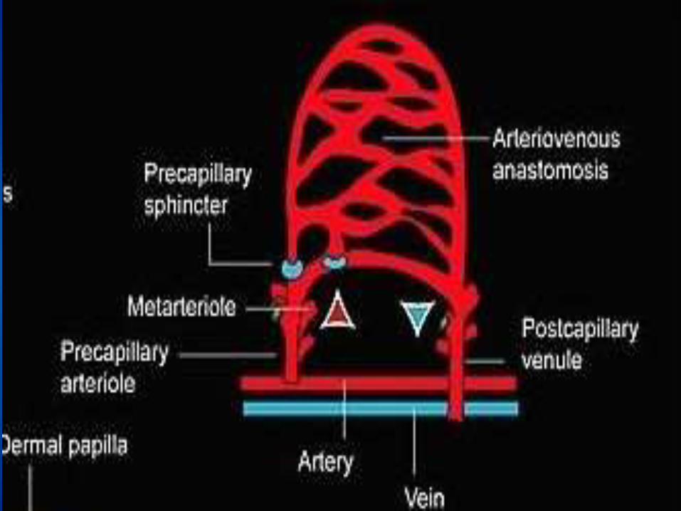
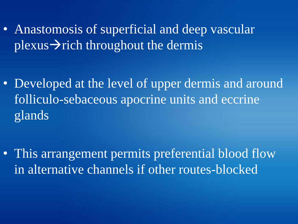
• Anastomosis of superficial and deep vascular
plexusrich throughout the dermis
• Developed at the level of upper dermis and around
folliculo-sebaceous apocrine units and eccrine
glands
• This arrangement permits preferential blood flow
in alternative channels if other routes-blocked

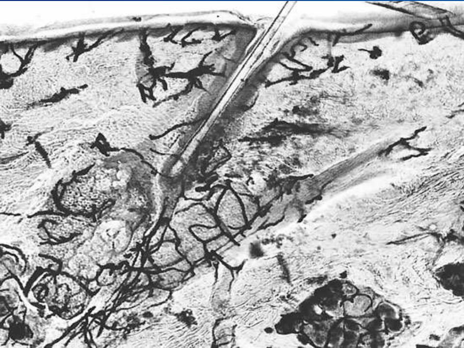
• Histo-chemical detection of alkaline
phosphatase activity indicates the presence of
arborizing nature of cutaneous circulation.
• Numerous capillaries in the adventitial dermis,
here shown enveloping sebaceous and eccrine
glands.


HISTOLOGY
• Arteries in subcutaneous fat and large
arterioles(deep part of dermis)
• Smaller arterioles(Superficial dermis)
• Papillary capillary loop
Ascending arterial segment
. Descending venous segment
• Post capillary venules
• Large venules and vein
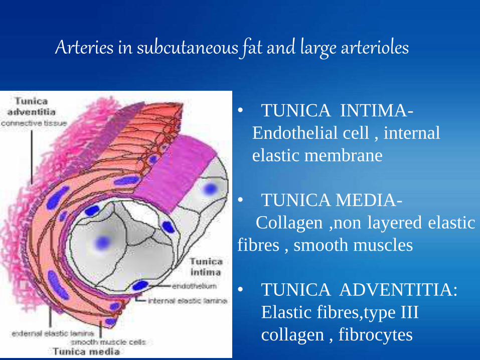
Arteries in subcutaneous fat and large arterioles
• TUNICA INTIMA-
Endothelial cell , internal
elastic membrane
• TUNICA MEDIA-
Collagen ,non layered elastic
fibres , smooth muscles
• TUNICA ADVENTITIA:
Elastic fibres,type III
collagen , fibrocytes
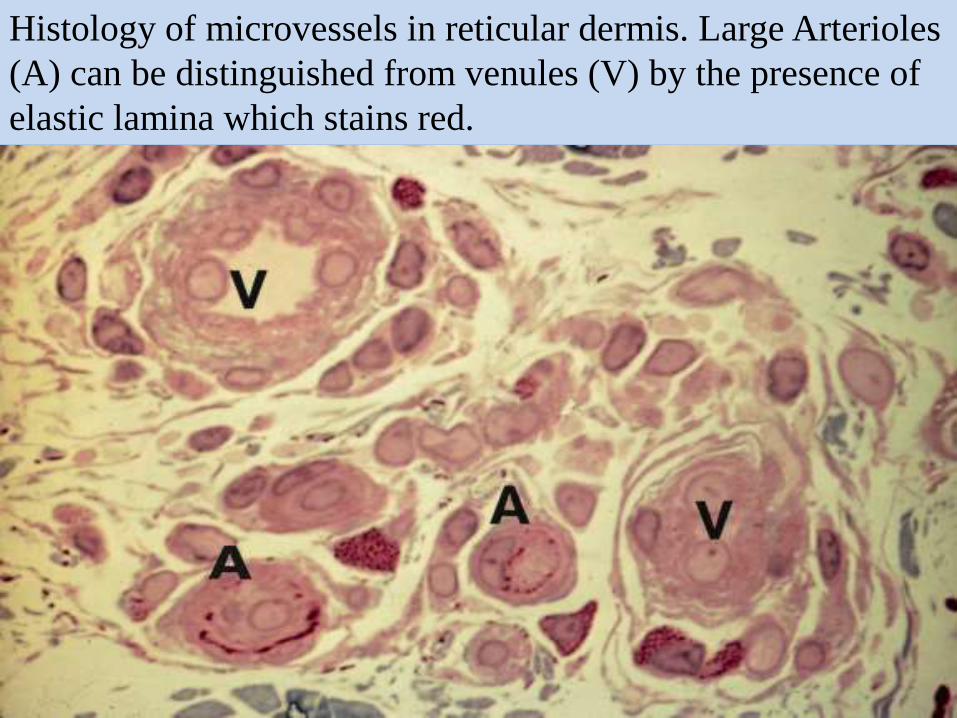
Histology of microvessels in reticular dermis. Large Arterioles
(A) can be distinguished from venules (V) by the presence of
elastic lamina which stains red.

SMALLER ARTERIOLES
• Absence of internal and external elastic
membrane
• Walls contain-Discontinued layer of
elastic fibres and smooth muscle

Transmission E.M of cross-section through a small arteriole.
Endothelial cell (E) surrounding the lumen (L) and the
presence of smooth muscle (SM).Small amount of elastic
tissue (el) next to the endothelial basement membrane (bm).

PAPILLARY CAPILLARY LOOP
• ASCENDING ARTERIAL SEGMENT

• DESCENDING VENOUS SEGMENT
–Lumen Wider
–Numerous pericytes
–Multilayered basal lamina.

POST CAPILLARY VENULES
–Endothelial cells
–Pericytes
–Basal lamina
–Type III collagen fibres

T.E.M of a transverse section through a venule.Endothelial
cells (E) in the lumen (L) is more convoluted. The endothelial
cells are surrounded by pericytes (P), and not smooth muscle
cells, and the basement membrane (bm)

• LARGER VENULES AND VEINS
– Variable amount of smooth muscle and elastic fibres
– No elastic membrane
– Valves
• VEIL CELLS
– Flat adventitial cells encircling arterioles,capillaries and
venules
– Demarcates the vessel wall from surrounding dermis

• BY ELECTRON MICROSCOPY,
Endothelial cell contains:
1. Cytoplamic filaments-7.5 -10nm
2. Pinocytic vesicles-50-70nm
3. Weibel-Palade bodies-Electron dense rod
shaped cytoplasmic structures

High-magnification view of Weibel–Palade bodies revealing
tubular profiles in cross-section.

CAPILLARIES:
1. TYPES : Continuous and fenestrated
CONTINUOUS:
• Lumina - continuous circumferential layer of
endothelial cells.
• Specialized intracellular junctions.
• Exchange of fluid and small water soluble
molecules –Via within micropinocytotic
vesicles.

Cross section through a capillary. Part of five endothelial cells
are pictured(E,E’),Basement membrane is seen surounding
and pericyte(P).within the lumen (L) is a platelet (Pl)
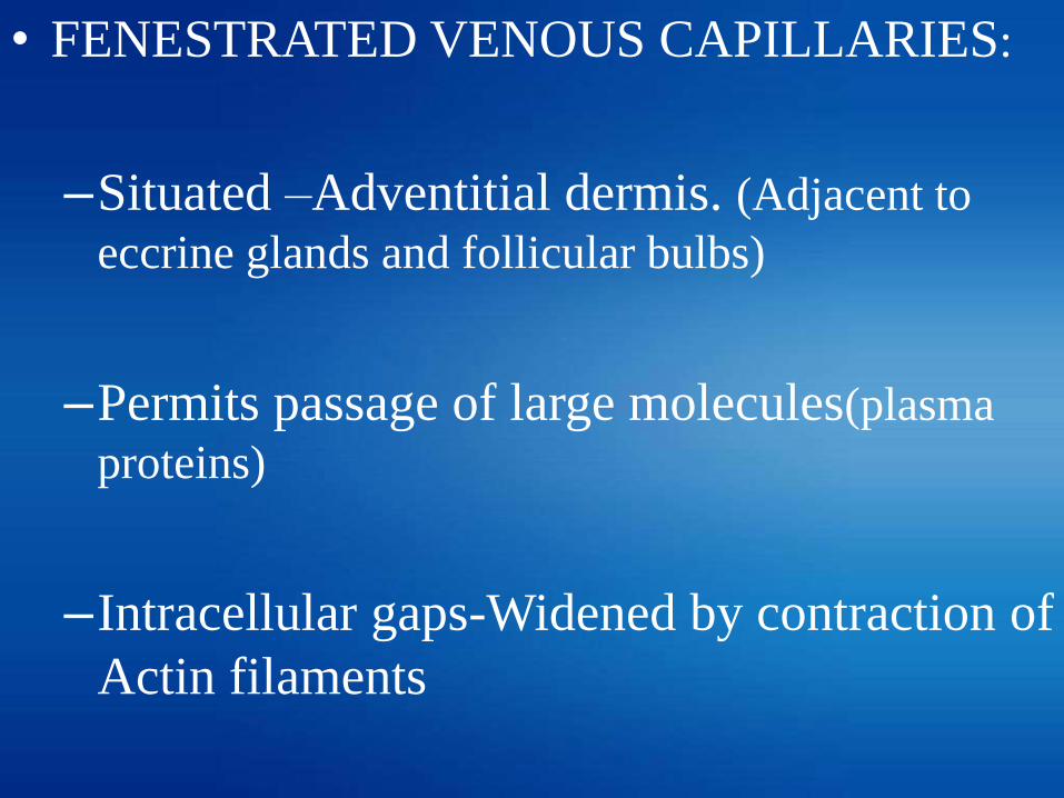
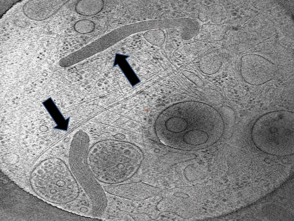
• FENESTRATED VENOUS CAPILLARIES:
–Situated –Adventitial dermis. (Adjacent to
eccrine glands and follicular bulbs)
–Permits passage of large molecules(plasma
proteins)
–Intracellular gaps-Widened by contraction of
Actin filaments
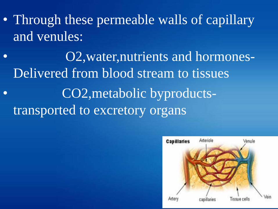
• Through these permeable walls of capillary
and venules:
• O2,water,nutrients and hormones-
Delivered from blood stream to tissues
• CO2,metabolic byproducts-
transported to excretory organs

Glomus bodies
• Specialized arterio-venous shunts
• Most abundant in recticular dermis of acral skin
• Regulation of temperature.
• Arterial segment- Sucquet Hoyer canal.
- Narrow lumen, thick wall
-endothelium,3-6 rows of glomus cells.
• Venous segment- thin wall
• - wide lumen.

• SUCQUET-HOYER CANAL:
–Single layer endothelium
–PAS positive, diastase resistant
basement membrane zone
–Media-4-6 layers of glomus cells
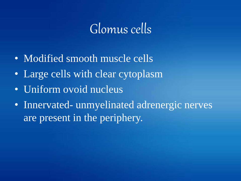
Glomus cells
• Modified smooth muscle cells
• Large cells with clear cytoplasm
• Uniform ovoid nucleus
• Innervated- unmyelinated adrenergic nerves
are present in the periphery.

Glomus cells surround arterioles and venules of
glomus bodies.
Physiological functions
• Nutritional support
• Immune surveillance
• Thermal regulation
• Wound healing
• Hemostasis
• Inflammation

Nerve Supply
• Controlled by adrenergic sympathetic nerves.
• These nerves are the efferent arm of
1. Baroreflex that originate in both arterial and
cardiopulmonary baroreceptors
2. Reflex baro response to upright posture and
exercise
3. Chemoreceptor reflex
4. Thermoregulatory reflexes

HUMORAL SUBSTANCES
• Direct effect upon arteriolar smooth muscle.
VASOCONSTRICTION VASODILATATION
Angiotensin II Histamine
Vasopressin Ethanol
Epinephrine Prostaglandin
Clinical significance
• Cutaneous capillary malformation-
– STURGE-WEBER SYNDROME-Seen in thelips,tongue,nasal and buccal mucosa
– Hereditary Hemorhhagic Telangiectasia
• Cutaneous vascular malformation-
- Klippel Trenaunay Syndrome
(Venous varicosities, edema, Hypertrophyof asssociated soft tissue and bone).


Contents
• Introduction.
• Histology.
• Physiological functions.
• Clinical significance.

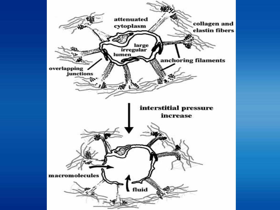
• Parallel to the major vascular network.
• Walls are not well developed.
• From superficial plexus of lymphatic capillaries
thicker walled lymphatic vessels with Valves
venous circulation.
introduction
• SITE:-• Upper part – recticular dermis
• Below the superficial plexus of venules
• At the zone - orientation of elastic fibres
changes from vertical – horizontal
• Supported by elastic fibers & anchoring filaments.

Superficial Dermis
Parallel structures to Vascular plexus.
Upper recticular Dermis
Single layer endothelium, discontinuous basal lamina
Deep part of dermis
Mesh Like
Subcutaneous fat
Single layer endothelium, discontinuous basal lamina, layers of smooth muscle cells
Lesser number of valves More number of valves
Increased lymphatic
clearence
Diminished lymphatic
clearence
Physiological functions
• Clearence of fluids, macromolecules,cells,
foreign materials from the interstitium.
• Maintains homeostasis.

Clinical significance
Failure of lymphatic system(burns, insect
bite,localisation of haematogenous
distribution infection) causes:
• Impaired immune function recurrent
infections.
• Lymphoedemas
• Fibrosis.
Summary
• BLOOD VESSEL.
1. Angioblast- Stem cells which forms the
endothelium, derived from the mesoderm.
2. Vascular architecture- Superficial and deep plexus,
connected to each other by communicating vessles.
3. From the superficial plexus, arises cascade of
capillaries that loops into each dermal papillae.
4. Anastomosis of superficial and deep plexus rich
through out the dermis.

5.HISTOLOGY-
Large arterioles & Arteries-
a. tunica intima- endothelium cells, int. elastic
membrane.
b. tunica media- Smooth muscle, external elastic
membrane
c. tunica adventitia- connective tissue.
Small arterioles-
a. absence of internal and external elastic membrane.
b. discontinuous smooth muscle & elastic fibres.

Ascending arterial limb- epithelial cell, pericytes,
basement membrane.
Descending venous segment- epithelial cells,
with numerous pericytes , multilayered basal lamina.
Post Capillary venule- epithelial cells, basement
membrane, collagen fibres.
Large venules & vein- variable amount of smooth
muscle, No ELASTIC Membrane.
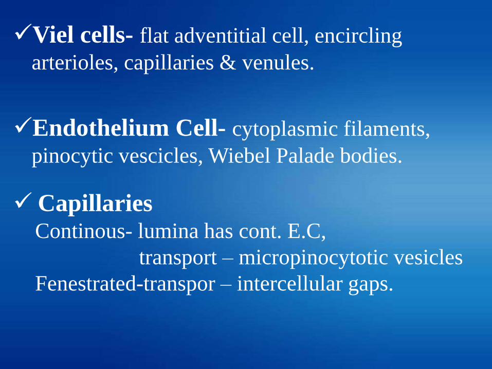
Viel cells- flat adventitial cell, encircling
arterioles, capillaries & venules.
Endothelium Cell- cytoplasmic filaments,
pinocytic vescicles, Wiebel Palade bodies.
CapillariesContinous- lumina has cont. E.C,
transport – micropinocytotic vesicles
Fenestrated-transpor – intercellular gaps.

6.Glomus bodies - specialised A-V shunts with out
interposition capillaries.
-regulation of temperature.
7.Functions - nutritional support, immune survielence,
thermal regulation, wound healing, hemostasis,
inflamation.
8.They are supplied by adrenergic sympathetic nerves.

9. Malformed cutaneous vascular– Klippel
trenaunay syndrome
10.Malformed capillaries –
Hereditary Hemorhhagic Telangiectasia
Sturge-Weber syndrome

• DERMAL LYMPHATICS
1. Upper part of reticular dermis, below superficial
plexus of venules.
2. Supported by elastic fibres & anchoring filaments.
3. Superficial lymphatics-Single layer endothelium,
discontinuous basal lamina.
4. Deep part of dermis-Single layer endothelium,
discontinuous basal lamina, layers of smooth
muscle cells.

• Function- clearence of fluid, macro mloecules,
foreign material & maintain homeostasis.
• Failure of lymphatic system- impaired
immune function, lymphoedemas.
