blood flukes of asiatic softshell ... - folia.paru.cas.cz · doi: 10.14411/fp.2016.031 roberts et...
TRANSCRIPT

http://folia.paru.cas.cz
This is an Open Access article distributed under the terms of the Creative Commons Attribution License (http://creativecommons.org/licenses/by/4.0), which permits unrestricted use, distribution, and reproduction in any medium, provided the original work is properly cited.
Research Article
Address for correspondence: J. Roberts, Auburn University, 203 Swingle Hall, Auburn, AL, 36849, USA. Phone: 334-844-9278; E-mail: [email protected] number for article: urn:lsid:zoobank.org:pub:D060C500-3A11-4FC7-8ED9-9523EE402E63
© Institute of Parasitology, Biology Centre CASFolia Parasitologica 2016, 63: 031doi: 10.14411/fp.2016.031
Blood flukes of Asiatic softshell turtles: revision of Coeuritrema Mehra, 1933 (Digenea: Schistosomatoidea) and a new species infecting Chinese softshell turtles, Pelodiscus sinensis (Trionychidae), from Vietnam
Jackson R. Roberts1, Raphael Orélis-Ribeiro1, Binh T. Dang2, Kenneth M. Halanych3 and Stephen A. Bullard1
1 Auburn University, School of Fisheries, Aquaculture & Aquatic Sciences and Aquatic Parasitology Laboratory, Auburn, AL, USA;2 Nha Trang University, Department of Biological Sciences, Institute for Biotechnology and Environment, Nha Trang, Vietnam;3 Auburn University, Department of Biological Sciences and Molette Biology Laboratory for Environmental & Climate Change Studies, Auburn, AL, USA
Abstract: Coeuritrema Mehra, 1933, previously regarded as a junior subjective synonym of Hapalorhynchus Stunkard, 1922, herein is revised to include Coeuritrema lyssimus Mehra, 1933 (type species), Coeuritrema rugatus (Brooks et Sullivan, 1981) comb. n., and Coeuritrema platti Roberts et Bullard sp. n. These genera are morphologically similar by having a ventral sucker, non-fused caeca, two testes, a pre-testicular cirrus sac, an intertesticular ovary, and a common genital pore that opens dorsally and in the sinistral half of the body. Phylogenetic analysis of the D1–D3 domains of the nuclear large subunit ribosomal DNA (28S) suggested that Coeuritrema and Hapalorhynchus share a recent common ancestor. Coeuritrema is morphologically most easily differentiated from Hapalorhynchus by having ventrolateral tegumental papillae and a definitive metraterm that is approximately 3−7× longer than the uterus. Coeuritrema comprises species that reportedly infect Asiatic softshell turtles (Testudines: Trionychidae) only, whereas Hapalorhynchus (as current-ly defined) comprises blood flukes that reportedly infect those hosts plus North American musk turtles (Sternotherus Bell in Gray) and mud turtles (Kinosternon Spix), both Kinosternidae, North American snapping turtles (Chelydridae), Asiatic hard-shelled turtles (Geoemydidae) and African pleurodirans (Pelomedusidae). Coeuritrema platti sp. n. infects the blood of Chinese softshell turtles, Pel-odiscus sinensis (Wiegmann), cultured in the Da Rang River Basin (Phu Yen Province, Vietnam). It differs from C. lyssimus by having a narrow hindbody (< 1.6× forebody width), ventrolateral tegumental papillae restricted to the hindbody, a short cirrus sac (< 10% of corresponding body length), a transverse ovary buttressing the caeca, a short, wholly pre-ovarian metraterm (~ 10% of corresponding body length), and a submarginal genital pore. It differs from C. rugatus by having small ventrolateral tegumental papillae, testes with-out deep lobes, and a Laurer’s canal pore that opens posterior to the vitelline reservoir and dorsal to the oviducal seminal receptacle. The new species is only the second turtle blood fluke reported from Vietnam.
Keywords: taxonomy, Spirorchiidae, 28S rDNA, molecular phylogeny, Griphobilharzia
The turtle blood flukes (TBFs; paraphyletic ‘Spirorchii-dae’) comprise 84 accepted species (54 freshwater; 30 ma-rine) assigned to 20 genera and that infect 37 freshwater and three marine turtle species (Platt 1993, 2002, Smith 1997a,b, Tkach et al. 2009, Platt and Sharma 2012, Ore-lis-Ribeiro et al. 2014, Roberts et al. 2016). Seventeen TBFs of five genera (Hapalorhynchus Stunkard, 1922; Vasotrema Stunkard, 1926; Coeuritrema Mehra, 1933; Enterohaematotrema Mehra, 1940, Cardiotrema Dwivedi, 1967) infect softshell turtles (Testudines: Trionychidae) (Table 1), but only nine of 31 (29%) trionychids host a TBF, suggesting that perhaps numerous innominate TBFs infect this turtle lineage. Softshell turtles of Pelodiscus Fitzinger (P. axenaria [Zhou, Zhang et Fang], P. maackii [Brandt],
P. parviformis Tang, P. sinensis [Wiegmann]) are commer-cially prized and cultured for food and magic throughout Asia (Alves et al. 2008, Fritz et al. 2010, Stuckas and Fritz 2011). A survey of Chinese aquaculture facilities indicated that > 300 million Chinese softshell turtles are in captivi-ty now and that ~ 125 million individuals are available for sale now in China alone (Haitao et al. 2008). Even though they are abundant in southeast Asian aquaculture, several of these species have become scarce in the wild (van Dijk et al. 2000). Despite their commercial importance, the tax-onomy of their blood parasites remains as underexplored as the infectious diseases that afflict these turtles in aqua-culture.

doi: 10.14411/fp.2016.031 Roberts et al.: Coeuritrema spp.
Folia Parasitologica 2016, 63: 031 Page 2 of 15
During a recent parasitological expedition to Vietnam, we encountered a new TBF species infecting Chinese softshell turtles, which led to a consideration of all TBFs infecting softshell turtles. Herein, we revise Coeuritrema, emend its diagnosis, redescribe Coeuritrema rugatus (Brooks et Sullivan, 1981) comb. n. (formerly Hapalo-rhynchus) based on museum specimens, describe a new species, and provide an updated phylogeny for the TBFs inclusive of the new taxon. This is the second TBF species reported from Vietnam.
MATERIALS AND METHODSTurtles were purchased from commercial turtle trappers in Nha
Trang, Vietnam and from a turtle farm in Phu Yen Province dur-ing 1−16 June 2015. Turtles were killed by decapitation. Necrop-sies were performed using 7.0 g/l sodium citrate saline solution. Living flukes intended as whole-mounts were killed with a butane hand lighter under little or no coverslip pressure and transferred to a vial of 5% neutral buffered formalin (n.b.f.). TBFs were maintained in 5% n.b.f. until staining. After washing with dis-tilled water, specimens intended as whole mounts were stained in Van Cleave’s hematoxylin with several additional drops of Ehr-lich’s hematoxylin, dehydrated using an ethanol series, cleared in clove oil, and permanently mounted in Canada balsam.
Whole mounts were examined using a Leica DM 2500 micro-scope with differential interference contrast optical components. Parasite measurements are reported in micrometres (µm) followed by the mean and number of specimens measured in parentheses. Turtle scientific names and taxonomic authorities follow van Dijk et al. (2014). Classification and anatomical terms for TBFs follow Luhman (1935 – genital sucker), Byrd (1939 – uterine pouch [see also Roberts et al. 2016]), Yamaguti (1971 – Manter’s organ), and Platt (1993 – median oesophageal pouch; 1998, 2002 – genital spines, plicate organ and most other terms).
Specimens for molecular analyses were handled with cam-el-hair brushes, pipettes or fine forceps, and immediately pre-served in a vial of absolute EtOH and stored at -20 °C. Total genomic DNA (gDNA) was extracted using DNeasyTM Blood and Tissue Kit (Qiagen, Valencia, California, USA) according to the manufacturer’s protocol, except for the incubation period with proteinase-K that was extended to overnight, and the final elution step wherein only 100 μl of elution buffer was used to increase the final DNA concentration in the eluate.
Partial 28S rDNA (domains D1–D3; ~ 1 400 bp) was ampli-fied using the forward primer ‘U178’ (5'-GCACCCGCTGAAY-TTAAG-3') and the reverse primer ‘L1642’ (5'-CCAGCGC-CATCCATTTTCA-3') (Lockyer et al. 2003). PCR amplifications were performed using a total volume of 25 μl containing ap-proximately 2 μl of DNA template, 0.4 μM of each primer along with 1× buffer, 2.5 mM MgCl2 (New England Biolabs, Ipswich, Massachusetts, USA), 1 mM dNTP mixture and 0.3 μl Taq poly-merase (5 U/μl) (New England Biolabs). Thermocycling profile consisted of an initial 4 min at 94 °C for denaturation, followed by 40 repeating cycles of 94 °C for 30 s for denaturation, 50 °C for 30 s for annealing, and 72 °C for 2 min for extension, fol-lowed by a final 5 min at 72 °C for extension. All PCR reactions were carried out in a Veriti Thermal Cycler (Applied Biosystems, Waltham, Massachusetts, USA). PCR products (5 μl) were veri-fied on a 1% agarose gel and stained with ethidium bromide. PCR
amplicons were gel-excised using QIAquickTM Gel Extraction Kit (Qiagen, Valencia, California, USA) following the manufac-turer’s protocol. DNA sequencing was performed by GENEWIZ with ABI Prism 3730xl DNA analyser (GENEWIZ, Inc., South Plainfield, New Jersey, USA). Primers used in sequencing of 28S rDNA included the PCR primers and the internal forward prim-ers 300F (5'-CAA GTACCGTGAGGGAAAGTTG-3') and 900F (5'-CCGTCTTGAAACACGGACCAAG-3') and reverse primer 1200R (5'-GCATAGTTCACCATCTTTCGG-3') (Lockyer et al. 2003). Sequence assembling and analysis of chromatograms were conducted using BioNumerics version 7.0 (Applied Maths, Sint-Martens-Latem, Belgium).
The sequence data for nuclear 28S rDNA generated during this study were aligned with those for selected blood flukes (Schistoso-matoidea) available on GenBank. Outgroups were selected from representative fish blood flukes, Aporocotylidae Odhner, 1912 (see Bullard et al. 2009). The ingroup comprised newly-generated sequence data from the new species (KX712243) and Hapalorhy-nchus foliorchis Brooks et Mayes, 1975 (KX712242) plus public-ly available sequences from Baracktrema obamai Roberts, Platt et Bullard, 2016 (KX061500), Griphobilharzia amoena Platt et Blair, 1991 (AY899914), and ten other TBFs (Orelis-Ribeiro et al. 2014). The specimens of H. foliorchis were collected in Ala-bama from Chelydra serpentina (Linnaeus) during October 2014 (Roberts et al. 2016). Sequences were aligned using MAFFT (Ka-toh and Toh 2010) with default settings implemented in the CIP-RES Science Gateway V.3.3 (Miller et al. 2010). The resulting alignment was refined by eye using MEGA version 5.2.2 (Tamura et al. 2011) and the ends of each fragment were trimmed to match the shortest sequence. Ambiguous positions were identified and removed using the Gblocks server (Castresana 2000) with set-tings for a less stringent selection. Bayesian inference (BI) was performed using the Metropolis-coupled Markov chain Monte Carlo method (MC3) in MrBayes version 3.2.6 (Huelsenbeck et al. 2001, Ronquist and Huelsenbeck 2003, Huelsenbeck and Ron-quist 2005) and run on CIPRES (Miller et al. 2010). The model of nucleotide substitution was selected based on the Akaike In-formation Criterion (Posada and Buckley 2004) as implement-ed in the jModelTest version 2.1.4 (Guindon and Gascuel 2003, Darriba et al. 2012). The GTR + I + G (proportion of invariable sites = 0.385 and gamma distribution = 1.393) model was inferred as the best estimator. Therefore, BI used the following param-eters: nst = 6, rates = invgamma, ngammacat = 4, and default priors. Analyses were run in duplicate each containing 4 inde-pendent chains (three heated and one cold chain) (nchains = 4) for 1.0 × 10⁷ generations (ngen = 10 000 000) sampled at intervals of 1 000 generations (samplefreq = 1 000). Results of the first 2 500 sampled trees were discarded as ‘burn-in’ based on the station-arity of the likelihood values, assessed by plotting the log-like-lihood values of the sample points against generation time using Tracer version 1.5 (Rambaut and Drummond 2009). All retained trees were used to estimate posterior probability of each node. A majority rule consensus tree with average branch lengths was constructed for the remaining trees using ‘summarise the trees’ (sumt) in MrBayes. Resulting phylogenetic trees were visualised using FigTree v1.3.1 (Rambaut 2009) and further edited with Adobe Illustrator CS3. Branch supports were considered as sig-nificant when posterior probabilities were > 0.95.

doi: 10.14411/fp.2016.031 Roberts et al.: Coeuritrema spp.
Folia Parasitologica 2016, 63: 031 Page 3 of 15
RESULTS
Coeuritrema Mehra, 1933 emended Figs. 1−7Diagnosis. Body dorsoventrally flattened (not cylindri-
cal), 3−7× longer than wide, constricted at level of ventral sucker, having hindbody 1−3× longer than forebody, as-pinous, papillate; ventrolateral tegumental papillae pres-ent, distributing from oral sucker posteriad or from ven-tral sucker posteriad. Dorsolateral and ventrolateral nerve chords present.
Oral sucker robust, aspinous, demarcated from body by posterior constriction. Ventral sucker present, aspinous. Pharynx present, enveloping anterior extremity of oe-sophagus. Oesophagus extending posteriad <1/4 of body length, ventral to anterior nerve commissure, lacking di-verticula, plicate organ, or median oesophageal pouch, straight or slightly sinuous; oesophageal gland surrounding oesophagus from posterior margin of pharynx to oesopha-geal-intestinal junction, strongly basophilic, widest at level of caecal bifurcation. Intestine comprising non-fused cae-ca, inverse U-shaped, smooth (lacking diverticula or sec-ondary rami), extending 1/2−3/4 of body length directly posteriad, not extensively convoluted, bifurcating anterior to ventral sucker, terminating in posterior body extremity.
Testes comprising one anterior testis and one posterior testis, in posterior half of body, intercaecal, having deep lobes or slightly irregular margins. Vas deferens extending directly anteriad and ventral to cirrus sac before turning posteriad and expanding to form external seminal vesicle; external seminal vesicle posterior to ventral sucker, inter-caecal, abutting anterodextral margin of cirrus sac, at level of common genital pore. Cirrus sac robust, pre-testicular, containing variously-sized secretory cells and large pars prostatica.
Ovary single, wider than long, triangular, lacking lobes, intercaecal, intertesticular. Oviduct emerging from dextral margin of ovary, directing posteriad; oviducal seminal re-ceptacle comprising middle portion of oviduct between ovary and posterior testis. Laurer’s canal intercaecal, intert-esticular, post-ovarian, extending anteriad or posteriad from oviduct at level of vitelline reservoir, opening dorsally. Vi-tellarium follicular, occupying space from caecal bifurca-tion to caecal termination; vitelline reservoir intercaecal, intertesticular, ventral to ovary. Ootype a weakly glandular and thin-walled chamber, intercaecal, occupying space be-tween anterior testis and ovary, posterior to cirrus sac.
Uterus intercaecal, intertesticular, straight (not coiled); uterine pouch absent. Egg single, occupying female repro-ductive tract proximal to metraterm. Metraterm massive, approximately 3−7× uterus length, longitudinal (extending anteriad in parallel with body margin), between ventral sucker and vitelline reservoir, sinistral to anterior testis, having an obviously muscular thick wall, apparently not storing egg(s). Common genital pore dorsal, sinistral, pos-terior to ventral sucker, predominantly anterior to genitalia, aspinous, lacking suckers.
Excretory vesicle globular, extensively lobed, extending posteriad from distal ends of caeca to posterior body end.
Excretory pore terminal. Manter’s organ absent. In blood of Asiatic trionychids.
Differential diagnosis. Body 3−7× longer than wide, aspinous; ventrolateral tegumental papillae present. Ventral sucker present. Oesophagus < 1/4 of body length, lacking diverticula, plicate organ, or median oesophageal pouch. Intestine comprising non-fused caeca. Testes comprising one anterior and one posterior testis. External seminal ves-icle abutting anterodextral margin of cirrus sac and at level of common genital pore. Ovary intertesticular. Oviducal seminal receptacle comprising middle portion of oviduct between ovary and posterior testis. Laurer’s canal intercae-cal, intertesticular, post-ovarian. Vitelline reservoir intert-esticular. Uterine pouch absent. Egg single, occupying fe-male reproductive tract proximal to metraterm. Metraterm massive, approximately 3−7× uterus length, apparently not storing egg(s). Common genital pore dorsal, sinistral, pre-dominantly anterior to genitalia. T y p e s p e c i e s : Coeuritrema lyssimus Mehra, 1933 from
heart ventricle of Indian flapshell turtle, Lissemys punctata (Bonnaterre) (Testudines: Trionychidae), in the Ganges River, India.Remarks. Mehra (1933) proposed Coeuritrema for
C. lyssimus and Hapalorhynchus odhnerensis (Mehra, 1933) Byrd, 1939. That diagnosis included “body wall with or without small papillae” (= ventrolateral tegumental pa-pillae) such that H. odhnerensis, which lacks ventrolateral tegumental papillae, could be included. As emended here-in, however, Coeuritrema includes papillate TBFs only. This genus has long been regarded as a junior subjective synonym of Hapalorhynchus (see Byrd 1939, Hughes et al. 1942, Brooks and Mayes 1976, Platt 1988, 2002, Bour-gat 1990), which lacks ventrolateral tegumental papillae. Coeuritrema further differs from Hapalorhynchus by hav-ing an external seminal vesicle that abuts the anterodextral margin of the cirrus sac and a definitive metraterm that is approximately 3−7× the uterus length.
The ootype and uterus are both relatively short and to-gether, rather than the metraterm, may store the egg in spe-cies of Coeuritrema. If a large egg, presumably one that is developed and ready for passage to the metraterm and/or for ejection, is present in the proximal portion of the female reproductive tract, the egg occupies the luminal spaces of both the ootype and uterus (Fig. 4). In that case, the ootype and uterus are difficult to differentiate (Fig. 4). Without an egg, the clear distinction between the ootype and the uter-us is evident: the ootype comprises the laterally-expanded chamber immediately distal to the ovo-vitelline duct and the uterus is the narrow, short duct connecting it with the metraterm (Fig. 6). If our understanding is correct, it may be useful to consider the ootype and uterus functioning to-gether as an ‘egg chamber’. Noteworthy along these lines also is that we did not observe an egg in the metraterm of any specimen, indicating that perhaps the metraterm of Coeuritrema spp. does not retain the egg.
These observations match those of previous workers handling specimens of the type species and Coeuritrema rugatus. Mehra (1933) explicitly reported that he never ob-

doi: 10.14411/fp.2016.031 Roberts et al.: Coeuritrema spp.
Folia Parasitologica 2016, 63: 031 Page 4 of 15
served more than a single egg (“ovum”) in the female re-productive tract of C. lyssimus (type species) and that each egg resided “in the proximal portion of the metraterm,” which, based on his illustrations (see plate 1, fig. 1 of Meh-ra 1933) could be interpreted as the putative egg chamber. Brooks and Sullivan (1981) likewise reported that only a single egg occurred in the uterus of ovigerous specimens. Perhaps this is related to the gestation time for the large egg.
The metraterm of other whole-mounted blood flukes does hold eggs (Bullard and Overstreet 2006, Bullard et al. 2006, Bullard and Jensen 2008); however, eggs in those species are smaller, much more numerous in the uterus and metraterm, and seemingly less developed. Perhaps this is because they must infect minute branchial vessels be-fore transiting to the gill epithelium and hatching (Bullard and Overstreet 2008). The extent to which the metraterm serves as a storage organ for TBF eggs is indeterminate, but if particular genera have or lack such an ‘egg chamber’ (= ootype + uterus), it is likely a significant differential di-agnostic feature relating genera. Resin-based ultramicroto-my of the wall of the proximal portion of the female repro-ductive tract could reveal cellular changes to the putative egg chamber wall in the presence/absence of the large egg.
Coeuritrema and Hapalorhynchus are similar morpho-logically by having a ventral sucker, non-fused caeca, two testes, a pre-testicular cirrus sac, an intertesticular ova-ry, and a common genital pore that opens dorsally and in the sinistral half of the body. Phylogenetic analysis of the D1–D3 domains of the nuclear large subunit riboso-mal DNA (28S) suggests that Coeuritrema and Hapalo-rhynchus indeed share a recent, common ancestor (Fig. 7; see below). We concur with Platt (2002) in that several apapillate turtle blood fluke species originally assigned to Coeuritrema should be assigned to Hapalorhynchus: H. odhnerensis, Hapalorhynchus ocadiae (Takeuti, 1942), Hapalorhynchus oschmarini (Belous, 1963), Hapalorhyn-chus macrotesticularis (Rohde, Lee et Lim, 1968), Hapalo-rhynchus mica (Oshmarin, 1971), Hapalorhynchus sheilae (Mehrotra, 1973) and Hapalorhynchus sutlejensis (Mehro-tra, 1973) (see Takeuti 1942, Rohde et al. 1968, Oshmarin 1971, Mehrotra 1973). This brings the total number of ac-cepted species of Coeuritrema and Hapalorhynchus to 3 and 19, respectively.
Yamaguti (1958) considered Coeuritrema a junior syn-onym of Tremarhynchus Thapar, 1933; however, the diag-nosis of Coeuritrema was published in May 1933 and that of Tremarhynchus was published immediately thereafter in June. Hence, the former genus has taxonomic priority (Dwivedi 1967). Byrd (1939) and Platt (2002) considered Tremarhynchus (and Coeuritrema) a junior subjective syn-onym of Hapalorhynchus. No author has treated Tremar-hynchus since 2002.
Tremarhynchinae Yamaguti, 1958 included Enterohae-matotrema and Tremarhynchus (type genus) (see Yamagu-ti 1958). Dwivedi (1967) commented that the subfamily should be renamed Coeuritrematinae Dwivedi, 1967 (type genus Coeuritrema) to correct Yamaguti’s (1958) name for the subfamily based on his erroneous acceptance of
Tremarhynchus as a senior synonym of Coeuritrema by taxonomic priority. This decision was probably based upon the International Code of Zoological Nomenclature’s Ar-ticle 61.1.2. (under “Statement of the Principle of Typifi-cation”), which states that “a nominal family-group taxon is the nominal genus on which its name is based.” (Inter-national Commission on Zoological Nomenclature 2000). Hence, because post-1967 Coeuritrema was correctly un-derstood to have taxonomic priority over Tremarhynchus, the subfamily name had to be changed.
Yamaguti (1971) accepted Coeuritrematinae Dwivedi, 1967 (as “1968”) and included Coeuritrema, Enterohae-matotrema, and Cardiotrema based on the presence of a ventral sucker, paired caeca not united posteriorly, two testes, a cirrus sac “more or less strongly developed” be-tween the ventral sucker and anterior testis, an intertesticu-lar ovary, a vitellarium as long as caeca, and a genital pore between the ventral sucker and testes. These genera plus Hapalorhynchus and Vasotrema include species infecting softshell turtles (Trionychidae) (Table 1). No molecular se-quence data exist for any species of Enterohaematotrema or Cardiotrema, and Vasotrema does not group with the Coeuritrema + Hapalorhynchus clade (Fig. 7). Hence, presently, no support exists for monophyly of softshell TBFs, suggesting that TBFs have repeatedly, independent-ly colonised softshell turtles.
Coeuritrema differs from Enterohaematotrema by hav-ing a dorsal, sinistral genital pore (rather than a ventral, medial genital pore), from Cardiotrema by having an inter-testicular ovary (rather than a sinistral ovary) and a large ventral sucker >1/2 body width (rather than <1/5 body width for Cardiotrema), and from Vasotrema by having two testes and a dorsal genital pore (rather than one testis posterior to the ovary and a ventral genital pore). Mehra (1934) provided a key to TBF genera and differentiated Coeuritrema and Hapalorhynchus by the presence or ab-sence of a cirrus sac and cirrus. These features have been the focus of intense study because of their stated value as differential features. Our observations herein confirmed the presence of an obvious cirrus sac among the accepted species of Coeuritrema, but this feature may or may not be of generic importance in other TBF genera. For example, within the closely-related Hapalorhynchus the presence or absence of the cirrus sac and cirrus have been accepted as generic features by some (Mehra 1939, 1940, Yamaguti 1958, 1971) and rejected by others (Byrd 1939, Brooks and Mayes 1976, Platt 1988, 2002, Bourgat 1990).
Brooks and Mayes (1976) provided supplemental ob-servations of the male genitalia of Hapalorhynchus stunk-ardi Byrd, 1939 and emended Hapalorhynchus to include “well-developed or poorly-developed cirrus.” Platt (1988) observed, in histological sections, prostate cells not bound by a limiting membrane but scattered in the surrounding parenchyma in Hapalorhynchus gracilis Stunkard, 1922, H. foliorchis and H. stunkardi. He also observed that the prostatic complex of Hapalorhynchus albertoi Lam-othe-Argumedo, 1978 and Hapalorhynchus brooksi Platt, 1988 was membrane-bound (= cirrus sac) and that the dis-tal portion of the ejaculatory duct (= cirrus) was eversi-

doi: 10.14411/fp.2016.031 Roberts et al.: Coeuritrema spp.
Folia Parasitologica 2016, 63: 031 Page 5 of 15
Table 1. Blood flukes infecting softshell turtles (Testudines: Trionychidae)
Turtle Fluke Site in host Locality Museum nos. Reference
Amyda cartilaginea (Boddaert), Asiatic softshell
Coeuritrema rugatus (Brooks et Sullivan, 1981) comb. n.
mesenteric blood vessels
Sungei Jempol, Ulu Jempol, State of Negeri Sembilan, West Malaysia
HWML 21339 Brooks and Sullivan (1981)
Apalone ferox (Schneider), Florida softshell
Vasotrema amydae Stunk-ard, 1926 (type species)
blood probably a Gulf of Mexico river drainage, Florida, USA
AMNH 791 Stunkard (1926)
blood probably a Gulf of Mexico river drainage, Florida, USA
AMNH 791 Stunkard (1928)
Vasotrema attenuatum Stunkard, 1928
blood probably a Gulf of Mexico river drainage, Florida, USA
AMNH 806, 807 Stunkard (1928)
Vasotrema sp. not specified Apalachicola River, Franklin County, Florida, USA
not specified Loftin (1960)
Apalone mutica (LeSueur), smooth softshell
Vasotrema attenuatum blood Missouri River Drainage, Nebraska, USA not specified Brooks and Mayes (1975)Vasotrema brevitestis Brooks et Mayes, 1975
blood Missouri River, 2.4 km south of Blair, Nebraska, USA
USNM 73817−73819; HWML 20077
Brooks and Mayes (1975)
Vasotrema robustum blood Missouri River Drainage, Nebraska, USA not specified Brooks and Mayes (1975)Apalone spinifera (LeSueur), spiny softshell
Vasotrema amydae (type species)
blood Ohio River Drainage, Indiana, USA AMNH 791 Stunkard (1926)
blood Ohio River Drainage, Indiana, USA AMNH 791 Stunkard (1928)Vasotrema attenuatum blood Ohio River Drainage, Indiana, USA AMNH 806, 807 Stunkard (1928)
mesenteric blood vessels
Reelfoot Lake, Tennessee, USA HWML9227 Byrd (1939) (see also Platt and Snyder 2007)
not specified Missouri River Drainage, Nebraska, USA not specified Brooks and Mayes (1975)Vasotrema brevitestis not specified Atkinson Lake, Atkinson Recreation Area,
0.8 km west of Atkinson, Nebraska, USAUSNM 73817−73819; HWML 20076
Brooks and Mayes (1975)
Vasotrema longitestis Byrd, 1939
arterial circulation Reelfoot Lake, Tennessee, USA USNM 9229, 80656-8; HWML 31121-8
Byrd (1939) (see Platt and Prestwood 1990)
Vasotrema robustum blood Ohio River Drainage, Indiana, USA AMNH 808, 809 Stunkard (1928)arterial system Reelfoot Lake, Tennessee, USA USNM 80657-2;
HWML 31122-2Byrd (1939) (see Platt and Prestwood 1990)
heart, large blood vessels
Huron River, Washtenaw County, Michigan, USA
USNM 37306 Wall (1951)
not specified Missouri River Drainage, Nebraska, USA HWML 20075 Brooks and Mayes (1975) not specified Nishnabotna River, Floyd County, Iowa, USA HWML 45795 Snyder (2004)
Dogania subplana (Saint-Hilaire), Malayan softshell turtle
Hapalorhynchus macrotesticularis (Rohde, Lee et Lim, 1968)
heart, arteries near heart
unknown drainage, Kuala Lumpur (purchased from Chinese merchant), Malaysia
Helm. Coll. No. R. 769, Zoology Dept., University of Malaysia, Kuala Lumpur
Rohde et al. (1968)
Lissemys punctata (Bonna-terre), Indian flapshell
Cardiotrema roparensis (Mehrotra, 1973)
not specified Sutlej River, Ropar, Punjab State, India not specified Mehrotra (1973)
body wash, hepatic blood vessels
Sutlej River, Ropar, Punjab State; Yamuna River, Karnal, Haryana State, India
not specified Tandon and Gupta (1985)
Enterohaematotrema palaeorticum Mehra, 1940 (type species)
small intestine (probably mesenter-ic blood vessels)
Ganges River, Allahabad, Uttar Pradesh State, India
not specified Mehra (1940)
Coeuritrema lyssimus Mehra, 1940 (type species)
ventricle of heart (adult)
Ganges River, Allahabad, Uttar Pradesh State, India
not specified Mehra (1933)
heart (adult) Ganges River Drainage, Rudrapur, Uttarakhand State, India
not specified Tandon and Gupta (1982)
Hapalorhynchus odhnerensis (Mehra, 1940)
ventricle of heart Ganges River, Allahabad, Uttar Pradesh State, India
not specified Mehra (1933)
Hapalorhynchus sheilae (Mehrotra, 1973)
not specified Patiala River (Ghaggar River Drainage), Patiala, Punjab State; Sutlej River, Ropar, Punjab State; Ghaggar River, Sangrur, Punjab State; Ganges River Drainage, Rudrapur, Uttarakhand State, India
not specified Mehrotra (1973)
heart, blood, liver Ganges River Drainage, Rudrapur, Uttara-khand State; Patiala River (Ghaggar River Drainage), Patiala, Punjab State; Ghaggar River, Sangrur, Punjab State, India
not specified Tandon and Gupta (1982)
Hapalorhynchus sutlejensis (Mehrotra, 1973)
not specified Sutlej River, Ropar, Punjab State; Ghaggar River, Sangrur, Punjab State, India
not specified Mehrotra (1973)
ventricle of heart Gomti River, Lucknow, Uttar Pradesh State; Ghaggar River, Sangrur, Punjab State, India
not specified Tandon and Gupta (1982)
Nilssonia gangetica (Cuvi-er), Indian softshell
Hapalorhynchus indicus (Thapar, 1933)
heart, large blood vessels
Gomti River, Lucknow, Uttar Pradesh State, India
not specified Thapar (1933)
Nilssonia hurum (Gray), Indian peacock softshell
Enterohaematotrema palaeorticum (type species)
intestine (probably mesenteric blood vessels)
pond in Kotah, Raipur, Madhya Pradesh State, India
not specified Shrivastava (1959)
Pelodiscus sinensis (Wieg-mann), Chinese softshell
Coeuritrema platti sp. n. heart, mesentery, lung
Da Rang River, Phuyen Province, Vietnam USNM 1411790, 1411791
Present study
Hapalorhynchus mica (Oshmarin, 1971)
hepatic blood vessels
Duong River, Hai Phong, Vietnam not specified Oshmarin (1971)
Hapalorhynchus oschmarini (Belous, 1963)
hepatic blood vessels
Lake Khanka, Russia not specified Belous (1963)
doi: 10.14411/fp.2016.031 Roberts et al.: Coeuritrema spp.
Folia Parasitologica 2016, 63: 031 Page 6 of 15
ble (Lamothe-Argumedo 1978, Platt 1988). As a result, he emended the diagnosis as “cirrus sac present or absent.” As additional species of Coeuritrema are documented, special care should be taken to assess the cirrus sac and cirrus.
Coeuritrema rugatus (Brooks et Sullivan, 1981) comb. n. Figs. 1, 2
ZooBank number for species: urn:lsid:zoobank.org:act:36DE9E3C-2809-476B-978A-67B2CC3B122F
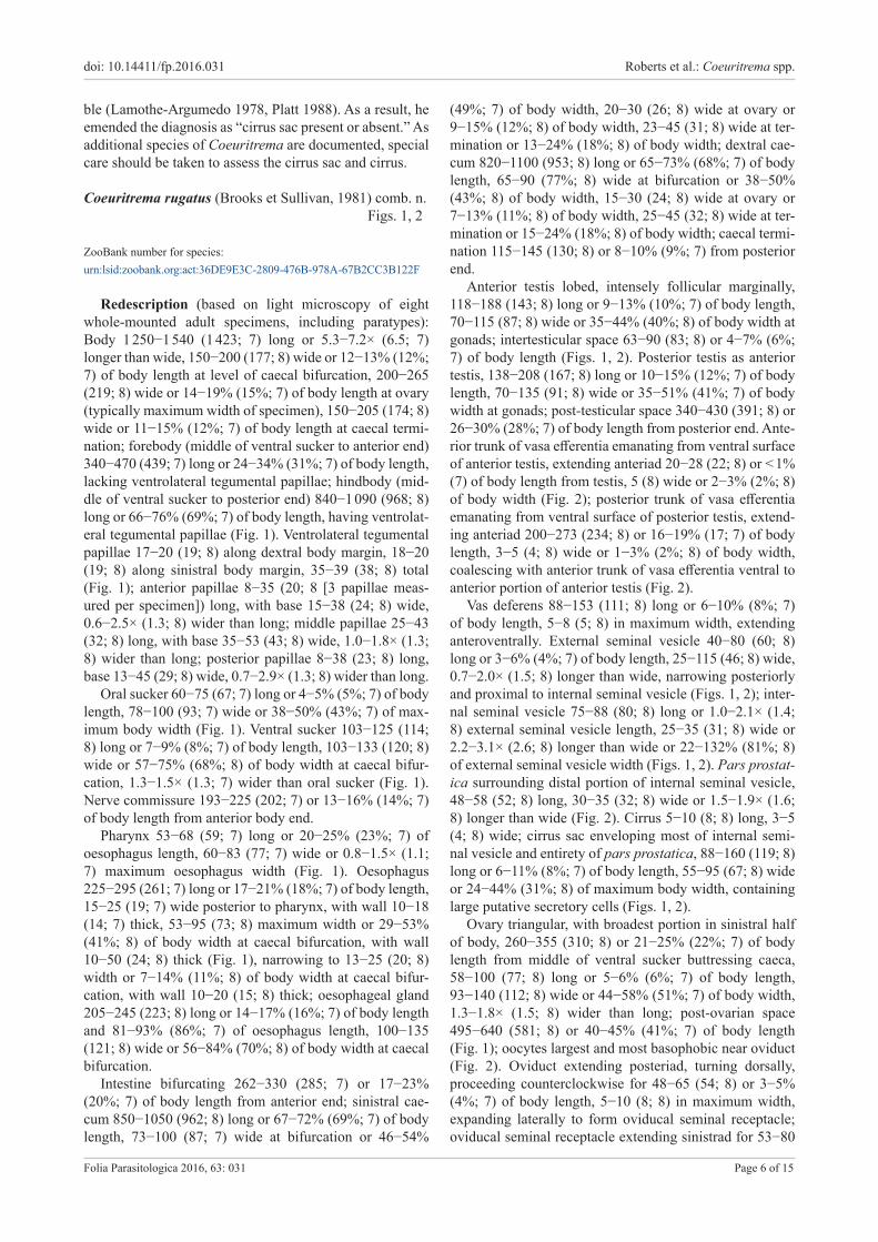
Redescription (based on light microscopy of eight whole-mounted adult specimens, including paratypes): Body 1 250−1 540 (1 423; 7) long or 5.3−7.2× (6.5; 7) longer than wide, 150−200 (177; 8) wide or 12−13% (12%; 7) of body length at level of caecal bifurcation, 200−265 (219; 8) wide or 14−19% (15%; 7) of body length at ovary (typically maximum width of specimen), 150−205 (174; 8) wide or 11−15% (12%; 7) of body length at caecal termi-nation; forebody (middle of ventral sucker to anterior end) 340−470 (439; 7) long or 24−34% (31%; 7) of body length, lacking ventrolateral tegumental papillae; hindbody (mid-dle of ventral sucker to posterior end) 840−1 090 (968; 8) long or 66−76% (69%; 7) of body length, having ventrolat-eral tegumental papillae (Fig. 1). Ventrolateral tegumental papillae 17−20 (19; 8) along dextral body margin, 18−20 (19; 8) along sinistral body margin, 35−39 (38; 8) total (Fig. 1); anterior papillae 8−35 (20; 8 [3 papillae meas-ured per specimen]) long, with base 15−38 (24; 8) wide, 0.6−2.5× (1.3; 8) wider than long; middle papillae 25−43 (32; 8) long, with base 35−53 (43; 8) wide, 1.0−1.8× (1.3; 8) wider than long; posterior papillae 8−38 (23; 8) long, base 13−45 (29; 8) wide, 0.7−2.9× (1.3; 8) wider than long.
Oral sucker 60−75 (67; 7) long or 4−5% (5%; 7) of body length, 78−100 (93; 7) wide or 38−50% (43%; 7) of max-imum body width (Fig. 1). Ventral sucker 103−125 (114; 8) long or 7−9% (8%; 7) of body length, 103−133 (120; 8) wide or 57−75% (68%; 8) of body width at caecal bifur-cation, 1.3−1.5× (1.3; 7) wider than oral sucker (Fig. 1). Nerve commissure 193−225 (202; 7) or 13−16% (14%; 7) of body length from anterior body end.
Pharynx 53−68 (59; 7) long or 20−25% (23%; 7) of oesophagus length, 60−83 (77; 7) wide or 0.8−1.5× (1.1; 7) maximum oesophagus width (Fig. 1). Oesophagus 225−295 (261; 7) long or 17−21% (18%; 7) of body length, 15−25 (19; 7) wide posterior to pharynx, with wall 10−18 (14; 7) thick, 53−95 (73; 8) maximum width or 29−53% (41%; 8) of body width at caecal bifurcation, with wall 10−50 (24; 8) thick (Fig. 1), narrowing to 13−25 (20; 8) width or 7−14% (11%; 8) of body width at caecal bifur-cation, with wall 10−20 (15; 8) thick; oesophageal gland 205−245 (223; 8) long or 14−17% (16%; 7) of body length and 81−93% (86%; 7) of oesophagus length, 100−135 (121; 8) wide or 56−84% (70%; 8) of body width at caecal bifurcation.
Intestine bifurcating 262−330 (285; 7) or 17−23% (20%; 7) of body length from anterior end; sinistral cae-cum 850−1050 (962; 8) long or 67−72% (69%; 7) of body length, 73−100 (87; 7) wide at bifurcation or 46−54%
(49%; 7) of body width, 20−30 (26; 8) wide at ovary or 9−15% (12%; 8) of body width, 23−45 (31; 8) wide at ter-mination or 13−24% (18%; 8) of body width; dextral cae-cum 820−1100 (953; 8) long or 65−73% (68%; 7) of body length, 65−90 (77%; 8) wide at bifurcation or 38−50% (43%; 8) of body width, 15−30 (24; 8) wide at ovary or 7−13% (11%; 8) of body width, 25−45 (32; 8) wide at ter-mination or 15−24% (18%; 8) of body width; caecal termi-nation 115−145 (130; 8) or 8−10% (9%; 7) from posterior end.
Anterior testis lobed, intensely follicular marginally, 118−188 (143; 8) long or 9−13% (10%; 7) of body length, 70−115 (87; 8) wide or 35−44% (40%; 8) of body width at gonads; intertesticular space 63−90 (83; 8) or 4−7% (6%; 7) of body length (Figs. 1, 2). Posterior testis as anterior testis, 138−208 (167; 8) long or 10−15% (12%; 7) of body length, 70−135 (91; 8) wide or 35−51% (41%; 7) of body width at gonads; post-testicular space 340−430 (391; 8) or 26−30% (28%; 7) of body length from posterior end. Ante-rior trunk of vasa efferentia emanating from ventral surface of anterior testis, extending anteriad 20−28 (22; 8) or < 1% (7) of body length from testis, 5 (8) wide or 2−3% (2%; 8) of body width (Fig. 2); posterior trunk of vasa efferentia emanating from ventral surface of posterior testis, extend-ing anteriad 200−273 (234; 8) or 16−19% (17; 7) of body length, 3−5 (4; 8) wide or 1−3% (2%; 8) of body width, coalescing with anterior trunk of vasa efferentia ventral to anterior portion of anterior testis (Fig. 2).
Vas deferens 88−153 (111; 8) long or 6−10% (8%; 7) of body length, 5−8 (5; 8) in maximum width, extending anteroventrally. External seminal vesicle 40−80 (60; 8) long or 3−6% (4%; 7) of body length, 25−115 (46; 8) wide, 0.7−2.0× (1.5; 8) longer than wide, narrowing posteriorly and proximal to internal seminal vesicle (Figs. 1, 2); inter-nal seminal vesicle 75−88 (80; 8) long or 1.0−2.1× (1.4; 8) external seminal vesicle length, 25−35 (31; 8) wide or 2.2−3.1× (2.6; 8) longer than wide or 22−132% (81%; 8) of external seminal vesicle width (Figs. 1, 2). Pars prostat-ica surrounding distal portion of internal seminal vesicle, 48−58 (52; 8) long, 30−35 (32; 8) wide or 1.5−1.9× (1.6; 8) longer than wide (Fig. 2). Cirrus 5−10 (8; 8) long, 3−5 (4; 8) wide; cirrus sac enveloping most of internal semi-nal vesicle and entirety of pars prostatica, 88−160 (119; 8) long or 6−11% (8%; 7) of body length, 55−95 (67; 8) wide or 24−44% (31%; 8) of maximum body width, containing large putative secretory cells (Figs. 1, 2).
Ovary triangular, with broadest portion in sinistral half of body, 260−355 (310; 8) or 21−25% (22%; 7) of body length from middle of ventral sucker buttressing caeca, 58−100 (77; 8) long or 5−6% (6%; 7) of body length, 93−140 (112; 8) wide or 44−58% (51%; 7) of body width, 1.3−1.8× (1.5; 8) wider than long; post-ovarian space 495−640 (581; 8) or 40−45% (41%; 7) of body length (Fig. 1); oocytes largest and most basophobic near oviduct (Fig. 2). Oviduct extending posteriad, turning dorsally, proceeding counterclockwise for 48−65 (54; 8) or 3−5% (4%; 7) of body length, 5−10 (8; 8) in maximum width, expanding laterally to form oviducal seminal receptacle; oviducal seminal receptacle extending sinistrad for 53−80

doi: 10.14411/fp.2016.031 Roberts et al.: Coeuritrema spp.
Folia Parasitologica 2016, 63: 031 Page 7 of 15
os
ph
nc
og
oe
dln
vs
esv
vdisvcs
ave
pve
cgp
mt
ov
pt
od
lvd
osr
vr
vr
ev
ep
at
dc sc
vlp
250 µm
1
ovov
atat
mtmt
isvisv
esvesv
osrosr
ptpt
50 µm
2
pve
od
Lc
vt
oooo
utut
cgp
vd
ave
cscs
pppp
ec
eggegg
vrsvrs
ovov
tvdtvd
cb
pppp
lvdlvd
lvdlvdlvdlvd
lvdlvd
Figs. 1−2. Coeuritrema rugatus (Brooks et Sullivan, 1981) comb. n. (Digenea: Schistosomatoidea) from the mesenteric vessels of Amyda cartilaginea (Boddaert) (Testudines: Trionychidae) from Malaysia, paratype (HWML Coll. No. 21339). Fig. 1. Total view, ven-trally. Fig. 2. Genitalia, ventral view. Abbreviations: at − anterior testis; ave − anterior trunk of vasa efferentia; cb − caecal bifurcation; cgp − common genital pore; cs − cirrus sac; dc − dextral caecum; dln − dorsolateral nerve chord; ec − eversible cirrus; egg − egg in utero; ep − excretory pore; esv − external seminal vesicle; ev − excretory vesicle; isv − internal seminal vesicle; Lc − Laurer’s canal; lvd − lateral vitelline collecting duct; mt − metraterm; nc − nerve commissure; od − oviduct; oe − oesophagus; og − oesophageal gland; oo − ootype; os − oral sucker; osr − oviducal seminal receptacle; ov − ovary; ph − pharynx; pp − pars prostatica; pt − posterior testis; pve − posterior trunk of vasa efferentia; sc − sinistral caecum; tvd − transverse vitelline duct; ut − uterus; vd − vas deferens; vlp − ven-trolateral tegumental papillae; vr − vitellarium; vrs − vitelline reservoir; vs − ventral sucker; vt − vitelline duct.
250
µm
50 µ
m
doi: 10.14411/fp.2016.031 Roberts et al.: Coeuritrema spp.
Folia Parasitologica 2016, 63: 031 Page 8 of 15
(63; 8) or 58−112% (84%; 8) of ovary width, 25−33 (28; 8) in maximum width at origin or 9−16% (13%; 8) of maxi-mum body width, narrowing before turning dorsal, extend-ing anterodextrad 58−95 (82; 8) or 5−7% (6%; 7) of body length, 10−13 (12; 8) in maximum width or 5−7% (5%; 8) of body width (Fig. 2). Laurer’s canal a narrow duct ex-tending 23−43 (31; 8) anterosinistrad from middle portion of oviduct, 8−13 (11; 8) wide, opening dorsally at level of middle portion of ovary (Fig. 2).
Vitellarium comprising a series of interconnected sphe-roid masses of follicles, distributing from level of caecal bifurcation to distal ends of caeca, lateral collecting ducts coalescing at level of posterior margin of ovary to form transverse vitelline duct; transverse vitelline duct ventral to ovary, 315−415 (369; 8) or 25−29% (27%; 7) of body length from middle of ventral sucker (Figs. 1, 2); vitelline reservoir sac-like, ventral to oviducal seminal receptacle; vitelline duct extending anterodextrad and dorsal 55−85 (64; 8) or 4−6% (5%; 7) of body length before connecting with oviduct at ootype (Fig. 2). Ootype difficult to discern in gravid specimens, 30−43 (36; 8) long, 28−43 (37; 8) wide, dorsal to anterior margin of ovary (Fig. 2).
Uterus comprising proximal portion and metraterm (Fig. 2), with a single egg in seven of eight specimens; proximal portion of uterus extending anterosinsitrad from ootype, 28−50 (39; 8) long or 2−4% (3%; 7) of body length, 18−30 (27; 8) wide or 9−15% (12%; 8) of max-imum body width; metraterm extending anterosinistrad, 148−218 (175; 8) long or 11−15% (12%; 7) of body length, maximum width of 25−45 (34; 8) or 12−21% (16%; 8) of maximum body width, 3.2−6.6× (4.7; 8) proximal uterus length. Uterine egg ovoid, 63−78 (72; 7) long or 5% (6) of body length, 23−33 (27; 7) wide or 9−16% (12%; 7) of maximum body width, 1.9−3.3× (2.7; 7) longer than wide (Fig. 2). Common genital pore 95−150 (123; 8) or 8−10% (9%; 7) of body length posterior to middle of ventral suck-er (Fig. 1).
Excretory vesicle 130−165 (144; 8) long or 10−12% (10%; 7) of body length, 78−100 (87; 8) wide or 45−55% (50%; 8) of body width at caecal termination; wall 8−13 (10; 8) thick (Fig. 1).
T y p e a n d o n l y k n o w n h o s t : Asiatic softshell turtle, Amyda cartilaginea (Boddaert) (Testudines: Trionychidae).
T y p e l o c a l i t y : Sungei Jempol, Ulu Jempol, State of Negeri Sembilan, Malaysia.
S i t e i n h o s t : Mesenteric blood vessels.S p e c i m e n s e x a m i n e d : Hapalorhynchus rugatus – Har-
old W. Manter Laboratory (HWML) of Parasitology Coll. No. 21339, paratypes, eight slides comprising eight whole-mount-ed specimens, seven of the eight slides labeled as Hapalo-rhynchus rugosus, ex Amyda cartilaginea (as Trionyx carti-lageneus) from Sungei Jempol, Ulu Jempol, State of Negeri Sembilan, Malaysia (Brooks and Sullivan 1981).
Remarks. Our interpretation of some features associat-ed with type materials (see above) of C. rugatus contradict-ed those of Brooks and Sullivan (1981), who did not detail the vasa efferentia, vas deferens, ootype or Laurer’s canal. They reported that C. rugatus lacked a pharynx; however,
the paratypes have a large pharynx surrounding the ante-rior extremity of the oesophagus immediately posterior to the mouth (Fig. 1). Coeuritrema and Baracktrema Roberts, Platt et Bullard, 2016 are the only accepted TBF genera described as having a pharynx, and the pharynx is immedi-ately posterior to the mouth and muscular oral sucker. Sev-eral species of Hapalorhynchus, Spirorchis MacCallum, 1918 and Vasotrema also have this configuration and will be treated in later taxonomic works (J.R.R., S.A.B. – un-publ. data).
Regarding the oviduct, Brooks and Sullivan (1981) il-lustrated it as originating from the posteromedial aspect of the ovary and turning anterodorsally, but we confirmed that it originates from the dextral side of the ovary. Regarding the oviducal seminal receptacle, they illustrated a sac-like structure branching from the oviduct and filled with sperm proximal to the uterus (fig. 3 in Books and Sullivan 1981, p. 1336). No paratype had such a structure. They stated that the vitelline reservoir was immediately postovarian and ventral to the ovary, but we observed that the reservoir coalesces sinistro-dorsal to the ovary (Fig. 2).
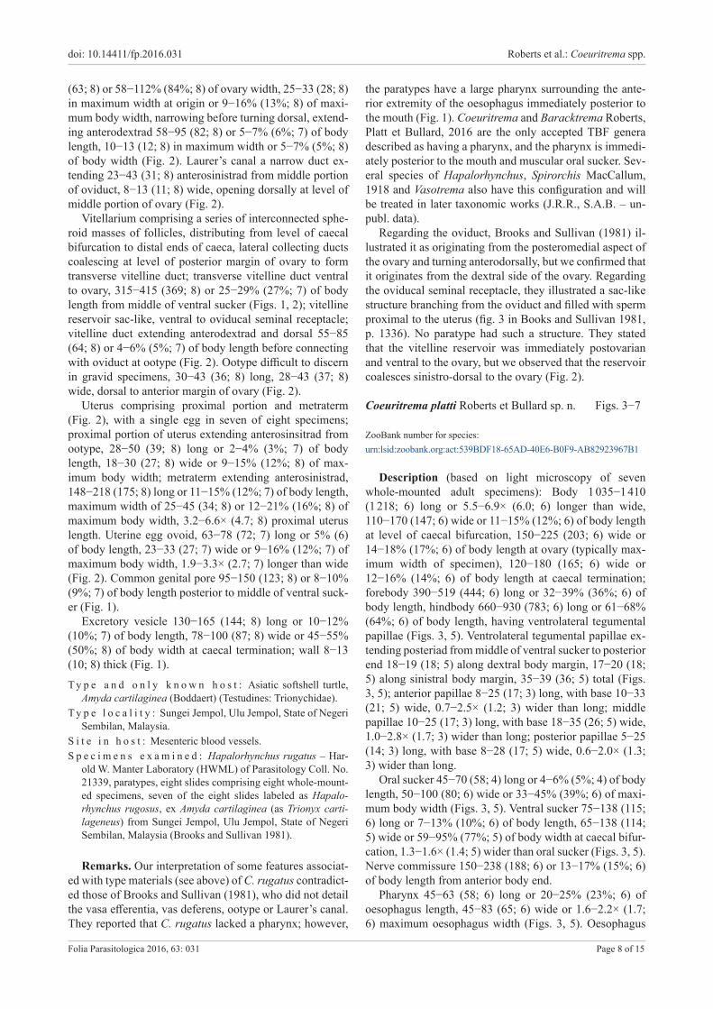
Coeuritrema platti Roberts et Bullard sp. n. Figs. 3−7
ZooBank number for species: urn:lsid:zoobank.org:act:539BDF18-65AD-40E6-B0F9-AB82923967B1
Description (based on light microscopy of seven whole-mounted adult specimens): Body 1 035−1 410 (1 218; 6) long or 5.5−6.9× (6.0; 6) longer than wide, 110−170 (147; 6) wide or 11−15% (12%; 6) of body length at level of caecal bifurcation, 150−225 (203; 6) wide or 14−18% (17%; 6) of body length at ovary (typically max-imum width of specimen), 120−180 (165; 6) wide or 12−16% (14%; 6) of body length at caecal termination; forebody 390−519 (444; 6) long or 32−39% (36%; 6) of body length, hindbody 660−930 (783; 6) long or 61−68% (64%; 6) of body length, having ventrolateral tegumental papillae (Figs. 3, 5). Ventrolateral tegumental papillae ex-tending posteriad from middle of ventral sucker to posterior end 18−19 (18; 5) along dextral body margin, 17−20 (18; 5) along sinistral body margin, 35−39 (36; 5) total (Figs. 3, 5); anterior papillae 8−25 (17; 3) long, with base 10−33 (21; 5) wide, 0.7−2.5× (1.2; 3) wider than long; middle papillae 10−25 (17; 3) long, with base 18−35 (26; 5) wide, 1.0−2.8× (1.7; 3) wider than long; posterior papillae 5−25 (14; 3) long, with base 8−28 (17; 5) wide, 0.6−2.0× (1.3; 3) wider than long.
Oral sucker 45−70 (58; 4) long or 4−6% (5%; 4) of body length, 50−100 (80; 6) wide or 33−45% (39%; 6) of maxi-mum body width (Figs. 3, 5). Ventral sucker 75−138 (115; 6) long or 7−13% (10%; 6) of body length, 65−138 (114; 5) wide or 59−95% (77%; 5) of body width at caecal bifur-cation, 1.3−1.6× (1.4; 5) wider than oral sucker (Figs. 3, 5). Nerve commissure 150−238 (188; 6) or 13−17% (15%; 6) of body length from anterior body end.
Pharynx 45−63 (58; 6) long or 20−25% (23%; 6) of oesophagus length, 45−83 (65; 6) wide or 1.6−2.2× (1.7; 6) maximum oesophagus width (Figs. 3, 5). Oesophagus

doi: 10.14411/fp.2016.031 Roberts et al.: Coeuritrema spp.
Folia Parasitologica 2016, 63: 031 Page 9 of 15
og
3
mµ 0
5
oe
cb
vs
od
cgp
mt
esv
at
pt
isvcs
osrvt
lvd
ov
dcsc
vr
ev
cgp
ave
pp
os
ph
nc
4
vlp
ep
250 µm
ptpt
osrosr
mtmt
esvesv
cscs isvisv
atat
ovov
eggegg
eggegg
vd
ec
pve
Lc
od
vr
vln
vtvt
vrs
tvdtvd
tvdtvd
50 µm
Figs. 3, 4. Coeuritrema platti sp. n. (Digenea: Schistosomatoidea) from viscera of Pelodiscus sinensis (Wiegmann) (Testudines: Tri-onychidae) from Vietnam, holotype (USNM Coll. No. 1411790). Fig. 3. Total view, dorsally. Fig. 4. Genitalia, dorsal view. Abbrevi-ations: at − anterior testis; ave − anterior trunk of vasa efferentia; cb − caecal bifurcation; cgp − common genital pore; cs − cirrus sac; dc − dextral caecum; ec − eversible cirrus; egg − egg in utero; ep − excretory pore; esv − external seminal vesicle; ev − excretory vesi-cle; isv − internal seminal vesicle; Lc − Laurer’s canal; lvd − lateral vitelline collecting duct; mt − metraterm; nc − nerve commissure; od − oviduct; oe − oesophagus; og − oesophageal gland; os − oral sucker; osr − oviducal seminal receptacle; ov − ovary; ph − pharynx; pp − pars prostatica; pt − posterior testis; pve − posterior trunk of vasa efferentia; sc − sinistral caecum; tvd − transverse vitelline duct; vd − vas deferens; vln − ventrolateral nerve chord; vlp − ventrolateral tegumental papillae; vr − vitellarium; vrs − vitelline reservoir; vs − ventral sucker; vt − vitelline duct.
250
µm
50 µ
m

doi: 10.14411/fp.2016.031 Roberts et al.: Coeuritrema spp.
Folia Parasitologica 2016, 63: 031 Page 10 of 15
Figs. 5, 6. Coeuritrema platti sp. n. (Digenea: Schistosomatoidea) from viscera of Pelodiscus sinensis (Wiegmann) (Testudines: Tri-onychidae) from Vietnam, paratype (USNM 1411791). Fig. 5. Total view, ventrally. Fig. 6. Genitalia, ventral view. Abbreviations: at − anterior testis; ave − anterior trunk of vasa efferentia; cb − caecal bifurcation; cgp − common genital pore; cs − cirrus sac; dc − dextral caecum; ec − eversible cirrus; ep − excretory pore; esv − external seminal vesicle; ev − excretory vesicle; isv − internal seminal vesicle; Lc − Laurer’s canal; lvd − lateral vitelline collecting duct; mt − metraterm; nc − nerve commissure; od − oviduct; oe − oesophagus; og − oesophageal gland; os − oral sucker; oo − ootype; osr − oviducal seminal receptacle; ov − ovary; ph − pharynx; pp − pars prostatica; pt − posterior testis; pve − posterior trunk of vasa efferentia; sc − sinistral caecum; tvd − transverse vitelline duct; ut − uterus; vd − vas deferens; vln − ventrolateral nerve chord; vlp − ventrolateral tegumental papillae; vr − vitellarium; vrs − vitelline reservoir; vs − ventral sucker; vt − vitelline duct.
os
ph
nc
og
oe
cbvln
vs
esvvd
csisv
ave
pve
cgp
mt
ov
pt
od
lvd
vtosr
vrvr
ev
ep
at
dcsc
vlp
250 µm
5
ovov
vrsvrs
lvdlvd
atat
mtmtisvisv
esvesv
osrosr
ptpt
50 µm6
pve
od
Lc
oooo
utut
ec
cgppp
vd
vt
ave
cscs
vtlvdlvd
lvdlvd
lvdlvd
tvdtvd
250
µm
50 µ
m

doi: 10.14411/fp.2016.031 Roberts et al.: Coeuritrema spp.
Folia Parasitologica 2016, 63: 031 Page 11 of 15
straight, 229−301 (259; 6) long or 18−23% (21%; 6) of body length, 10−15 (13; 6) wide posterior to pharynx, with wall 7−10 (9; 6) thick, 25−50 (39; 6) maximum width or (26%; 6) of body width at caecal bifurcation, with wall 10−27 (20; 6) thick, constricting to 13−18 (16; 6) width or 8−16% (11%; 6) of body width at caecal bifurcation, with wall 10−15 (12; 6) thick; oesophageal gland 198−288 (254; 6) long or 17−24% (21%; 6) of body length and 0.9−1.1× (1.0; 6) oesophagus length, 75−125 (104; 6) wide or 68−74% (71%; 6) of body width at caecal bifur-cation (Figs. 3, 5). Intestine bifurcating 235−365 (285; 6) or 17−27% (24%; 6) of body length from anterior end; sin-istral caecum 585−940 (754; 6) long or 51−68% (62%; 6) of body length, 35−45 (39; 6) wide or 24−32% (27%; 6) of body width at bifurcation, 13−30 (20; 6) wide or 6−15% (10%; 6) of body width at ovary, 20−35 (27; 6) wide or 12−19% (16%; 6) of body width at termination; dextral caecum 585−940 (743; 6) long or 52−67% (61%; 6) of body length, 38−45 (41; 6) wide or 24−36% (29%; 6) of body width at bifurcation, 18−35 (25; 6) wide or 10−18% (12%; 6) of body width at ovary, 18−35 (27; 6) wide or 11−20% (16%; 6) of body width at termination; caecal ter-mination 92−140 (120; 6) or 8−14% (10%; 6) from poste-rior end.
Anterior testis follicular throughout, lacking lobes, 60−143 (106; 6) long or 5−11% (9%; 6) of body length, 50−95 (71; 6) wide or 25−43% (35%; 6) of body width at gonads; intertesticular space 30−103 (74; 6) or 3−7% (6%; 6) of body length (Figs. 3−6). Posterior testis as an-terior testis, 90−158 (130; 6) long or 8−15% (11%; 6) of body length, 55−125 (89; 6) wide or 28−57% (44%; 6) of body width at gonads; post-testicular space 293−335 (310; 6) or 24−28% (27%; 6) of body length from poste-rior end. Anterior trunk of vasa efferentia emanating from ventral surface of anterior testis, extending anteriad 10−35 (26; 5) or 1−3% (2%; 5) of body length from testis, 3 (5) wide or 1−2% (1%; 5) of body width (Figs. 4−6); posteri-or trunk of vasa efferentia emanating from ventral surface of posterior testis, extending anteriad 188−255 (224; 5) or 17−21% (18%; 5) of body length, 3−5 (4; 5) wide or 1−3% (2%; 5) of body width, coalescing with anterior trunk of vasa efferentia ventral to anterior portion of anterior testis (Figs. 4−6).
Vas deferens 80−138 (101; 5) long or 6−10% (8%; 5) of body length, 3−5 (4; 3) in maximum width, extending an-teroventrally. External seminal vesicle 63−88 (75; 5) long or 6% (5) of body length, 25−38 (33; 5) wide, 1.9−2.5× (2.3; 5) longer than wide, constricting posteriorly prior to internal seminal vesicle (Figs. 3−6); internal seminal vesi-cle 88−118 (100; 5) long or 1.1−1.6× (1.4; 5) external sem-inal vesicle length, 13−35 (23; 6) wide or 3.4−6.8× (4.4; 5) longer than wide or 52−92% (73%; 5) of external seminal vesicle width (Figs. 3−6). Pars prostatica surrounding dis-tal portion of internal seminal vesicle (Figs. 4, 6), 53−55 (54; 3) long, 20−30 (25; 3) wide or 1.8−2.7× (2.2; 3) longer than wide. Cirrus 5−8 (6; 3) long, 3 (3) wide; cirrus sac enveloping most of internal seminal vesicle and entirety of pars prostatica, 78−128 (100; 6) long or 7−9% (8%; 6) of body length, 45−65 (52; 6) wide or 22−30% (26%; 6) of
maximum body width, containing large putative secretory cells (Figs. 3−6).
Ovary triangular, with broadest portion in sinistral half of body, 160−320 (246; 6) or 15−23% (20%; 6) of body length from middle of ventral sucker, buttressing caeca, 35−115 (84; 6) long or 3−9% (7%; 6) of body length, 65−153 (111; 6) wide or 42−70% (54%; 6) of body width, 1.1−1.9× (1.4; 6) wider than long; post-ovarian space 425−510 (458; 6) or 35−45% (28%; 6) of body length (Figs. 3−6); oocytes uniform in size and basophilic throughout ovary (Figs. 4, 6). Oviduct extending posteriad, turning dorsally, proceed-ing sinistrad, turning dorsal again before extending poste-riad for 35−105 (61; 6) or 3−7% (5%; 6) of body length, 5−10 (8; 6) in maximum width, expanding laterally to form oviducal seminal receptacle; oviducal seminal receptacle extending sinistrad for 65−80 (71; 3) or 63−78% (73%; 3) of ovary width, 25−40 (34; 5) in maximum width at origin or 11−20% (16%; 5) of maximum body width, narrowing before turning dorsal, extending anterodextrad 78−80 (79; 3) or 6−7% (7%; 3) of body length, 10−13 (11; 3) in max-imum width or 5−6% (5%; 3) of body width (Figs. 3−6). Laurer’s canal a narrow duct extending 15−25 (21; 3) an-terosinistrad from middle portion of oviduct, 5−8 (6; 3) wide, opening dorsal and over proximal portion of ovidu-cal seminal receptacle (Figs. 4, 6).
Vitellarium comprising a series of interconnected sphe-roid masses of follicles, distributing from level of caecal bifurcation to distal ends of caeca, lateral collecting ducts coalescing at level of posterior margin of ovary to form transverse vitelline duct; transverse vitelline duct ventral to ovary, 245−385 (297; 3) or 21−27% (24%; 3) of body length from middle of ventral sucker, lobed dorsally (Figs. 3−6); vitelline reservoir sac-like, ventral to oviducal seminal re-ceptacle; vitelline duct extending anterodextrad and dorsal 30−58 (47; 5) or 2−5% (4%; 5) of body length before con-necting with oviduct at ootype (Figs. 3−6). Ootype difficult to discern in gravid specimens, 25−40 (32; 3) long, 33−43 (38; 3) wide, dorsal to dextral half of ovary (Fig. 6).
Uterus comprising proximal portion and metraterm (Figs. 3−6), with a single egg in four of seven specimens; proximal portion of uterus extending anterosinistrad from ootype, 25−45 (36; 3) long or 2−4% (3% 3) of body length, 23−38 (29; 3) wide or 12−18% (14%; 3) of maximum body width; metraterm extending anterosinistrad, 128−163 (147; 5) long or 11−13% (12%; 5) of body length, 45−63 (53; 3) wide or 13−32% (23%; 3) of maximum body width, 2.8−5.8× (4.3; 3) proximal uterus length. Uterine egg ovoid, 80−95 (86; 4) long or 6−8% (7%; 4) of body length, 30−38 (35; 4) wide or 14−17% (16%; 4) of maximum body width, 2.3−2.7× (2.5; 4) longer than wide (Fig. 4). Com-mon genital pore 63−138 (103; 6) or 6−11% (8%; 6) of body length posterior to middle of ventral sucker.
Excretory vesicle 120−180 (165; 6) long or 12−17% (14%; 6) of body length, 38−90 (74; 6) wide or 32−53% (44%; 6) of body width at caecal termination, with wall 5−15 (9; 4) thick (Figs. 3, 5).
T y p e a n d o n l y k n o w n h o s t : Chinese softshell turtle, Pelodiscus sinensis (Wiegmann) (Testudines: Trionychidae).

doi: 10.14411/fp.2016.031 Roberts et al.: Coeuritrema spp.
Folia Parasitologica 2016, 63: 031 Page 12 of 15
0.05 substitutions per site
AY604708 Hapalotrema mehrai
AY222178 Plethorchis acanthus
AY604705 Spirhapalum polesianum
AY222179 Neoparacardicola nasonis
AY604709 Carettacola hawaiiensis
FJ481164 Spirorchis haematobiusAY222174 Spirorchis scripta
KX061500 Baracktrema obamai
AY222177 Aporocotyle spinosicanalisAY604711 Unicaecum sp.
FJ481166 Spirhapalum siamensis
AY604706 Vasotrema robustum
AY604707 Learedius learedi
AY157239 Chimaerohemecus trondheimensis
1
11
1
1
1
1
1
1
0.85
0.98
AY899914 Griphobilharzia amoena
AY604710 Hapalorhynchus gracilisKX712243 Coeuritrema platti sp. n.
KX712242 Hapalorhynchus foliorchis11
1
1
7
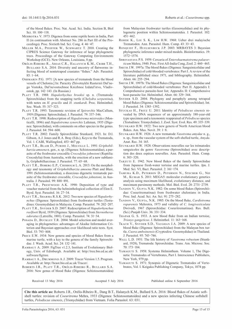
Fig. 7. Phylogenetic relationships of blood flukes reconstructed by Bayesian interference and based on partial D1−D3 domains of 28S from 18 taxa (13 TBFs, one crocodilian blood fluke, four fish blood flukes). Numbers aside tree nodes indicate posterior probability. Definitive hosts are indicated by icons aside tree nodes.
T y p e l o c a l i t y : Da Rang River Basin, Phu Yen Province, Vietnam.
S i t e i n h o s t : Heart, mesentery, lung.P r e v a l e n c e a n d i n t e n s i t y o f i n f e c t i o n : Two
(100%) P. sinensis from an aquaculture facility in the Da Rang River Basin had four and five specimens of C. platti. None of two P. sinensis collected from the Cai River Basin in Nha Trang yielded specimens.
S p e c i m e n s / m a t e r i a l s d e p o s i t e d : Holotype, United States National Museum (USNM) Coll. No. USNM 1411790; one paratype, United States National Museum (USNM) Coll. No. USNM 1411791; one paratype (Institute of Parasitology, Academy of Sciences of the Czech Republic, České Budě-jovice (IPCAS) Coll. No. D-727; GenBank Accession No. KX712243.
M a t e r i a l e x a m i n e d : Hapalorhynchus gracilis – Ameri-can Museum of Natural History (AMNH) Coll. No. 125, holo-type, one slide comprising one whole-mounted specimen, ex Chelydra serpentina from North Judson, Indiana (Stunkard 1922); AMNH 1269, paratypes, four slides (three of the four la-beled Hapalorhychus gracilis) comprising four whole-mount-ed specimens, ex Chelydra serpentina from North Judson, Indiana (Stunkard 1922); Vasotrema attenuatum − AMNH 806, syntype, 17 slides comprising 25 whole-mounted spec-imens, ex Apalone ferox and A. spinifera from Florida and Indiana respectively (Stunkard 1928). Hapalorhynchus rug-atus − HWML 21339.
E t y m o l o g y : The specific epithet platti honours our friend and colleague Thomas R. Platt (Professor Emeritus, Saint Mary’s College, Notre Dame, Indiana) for his extensive, sus-
tained contributions to the taxonomy and systematics of turtle blood flukes.
Remarks. Coeuritrema platti is most similar to C. rug-atus by the combination of having ventrolateral tegumental papillae restricted to the hindbody as well as a hindbody < 1.6× forebody width. The new species is most easily dis-tinguished from C. rugatus by having small ventrolateral tegumental papillae (35 μm maximum base width), tes-tes without deep lobes, and a Laurer’s canal pore open-ing posterior to the vitelline reservoir and dorsal to the oviducal seminal receptacle. Coeuritrema rugatus has large ventrolateral tegumental papillae (53 μm maximum base width), deeply lobed testes, and a Laurer’s canal pore opening anterior to the vitelline reservoir and oviducal seminal receptacle. Coeuritrema platti differs from C. lys-simus by having a narrow hindbody (1.1−1.4× forebody width), ventrolateral tegumental papillae restricted to the hindbody, a short cirrus sac extending 7−9% of body length, a transverse ovary buttressing the caeca, a short, wholly pre-ovarian metraterm (11–13% of body length), and a submarginal genital pore. Coeuritrema lyssimus has a wide hindbody (1.7× forebody width), ventrolateral tegumental papillae distributing from the oral sucker to the excretory pore, a long cirrus sac extending 10−13% of body length, a sinistral ovary that does not buttress the caeca, a metraterm that begins lateral to the ovary and ex-tends anteriad 17−18% of the body length, and a marginal genital pore.

doi: 10.14411/fp.2016.031 Roberts et al.: Coeuritrema spp.
Folia Parasitologica 2016, 63: 031 Page 13 of 15
Molecular phylogenetic results Fig. 7The results of our phylogenetic analysis of the D1–D3
domains of the nuclear large subunit ribosomal DNA (28S) produced three clades: the chondrichthyan blood fluke Chimaerohemecus trondheimensis van der Land, 1967, the blood flukes of bony fishes (Aporocotyle spinosican-alis Williams, 1958, Plethorchis acanthus Martin, 1975 and Neoparacardicola nasonis Yamaguti, 1970), and the TBFs, including the enigmatic crocodilian blood fluke Gri-phobilharzia amoena (Fig. 7). Within TBFs, and as already reported elsewhere (Snyder 2004, Orelis-Ribeiro et al. 2014, Roberts et al. 2016), the marine (Carettacola Manter et Larson, 1950 clade) and freshwater TBFs clustered sep-arately and with high nodal support. Within the freshwater TBFs, we recovered separate Hapalorhynchus and Spiror-chis clades. The new species was sister to Hapalorhynchus spp., and that clade was sister to G. amoena. The phyloge-netic distance (branch lengths) within the Hapalorhynchus clade supported our conclusions from morphology in that Coeuritrema is distinctive from other accepted TBF gen-era. Additional sequences from Enterohaematotrema and Cardiotrema are required to test monophyly of Coeuri-trematinae (see above).
DISCUSSIONOur morphological descriptions and comparisons cou-
pled with our molecular phylogenetic results indicated that Coeuritrema shares a recent common ancestor with Hap-alorhynchus (Fig. 7). However, definitive host ecology, phylogenetic affiliation and geographic distribution do not explain the observed sister-taxa relationships. In specif-ic, both H. gracilis (type species) and H. foliorchis infect the common snapping turtle, Chelydra serpentina (Testu-dines: Chelydridae), whereas C. platti infects P. sinensis (Trionychidae). Chelydra serpentina and P. sinensis are as-signed to different families and those families are not phy-logenetically closely related (Guillon et al. 2012, Crawford et al. 2015). Moreover, none of these TBFs nor their turtle hosts have overlapping geographic distributions: H. graci-lis and H. foliorchis range in North America only, where-as C. platti ranges in Vietnam only (Table 1). Excluding definitive host ancestry and biogeography, we predict that knowledge of the life cycles of species of Hapalorhynchus, Coeuritrema and Griphobilharzia could help explain the observed topology. Unfortunately, no life cycle is known for any species of the Hapalorhynchus clade, precluding a deeper discussion of this matter herein. Orelis-Ribeiro et al. (2014) concluded that blood fluke clades can be iden-tified by their molluscan intermediate hosts, with marine flukes infecting bivalves plus freshwater and estuarine flukes infecting snails. Additional morphological, life his-tory and sequence data sourced from the other 17 accept-ed species of Hapalorhynchus and additional species of
Coeuritrema are required to further test the phylogenetic pattern we recovered herein (Fig. 7).
Especially noteworthy herein is the sister relationship between Griphobilharzia amoena and the Coeuritrema + Hapalorhynchus clade. Morphologically, G. amoena ap-pears as a schistosome (although having a single testis and lacking fused caeca), with markedly distinctive morpho-logical features that do not intuitively align it with Coeuri-trema nor Hapalorhynchus or other TBFs for that matter. Griphobilharzia Platt et Blair, 1991 resembles schistoso-matids by dioecity and having a ventral sucker (Platt et al. 1991, 2013). Further, by having a well-developed gynaeco-phoric canal (see Khalil 2002), it resembles several genera of Schistosomatinae Stiles et Hassall, 1898: Schistosoma Weinland, 1858, Ornithobilharzia Odhner, 1912, Austro-bilharzia Johnston, 1917, Macrobilharzia Travassos, 1922, Schistosomatium Tanabe, 1923, Heterobilharzia Price, 1929, Bivitellobilharzia Vogel et Minning, 1940 and Ori-entobilharzia Dutt et Srivastava, 1955.
The stark discordance between comparative morphol-ogy and gene sequence analysis reiterate the need for ad-ditional molecular gene sequence data from specimens identified as G. amoena. Griphobilharzia amoena differs from all other TBFs by being dioecious and by maturing in the freshwater crocodile, Crocodylus johnstoni Krefft. Griphobilharzia resembles all TBFs, except Baracktrema (single caecum), Neospirorchis Price, 1934 (fused caeca) and Unicaecum Stunkard, 1925 (single caecum), by hav-ing two non-fused caeca. Griphobilharzia resembles Ba-racktrema, Neospirorchis, Unicaecum, Uterotrema Platt et Pichelin, 1994, and Vasotrema by having a single testis.
Regarding the systematics of blood flukes sensu lato, for now, this clade as well as the marine TBF clade (Caret-tacola, Hapalotrema Looss, 1899 and Learedius Price, 1934), must continue to remain in systematic limbo, with-out a familial assignment (Looss 1899, Price 1934, Manter and Larson 1950, Orelis-Ribeiro et al. 2014, Roberts et al. 2016).
Acknowledgements. We thank Dang Nguyen Anh Tuan and Tran Quang Sang (Nha Trang University) as well as Matthew R. Womble (National Oceanic and Atmospheric Administration, Washington, DC) for helping collect turtles and blood flukes in Vietnam, and Gabor Racz (HWML) and Estefania Rodriguez (AMNH) for loaning museum specimens. The present study is a contribution of the Southeastern Cooperative Fish Parasite and Disease Project (Auburn University) and was supported in part by a grant from the York International Scholars Program (Auburn University) awarded to JRR, ROR and SAB and by the National Science Foundation Division of Environmental Biology via grant nos. 1112729, 1051106 (also with KHM) and 1048523 awarded to SAB. We thank and are indebted to Tom Platt for his dona-tion of his library and helminthological collection to SAB, which made possible the revisionary systematics work presented herein and that which is forthcoming.

doi: 10.14411/fp.2016.031 Roberts et al.: Coeuritrema spp.
Folia Parasitologica 2016, 63: 031 Page 14 of 15
Alves R.R.N., Vieira W.L.S., Santana G.G. 2008: Reptiles used in traditional folk medicine: conservation implications. Bi-odivers. Conserv. 17: 2037−2049.
Belous E.V. 1963: [Helminth fauna of water turtles, Amyda sinen-sis, of the Far East.] Helminthologia 4: 79−99. (In Russian.)
Bourgat R. 1990: Extension taxonomique et biogeographique du genre Hapalorhynchus (Trematoda, Spirorchiidae). Bull. Soc. Fr. Parasitol. 8: 289−294.
Brooks D.R., Mayes M.A. 1975: Platyhelminths of Nebraska tur-tles with descriptions of two new species of spirorchiids (Trem-atoda: Spirorchiidae). J. Parasitol. 61: 403−406.
Brooks D.R., Mayes M.A. 1976: Telorchis gutturosi sp. n. (Trem-atoda: Telorchiidae) from Graptemys pseudogeographica Gray in Nebraska, with reports of additional species of trematodes from Nebraska turtles. J. Parasitol. 62: 901−905.
Brooks D.R., Sullivan J.J. 1981. Hapalorhynchus rugatus sp. nov. (Digenea: Spirorchidae) from a Malaysian freshwater turtle. Can. J. Zool. 59: 1335−1338.
Bullard S.A., Jensen K. 2008. Blood flukes (Digenea: Aporo-cotylidae) of stingrays (Myliobatiformes: Dasyatidae): Orchis-pirium heterovitellatum from Himantura imbricata in the Bay of Bengal and a new genus and species from Dasyatis sabina in the northern Gulf of Mexico. J. Parasitol. 94: 1311−1321.
Bullard S.A., Jensen K., Overstreet R.M. 2009. Historical account of the two family-group names in use for the single ac-cepted family comprising the “fish blood flukes.” Acta Parasitol. 54: 78−84.
Bullard S.A., Overstreet R.M. 2006. Psettarium anthicum sp. n. (Digenea: Sanguinicolidae) from the heart of cobia Rachycen-tron canadum (Rachycentridae) in the northern Gulf of Mexico. Folia Parasitol. 53: 117−124.
Bullard S.A., Overstreet R.M. 2008. Chapter 14: Digeneans as enemies of fishes. In: J. Eiras, H. Segner, T. Wahil and B.G. Kapoor (Eds.), Fish Diseases. Science Publishers, New Hamph-sire, pp. 817–976.
Bullard S.A., Overstreet R.M., Carlson, J.K. 2006. Se-lachohemecus benzi n. sp. (Digenea: Sanguinicolidae) from the blacktip shark Carcharhinus limbatus in the northern Gulf of Mexico. Syst. Parasitol. 63: 143−154.
Byrd E.E. 1939: Studies on the blood flukes of the family Spirorchi-dae. Part II. Revision of the family and description of new spe-cies. J. Tenn. Acad. Sci. 14: 116−161.
Castresana J. 2000: Selection of conserved blocks from multi-ple alignments for their use in phylogenetic analysis. Mol. Biol. Evol. 17: 540−552.
Crawford N.G., Parham J.F., Sellas A.B., Faircloth B.C., Glenn T.C., Papenfuss T.J., Henderson J.B., Hansen M.H., Simison W.B. 2015: A phylogenomic analysis of turtles. Mol. Phylogenet. Evol. 83: 250−257.
Darriba D., Taboada G.L., Doallo R., Posada D. 2012: jMod-elTest 2: more models, new heuristics and parallel computing. Nat. Methods 9: 772.
van Dijk P.P., Iverson J.B., Rhodin A.G. J., Shaffer H.B., Bour R. 2014: Turtles of the world: annotated checklist of tax-onomy, synonymy, distribution with maps, and conservation sta-tus. Seventh Edition. Chel. Res. Monogr. 5: 329−479.
van Dijk P.P., Stuart B.L., Rhodin A.G.L. 2000: Asian Turtle trade. Proceedings of a Workshop on Conservation and Trade of Freshwater Turtles and Tortoises in Asia, Phnom Penh, Cam-bodia, 1–4 December 1999. Chelonian Research Foundation, Lunenburg, Massachusetts, 164 pp.
Dwivedi M.P. 1967: Contribution to the family Spirorchiidae Stunkard, 1921 (Digenea: Trematoda). Ind. J. Helminthol. 19: 1−14.
Fritz U., Gong S., Auer M., Kuchling G., Schneeweif, Hundsdörfer A. K. 2010: The world’s economically most important chelonians represent a species complex (Testudines: Trionychidae: Pelodiscus). Org. Divers. Evol. 10: 227−242.
Guillon J., Guéry L., Hulin V., Girondot M. 2012: A large phylogeny of turtles (Testudines) using molecular data. Contr. Zool. 81: 147−158.
Guindon S., Gascuel O. 2003: A simple, fast and accurate meth-od to estimate large phylogenies by maximum-likelihood. Syst. Biol. 52: 696−704.
Haitao S., Parham J.F., Zhiyong F., Meiling H., Feng Y. 2008. Evidence for the massive scale of turtle farming in China. Oryx 42: 147−150.
Huelsenbeck J.P., Ronquist F. 2005: Bayesian analysis of mo-lecular evolution using MrBayes. In: R. Nielsen (Ed.), Statistical Methods in Molecular Evolution. Springer Verlag, New York, pp. 183−232.
Huelsenbeck J.P., Ronquist F., Nielsen R., Bollback J.P. 2001: Bayesian inference of phylogeny and its impact on evolu-tionary biology. Science 294: 2310−2314.
Hughes R.C., Higginbotham J.W., Clary J.W. 1942: The trem-atodes of reptiles, Part I. Am. Mid. Nat. 27: 109−134.
International Commission on Zoological Nomenclature 2000: International Code of Zoological Nomenclature (ICZN), Fourth Edition, The Natural History Museum, London, 306 pp.
Katoh K., Toh. H. 2010: Parallelization of the MAFFT multiple sequence alignment program. Bioinformatics 26: 1899−1900.
Khalil L.F. 2002: Family Schistosomatidae Stiles & Hassall, 1898. In: D.I. Gibson, A.J. Jones and R.A. Bray (Eds.), Keys to the Trematoda, Vol. 1. CABI, Wallingford, pp. 419−432.
Lamothe-Argumedo R. 1978: Tremátodes de reptiles 1. Descrip-tion de una especie nueve de la familia Spirorchidae, parásita de Kinosternon leucostomum de Villahermosa, Tabasco, México. An. Inst. Biol. Univ. Nal. Autón. Méx. 49: 19−24.
Lockyer A.E., Olson P.D., Ostergaard P., Rollinson D., Johnston D.A., Attwood S.W., Southgate V.R., Horák P., Snyder S.D., Le T. H., Agatsuma T., McManus D.P., Carmichael A.C., Naem S., Littlewood D.T.J. 2003: The phylogeny of the Schistosomatidae based on three genes with emphasis on the interrelationships of Schistosoma Weinland, 1858. Parasitology 126: 203−224.
Loftin H. 1960: An annotated check-list of trematodes and ces-todes and their vertebrate hosts from northwest Florida. Q. J. Florida Acad. Sci. 23: 302−314.
Looss A. 1899: Weitere Beiträge zur kenntnis der Trematoden-Fau-na Aegyptens, zugleich versuch einer natürlichen Gleiderung des genus Distomum Retzius. Zool. Jahrb. 12: 521−784.
Luhman M. 1935: Two new trematodes from the loggerhead turtle (Caretta caretta). J. Parasitol. 21: 274−276.
Manter H.W., Larson M.I. 1950: Two new blood flukes from a marine turtle, Caretta caretta. J. Parasitol. 36: 595−599.
Mehra H.R. 1933: New blood flukes of the family Spirorchidae Stunkard from Indian fresh-water tortoises with discussion on the synonymy of certain genera and the relationships of the fam-ilies of blood flukes. Part I. Bull. Acad. Sci. United Prov. Agra Oudh, India 2: 203−225
Mehra H.R. 1934: New blood flukes of the family Spirorchidae Stunkard from Indian fresh-water tortoises with discussion on the synonymy of certain genera and the relationships of the fam-ilies of blood flukes. Part II. Bull. Acad. Sci. United Prov. Agra Oudh, India 3: 169−196.
Mehra H.R. 1939: New blood flukes of the family Spirorchidae Stunkard (Trematoda) from the marine turtle Chelone mydas of the Arabian Sea with observations on the synonymity of certain genera and classification of the family. Proc. Nat. Acad. Sci. In-dia 9: 155−167.
Mehra H.R. 1940: A new distome Enterohaematotrema n. g. and a new blood fluke Hemiorchis bengalensis n. sp. belonging to the family Spirorchidae Stunkard, and a new species of the ge-nus Dendritobilharzia Skrjabin and Zakharow belonging to the family Schistosomatidae Poche, with remarks on the evolution
REFERENCES
doi: 10.14411/fp.2016.031 Roberts et al.: Coeuritrema spp.
Folia Parasitologica 2016, 63: 031 Page 15 of 15
of the blood flukes. Proc. Nat. Acad. Sci. India. Section B, Biol Sci. 10: 100−118.
Mehrotra V. 1973: Digenea from some reptile hosts in India, Part II (in continuation with Abstract No. 286 in Part III of the Pro-ceedings). Proc. Sixtieth Ind. Sci. Cong. 4: 46−47.
Miller M.A., Pfeiffer W., Schwartz T. 2010. Creating the CIPRES Science Gateway for inference of large phylogenetic trees. Proceedings of the Gateway Computing Environments Workshop (GCE). New Orleans, Louisiana, 8 pp.
Orélis-Ribeiro R., Arias C.R., Halanych K.M., Cribb T.H., Bullard S.A. 2014: Diversity and ancestry of flatworms in-fecting blood of nontetrapod craniates “fishes.” Adv. Parasitol. 85: 1−64.
Oshmarin P.G. 1971: [A new species of trematode from the blood vessels of Chelonia.] In: Parazity Zhivotnykhi Rasternii Dal’ne-go Vostoka, Dal’nevostochnoe Knizhnoe Izdatel’stvo, Vladiv-ostok, pp. 142−143. (In Russian.)
Platt T.R. 1988: Hapalorhychus brooksi sp. n. (Trematoda: Spirorchiidae) from the snapping turtle (Chelydra serpentina), with notes on H. gracilis and H. stunkardi. Proc. Helminthol. Soc. Wash. 55: 317−323.
Platt T.R. 1993: Taxonomic revision of Spirorchis MacCallum, 1919 (Digenea: Spirorchidae). J. Parasitol. 79: 337−346.
Platt T.R. 1998: Redescription of Hapalotrema mistroides (Mon-ticelli, 1896) and Hapalotrema synorchis Luhman, 1935 (Dige-nea: Spirorchidae), with comments on other species in the genus. J. Parasitol. 84: 594−600.
Platt T.R. 2002: Family Spirorchiidae Stunkard, 1921. In: D.I. Gibson, A.J. Jones and R.A. Bray (Eds.), Keys to the Trematoda, Vol. 1. CABI, Wallingford, 453−467 pp.
Platt T.R., Blair D., Purdie J., Melville L. 1991: Griphobil-harzia amoena n. gen., n. sp. (Digenea: Schistosomatidae), a par-asite of the freshwater crocodile Crocodylus johnstoni (Reptilia: Crocodylia) from Australia, with the erection of a new subfami-ly, Griphobilharzinae. J. Parasitol. 77: 65−68.
Platt T.R., Hoberg E.P., Chisholm L.A. 2013: On the morphol-ogy and taxonomy of Griphobilharzia amoena Platt and Blair, 1991 (Schistosomatoidea), a dioecious digenetic trematode par-asite of the freshwater crocodile, Crocodylus johnstoni, in Aus-tralia. J. Parasitol. 99: 888−891.
Platt T.R., Prestwood A.K. 1990: Deposition of type and voucher material from the helminthological collection of Elon E. Byrd. Syst. Parasitol. 16: 27−34.
Platt T.R., Sharma R.S.K. 2012: Two new species of Hapalorhy-nchus (Digenea: Spirorchiidae) from freshwater turtles (Testu-dines: Geoemydidae) in Malaysia. Comp. Parasitol. 79: 202−207.
Platt T.R., Snyder S.D. 2007: Redescription of Hapalorhynchus reelfooti Byrd, 1939 (Digenea: Spirorchiidae) from Sternotherus odoratus (Latreille, 1801). Comp. Parasitol. 74: 31−34.
Posada D., Buckley T.R. 2004: Model selection and model aver-aging in phylogenetics: advantages of Akaike Information Cri-terion and Bayesian approaches over likelihood ratio tests. Syst. Biol. 53: 793−808.
Price E.W. 1934: New genera and species of blood flukes from a marine turtle, with a key to the genera of the family Spirorchi-dae. J. Wash. Acad. Sci. 24: 132−141.
Rambaut A. 2009: FigTree v1.2.3, Institute of Evolutionary Biol-ogy, Univ. of Edinburgh. Available at: http://tree.bio.ed.ac.uk/software/figtree.
Rambaut A., Drummond A.J. 2009: Tracer Version 1.5. Program. Available at: http://beast.bio.ed.ac.uk/Tracer/.
Roberts J.R., Platt T.R., Orélis-Ribeiro R., Bullard S.A. 2016: New genus of blood fluke (Digenea: Schistosomatoidea)
from Malaysian freshwater turtles (Geoemydidae) and its phy-logenetic position within Schistosomatoidea. J. Parasitol. 102: 451−462.
Rohde K., Lee S. K., Lim H.W. 1968: Ueber drei malayische Trematoden. Ann. Parasitol. Hum. Comp. 43: 33−43.
Ronquist F., Huelsenbeck J.P. 2003: MRBAYES 3: Bayesian phylogenetic inference under mixed models. Bioinformatics. 19: 1572−1574.
Shrivastava P.S. 1959: Cercaria of Enterohaematotrema palaeor-ticum Mehra, 1940. Proc. First All-India Cong, Zool. 2: 460−465.
Smith J.W. 1997a: The blood flukes (Digenea: Sanguinicolidae and Spirorchidae) of cold-blooded vertebrates: Part I. A review of the literature published since 1971, and bibliography. Helminthol. Abstr. 66: 255−294.
Smith J.W. 1997b: The blood flukes (Digenea: Sanguinicolidae and Spirorchidae) of cold-blooded vertebrates: Part II. Appendix I: Comprehensive parasite-host list; Appendix II: Comprehensive host-parasite list. Helminthol. Abstr. 66: 329−344.
Snyder S.D. 2004. Phylogeny and paraphyly among tetrapod blood flukes (Digenea: Schistosomatidae and Spirorchiidae). Int. J. Parasitol. 34: 1385−1392.
Stuckas H., Fritz U. 2011: Identity of Pelodiscus sinensis re-vealed by DNA sequences of an approximately 180-year-old type specimen and a taxonomic reappraisal of Pelodiscus species (Testudines: Trionychidae). J. Zool. Syst. Evol. Res. 49: 335−339.
Stunkard H.W. 1922: Two new genera of North American blood flukes. Am. Mus. Novit. 39: 1−8.
Stunkard H.W. 1926: A new trematode Vasotrema amydae n. g., n. sp., from the vascular system of the soft-shelled turtle, Amyda. Anat. Rec. 34: 165.
Stunkard H.W. 1928: Observations nouvelles sur les trematodes sanguicoles du genre Vasotrema (Spirorchidae) avec descrip-tion des deux espèces nouvelles. Ann. Parasitol. Hum. Comp. 6: 303−320.
Takeuti E. 1942. New blood flukes of the family Spirorchidae from Japanese fresh-water tortoise and marine turtles. Jpn. J. Med. Sci. VI, Bact. Parasitol. 2: 161−174.
Tamura K.D., Peterson D., Peterson N., Stecher G., Nei M., Kumar S. 2011: MEGA5: molecular evolutionary genetics analysis using maximum likelihood, evolutionary distance, and maximum parsimony methods. Mol. Biol. Evol. 28: 2731−2739.
Tandon V., Gupta N.K. 1982: On some blood flukes (Spirorchii-dae: Coeuritrematinae) from freshwater chelonians in India. Proc. Ind. Acad. Sci. An. Sci. 91: 275−282.
Tandon V., Gupta, N.K. 1985: On the blood fluke, Cardiotrema roparensis Mehrotra, 1973 and validity of C. longivesticulata Dwivedi, 1967 (Spirorchiidae: Coeuritrematinae). Res. Bull. (Sci.) Panjab Univ. 36: 331−334.
Thapar G. S. 1933. A new blood fluke from an Indian tortoise, Trionyx gangeticus. J. Helminthol. 11: 163−168.
Tkach V., Snyder S.D., Vaughan J.A. 2009: A new species of blood fluke (Digenea: Spirorchiidae) from the Malayan box tur-tle, Cuora amboinensis (Cryptodira: Geoemydidae) in Thailand. J. Parasitol. 95: 743−746.
Wall L.D. 1951: The life history of Vasotrema robustum (Stunk-ard, 1928), Trematoda: Spirorchiidae. Trans. Am. Microsc. Soc. 70: 173−184.
Yamaguti S. 1958: Systema Helminthum. Volume 1, The Dige-netic Trematodes of Vertebrates, Part I. Interscience Publishers, New York, 979 pp.
Yamaguti S. 1971: Synopsis of Digenetic Trematodes of Verte-brates, Vol. I. Keigaku Publishing Company, Tokyo, 1074 pp.
Received 13 May 2016 Accepted 5 July 2016 Published online 6 September 2016
Cite this article as: Roberts J.R., Orélis-Ribeiro R., Dang B.T., Halanych K.M., Bullard S.A. 2016: Blood flukes of Asiatic soft-shell turtles: revision of Coeuritrema Mehra, 1933 (Digenea: Schistosomatoidea) and a new species infecting Chinese softshell turtles, Pelodiscus sinensis, (Trionychidae) from Vietnam. Folia Parasitol. 63: 031.