blood film abnormalities
DESCRIPTION
paediatricsTRANSCRIPT

BLOOD FILM ABNORMALITIES
BY DR SANA BUSHRA


Target cells
• These contain excess membrane or insufficient haemoglobin and are recognized as ‘target –like’ with red peripheries, central pallor and with a dot of haemoglobin at the centre
Post splenectomy Haemoglobinopathies(sickle cell,thalassemia
disease plus trait) Severe iron deficiency Obstructive jaundice


Anisocytosis
• Variation in size of erythrocytes Iron deficiency anemia Beta thalassemia Megaloblastic anemia


Howell- jolly bodies
• Dense nuclear round remnants found within erythrocytes . Remnants of DNA and RNA
Beta –thalassemia Post splenectomy Megaloblastic anemia Sometimes found in premature infants
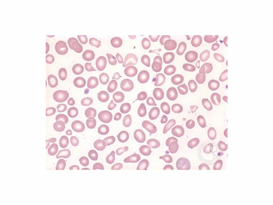

Poikilocytosis
• Variation in shape of erythrocytes Severe iron deficiency Beta thalassemia


Heinz bodies
• Denatured hemoglobin / hemoglobin remnant stained by supravital staining seen as a dark dot in erythrocyte.
Red cell enzyme defects ( e.g G6PD deficiency , pyruvate kinase deficiency)
Drugs and chemicals causing haemolytic anaemia


Left shift and leukaemoid reactions
Less mature white cells released prematurely from the bone marrow . Careful examination may be needed to distinguish from leukemia . Also refers to increased neutrophil/lymphocyte ratio.
Sepsis ( caused by increased demand on neutrophils , less mature cells are released prematurely )
Tuberculosis Syphilis Toxoplasmosis Down syndrome
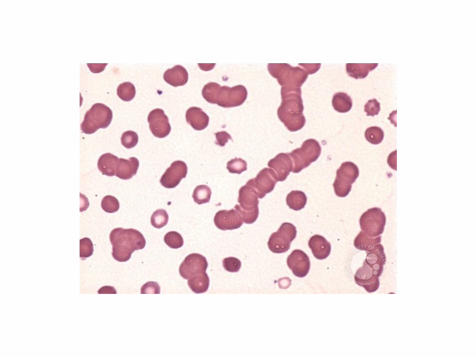

Rouleaux formation
• Erythrocytes stacked in rows one on another Inflammation Malignancy
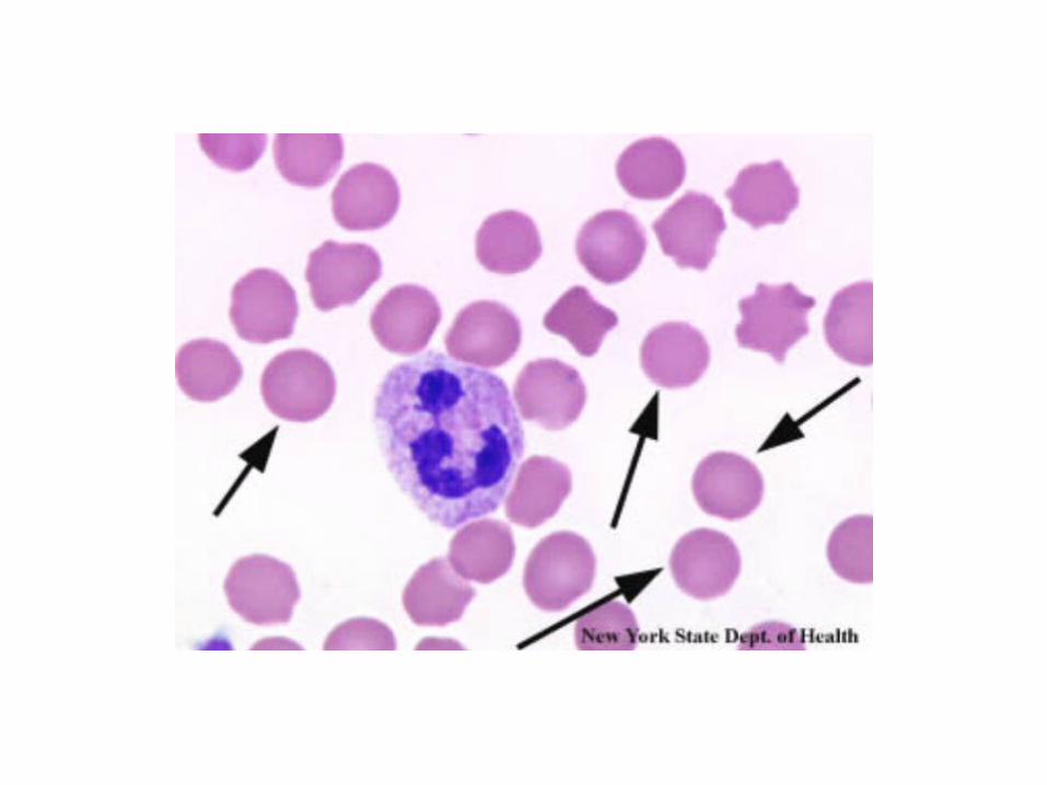

Spherocytes
• Seen as spherical cells with no central pallor; give rise to a low MCV
Hereditary spherocytosis Immune haemolytic anemia Severe burns Post transfusion


Dimorphic blood film
• The presence of two differently sized populations of red cells , with increased red cell distribution width(RDW)
Sideroblastic anemia(bone marrow cells unable to utilise iron to form haemoglobin characterized by ring sideroblasts in bone marrow; caused by inherited and acquired causes such as lead poisoning , anti TB drugs)

Continued
Combination of iron deficiency with a B12/folate deficiency
Iron deficiency anaemia (with microcytosis) and having received a blood transfusion


Basophil stippling
• Seen as numerous small dots in the red cell due to alpha chain clumping
Lead poisoning Beta thalassemia


Tear drop cells


Acanthocytes
• Red cells with spiny projections protruding from the surface
Post splenectomy Abetalipoprotenemia