blood anatomy & physiology. functions of blood transportation transportation heat regulation...
TRANSCRIPT
BloodBlood
Anatomy & PhysiologyAnatomy & Physiology
Functions of bloodFunctions of blood
TransportationTransportationHeat regulation Heat regulation
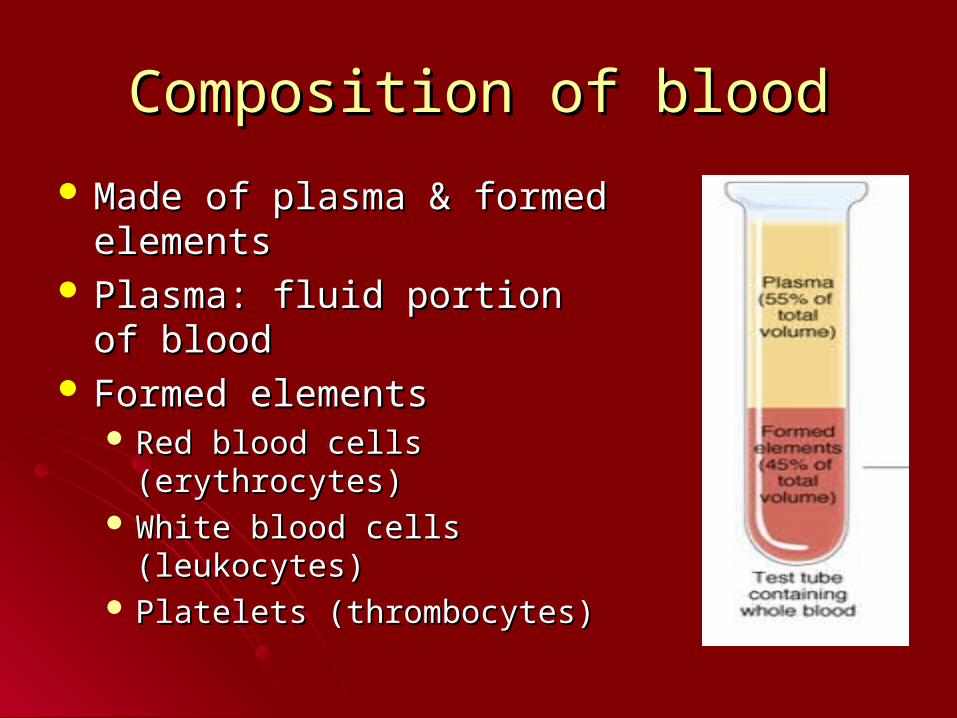
Composition of bloodComposition of blood
Made of plasma & formed Made of plasma & formed elementselements
Plasma: fluid portion of bloodPlasma: fluid portion of blood Formed elementsFormed elements
Red blood cells (erythrocytes)Red blood cells (erythrocytes) White blood cells (leukocytes)White blood cells (leukocytes) Platelets (thrombocytes)Platelets (thrombocytes)
Watch US blood cells clipWatch US blood cells clip
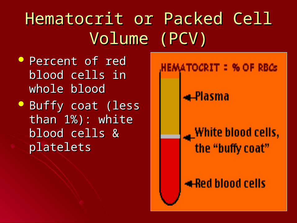
Hematocrit or Packed Cell Volume Hematocrit or Packed Cell Volume (PCV)(PCV)
Percent of red blood Percent of red blood cells in whole bloodcells in whole blood
Buffy coat (less than Buffy coat (less than 1%): white blood cells 1%): white blood cells & platelets & platelets
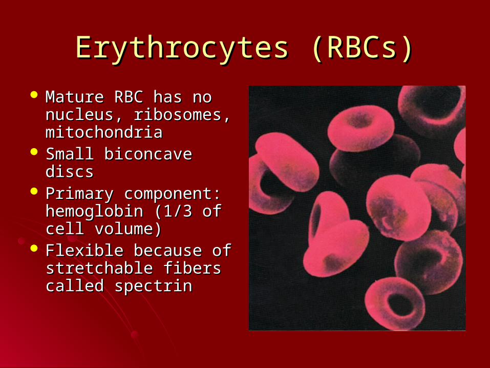
Erythrocytes (RBCs)Erythrocytes (RBCs)
Mature RBC has no Mature RBC has no nucleus, ribosomes, nucleus, ribosomes, mitochondriamitochondria
Small biconcave discs Small biconcave discs Primary component: Primary component:
hemoglobin (1/3 of hemoglobin (1/3 of cell volume)cell volume)
Flexible because of Flexible because of stretchable fibers stretchable fibers called spectrincalled spectrin
Function of RBCsFunction of RBCs
Transportation of oxygen & carbon dioxide Transportation of oxygen & carbon dioxide depends on hemoglobin & an enzyme depends on hemoglobin & an enzyme carbonic anhydrasecarbonic anhydrase
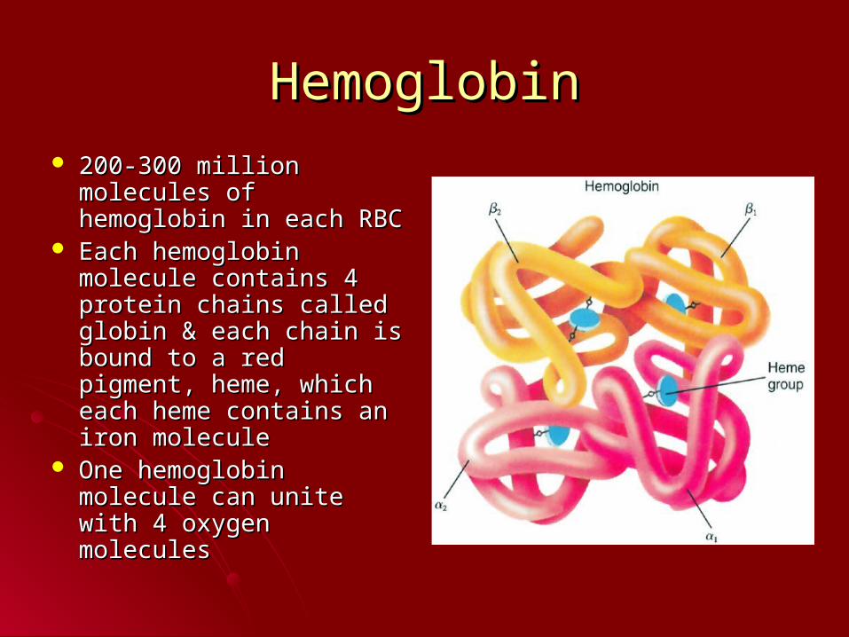
HemoglobinHemoglobin 200-300 million 200-300 million
molecules of hemoglobin molecules of hemoglobin in each RBCin each RBC
Each hemoglobin Each hemoglobin molecule contains 4 molecule contains 4 protein chains called protein chains called globin & each chain is globin & each chain is bound to a red pigment, bound to a red pigment, heme, which each heme heme, which each heme contains an iron moleculecontains an iron molecule
One hemoglobin One hemoglobin molecule can unite with 4 molecule can unite with 4 oxygen moleculesoxygen molecules
ErythropoiesisErythropoiesis
Formation of RBCFormation of RBCBegins in the bone marrow from Begins in the bone marrow from
hematopoietic stem cells which form all hematopoietic stem cells which form all blood cellsblood cells
In series of steps lose nuclei to become In series of steps lose nuclei to become reticulocyte which is released into reticulocyte which is released into circulation which become mature RBC circulation which become mature RBC which is smallerwhich is smaller
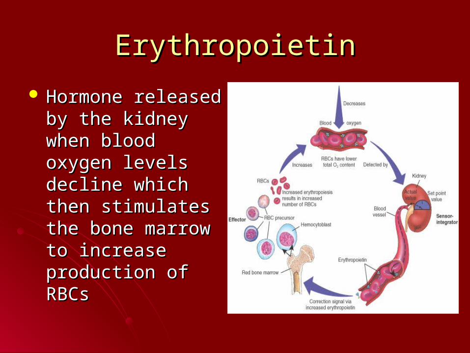
ErythropoietinErythropoietin
Hormone released by Hormone released by the kidney when the kidney when blood oxygen levels blood oxygen levels decline which then decline which then stimulates the bone stimulates the bone marrow to increase marrow to increase production of RBCsproduction of RBCs
Destruction of RBCsDestruction of RBCs
Life span of RBC is about 105-120 daysLife span of RBC is about 105-120 daysMacrophages in lining of blood vessels in Macrophages in lining of blood vessels in
spleen & liver phagocytose old or spleen & liver phagocytose old or damaged RBCdamaged RBC
Hemoglobin broken down & amino acids, Hemoglobin broken down & amino acids, iron & pigment bilirubin releasediron & pigment bilirubin released
Iron used to form new hemoglobin & Iron used to form new hemoglobin & bilirubin transported to liver & excreted into bilirubin transported to liver & excreted into the intestines in bilethe intestines in bile
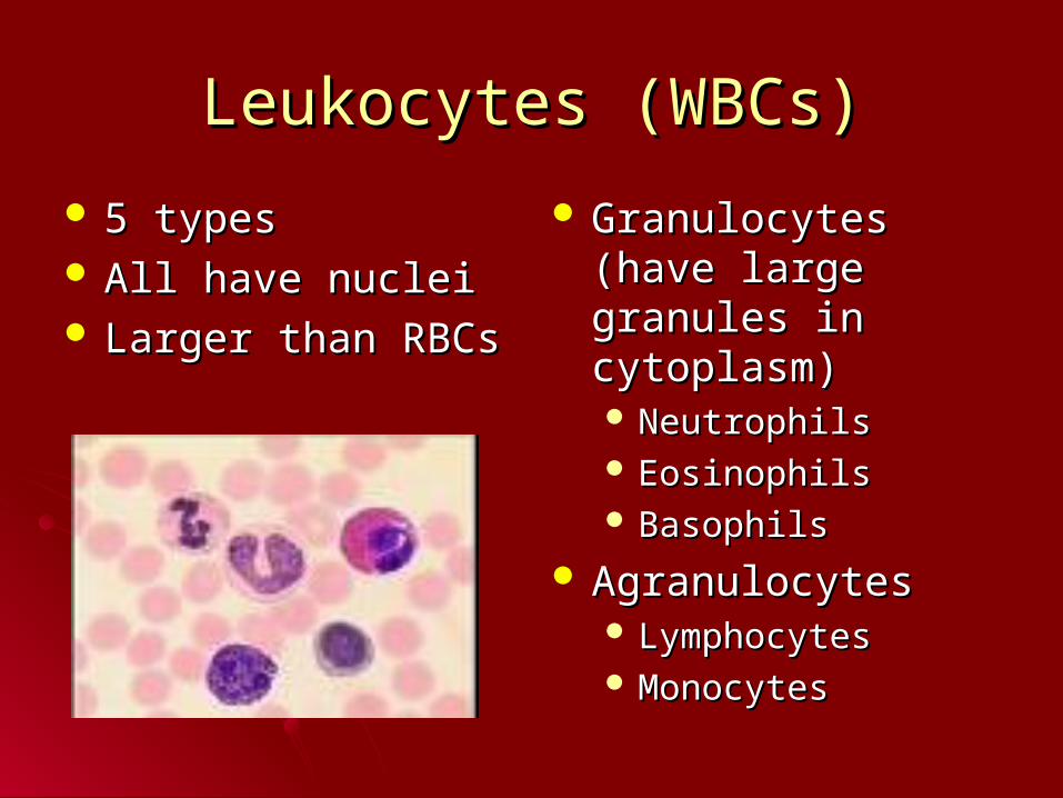
Leukocytes (WBCs)Leukocytes (WBCs)
5 types5 types All have nucleiAll have nuclei Larger than RBCsLarger than RBCs
Granulocytes (have Granulocytes (have large granules in large granules in cytoplasm)cytoplasm) NeutrophilsNeutrophils EosinophilsEosinophils BasophilsBasophils
AgranulocytesAgranulocytes LymphocytesLymphocytes MonocytesMonocytes
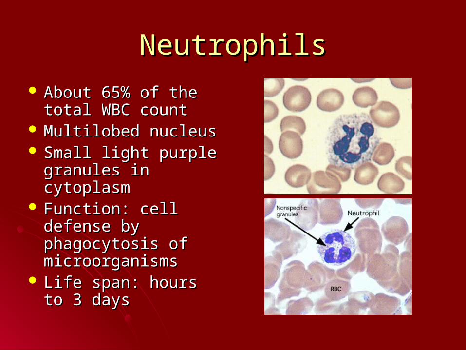
NeutrophilsNeutrophils
About 65% of the total About 65% of the total WBC countWBC count
Multilobed nucleusMultilobed nucleus Small light purple Small light purple
granules in cytoplasmgranules in cytoplasm Function: cell defense Function: cell defense
by phagocytosis of by phagocytosis of microorganismsmicroorganisms
Life span: hours to 3 Life span: hours to 3 daysdays
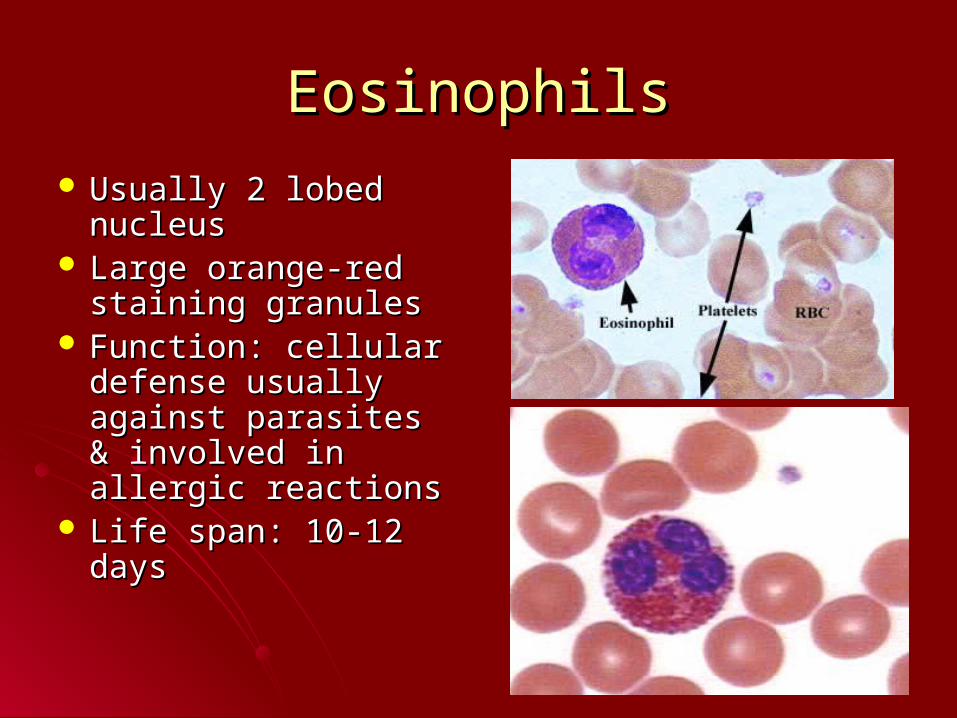
EosinophilsEosinophils
Usually 2 lobed Usually 2 lobed nucleusnucleus
Large orange-red Large orange-red staining granulesstaining granules
Function: cellular Function: cellular defense usually defense usually against parasites & against parasites & involved in allergic involved in allergic reactionsreactions
Life span: 10-12 daysLife span: 10-12 days
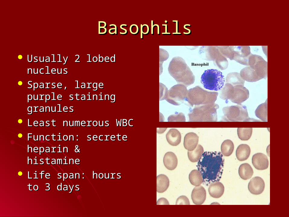
BasophilsBasophils
Usually 2 lobed Usually 2 lobed nucleusnucleus
Sparse, large purple Sparse, large purple staining granulesstaining granules
Least numerous WBCLeast numerous WBC Function: secrete Function: secrete
heparin & histamineheparin & histamine Life span: hours to 3 Life span: hours to 3
daysdays
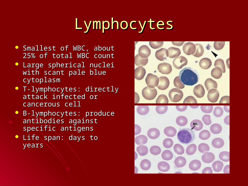
LymphocytesLymphocytes Smallest of WBC, about Smallest of WBC, about
25% of total WBC count25% of total WBC count Large spherical nuclei Large spherical nuclei
with scant pale blue with scant pale blue cytoplasmcytoplasm
T-lymphocytes: directly T-lymphocytes: directly attack infected or attack infected or cancerous cellcancerous cell
B-lymphocytes: produce B-lymphocytes: produce antibodies against antibodies against specific antigensspecific antigens
Life span: days to yearsLife span: days to years
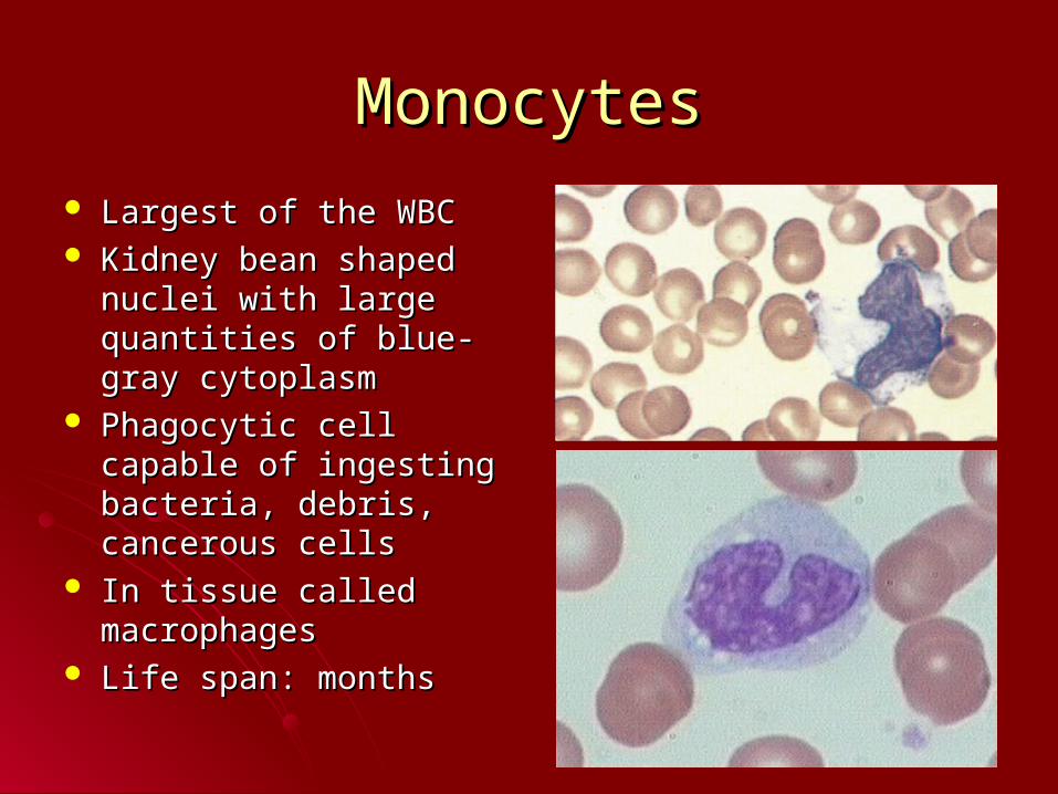
MonocytesMonocytes
Largest of the WBCLargest of the WBC Kidney bean shaped Kidney bean shaped
nuclei with large nuclei with large quantities of blue-gray quantities of blue-gray cytoplasmcytoplasm
Phagocytic cell capable Phagocytic cell capable of ingesting bacteria, of ingesting bacteria, debris, cancerous cellsdebris, cancerous cells
In tissue called In tissue called macrophagesmacrophages
Life span: monthsLife span: months
WBC FormationWBC Formation
Neutrophils, eosinophils, basophils Neutrophils, eosinophils, basophils originate in bone marroworiginate in bone marrow
Most lymphocytes & monocytes originate Most lymphocytes & monocytes originate in lymphatic tissuein lymphatic tissue

Watch US platelets clipWatch US platelets clip
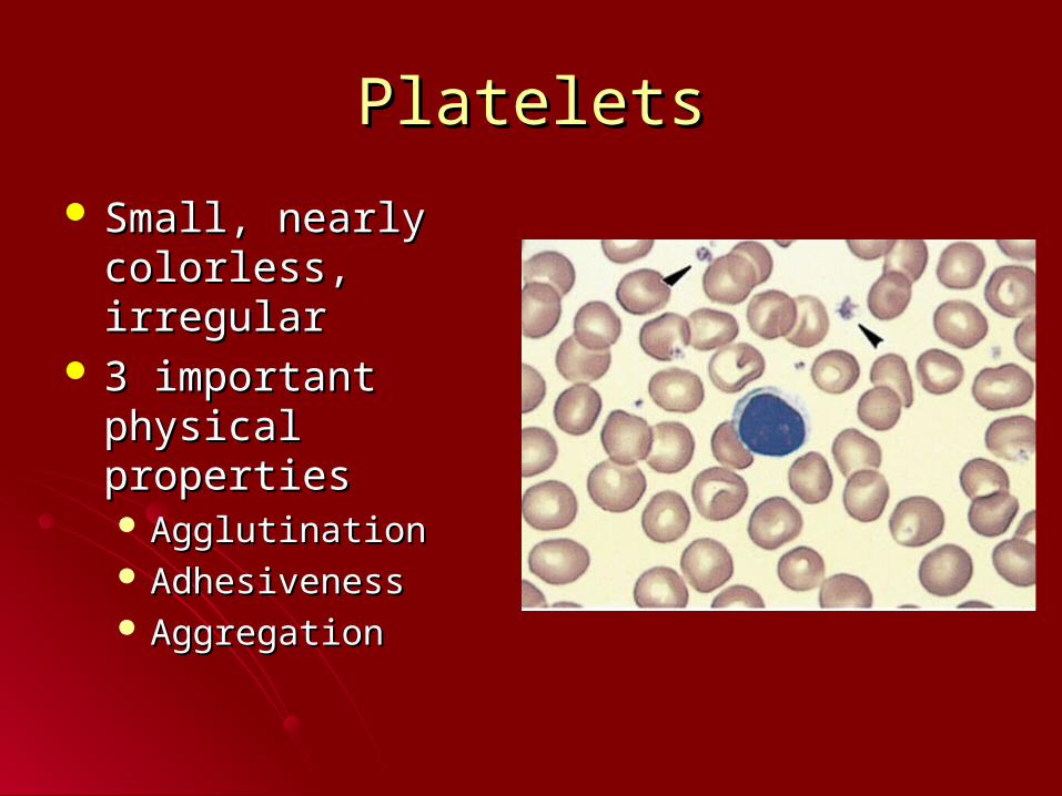
PlateletsPlatelets
Small, nearly Small, nearly colorless, irregularcolorless, irregular
3 important physical 3 important physical propertiesproperties AgglutinationAgglutination AdhesivenessAdhesiveness AggregationAggregation
Functions of plateletsFunctions of platelets
Hemostasis: stoppage of blood flowHemostasis: stoppage of blood flowDamage to blood vesselsDamage to blood vesselsvascular spasmvascular spasm
temporary platelet plug by sticky plateletstemporary platelet plug by sticky platelets secrete ADP, thromboxane & fatty acid secrete ADP, thromboxane & fatty acid (arachidonic acid) which are involved in (arachidonic acid) which are involved in coagulationcoagulation
Coagulation: blood clottingCoagulation: blood clotting
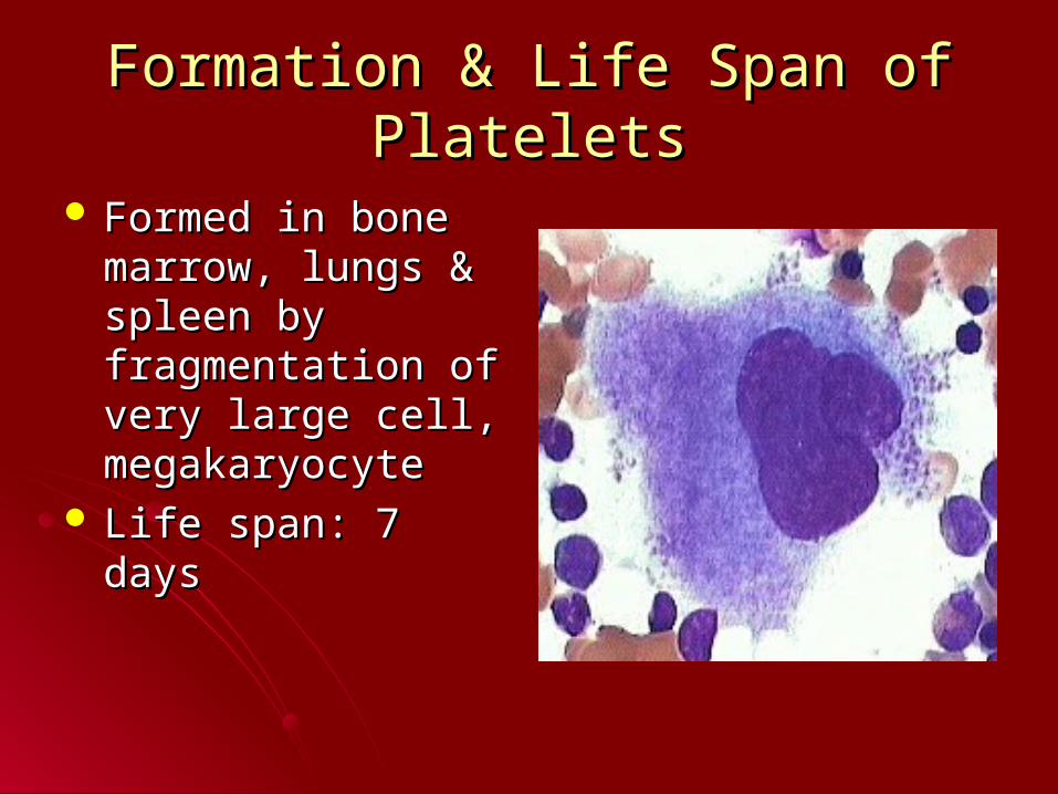
Formation & Life Span of PlateletsFormation & Life Span of Platelets
Formed in bone Formed in bone marrow, lungs & marrow, lungs & spleen by spleen by fragmentation of very fragmentation of very large cell, large cell, megakaryocytemegakaryocyte
Life span: 7 daysLife span: 7 days
Watch US blood type clipWatch US blood type clip
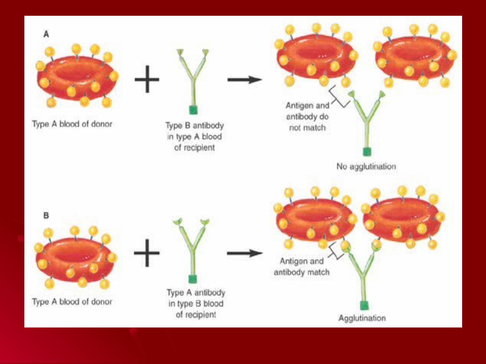
Blood TypesBlood Types
Refers to the type of antigens, called Refers to the type of antigens, called agglutinogensagglutinogens, present on RBC membrane, present on RBC membrane
Important blood antigens: A, B, RhImportant blood antigens: A, B, RhAgglutininsAgglutinins: antibodies dissolved in : antibodies dissolved in
plasma that react with specific blood group plasma that react with specific blood group antigensantigens
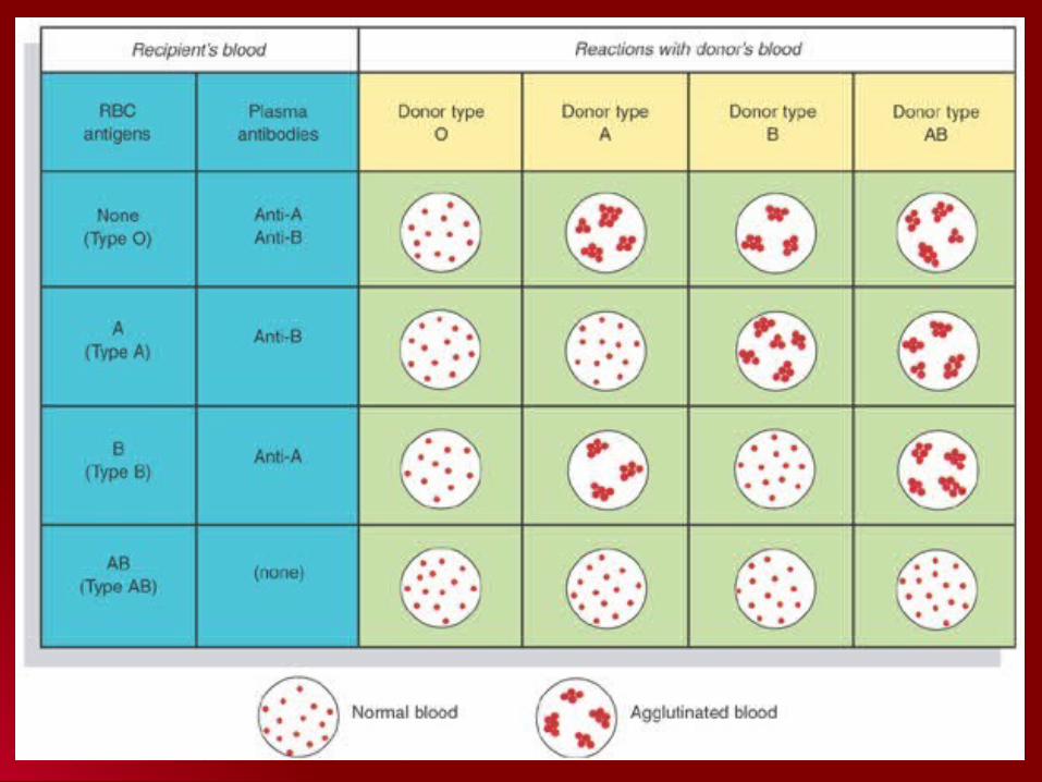
ABO SystemABO System
Type A: Antigen A on RBCsType A: Antigen A on RBCsType B: Antigen B on RBCsType B: Antigen B on RBCsType AB: Antigen A & B on RBCsType AB: Antigen A & B on RBCsType O: Neither A nor B on RBCsType O: Neither A nor B on RBCsPlasma never contains Ab against Ag Plasma never contains Ab against Ag
present on it own RBCs present on it own RBCs Plasma does contain AB against those Ag Plasma does contain AB against those Ag
notnot present on its RBCs present on its RBCs
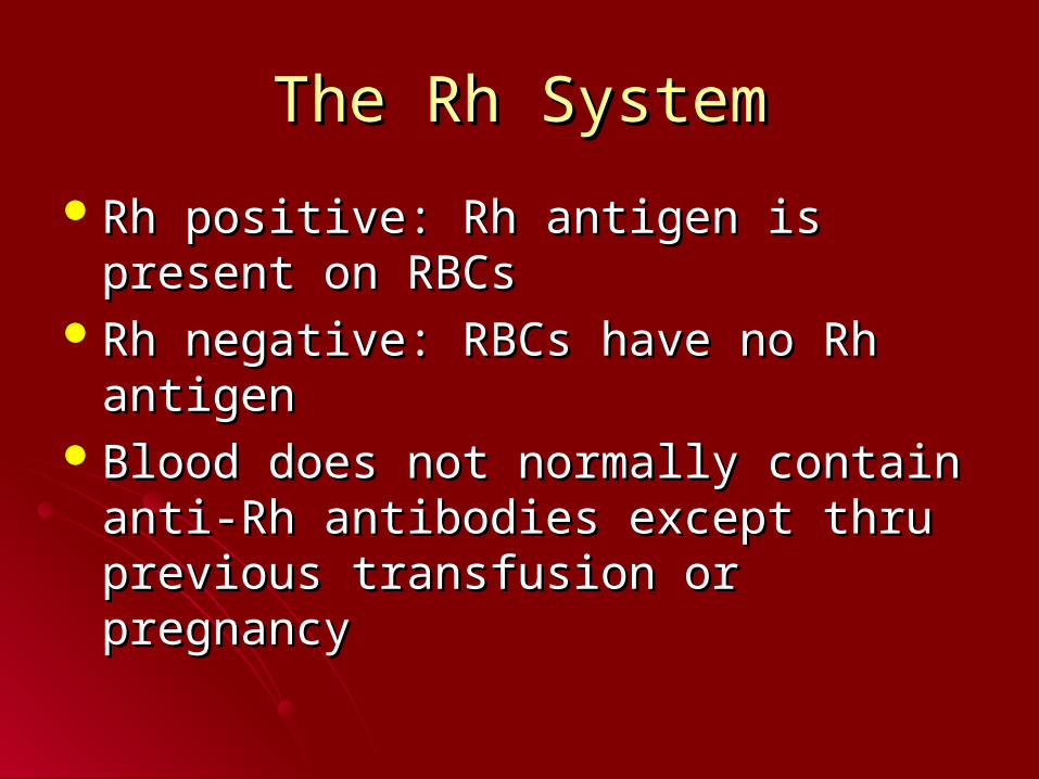
The Rh SystemThe Rh System
Rh positive: Rh antigen is present on Rh positive: Rh antigen is present on RBCsRBCs
Rh negative: RBCs have no Rh antigenRh negative: RBCs have no Rh antigenBlood does not normally contain anti-Rh Blood does not normally contain anti-Rh
antibodies except thru previous antibodies except thru previous transfusion or pregnancytransfusion or pregnancy
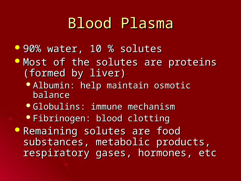
Blood PlasmaBlood Plasma
90% water, 10 % solutes90% water, 10 % solutesMost of the solutes are proteins (formed Most of the solutes are proteins (formed
by liver)by liver)Albumin: help maintain osmotic balanceAlbumin: help maintain osmotic balanceGlobulins: immune mechanismGlobulins: immune mechanismFibrinogen: blood clottingFibrinogen: blood clotting
Remaining solutes are food substances, Remaining solutes are food substances, metabolic products, respiratory gases, metabolic products, respiratory gases, hormones, etchormones, etc
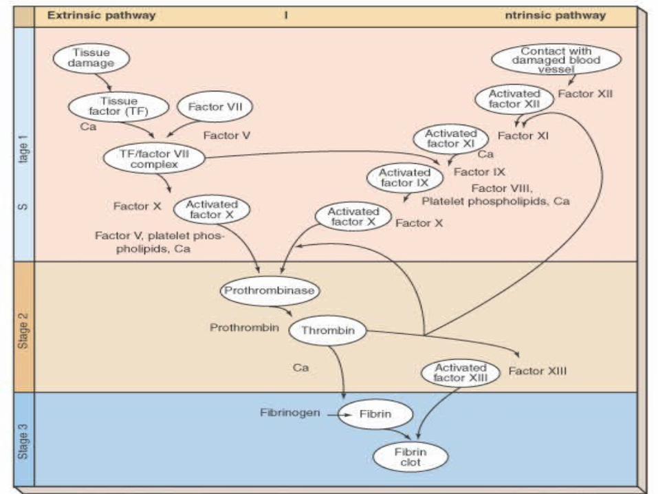
CoagulationCoagulation
Four components Four components critical to coagulationcritical to coagulation ProthrombinProthrombin ThrombinThrombin FibrinogenFibrinogen FibrinFibrin
Three stagesThree stages Stage IStage I Stage IIStage II Stage IIIStage III
Stage IStage I
Production of thromboplastin activator by Production of thromboplastin activator by either:either:Extrinsic pathway: chemicals released from Extrinsic pathway: chemicals released from
damaged tissuesdamaged tissues Intrinsic pathway: chemicals present in the Intrinsic pathway: chemicals present in the
bloodblood
Stage IIStage II
Conversion of prothrombin to thrombin by Conversion of prothrombin to thrombin by the prothrombin activator produced in the prothrombin activator produced in stage Istage I
Stage IIIStage III
Conversion of fibrinogen to fibrin and Conversion of fibrinogen to fibrin and production of fibrin clot by thrombin production of fibrin clot by thrombin produced in Stage IIproduced in Stage II
Coagulation factsCoagulation facts
Many of clotting factors require calcium ion Many of clotting factors require calcium ion as a cofactoras a cofactor
Liver synthesizes both prothrombin & Liver synthesizes both prothrombin & fibrinogen. Vitamin K is necessary for fibrinogen. Vitamin K is necessary for production of prothrombinproduction of prothrombin
Conditions that oppose clottingConditions that oppose clotting
Smooth surface of lining of blood vessels Smooth surface of lining of blood vessels does not allow platelets to stickdoes not allow platelets to stick
Antithrombins: substances in blood that Antithrombins: substances in blood that oppose or inactivate thrombinoppose or inactivate thrombinEx: HeparinEx: Heparin
Conditions that hasten clottingConditions that hasten clotting
Rough spot in the blood vessel liningRough spot in the blood vessel liningAbnormally slow blood flowAbnormally slow blood flow
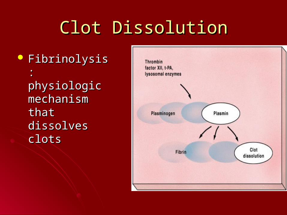
Clot DissolutionClot Dissolution
Fibrinolysis: Fibrinolysis: physiologic physiologic mechanism that mechanism that dissolves clotsdissolves clots

Image CitationsImage Citations Slide 5: Hematocrit, 12/20/06, Slide 5: Hematocrit, 12/20/06,
http://www.drstandley.com/labvalues_hematology.shtmlhttp://www.drstandley.com/labvalues_hematology.shtml Slide 13: Neutrophil, 12/27/06, Slide 13: Neutrophil, 12/27/06,
http://faculty.une.edu/com/abell/histo/histolab3a.htmhttp://faculty.une.edu/com/abell/histo/histolab3a.htm Slide 14: Eosinophil, 12/27/06, Slide 14: Eosinophil, 12/27/06,
http://faculty.une.edu/com/abell/histo/histolab3a.htmhttp://faculty.une.edu/com/abell/histo/histolab3a.htm Slide 14: eosinophil1a, 12/27/06, Slide 14: eosinophil1a, 12/27/06,
http://cellbio.utmb.edu/microanatomy/blood/more_eosinophils.htmhttp://cellbio.utmb.edu/microanatomy/blood/more_eosinophils.htm Slide 15: Basophils, 12/27/06, Slide 15: Basophils, 12/27/06,
http://faculty.une.edu/com/abell/histo/histolab3a.htmhttp://faculty.une.edu/com/abell/histo/histolab3a.htm Slide 16: Medium lymphocyte, 12/27/06, Slide 16: Medium lymphocyte, 12/27/06,
http://www.anatomy.dal.ca/Human_Histology/Lab7/61LO4.htmlhttp://www.anatomy.dal.ca/Human_Histology/Lab7/61LO4.html Slide 17: Monocyte, 12/27/06, Slide 17: Monocyte, 12/27/06,
http://www.med-ed.virginia.edu/courses/path/innes/nh/wcbmaturatiohttp://www.med-ed.virginia.edu/courses/path/innes/nh/wcbmaturation.cfmn.cfm
Slide 21: Megakaryocyte, 12/30/06, http://www.med-Slide 21: Megakaryocyte, 12/30/06, http://www.med-ed.virginia.edu/courses/path/innes/nh/platelets.cfmed.virginia.edu/courses/path/innes/nh/platelets.cfm