blockade of tnf-α rapidly inhibits pain responses in … · blockade of tnf-α rapidly inhibits...
TRANSCRIPT

Blockade of TNF-α rapidly inhibits pain responses inthe central nervous systemAndreas Hessa,1, Roland Axmannb,1, Juergen Rechb,1, Stefanie Finzelb, Cornelia Heindla, Silke Kreitza, Marina Sergeevaa,Marc Saakec, Meritxell Garciac, George Kolliasd, Rainer H. Straube, Olaf Spornsf, Arnd Doerflerc, Kay Brunea,and Georg Schettb,2
aInstitute of Experimental and Clinical Pharmacology and Toxicology, bDepartment of Internal Medicine 3, and cDivision of Neuroradiology, University ofErlangen-Nuremberg, 91054 Erlangen, Germany; dInstitute of Immunology, Alexander Fleming Biomedical Sciences Research Center, 16672 Vari, Greece;eDepartment of Internal Medicine I, University of Regensburg, 93053 Regensburg, Germany; and fDepartment of Psychological and Brain Sciences, Programsin Neuroscience and Cognitive Science, Indiana University, Bloomington, IN 47405
Edited* by Charles A. Dinarello, University of Colorado Denver, Aurora, CO, and approved December 29, 2010 (received for review August 19, 2010)
There has been a consistent gap in understanding how TNF-α neu-tralization affects the disease state of arthritis patients so rapidly,considering that joint inflammation in rheumatoid arthritis is achronic conditionwith structural changes.We thushypothesized thatneutralizationofTNF-α acts through theCNSbeforedirectly affectingjoint inflammation. Through use of functional MRI (fMRI), we dem-onstrate that within 24 h after neutralization of TNF-α, nociceptiveCNS activity in the thalamus and somatosensoric cortex, but also theactivation of the limbic system, is blocked. Brain areas showingblood-oxygen level-dependent signals, a validated method to assessneuronal activity elicited by pain, were significantly reduced as earlyas 24 h after an infusion of a monoclonal antibody to TNF-α. In con-trast, clinical and laboratory markers of inflammation, such as jointswelling and acute phase reactants, were not affected by anti-TNF-αat these early time points. Moreover, arthritic mice overexpressinghuman TNF-α showedan alteredpain behavior and amore intensive,widespread, and prolonged brain activity upon nociceptive stimulicompared with wild-type mice. Similar to humans, these changes,as well as the rewiring of CNS activity resulting in tight clusteringin the thalamus, were rapidly reversed after neutralization of TNF-α.These results suggest that neutralization of TNF-α affects nociceptivebrain activity in the context of arthritis, long before it achieves anti-inflammatory effects in the joints.
cytokines | antiinflammatory therapy
Arthritis is one of the most disabling chronic human diseases.Rheumatoid arthritis (RA) affects up to 1%of the population
and is characterized by pain, swelling, and stiffness of joints,leading to a serious decay of life quality. Pain is the initial andprevailing symptom of the disease, leading to immobility, which inturn causes complications such as osteoporosis and cardiovasculardisease. During the last 10 y the pharmacologic treatment of dis-eases such as RA has substantially improved because of the de-velopment of cytokine blocking agents (1).Although inflamed joints express a multitude of mediators, in-
cluding cytokines, chemokines, and growth factors, which con-tribute to the pathogenesis of arthritis, inhibition of TNF-α hasemerged as a particularly successful therapeutic strategy (2, 3).The therapeutic success of TNF-α blockade in RA is unique andhas been largely considered to result from rapid and efficientneutralization of joint inflammation based on breakdown of theinflammatory cytokine network in the affected joint, which resultsin an improvement of the signs and symptoms of the disease (4).It has, however, always been stunning, how fast the blockade ofTNF-α improves the patient condition, in particular because dis-eases like RA are highly chronic, building up a vast amount ofinflammatory tissue, and leading to irreversible damage of thecartilage and the bone. Thus, rapid resolution of this highly or-ganized inflammatory tissue or tissue damage is very unlikely toexplain the fast effect of TNF-α blockade. Evenmore importantly,neutralization of other inflammatorymediators, in particular IL-1,
which is a central inducer of inflammation and structural damagein arthritis (5), appears to have less-pronounced and less-rapideffects on the symptoms of RA, although its role in protectingstructural changes in the joints is substantial (6, 7).These observations, and the fact that traditional antirheumatic
drugs such as methotrexate achieve far slower clinical responsesthan TNF-α blockers, have led us to suspect that blockade ofTNF-α could has additional effects that go beyond the sole in-hibition of joint inflammation.We hypothesized that TNF-α block-ade could influence processes in the CNS, which control thepatient’s perception of the disease state. Indeed, earlier observa-tions inmice have suggested thatTNF-α can induce a depressive-likebehavior in mice (8). Pain processing and sensation are key mech-anisms in the CNS elicited by arthritis, and pain is the dominantsymptom of disease, which by far has the strongest impact on thedisease burden of arthritis. Importantly, effects onpain also drive thestandard read-out parameters formeasuring therapeutic response inarthritis. Thus, if TNF-α blockade has unique pain-reducing prop-erties, its efficacy in reducing clinical disease activity of RA willbe high, as such disease-activity measures are strongly influencedby pain. In consequence, other treatment strategies, lacking suchCNSeffects, will less strongly affect the patient’s disease state, even ifshowing similar anti-inflammatory or structure-sparing effects.Interestingly, inflammation is considered to lower the thresh-
old for pain perception in the CNS, leading to hyperalgesia (9).In contrast to the well-documented role of TNF-α as a proin-flammatory cytokine, its role as a mediator of pain is in-completely characterized. It is known that both receptors forTNF-α are expressed in dorsal root ganglion neurons and thatintra-articular injection of TNF-α blocking agents reduces noci-ception elicited by joint inflammation (10). TNF-α is also in-volved in development of mechanical hyperalgesia followinginjury (11, 12). Hence, we hypothesized that TNF-α leads toa major change in pain perception in the CNS during arthritis.To search for the rapid effects of TNF-α blockade, we visual-
ized cerebral nociception elicited by arthritis and its reversibilityupon TNF-α blockade using blood-oxygen level-dependent(BOLD) functional MRI (fMRI) as early as 24 h after initiation ofthe treatment. The BOLD signal reflects changes in hemody-namics linked to increased or decreased neuronal metabolic ac-tivity in response to outer sensory stimulation, thus allowing
Author contributions: K.B. and G.S. designed research; A.H., R.A., J.R., S.F., C.H., S.K.,M. Sergeeva, M.G., and G.S. performed research; A.H., G.K., and G.S. contributed newreagents/analytic tools; A.H., R.A., J.R., M. Saake, R.H.S., O.S., A.D., K.B., and G.S. analyzeddata; and A.H. and G.S. wrote the paper.
The authors declare no conflict of interest.
*This Direct Submission article had a prearranged editor.1A.H., R.A., and J.R. contributed equally to this work.2To whom correspondence should be addressed. E-mail: [email protected].
This article contains supporting information online at www.pnas.org/lookup/suppl/doi:10.1073/pnas.1011774108/-/DCSupplemental.
www.pnas.org/cgi/doi/10.1073/pnas.1011774108 PNAS Early Edition | 1 of 6
MED
ICALSC
IENCE
S
localization of brain areas activated by stimuli (13, 14). Asa proof-of-concept, we first evaluated whether TNF-α blockaderapidly reverses the hypernociception in patients with RA. Wethen performed an in-depth analysis of this rapid effect of TNF-αblockade in a mouse model of arthritis based on the over-expression of human TNF-α.
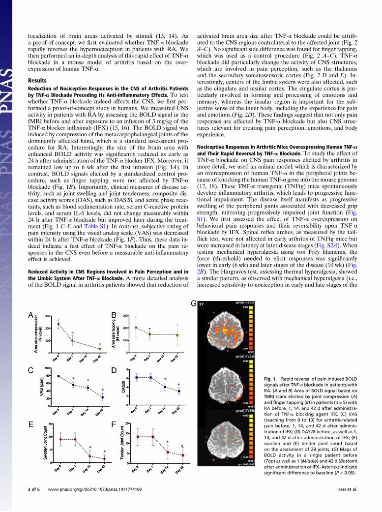
ResultsReduction of Nociceptive Responses in the CNS of Arthritis Patientsby TNF-α Blockade Preceding Its Anti-Inflammatory Effects. To testwhether TNF-α blockade indeed affects the CNS, we first per-formed a proof-of-concept study in humans. We measured CNSactivity in patients with RA by assessing the BOLD signal in thefMRI before and after exposure to an infusion of 3 mg/kg of theTNF-α blocker infliximab (IFX) (15, 16). The BOLD signal wasinduced by compression of the metacarpophalangeal joints of thedominantly affected hand, which is a standard assessment pro-cedure for RA. Interestingly, the size of the brain area withenhanced BOLD activity was significantly reduced as early as24 h after administration of the TNF-α blocker IFX. Moreover, itremained low up to 6 wk after the first infusion (Fig. 1A). Incontrast, BOLD signals elicited by a standardized control pro-cedure, such as finger tapping, were not affected by TNF-αblockade (Fig. 1B). Importantly, clinical measures of disease ac-tivity, such as joint swelling and joint tenderness, composite dis-ease activity scores (DAS), such as DAS28, and acute phase reac-tants, such as blood sedimentation rate, serum C-reactive proteinlevels, and serum IL-6 levels, did not change measurably within24 h after TNF-α blockade but improved later during the treat-ment (Fig. 1 C–E and Table S1). In contrast, subjective rating ofpain intensity using the visual analog scale (VAS) was decreasedwithin 24 h after TNF-α blockade (Fig. 1F). Thus, these data in-deed indicate a fast effect of TNF-α blockade on the pain re-sponses in the CNS even before a measurable anti-inflammatoryeffect is achieved.
Reduced Activity in CNS Regions Involved in Pain Perception and inthe Limbic System After TNF-α Blockade. A more detailed analysisof the BOLD signal in arthritis patients showed that reduction of
activated brain area size after TNF-α blockade could be attrib-uted to the CNS regions contralateral to the affected joint (Fig. 2A–C). No significant side difference was found for finger tapping,which was used as a control procedure (Fig. 2 A–C). TNF-αblockade did particularly change the activity of CNS structures,which are involved in pain perception, such as the thalamusand the secondary somatosensoric cortex (Fig. 2 D and E). In-terestingly, centers of the limbic system were also affected, suchas the cingulate and insular cortex. The cingulate cortex is par-ticularly involved in forming and processing of emotions andmemory, whereas the insular region is important for the sub-jective sense of the inner body, including the experience for painand emotions (Fig. 2D). These findings suggest that not only painresponses are affected by TNF-α blockade but also CNS struc-tures relevant for creating pain perception, emotions, and bodyexperience.
Nociceptive Responses in Arthritic Mice Overexpressing Human TNF-αand Their Rapid Reversal by TNF-α Blockade. To study the effect ofTNF-α blockade on CNS pain responses elicited by arthritis inmore detail, we used an animal model, which is characterized byan overexpression of human TNF-α in the peripheral joints be-cause of knocking the human TNF-α gene into the mouse genome(17, 18). These TNF-α transgenic (TNFtg) mice spontaneouslydevelop inflammatory arthritis, which leads to progressive func-tional impairment. The disease itself manifests as progressiveswelling of the peripheral joints associated with decreased gripstrength, mirroring progressively impaired joint function (Fig.S1). We first assessed the effect of TNF-α overexpression onbehavioral pain responses and their reversibility upon TNF-αblockade by IFX. Spinal reflex arches, as measured by the tail-flick test, were not affected in early arthritis of TNFtg mice butwere increased in latency at later disease stages (Fig. S2A). Whentesting mechanical hyperalgesia using von Frey filaments, theforce (threshold) needed to elicit responses was significantlylower in early (6 wk) and later stages of the disease (10 wk) (Fig.2B). The Hargraves test, assessing thermal hyperalgesia, showeda similar pattern, as observed with mechanical hyperalgesia (i.e.,increased sensitivity to nociception in early and late stages of the
Fig. 1. Rapid reversal of pain induced BOLDsignals after TNF-α blockade in patients withRA. (A and B) Area of BOLD signal based onfMRI scans elicited by joint compression (A)and finger tapping (B) in patients (n = 5) withRA before, 1, 14, and 42 d after administra-tion of TNF-α blocking agent IFX. (C) VAS(reaching from 0 to 10) for arthritis-relatedpain before, 1, 14, and 42 d after adminis-tration of IFX; (D) DAS28 before, as well as 1,14, and 42 d after administration of IFX; (E)swollen and (F) tender joint count basedon the assessment of 28 joints. (G) Maps ofBOLD activity in a single patient before(Top) as well as 1 (Middle) and 42 d (Bottom)after administration of IFX. Asterisks indicatesignificant difference to baseline (P < 0.05).
2 of 6 | www.pnas.org/cgi/doi/10.1073/pnas.1011774108 Hess et al.

disease of TNFtg mice) (Fig. S2C). In contrast, visceral noci-ception, measured by intraperitoneal injection of MgSO4, wassignificantly reduced rather than enhanced in TNFtgmice, both inearly and late disease stages, suggesting a desensitization to vis-ceral nociception in TNFtg mice (Fig. S2D). The Rotarod testmeasuring motor activity was not impaired at an early diseasestage but significantly decreased at later stages (Fig. S3A). Similardata were obtained when the suspended-tail test was applied,which showed a significant decrease in overall activity in TNFtgmice but only in later stages of disease (Fig. S3B).To test whether this nociceptive sensitization is reversible, we
treated TNFtg mice with the TNF-α blocker IFX. Whereas thelatency of spinal pain reflex arches, as measured by the tail-flicktest, were normalized 72 h after TNF-α blockade (Fig. S2E, in-creased sensitivity to mechanical nociceptive stimuli in TNFtgmice was completely reversed as early as 24 h after injection (Fig.S2F). This profound and rapid antinociceptive effect of TNF-αblockade was further substantiated by the Hargreaves test, whichnormalized within 24 h after TNF-α blockade and remained atthe level of WT mice over 72 h (Fig. S2G). Within this short timeperiod after TNF-α blockade, no apparent change of clinic-analogparameters (such as paw swelling or grip strength) or histopath-ologic signs of arthritis (such as synovitis) could be observed.Moderate effects on visceral nociception were also observed, asthe reduced sensitivity of TNFtg mice to visceral nociception wasreversed to WT levels within 72 h after TNF-α blockade (Fig.S2H). TNF-α blockade also completely restored motor activitywithin the first 24 h (Rotarod test) (Fig. S3C). Similarly, butslightly less pronounced, decreased overall mobility and activityof TNFtg mice as measured by the suspended-tail test also im-proved upon TNF-α blockade(Fig. S3D).
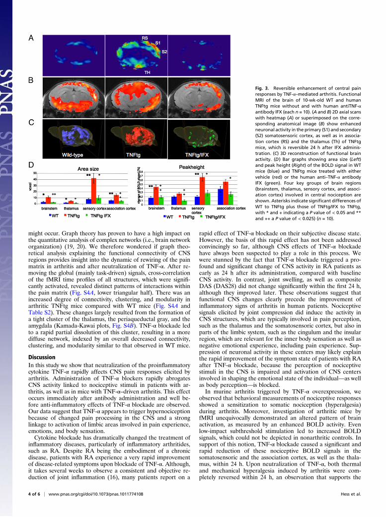
Mapping of Nociceptive Brain Responses Elicited by Arthritis andTheir Modulation by TNF-α Blockade.Comparative fMRI analysis ofthe brain-activity patterns elicited by heat stimulation of the hindpaw revealed a significant increase of the BOLD activity inTNFtg mice in the so-called pain matrix (19, 20) of the braincompared with the WT mice (Fig. 3 A–C). TNF-α blockaded toa fast and complete reversal of the increased brain activity in
TNFtgmice to the level ofWTmice within 24 h.When quantifyingthe BOLD activity by measuring the area size and peak amplitudeof the BOLD signal, we observed a consistent and significant in-crease in the somatosensoric cortex of TNFtg mice, which wascompletely abolished after TNF-α blockade (Fig. 3C). Smaller,but still significant, increases (Fig. 3C) were also observed in thethalamus, as well as in the association cortex in TNFtg comparedwith WT mice, which were again completely reversed uponblockade of TNF-α (Fig. 3D).On the basis of these observations, we aimed at precisely
mapping the interaction of the temporal structure of the BOLDsignals in arthritis (Fig. 4A). Again, amplitudes of BOLD signalswere generally higher in TNFtg mice than in WT mice. More-over, even low-impact subthreshold stimulation led to increasedbrain activity in TNFtg mice, which was not found in WT mice(red color in Fig. 4A at S1/S2 and peaks in Fig. 4B). This patternwas completely reversed upon TNF-α blockade as early as 24 hafter injection. Aside from higher amplitudes, brain activationwas more widespread in TNFtg mice, affecting many more brainregions than in WT mice. In the latter, the nociceptive responsewas localized to several well-defined brain areas (vertical patternin Fig. 4A, white circles). This spreading of brain activation inTNFtg mice was partially reversed upon blockade of TNF-α. Inaddition, the increased BOLD activity was maintained evenduring the intervals between stimulations and did not completelyregress to baseline levels (horizontal red lines in Fig. 4A; red linein Fig. 4B is permanently high above baseline). TNF-α blockaderapidly reversed this prolonged activation, and the BOLD signalin the time periods between the stimulations dropped dramati-cally, at times below the baseline level. This effect was particu-larly pronounced in somatosensoric cortical and limbic regions ofthe brain (asterisks at dark blue intervals in Fig. 4A and greenline Fig. 4B).
Rewiring of the Pain Matrix in the CNS by Chronic Arthritis and ItsReversibility by TNF-α Blockade. Higher-order changes in brainfunction may not only be reflected in changes of responseproperties of single brain structures. Instead, dynamic changes ofthe complex network connectivity (i.e., within the pain matrix)
Fig. 2. Mapping of BOLD changes after TNF-α blockingtherapy. (A–C) Changes in the area size of the BOLDsignal elicited by joint compression (blue bars) or fingertapping (red bars) comparing day 0 with day 1 (W2/W1)and day 0 with day 42 (W3/W1) after IFX treatment:overall (A) and specific for the contralateral (B) andipsilateral hemisphere (C). (D and E) Changes in thearea size of the BOLD signal elicited by joint compres-sion (D) or finger tapping (E) comparing day 0 with day1 (W2/W1) at the following brain regions: thalamus(Th), secondary (S2) and primary (S1), somatosensoriccortex, parietal cortex (Par), posterior cingulate cortex(PCC), anterior cingulate cortex (ACC), lateral (LPFC) andmedial (MPFC) prefrontal cortex, anterior (AIns) andposterior (PIns) insular cortex, cerebellum (Cb), motorcortex (M1), and periaqueductal gray (PAG).
Hess et al. PNAS Early Edition | 3 of 6
MED
ICALSC
IENCE
S
might occur. Graph theory has proven to have a high impact onthe quantitative analysis of complex networks (i.e., brain networkorganization) (19, 20). We therefore wondered if graph theo-retical analysis explaining the functional connectivity of CNSregions provides insight into the dynamic of rewiring of the painmatrix in arthritis and after neutralization of TNF-α. After re-moving the global (mainly task-driven) signals, cross-correlationof the fMRI time profiles of all structures, which were signifi-cantly activated, revealed distinct patterns of interactions withinthe pain matrix (Fig. S4A, lower triangular half). There was anincreased degree of connectivity, clustering, and modularity inarthritic TNFtg mice compared with WT mice (Fig. S4A andTable S2). These changes largely resulted from the formation ofa tight cluster of the thalamus, the periaqueductal gray, and theamygdala (Kamada-Kawai plots, Fig. S4B). TNF-α blockade ledto a rapid partial dissolution of this cluster, resulting in a morediffuse network, indexed by an overall decreased connectivity,clustering, and modularity similar to that observed in WT mice.
DiscussionIn this study we show that neutralization of the proinflammatorycytokine TNF-α rapidly affects CNS pain responses elicited byarthritis. Administration of TNF-α blockers rapidly abrogatesCNS activity linked to nociceptive stimuli in patients with ar-thritis, as well as in mice with TNF-α–driven arthritis. This effectoccurs immediately after antibody administration and well be-fore anti-inflammatory effects of TNF-α blockade are observed.Our data suggest that TNF-α appears to trigger hypernociceptionbecause of changed pain processing in the CNS and a stronglinkage to activation of limbic areas involved in pain experience,emotions, and body sensation.Cytokine blockade has dramatically changed the treatment of
inflammatory diseases, particularly of inflammatory arthritides,such as RA. Despite RA being the embodiment of a chronicdisease, patients with RA experience a very rapid improvementof disease-related symptoms upon blockade of TNF-α. Although,it takes several weeks to observe a consistent and objective re-duction of joint inflammation (16), many patients report on a
rapid effect of TNF-α blockade on their subjective disease state.However, the basis of this rapid effect has not been addressedconvincingly so far, although CNS effects of TNF-α blockadehave always been suspected to play a role in this process. Wewere stunned by the fact that TNF-α blockade triggered a pro-found and significant change of CNS activity in RA patients asearly as 24 h after its administration, compared with baselineCNS activity. In contrast, joint swelling, as well as compositeDAS (DAS28) did not change significantly within the first 24 h,although they improved later. These observations suggest thatfunctional CNS changes clearly precede the improvement ofinflammatory signs of arthritis in human patients. Nociceptivesignals elicited by joint compression did induce the activity inCNS structures, which are typically involved in pain perception,such as the thalamus and the somatosensoric cortex, but also inparts of the limbic system, such as the cingulum and the insularregion, which are relevant for the inner body sensation as well asnegative emotional experience, including pain experience. Sup-pression of neuronal activity in these centers may likely explainthe rapid improvement of the symptom state of patients with RAafter TNF-α blockade, because the perception of nociceptivestimuli in the CNS is impaired and activation of CNS centersinvolved in shaping the emotional state of the individual—as wellas body perception—is blocked.In murine arthritis triggered by TNF-α overexpression, we
observed that behavioral measurements of nociceptive responsesshowed a sensitization to somatic nociception (hyperalgesia)during arthritis. Moreover, investigation of arthritic mice byfMRI unequivocally demonstrated an altered pattern of brainactivation, as measured by an enhanced BOLD activity. Evenlow-impact subthreshold stimulation led to increased BOLDsignals, which could not be depicted in nonarthritic controls. Insupport of this notion, TNF-α blockade caused a significant andrapid reduction of these nociceptive BOLD signals in thesomatosensoric and the association cortex, as well as the thala-mus, within 24 h. Upon neutralization of TNF-α, both thermaland mechanical hyperalgesia induced by arthritis were com-pletely reversed within 24 h, an observation that supports the
Fig. 3. Reversible enhancement of central painresponses by TNF-α–mediated arthritis. FunctionalMRI of the brain of 10-wk-old WT and humanTNFtg mice without and with human antiTNF-αantibody IFX (each n = 10). (A and B) 2D axial scanswith heatmap (A) or superimposed on the corre-sponding anatomical image (B) show enhancedneuronal activity in the primary (S1) and secondary(S2) somatosensoric cortex, as well as in associa-tion cortex (RS) and the thalamus (Th) of TNFtgmice, which is reversible 24 h after IFX adminis-tration. (C) 3D reconstruction of functional brainactivity. (D) Bar graphs showing area size (Left)and peak height (Right) of the BOLD signal in WTmice (blue) and TNFtg mice treated with eithervehicle (red) or the human anti–TNF-α antibodyIFX (green). Four key groups of brain regions(brainstem, thalamus, sensory cortex, and associ-ation cortex) involved in central nociception areshown. Asterisks indicate significant differences ofWT to TNFtg plus those of TNFtg/IFX to TNFtg,with * and + indicating a P value of < 0.05 and **and ++ a P value of < 0.025) (n = 10).
4 of 6 | www.pnas.org/cgi/doi/10.1073/pnas.1011774108 Hess et al.

results obtained in RA patients. Within this short period noapparent effect on clinical analog parameters (such as pawswelling) or histopathologic signs of arthritis (such as synovitis)could be observed. This result indicates that the CNS effect ofTNF-α blockade precedes its anti-inflammatory effect.Interestingly, we even observed a decrease in the BOLD signal
below the normal levels observed in nonarthritic WT mice. Thiseffect was again particularly pronounced in somatosensoric cor-tical and limbic regions of the brain. One explanation for thisdecrease of the BOLD signal below the baseline level could be anincreased activity of the cerebral inhibitory network developedduring this chronic pain state (21). In this context, it is worthmentioning that brain network analysis detected a tight clusterconsisting of thalamic structures, the periaquaeductal gray, andlimbic areas, such as the amygdala in TNFtg mice. This findingsupports the concept of an increased efficacy of the cerebral in-hibitory network in arthritis. This rewiring may have developedas an adaptation to chronic pain preventing the abnormally in-creased flow of nociceptive signals to higher CNS structures, es-pecially to the cortex. Importantly, TNF-α blockade rapidlyinduced partial dissolution of this cluster; this happened within24 h after TNF-α blockade, in parallel with the reduction of thenociceptive BOLD activity and the restoration of the normalnociceptive behavior.In summary, these data show that TNF-α leads to more in-
tensive, widespread, and prolonged brain activity upon nocicep-tive stimulation, which is reversible upon neutralization of TNF-α.This reversal of pain sensitization occurs rapidly and is not pri-marily linked to the anti-inflammatory effects of TNF-α blockade.In patients with RA, a very similar rapid and profound down-regulation of nociceptive brain activity can be observed, whichdid precede the alleviation of joint inflammation. Although sub-clinical anti-inflammatory effects of TNF-α blockade within thefirst 24 h after exposure to TNF-α blockade cannot be ruled outcompletely, the lack of a drop in acute phase reactant and cyto-kine levels within this short time interval does at least not supportsuch a concept. Our findings emphasize that changes of nocicep-tive responses in the brain precede the anti-inflammatory effectof TNF-α blockers. Importantly, these findings may be a good
explanation for the rapid improvement of the symptom state in-duced by TNF-α blocking therapy. These data also suggest thatanticytokine therapy targetingTNF-αmay achieve its (fast or rapid)clinical benefit remote from the inflamed joint through influencingCNS processes. Our findings also extend previous observations onthe effects of TNF-α in the CNS (i.e., the regulation of expressionof clock genes), whichmay explain the high prevalence of fatigue ininflammatory diseases (22). Finally, our findings also raise thepossibility that the assessment of responsiveness of BOLD activityin the CNS of RA patients and patients with other forms of in-flammatory arthritis may be used as a surrogate marker for pre-dicting clinical responses to therapeutic intervention in a muchfaster way than it is currently realized.
Materials and MethodsMice. The human TNF-α and TNFtg mice (strain Tg197) were previously de-scribed (17). Treatment with IFX (Centocor), a neutralizing chimeric human-murine monoclonal antibody directed against human TNF-α, was done by asingle intravenous injection of a doseof 10mg/kg antibody 24hbefore testing(15). Clinical evaluation was performed weekly, starting at 4 wk after birth.Arthritis was evaluated in a blinded manner, as described previously (18). Allanimal experiments were approved by the local ethics committee of theUniversity of Erlangen-Nuremberg.
Tests for Nociceptive Behavior. The tail-flick test was conducted using a tail-flick analgesia meter (Columbus Instruments), Hargreaves test by IITC PlantarAnalgesia Meter, and measurement of paw-withdrawal latency upon heatstimulation. The mechanical nociception test was performed by applying anascending series of von Frey hairs (23). In visceral nociception tests, mice wereintraperitoneally injected with MgSO4 (120 mg/kg) (23). Depression wastested by the tail-suspension test and locomotor activitiy was tested by UgoBasile 7750 accelerating Rotarod (Ugo Basile) (24).
Functional MRI in Mice. WT and TNFtg mice (n = 10 per group, 10 wk) wereanesthetized with isoflurane and placed on a cradle inside the magneticresonance tomograph (Bruker BioSpec 47/40, quadrature head coil) (25). Thecontact heat stimuli sequences (40, 45, 50, and 55 °C, plateau for 5 s, ramp 15 s)were presented at the right hind paw with 3-min and 25-s intervals, threetimes, using a custom-made computer-controlled Peltier heating device. Aseries of 750 sets of functional images (matrix 64 × 64, field of view 15 × 15mm, slice thickness 0.5 mm, axial, 22 slices) were sampled using gradientecho-based Echo Planar Imaging Technique (single shot: TR = 4,000 ms,
Fig. 4. Temporal profiles and reversibility of TNF-α–induced changes of central pain response. (A)Temporal profiles showing the intensity of BOLDsignals obtained by fMRI of 10-wk-old WT miceand human TNFtg mice treated either with vehicleor with human anti–TNF-α antibody IFX. Colorsindicate peak height of the BOLD signal from verylow (dark blue) to very high (red). The x axis indi-cates time with at total of three sequences, each ofwhich comprises four pain stimuli (S1 to S4) withincreasing intensity (40, 45, 50, 55 °C). The y axisreflects 32 different brain regions as follows: mo-tor cortex (M1), cerebellum (Cb), ventral pallidum(VP), globus pallidus (GP), nucleus accumbens(Acb), striatum (CPu), periaqueductal gray (PAG),zona incerta (ZI), hypothalamus (HT), bed nucleusof stria terminalis (BST), amygdala (Amd), hippo-campus (Hip), septal area (Sep), piriform cortex(Pir), perirhinal/ectorhinal cortex (Prh/Ect), ento-rhinal cortex (Ent), insular cortex (Ins), frontal as-sociation cortex (FrA), cingulate cortex (Cg),retrosplenial cortex (RS), secondary (S2) and pri-mary somatosensory cortex (S1), ventral postero-lateral/posteromedial thalamic nucleus (VPL/VPM),medial thalamus (MT), lateral posterior thalamicnucleus (LP), lateral (LG) and medial geniculatenucleus (MG), pretectal area (PTA), superior (SC)and inferior colliculus (IC), substantia nigra (SN), ventral tegmental area (VTA). (B) Curves showing average time-dependent changes of the amplitudes ofBOLD signals in WT (blue), hTNFtg mice (red), and the latter treated with the human anti–TNF-α antibody IFX (green).
Hess et al. PNAS Early Edition | 5 of 6
MED
ICALSC
IENCE
S

TEef = 24.38 ms, NEX = 2) within 50 min. Finally, 22 corresponding ana-tomical T2 reference images (RARE, slice thickness 0.5 mm, field of view 15 ×15 mm, matrix 256 × 128, TR = 2,000 ms, TEef =56 ms) were taken as pre-viously described in detail (26).
Patients. Five female patients with RA, failing on standard treatment withdisease-modifying antirheumatic drugs and having active disease with jointtenderness and swelling, received the TNF-α blocker IFX at a dose of 3 mg/kgas an intravenous infusion. Mean (± SD) age was 56.3 ± 8.2 y and mean (±SD) disease duration was 8.5 ± 3.3 y. The number of tender and swollenjoints, joint pain (VAS ranging from 0 to 10), overall disease activity calcu-lated by the DAS based on 28 joints (DAS28) (27), C-reactive protein, and IL-6levels were assessed at baseline and 1, 14, and 42 d after the infusion.
Functional MRI in Humans.All anatomical and fMRI data were acquired on a 3Tscanner (Magnetom Trio; Siemens) using a standard eight-channel phased-array head coil. The ethics committee of the University Clinic of Erlangenapproved all procedures and written informed consent was obtained from allpatients. For anatomic datasets, we used a T1-weighted MPRAGE-sequence(field-of-view = 256 mm, matrix size = 256 × 256, voxel size = 1.0 × 1.0 × 1.0mm3, slices = 176, slice thickness = 1 mm, TR = 1,900 ms, TE = 1.13 ms). Foreach subject, two experiments with different stimulation conditions (first,finger-tapping and second, compression of the metacarpophalangeal joints)were performed. In each of them, 93 whole-brain images were obtainedwith a gradient-echo, echo-planar scanning sequence (TR = 3,000 ms, TE = 30ms, flip angle 90°; field-of-view, 220 mm2, acquisition matrix 64 × 64, 36 axialslices, slice thickness 3 mm, gap 0.75 mm).
Functional MRI Analysis. Functional analysis was performed for mice andhumans using Brain VoyagerQX (Version 10.3) and our own softwareMagnAn(25), as previously described (28). In summary, after preprocessing [motion-corrected using sinc interpolation Gaussian spatial (human: FWHM = 4 mm,mouse: 0.469 mm) and temporal (FWHM = 3 volumes) smoothing], generallinear modeling analysis with separate predictors for each stimulus was per-formed. The statistical parametric mapping obtained were corrected formultiple comparisons, false-discovery rate threshold at z-score level of 3.3, anddifferent groups of activated voxels were labeled as belonging to certain brainstructures based on (i) themouse atlas from Paxinos (29) or (ii) theMai atlas of
the human brain (30). The voxels, which were significantly activated by theabove criterion, were counted as the activated volume per given brain region.The mean corresponding peak activity was determined for each stimulationtemperature for mice and for tapping and compression in humans, averagedover all activated brain structures and finally over all subjects of one experi-mental group, respectively, to provide a global group comparison.
Graph Theoretical Analysis. Functional connectivity patterns were computedas cross-correlations of the residuals after the global signal mean was re-moved by linear regression and represented as correlationmatrices. Similarityacross these correlation matrices was calculated as Spearman rank correla-tion. Modularity and clustering coefficients, as well as several centralitymeasures, were computed from theweighted pattern of positive correlationspresent between brain regions (31). The network modularity index wasobtained by optimally subdividing the network into modules, such that mostconnections are made within modules and only few connections exist be-tween modules (32, 33). Clustering was computed from the weighted func-tional-connectivity matrix (34) and scaled relative to a population of 100random networks with identical degree distribution. Node-strength cen-trality was measured as the sum of each node’s positive cross-correlations.The networks are visualized using a force-based algorithm after Kamada-Kawai for achieving that all edges are of more or less equal length, and thereexist as few crossing edges as possible (35).
Statistical Analysis. Data are presented as mean ± SEM. Group mean valueswere compared by the Student’s t tests in a task-specific single-test frame-work to assess significant differences between the different mouse strains.
ACKNOWLEDGMENTS. This study was supported by the Deutsche For-schungsgemeinschaft FG 661/TP4 and SPP1468-Immunobone (to A.H. andG.S.); the Bundesministerium für Bildung und Forschung projects 01EM0514,01GQ0731, 0314102, Ankyloss and Immunopain (to G.S. and A.H.); the Mas-terswitch, Kinacept, and Adipoa projects of the European Union (to G.S.);the Interdisciplinary Centre for Clinical Research and the Erlanger Leistungs-bezogene Anschubfinanzierung und Nachwuchsförderung (ELAN) fund ofthe University of Erlangen-Nuremberg (to G.S. and A.H.); K.B. is DoerenkampProfessor for Innovations in Animal and Consumer Protection.
1. Smolen JS, Steiner G (2003) Therapeutic strategies for rheumatoid arthritis. Nat RevDrug Discov 2:473–488.
2. Firestein GS (2003) Evolving concepts of rheumatoid arthritis. Nature 423:356–361.3. McInnes IB, Schett G (2007) Cytokines in the pathogenesis of rheumatoid arthritis. Nat
Rev Immunol 7:429–442.4. Brennan FM, Jackson A, Chantry D, Maini R, Feldmann M (1989) Inhibitory effect of
TNFa antibodies on synovial cell interleukin-1 production in rheumatoid arthritis.Lancet 334:244–247.
5. Dinarello CA (2010) IL-1: Discoveries, controversies and future directions. Eur JImmunol 40:599–606.
6. Singh JA, et al. (2009) Biologics for rheumatoid arthritis: An overview of Cochranereviews. Cochrane Database Syst Rev 4:CD007848.
7. van den Berg WB (2001) Uncoupling of inflammatory and destructive mechanisms inarthritis. Semin Arthritis Rheum 30(5, Suppl 2):7–16.
8. O’Connor JC, et al. (2009) Interferon-gamma and tumor necrosis factor-alpha mediatethe upregulation of indoleamine 2,3-dioxygenase and the induction of depressive-likebehavior in mice in response to bacillus Calmette-Guerin. J Neurosci 29:4200–4209.
9. Reinold H, et al. (2005) Spinal inflammatory hyperalgesia is mediated by pros-taglandin E receptors of the EP2 subtype. J Clin Invest 115:673–679.
10. Boettger MK, et al. (2008) Antinociceptive effects of tumor necrosis factor alphaneutralization in a rat model of antigen-induced arthritis: evidence of a neuronaltarget. Arthritis Rheum 58:2368–2378.
11. Marchand F, et al. (2009) Effects of Etanercept and Minocycline in a rat model ofspinal cord injury. Eur J Pain 13:673–681.
12. Schäfers M, et al. (2008) Selective stimulation of either tumor necrosis factor receptordifferentially induces pain behavior in vivo and ectopic activity in sensory neurons invitro. Neuroscience 157:414–423.
13. Ogawa S, Lee TM, Kay AR, Tank DW (1990) Brain magnetic resonance imaging withcontrast dependent on blood oxygenation. Proc Natl Acad USA 87:9868–9872.
14. Thulborn KR, Waterton JC, Matthews PM, Radda GK (1982) Oxygenation dependenceof the transverse relaxation time of water protons in whole blood at high field.Biochim Biophys Acta 714:265–270.
15. Knight DM, et al. (1993) Construction and initial characterization of a mouse-humanchimeric anti-TNF antibody. Mol Immunol 30:1443–1453.
16. Maini R, et al.; ATTRACT Study Group (1999) Infliximab (chimeric anti-tumour necrosisfactor alpha monoclonal antibody) versus placebo in rheumatoid arthritis patientsreceiving concomitant methotrexate: A randomised phase III trial. Lancet 354:1932–1939.
17. Keffer J, et al. (1991) Transgenic mice expressing human tumour necrosis factor: Apredictive genetic model of arthritis. EMBO J 10:4025–4031.
18. Diarra D, et al. (2007) Dickkopf-1 is a master regulator of joint remodeling. Nat Med13:156–163.
19. Apkarian AV, Bushnell MC, Treede RD, Zubieta JK (2005) Human brain mechanisms ofpain perception and regulation in health and disease. Eur J Pain 9:463–484.
20. Borsook D, Becerra L (2007) Phenotyping central nervous system circuitry in chronicpain using functional MRI: considerations and potential implications in the clinic. CurrPain Headache Rep 11:201–207.
21. Shmuel A, Augath M, Oeltermann A, Logothetis NK (2006) Negative functional MRIresponse correlates with decreases in neuronal activity in monkey visual area V1. NatNeurosci 9:569–577.
22. Cavadini G, et al. (2007) TNF-alpha suppresses the expression of clock genes byinterfering with E-box-mediated transcription. Proc Natl Acad USA 104:12843–12848.
23. Whishaw IQ (1999) The Behavior of the Laboratory Rat: A Handbook with Tests, edsWhishaw IQ, Kolb B (Oxford University Press, Oxford), pp 478–486.
24. Jones BJ, Roberts DJ (1968) The quantiative measurement of motor inco-ordination innaive mice using an acelerating rotarod. J Pharm Pharmacol 20:302–304.
25. Hess A, SergejevaM, Budinsky L, Zeilhofer HU, Brune K (2007) Imaging of hyperalgesiain rats by functional MRI. Eur J Pain 11:109–119.
26. Hennig J, Nauerth A, Friedburg H (1986) RARE imaging: A fast imaging method forclinical MR. Magn Reson Med 3:823–833.
27. Prevoo ML, et al. (1995) Modified disease activity scores that include twenty-eight-joint counts. Development and validation in a prospective longitudinal study ofpatients with rheumatoid arthritis. Arthritis Rheum 38:44–48.
28. Knabl J, et al. (2008) Reversal of pathological pain through specific spinal GABAAreceptor subtypes. Nature 451:330–334.
29. Paxinos G, Franklin KBJ (2001) The Mouse Brain in Stereotactic Coordinates (ElsevierAcademic Press, Burlington, MA).
30. Mai JK, Paxinos G, Voss T (2008) Atlas of the Human Brain (Elsevier Academic Press,Burlington, MA).
31. Rubinov M, Sporns O (2010) Complex network measures of brain connectivity: Usesand interpretations. Neuroimage 52:1059–1069.
32. Girvan M, Newman ME (2002) Community structure in social and biological networks.Proc Natl Acad Sci USA 99:7821–7826.
33. Newman ME (2004) Analysis of weighted networks. Phys Rev E Stat Nonlin SoftMatter Phys 70:056131.
34. Onnela JP, Saramäki J, Kertész J, Kaski K (2005) Intensity and coherence of motifs inweighted complex networks. Phys Rev E Stat Nonlin Soft Matter Phys 71:065103.
35. Kamada T, Kawai S (1989) An algorithm for drawing general undirected graphs. InfProcess Lett 31:7–15.
6 of 6 | www.pnas.org/cgi/doi/10.1073/pnas.1011774108 Hess et al.