biosystematics of the genus dactylosporangium and some
TRANSCRIPT

Biosystematics of the Genus
Dactylosporangium and Some Other Filamentous Actinomycetes
Byung-Yong Kim (BSc., MSc. Agricultural Chemistry, Korea University, Korea)
Thesis submitted in accordance with the requirements of the Newcastle University for the Degree of Doctor of Philosophy
November 2010
School of Biology, Faculty of Science, Agriculture and Engineering,
Newcastle University, Newcastle upon Tyne, United Kingdom

ii
Dedicated to my mother who devoted her life to our family,
and to my brother who led me into science
"Whenever I found out anything remarkable, I have thought it my duty to
put down my discovery on paper, so that all ingenious people might be
informed thereof." - Antonie van Leeuwenhoek, Letter of June 12, 1716
“Freedom of thought is best promoted by the gradual illumination of
men’s minds, which follows from the advance of science.”- Charles Darwin,
Letter of October 13, 1880

iii
Abstract This study tested the hypothesis that a relationship exists between taxonomic diversity and antibiotic resistance patterns of filamentous actinomycetes. To this end, 200 filamentous actinomycetes were selectively isolated from a hay meadow soil and assigned to groups based on pigments formed on oatmeal and peptone-yeast extract-iron agars. Forty-four representatives of the colour-groups were assigned to the genera Dactylosporangium, Micromonospora and Streptomyces based on complete 16S rRNA gene sequence analyses. In general, the position of these isolates in the phylogenetic trees correlated with corresponding antibiotic resistance patterns. A significant correlation was found between phylogenetic trees based on 16S rRNA gene and vanHAX gene cluster sequences of nine vancomycin-resistant Streptomyces isolates. These findings provide tangible evidence that antibiotic resistance patterns of filamentous actinomycetes contain information which can be used to design novel media for the selective isolation of rare and uncommon, commercially significant actinomycetes, such as those belonging to the genus Dactylosporangium, a member of the family Micromonosporaceae. A culture-independent, nested PCR procedure based on genus-specific oligonucleotide primers detected the presence of Dactylosporangium strains in 14 out of 21 environmental samples. Clones generated from the 14 positive samples formed novel phyletic lines in the Dactylosporangium 16S rRNA gene tree. Presumptive dactylosporangiae were isolated from 7 of these samples using a medium designed to be selective for members of the genus Dactylosporangium. One hundred and two out of 219 representative presumptive dactylosporangiae were considered to be bona fide members of the genus Dactylosporangium as they gave PCR amplification products with primers specific for this taxon. Representatives of the Dactylsporangium isolates formed distinct phyletic lines in the Dactylosporangium 16S rRNA gene tree were designated as new species, namely Dactylosporangium luridum sp. nov. and Dactylosporangium luteum sp. nov., based on a polyphasic study. Similarly, “Dactylosporangium salmoneum” NRRL B-16294 was validly described as a new species, Dactylosporangium salmoneum sp. nov., nom. rev. In addition, “Dactylosporangium variesporum” NRRL B-16296 was transferred to the genus Saccharothrix as Saccharothrix variisporea corrig. (ex. Tomita et al. 1977) sp. nov., nom. rev. Some of the representative Dactylosporangium isolates inhibited the growth of Bacillus subtilis, Kocuria rhyzophila and Staphylococcus aureus strains, suggesting that novel Dactylosporangium strains might be a rich source of novel antibiotics. Verrucosispora maris AB-18-032, another member of the family Micromonosporaceae, produces atrop-abyssomicin C, the first natural inhibitor of the para-aminobenzoic acid pathway. The self-protective mechanism of this strain was sought by conjugating an atrop-abyssomicin C sensitive Streptomyces griseus strain against a genomic DNA library prepared from V. maris AB-18-032. Seven resultant resistant exconjugants were screened for atrop-abyssomicin C resistance genes using four designed PCR primers. The failure to detect PCR amplification products suggests that the resistance shown by the exconjugants is conferred by mutation within the S. griseus strain or by cloning of unidentified resistance genes from the V. maris strain.

iv
Acknowledgements I would like to express my deepest gratitude to my supervisor Prof. Michael Goodfellow MBE who has supported me with respect to both academic and private issues during my stay at Newcastle. This project would never have been accomplished without his consideration, encouragement and guidance. I am also grateful to Dr. Jem Stach and Dr. Gabriel Uguru for their valuable discussions and many helpful suggestions. I am very indebted to Prof. Hans-Peter Fiedler (University of Tübingen), Prof. David Labeda (USDA-ARS), Prof. David Hopwood (John Innes Centre) and Dr. Hee-Jeon Hong (University of Cambridge) for their advice and support. I would also like to thank all of the staff in the school of Biology, notably Dr. Heather Finlayson, Dr. Ian Singleton, Ms. Jan Fife, Mr. David Moir, Mr. Roger Furness, Ms. Nikki Morton and Ms. Deborah Bond for their many kindnesses and help. I am especially grateful to my former supervisors, Prof. Won-Gi Bang, Prof. In-Geol Choi and Dr. Heesang Song (Korea University), and to Prof. Jongsik Chun (Seoul National University) for their encouragement. My sincere gratitude goes to my colleagues in KACC (Korean Agricultural Culture Collection), Dr. Soon-Wo Kwon, Dr. Hang-Yeon Weon and Dr. Jaekyeong Song for their assistance and support. My Laboratory Managers, Mrs. Roselyn Brown and Mrs. Miriam Earnshaw shared their extensive experience of laboratory work and their wisdom of life with me. Indeed, my Newcastle life would have failed without them. I am grateful to laboratory members: Amanda, Ankur, Annie, Ashley, Cathy, Jeni, Liz, Madhav, Porntipa, Rakesh, Sanjay, Tiago, Yash and Yashodhara for helpful discussions and assistances, and to my colleagues in China: Dr. Hong Jiang, Dr. Kui Hong, Dr. Wen-Jun Li, Qingyi and Dylan. I also owe lots of thanks to my best friends, Daeik and Jonghun who have always stood by me as pillars of strength. I gratefully acknowledge financial support from the Newcastle University for an Overseas Research Student (ORS) Award, an International Postgraduate Scholarship (NUIPS) and for childcare funding. I am also indebted to the National Institute for International Education of the Korean Government (NIIED) for a National Scholarship. Attendance at several academic conferences was supported by the Society of General Microbiology. I take deep pleasure in thanking my dear wife, Munja and our two lovely sons, Soo-Young and Junyoung for their love and patience during this tough journey. Finally, this study and all my achievements are dedicated to my mother, brother and four sisters who have always been my source of energy. It is through their selfless sacrifices that I have made it this far.

v
Author’s Declaration
Except where acknowledgement has been given, this dissertation is the original work of
the author. The material presented has never been submitted to the Newcastle
University or to any other educational establishment for purposes of obtaining a higher
degree.
November 2010 Byung-Yong Kim

vi
Publications Related to the Thesis Publications:
Byung-Yong Kim, James E. M. Stach, Hang-Yeon Weon, Soon-Wo Kwon and Michael Goodfellow (2010). Dactylosporangium luridum sp. nov., Dactylosporangium luteum sp. nov. and Dactylosporangium salmoneum sp. nov., nom. rev., isolated from soil. International Journal of Systematic and Evolutionary Microbiology 60, 1813-1823. Byung-Yong Kim, Roselyn Brown, David P. Labeda and Michael Goodfellow (2010). Reclassification of ‘Dactylosporangium variesporum’ as Saccharothrix variisporea corrig. (ex Tomita et al. 1977) sp. nov., nom. rev. International Journal of Systematic and Evolutionary Microbiology (In press). Byung-Yong Kim, Jenileima Devi Kshetrimayum and Michael Goodfellow (2010) Detection, selective isolation and characterisation of Dactylosporangium strains from diverse environmental samples. Environmental Microbiology (submitted).
Oral presentation:
Byung-Yong Kim (2009). Detection, Selective Isolation and Characterisation of Dactylosporangium Strains. 15th International Symposium on Biology of Actinomycetes, Shanghai, China.
Poster presentations:
Byung-Yong Kim, Roselyn Brown, James E. M. Stach and Michael Goodfellow (2009). Taxonomic Approach to the Soil Resistome. Society of General Microbiology, Harrogate, UK. Byung-Yong Kim, Roselyn Brown, James E. M. Stach, Anthony G. O’Donnell and Michael Goodfellow (2007). Systematic Approach to Antibiotic Resistance of Actinomycetes isolated from Soil. 11th International Conference on Culture Collection, Goslar, Germany. Byung-Yong Kim, James E. M. Stach, Anthony G. O’Donnell and Michael Goodfellow (2007). Diversity of Antibiotic-Resistant Actinomycetes in Soil. 14th International Symposium on Biology of Actinomycetes, Newcastle upon Tyne, UK.

vii
Abbreviation
bp Base pair
ng Nanogram
μl Microliter
10× Tenfold concentration
no. Number
M Molar
nt Nucleotide
v/v Volume/volume
w/v Weight/volume
rpm Revolutions per minute
dNTP Deoxynucleoside triphosphate
DMSO Dimethylsulfoxide
EDTA Ethylenediamine tetraacetic acid
FAME Fatty acid methyl ester
%G+C Percentage of guanine and cytosine
RNase Ribonuclease
rRNA Ribosomal RNA
PCR Polymerase Chain Reaction
Taq DNA Polymerase Thermus aquaticus DNA polymerase
SSM Simple matching coefficient
Tm Melting temperature
Tor Optimal renaturation temperature
UPGMA Unweighted pair group method with arithmetic mean

viii
Table of Contents
Abstract iii
Acknowledgements iv
Author’s Declaration v
Publications Related to the Thesis vi
Abbreviation vii
Table of Contents viii
1. General Introduction 1
1.1 Aims of study 1
1.2 Prokaryotic systematics 5
1.3 Prokaryotic diversity and bioprospecting 17
1.4 Actinomycete diversity in natural habitats 22
1.5 Selective isolation of actinomycetes 24
1.6 The genus Dactylosporangium 29
1.7 Antibiotic resistance and self-protection mechanisms of actinomycetes 37
2. Materials and methods 42
2.1 Source and physico-chemical properties of samples 42
2.2 Selective isolation and enumeration 42
2.3 Molecular taxonomic methods 44
2.4 DNA-DNA relatedness experiments 49
2.5 Phenotypic characterisation 50
2.6 Chemotaxonomic tests 58
2.7 Antibiotic resistance profiles 64
2.8 Detection of dactylosporangial diversity using a culture-independent approach
65
2.9 Antimicrobial potential and activity of isolates 67
2.10 Self-resistance of Verrucosispora maris AB-18-032 to atrop-abyssomicin C
68
3. Comparative study of antibiotic resistance profiles and taxonomy of
representative soil actinomycetes 72
3.1 Abstract 72

ix
3.2 Introduction 72
3.3 Materials and methods 75
3.4 Results 78
3.5 Discussion 90
4. Detection, selective isolation and characterisation of Dactylosporangium strains
from environmental samples 92
4.1 Abstract 92
4.2 Introduction 92
4.3 Materials and methods 94
4.4 Results 102
4.5 Discussion 111
5. Three new species of Dactylosporangium isolated from soil: Dactylosporangium
luridum sp. nov., Dactylosporangium luteum sp. nov. and Dactylosporangium
salmoneum sp. nov., nom. rev.
115
5.1 Abstract 115
5.2 Introduction 115
5.3 Materials and methods 116
5.4 Results and discussion 119
5.5 Conclusion 131
6. Reclassification of “Dactylosporangium variesporum” as Saccharothrix
variisporea corrig. sp. nov., nom. rev. 134
6.1 Abstract 134
6.2 Introduction 134
6.3 Materials and methods 135
6.4 Results and discussion 136
6.5 Conclusion 140
7. Search for a self-resistance mechanism of Verrucosispora maris AB-18-032 to
atrop-abyssomicin C 142
7.1 Abstract 142
7.2 Introduction 142

x
7.3 Materials and Methods 144
7.4 Results 144
7.5 Discussion 147
8. General discussion and perspectives for future work 150
8.1 General discussion 150
8.2 Perspectives for future work 153
References 155
Appendix A: Medium formulations 192
Appendix B: Buffers and reagents 199

1
Chapter 1. General introduction
1.1 Aims of study
Actinomycetes are an integral part of the indigenous soil microflora (Goodfellow
& Williams, 1983; Ul-Hassan & Wellington, 2009; Goodfellow et al., 2010a) and are
well known as the primary source of commercially significant secondary metabolites,
notably antibiotics (Strohl, 2004; Bérdy, 2005). Many actinomycetes produce more than
one antibiotic (Baltz, 2008) and show resistance to multiple antibiotics including those
synthesised by other strains (D'Costa et al., 2006). Evidence from numerous studies
suggests that antibiotic-resistance genes probably originated from soil bacteria, notably
antibiotic producing actinomycetes (Benveniste & Davies, 1973; Pang et al., 1994;
Davies, 1997; Mindlin et al., 2006; Woo et al., 2006; Wright, 2010), which means that
soil may well be the reservoir of antibiotic resistance genes that have emerged or have
the potential to be distributed amongst pathogenic microorganisms (Riesenfeld et al.,
2004; Wright, 2007; Dantas et al., 2008). There is evidence that resistance genes of
actinomycetes are genetically related to mechanisms of self-protection (Cundliffe, 1989;
Hopwood, 2007; Cundliffe & Demain, 2010) and horizontal gene transfer (Wiener et al.,
2002; Laskaris et al., 2010).
There may be a link between antibiotic resistance profiles and the production of
secondary metabolites from soil bacteria, notably actinomycetes (Yamashita et al.,
1985; Bibikova et al., 1989; Alderson et al., 1993; Phillips et al., 1994; Hotta & Okami,
1996; Hotta, 1999; Nodwell, 2007). This raises the possibility that antibiotic resistant
patterns may contain useful information for the selective isolation of actinomycetes
from microbial communities, which have the capacity to produce certain classes of
biologically active compounds. Antibiotic resistance patterns of actinomycetes have
been used both as taxonomic markers (Phillips et al., 1994; Kumar & Goodfellow,
2008) and to choose antibiotics which are selective for the isolation of specific
actinomycete genera or species from environmental samples when added to basal media
(Suzuki et al., 1994; Hayakawa, 2008; Goodfellow, 2010). Such selective agents are
chosen to inhibit unwanted bacteria while showing little or no deleterious effects on
target groups. However, it has still to be established whether there is a correlation
between antibiotic resistance patterns and the taxonomy of indigenous soil bacteria
(Shomura, 1993; Phillips et al., 1994; Wright, 2007).
2
There is an urgent need to find new drugs, especially antibiotics, to control the
spread of antibiotic resistant pathogens (Payne et al., 2007; Spellberg et al., 2008;
Fischbach & Walsh, 2009) and to treat life threatening diseases such as cancer (Olano et
al., 2009). Amongst prokaryotes, members of the class Actinobacteria, notably the
genus Streptomyces, remain a unique source of natural products (Bérdy, 2005; Newman
& Cragg, 2007; Goodfellow & Fiedler, 2010). Actinobacteria account for about 45% of
all bioactive secondary metabolites with 7,600 of them (30%) coming from
Streptomyces strains (Bérdy, 2005) Even so, it has been predicted that only about 10%
of the total number of natural products which can be produced by these organisms have
been discovered (Watve et al., 2001). It is evident from whole-genome sequencing
studies that members of other actinomycete taxa, such as the genera Amycolatopsis,
Saccharopolyspora and Salinispora, have the capacity to synthesise many unknown
secondary metabolites (Oliynyk et al., 2007; Udwary et al., 2007; Zhao et al., 2010).
Consequently, actinomycetes are back in favour as a potential source of novel, clinically
significant, natural products (Goodfellow & Fiedler, 2010).
It is becoming increasingly difficult to discover new bioactive compounds from
representatives of well known actinobacterial species as this leads to the costly
rediscovery of known metabolites (Busti et al., 2006; Lam, 2007). This problem is
being addressed by using standard procedures to selectively isolate novel actinomycetes
from poorly studied habitats (Bredholdt et al., 2007; Hong et al., 2009; Okoro et al.,
2009), by developing new methods to selectively isolate uncommon and rare
actinomycetes from soil (Suzuki et al., 2001a, b; Tan et al., 2006; Gontang et al., 2007)
and by devising innovative strategies for the cultivation of specific components of
previously uncultivated actinomycetes detected in natural habitats using culture-
independent techniques (Monciardini et al., 2002; Mincer et al., 2005; Hahn, 2009).
These strategies have lead to the isolation of novel actinomycetes found to produce a
range of novel bioactive compounds (Fiedler et al., 2005; Jensen et al., 2005, 2007;
Goodfellow & Fiedler, 2010), as exemplified by the discovering of salinosporamide, an
anticancer drug produced by Salinispora tropica, which is in clinical trials (Fenical et
al., 2009).
Improved strategies are needed to selectively isolate and characterize many of the
240 genera classified in the class Actinobacteria, including the genus like
3
Dactylosporangium which contains strains known to synthesise novel metabolites
(Shomura et al., 1980; Theriault et al., 1987; Kizuka et al., 2002; Tani et al., 2004). This
genus was proposed by Thiemann et al. (1967) and is one of the 27 genera classified in
the family Micromonosporaceae (Zhi et al., 2009; Xie et al., 2010). It encompasses
aerobic, filamentous actinomycetes which release motile spores from sporangia borne
on short sporangiosphores on substrate mycelia and is characterised by the presence of
meso-and/or hydroxy diaminopimelic acid in whole-organism hydrolysates and other
chemical characters (Vobis, 1989, 2006). Little is known about either the taxonomic
diversity or the ability of Dactylosporangium strains to produce novel bioactive
compounds as such studies have been hindered by difficulties in isolating, growing and
characterising members of this taxon (Hayakawa et al., 1991a, b; Shomura, 1993;
Hayakawa, 2008).
Resistance genes are generally associated with antibiotic biosynthetic gene clusters
(Hopwood, 2007; Nodwell, 2007; Cundliffe & Demain, 2010; Laskaris et al., 2010) as
antibiotic-producing actinomycetes have resistance mechanisms for self-protection, as
shown by the resistance of Streptomyces strains to spectinomycin (Lyutzkanova et al.,
1997; Kim et al., 2008) and Streptomyces hygroscopicus to hygromycin (Dhote et al.,
2008). Little is known about the self-resistance mechanism(s) shown by Verrucosispora
maris AB-18-032; this strain produces atrop-abyssomicin C, a polycyclic polyketide
antibiotic which acts as an inhibitor of para-aminobenzoic acid (pABA) biosynthesis
(Bister et al., 2004; Riedlinger et al., 2004; Keller et al., 2007a, b). This antibiotic
shows activity against Gram-positive bacteria, including Bacillus subtilis and
methicillin-resistant Staphylococcus aureus (MRSA), and was the first known natural
inhibitor of the pABA biosynthetic pathway.
The present study was designed to meet several objectives, notably to establish
whether a correlation exists between antibiotic resistance patterns and the taxonomy of
filamentous actinomycetes isolated from a hay meadow soil, to determine the
taxonomic diversity of Dactylosporangium strains in a range of natural habitats using
culture-dependent and culture-independent procedures, to establish the potential of
representative dactylosporangiae to produce novel antibiotics, and to unravel the
mechanism(s) whereby Verrucosispora maris AB-18-032 is protected against the action
of atrop-abyssomicin C. The results of these investigation are presented as a series of
papers. The thesis includes the following chapters:

4
Chapter 1. General introduction.
Chapter 2. Materials and methods.
Chapter 3. Comparative study of antibiotic resistance profiles and taxonomy of
representative soil actinomycetes
• Selective isolation of filamentous actinomycetes from the hay meadow soil.
• Antibiotic resistance profiling of dereplicated isolates.
• Phylogenetic analyses of representative isolates.
• Correlation between antibiotic resistance and 16S rRNA gene similarity data.
• Comparison of phylogenies between vanHAX gene cluster and 16S rRNA gene
sequences of vancomycin-resistant isolates.
Chapter 4. Detection, selective isolation and characterisation of Dactylosporangium
strains from environmental samples.
• Molecular detection and culture-independent Dactylosporangium diversity.
• Selective isolation of Dactylosporangium strains from environmental samples.
• Culture-dependent Dactylosporangium diversity.
• Antimicrobial activity and detection of nonribosomal peptide synthetase and
polyketide synthase genes in representative Dactylosporangium strains.
Chapter 5. Three new species of Dactylosporangium isolated from soil:
Dactylosporangium luridum sp. nov., Dactylosporangium luteum sp. nov. and
Dactylosporangium salmoneum sp. nov., nom. rev.
• Comparison of soil isolates assigned to the genus Dactylosporangium with one
another and with the type strains of Dactylosporangium species and
“Dactylosporangium salmoneum ” NRRL B-16294.
• Description of Dactylosporangium luridum sp. nov. and Dactylosporangium
luteum sp. nov.
• Proposal that “Dactylosporangium salmoneum” NRRL B-16294 be reclassified
as Dactylosporangium salmoneum sp. nov.
Chapter 6. Reclassification of “Dactylosporangium variesporum” as Saccharothrix
variisporea corrig. sp. nov., nom. rev.

5
• Polyphasic taxonomic study of “Dactylosporangium variesporum” NRRL B-
16296.
• Comparison of “Dactylosporangium variesporum” NRRL B-16296 with the
type strains of Saccharothrix species.
• Reclassification of “Dactylosporangium variesporum” NRRL B-16296 (Tomita
et al. 1977) as Saccharothrix variisporea sp. nov., nom. rev.
Chapter 7. Search for a self-resistance mechanism of Verrucosispora maris AB-18-032
to atrop-abyssomicin C.
• Construction of resistant mutants by conjugation.
• Selection of atrop-abyssomicin C resistant exconjugants.
• PCR primer design of putative self-resistance genes
• Detection of target genes of exconjugants by PCR reactions.
Chapter 8. General discussion and perspectives for future work
1.2 Prokaryotic systematics
Prokaryotic systematics, the scientific study of the kinds and diversity of Archaea
and Bacteria, is a scientific discipline which includes: Classification, Nomenclature and
Identification. The initial step, classification, is the process of assigning organisms to
taxonomic groups on the basis of similarities and differences. The outcome of this
process is an orderly arrangement or system which is intended to show natural
relationships between taxa and to serve as an information storage and retrieval system.
The term classification includes both the process and the outcome of the exercise
though outcomes are often referred to as taxonomies. Sound classification of
prokaryotes is essential for stable nomenclature and for reliable identification.
Taxonomies based on genotypic and phenotypic properties are termed phenetic
classifications. Such classifications involve the acquisition of measurable features of
prokaryotes (e.g., biochemical, chemical, morphological and physiological properties),
including genetic relationships (e.g., DNA-DNA homology values). Phenetic
classifications show relationships between organisms as they exist now, that is, without
reference to evolutionary pathways or ancestry. In contrast, phylogenetic classifications
6
express inferred evolutionary relatedness between organisms and reflect the extent of
change over time. In practice, phylogenetic classifications are usually found to be
phenetically coherent. Current approaches to prokaryotic classification based on 16S
rRNA gene sequences claim to be phylogenetic, but many are in fact phenetic measures
of affinity with homologous nucleotide sequences as characters.
The second step, nomenclature, deals with terms used to denote ranks in the
taxonomic hierarchy (e.g., species, genera, families) and with the practice of giving
correct, internationally recognised names to taxonomic groups according to rules laid
out in successive editions of the International Code of Nomenclature of Bacteria
(Lapage et al., 1975, 1992). Two reforms of the “Bacteriological Code” edited by
Lapage and his colleagues in 1975 have had a far reaching impact on the nomenclature
of prokaryotes. Firstly, a definitive document and starting date for the recognition of
names was introduced with the publication of the Approved Lists of Bacterial Names on
January 1, 1980 (Skerman et al., 1980); names published before this date and omitted
from the Approved Lists lost their standing in nomenclature, a development that cleared
away thousands of meaningless names. Secondly, it was established that names of new
taxa could only be validly published in the International Journal of Systematic and
Evolutionary Microbiology (IJSEM; formerly the International Journal of Systematic
Bacteriology), but could be effectively published in appropriate international journals
and then cited in Validation Lists published in the IJSEM. The correct use of names is
important as microbiologists need to know which organisms they are studying before
they can transmit information about them within and outside the scientific community.
In other words, an organism’s name is a key to its literature, an entry to what is known
about it.
Identification, the final stage of the taxonomic trinity, is sometimes seen as the
raison d’etre of prokaryotic systematics due to the importance of accurately identifying
unknown organisms, not least pathogenic bacteria (Priest & Williams, 1993). It is both
the act and the result of determining whether unknown organisms belong to established
and validly named taxa (Krieg, 2005). It involves determining the key characteristics of
unknown organisms and matching them against databases containing corresponding
information on established taxa (Priest, 2004). Organisms found to fall outside known
groups should be described and classified as new taxa.

7
Classifications of prokaryotes are data dependent and are in a continuous state of
development as high quality information becomes available from the application of new
and improved taxonomic methods. Such taxonomies are essentially pragmatic as they
are driven by practical imperatives not by theoretical considerations, as exemplified by
the biological species concept (Goodfellow et al., 1997; Schleifer, 2009). Current
approaches to prokaryotic taxonomy are based on the integrated use of genotypic and
phenotypic features acquired through the application of chemotaxonomic, molecular
systematic and numerical and non-numerical phenotypic methods. This practice, known
as polyphasic taxonomy, was introduced by Colwell (1970) to signify successive or
simultaneous studies on groups of prokaryotes using methods chosen to yield high
quality data. The polyphasic approach has provided a sound basis for stable
nomenclature and reliable identification, essential factors for a practical or utilitarian
taxonomy designed to serve different end users (Vandamme et al., 1996; Goodfellow et
al., 1997; Gillis et al., 2005; Schleifer, 2009).
The widespread application of polyphasic taxonomy led to significant
improvements in the classification of prokaryotes, notably in groups like the
Actinobacteria and Cyanobacteria where traditional approaches based on form and
function proved unreliable (Goodfellow & Maldonado, 2006; Kroppenstedt &
Goodfellow, 2006; Gupta, 2009). However, it has not been possible to draw up a
recommended set of methods to be used in polyphasic studies as taxonomic toolkits are
influenced by the biological properties and ranks of the taxa under study and by the
equipment available to investigators. Nevertheless, sequencing highly conserved
macromolecules, notably 16S rRNA genes, has provided valuable data for generating
phylogenies at and above the genus level (Ludwig & Klenk, 2005). In contrast, DNA-
DNA relatedness, molecular fingerprinting and phenotypic techniques are methods of
choice for delineating taxa at and below the rank of species (Rosselló-Mora & Amann,
2001). A schematic overview of methods commonly used in polyphasic taxonomic
studies is shown in Figure 1.1.

8
Fig. 1.1. Sources of taxonomic informations for polyphasic studies (modified from Vandamme et al., 1996). Abbreviations: AFLP, amplified fragment length polymorphism; ARDRA, amplified rDNA restriction analysis; PFGE, pulsed field gel electrophoresis; RAPD, random amplification of polymorphic DNA; RFLP, restriction fragment length polymorphism.
RRNNAA
• BASE SEQUENCES. • LOW MOLECULAR WEIGHT RNA
PROFILES
DDNNAA TOTAL DNA • BASE COMPOSITION (MOL% G+C)). • RESTRICTION PATTERNS (RFLP, PFGE). • GENOME SIZE. • DNA‐DNA HYBRIDIZATION. • WHOLE GENOME SEQUENCING.
DNA SEGMENTS • PCR BASED DNA FINGERPRINTING
(RIBOTYPING, ARDRA, RAPD, AFLP). • DNA PROBES. • GENE SEQUENCING. • MULTILOCUS SEQUENCE TYPING.
PPRROOTTEEIINNSS• ELECTROPHORETIC PATTERNS OF TOTAL CELLULAR OR CELL
ENVELOPE PROTEINS (1D or 2D). • ENZYME PATTERNS (MULTILOCUS ENZYME ELECTROPHORESIS). • MATRIX‐ASSISTED LASER DESORPTION/IONIZATION TIME‐OF‐FLIGHT
23S
16S
5S
tRNA
mRNA
PPhheennoottyyppiicc iinnffoorrmmaattiioonn
GGeennoommiicc iinnffoorrmmaattiioonn
EExxpprreesssseedd ffeeaattuurreess • MORPHOLOGY. • PHYSIOLOGY (BIOLOG, API). • ENZYME TESTS (API ZYM). • SEROLOGY.
CChheemmoottaaxxoonnoommiiccaall mmaarrkkeerrss• CELLULAR FATTY ACIDS. • MYCOLIC ACIDS. • POLAR LIPIDS. • QUINONES. • POLYAMINES. • CELL WALL COMPOUNDS. • EXOPOLYSACCHARIDES.
9
The basic taxonomic unit in prokaryotic systematics is the species though the
definition of species remains a source of controversy amongst microbiologists
(Rosselló-Mora & Amann, 2001; Staley, 2006; Schleifer, 2009). Although there is not a
universally accepted definition of species in prokaryotic systematics (Ward, 1998;
Stackebrandt et al., 2002b), an operational or utilitarian species concept has been
proposed for cultivable bacteria on the basis of developments in classification and
identification (Goodfellow et al., 1997; Schleifer 2009). Extensive taxonomic studies
have led to the recommendation that genomic species should include strains with
approximately 70% or more DNA-DNA relatedness with a difference of 5ºC or less in
thermal stability (ΔTm;(Wayne et al., 1987).
Polyphasic taxonomic methods driven approaches to the circumscription of
prokaryotic species are sound in an operational sense but are flawed from a theoretical
perspective, as they do not take into account that species are products of evolutionary
processes (Ward, 1998; Staley, 2006; Schleifer, 2009). Nevertheless, polyphasic studies
which draw upon information acquired from chemotaxonomic, numerical phenetic and
molecular systematic studies are of considerable practical value in applied microbiology
(Priest & Goodfellow, 2000; De Vos et al., 2009).
Chemotaxonomy. This is the study of the discontinuous distribution of chemical
macromolecules, notably amino acids, lipids, polysaccharides and related polymers,
proteins and isoprenoid quinones amongst members of different taxa and the use of such
information for classification and identification (Goodfellow & O'Donnell, 1994;
Schleifer, 2009). Chemotaxonomic analyses of macromolecules, particularly amino
acids and peptides, lipids (e.g., fatty acids, mycolic acids and polar lipids), and
polysaccarides and related polymers (e.g., sugars and teichoic acids) provide valuable
data for the classification of prokaryotes at various ranks in the taxonomic hierarchy
(Table 1.1).
The determination of amino acid and cell wall sugar composition and
peptidoglycan structure led to a radical reappraisal of actinomycete systematics
(Williams et al., 1989; Goodfellow & Fiedler, 2010). Recently, analyses of proteins
using sodium dodecyl sulfate polyacrylamide gel electrophoresis (SDS-PAGE;(Lanoot
et al., 2002) and matrix-assisted laser desorption/ionisation-time of flight (MALDI-
TOF;(Siegrist et al., 2007) have provided valuable information for the classification of
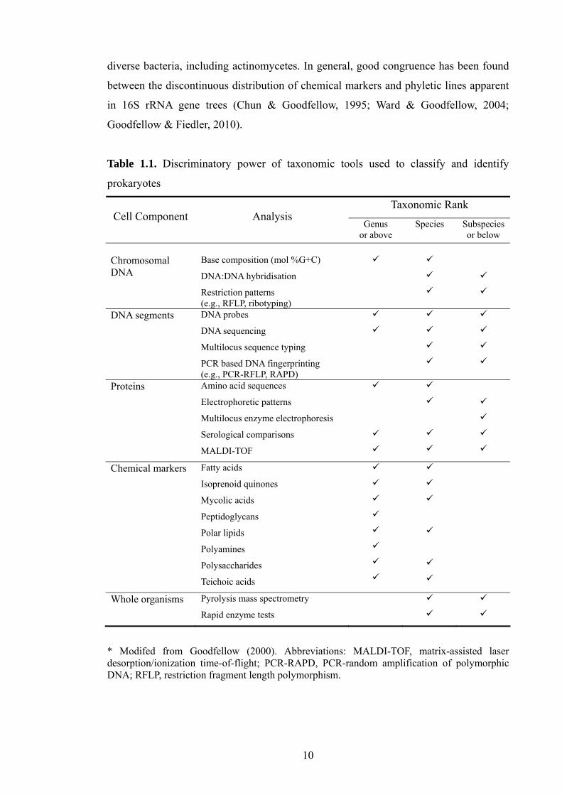
10
diverse bacteria, including actinomycetes. In general, good congruence has been found
between the discontinuous distribution of chemical markers and phyletic lines apparent
in 16S rRNA gene trees (Chun & Goodfellow, 1995; Ward & Goodfellow, 2004;
Goodfellow & Fiedler, 2010).
Table 1.1. Discriminatory power of taxonomic tools used to classify and identify
prokaryotes
Taxonomic Rank Cell Component Analysis
Genus or above
Species Subspeciesor below
Chromosomal DNA
Base composition (mol %G+C)
DNA:DNA hybridisation
Restriction patterns (e.g., RFLP, ribotyping)
DNA segments DNA probes
DNA sequencing
Multilocus sequence typing
PCR based DNA fingerprinting (e.g., PCR-RFLP, RAPD)
Proteins Amino acid sequences
Electrophoretic patterns
Multilocus enzyme electrophoresis
Serological comparisons
MALDI-TOF
Chemical markers Fatty acids
Isoprenoid quinones
Mycolic acids
Peptidoglycans
Polar lipids
Polyamines
Polysaccharides
Teichoic acids
Whole organisms Pyrolysis mass spectrometry
Rapid enzyme tests
* Modifed from Goodfellow (2000). Abbreviations: MALDI-TOF, matrix-assisted laser desorption/ionization time-of-flight; PCR-RAPD, PCR-random amplification of polymorphic DNA; RFLP, restriction fragment length polymorphism.

11
Numerical taxonomy. This is the classification by numerical methods of strains and
taxonomic units into taxa based on many shared characters (Sneath, 1957). The primary
aim of this method is to assign individual strains to homogeneous groups or clusters
(taxospecies) using large sets of phenotypic data. The organisms to be classified are
referred to as operational taxonomic units (OTUs;(Sneath & Johnson, 1972). It is
essential in such studies to use phenotypic characters that are genetically stable, hence
not susceptible to environmental changes, and which are not particularly sensitive to
experimental conditions or observational uncertainties. The usual practice is to take a
selection of biochemical, cultural, morphological, nutritional and physiological
characters to represent the phenome, that is, the genotype and phenotype. It is important
in numerical taxonomic studies to have sufficient information to discriminate between
taxa (Sneath & Sokal, 1973; Goodfellow et al., 1997). Most unit characters tend to be
binary or two-state; possession of a character is coded as 1 or + and absence by 0 or -.
Test results, once obtained, need to be evaluated and unreliable information discarded.
Many resemblance coefficients are available to estimate relationships between
pairs of organisms; the two most commonly used ones are the Jaccard (SJ;(Jaccard,
1908) and simple matching (SSM; Sokal & Michener, 1958) coefficients. The SSM
coefficient is used to calculate similarities based on both positive and negative
similarities hence the ratio of the total number of matches to the total number of unit
characters is determined. In the case of the SJ coefficient, negative matches are ignored
which means that this coefficient measures the ratio of the total number of positive
matches to the total number of characters minus the sum of negative matches. The SJ
coefficient is generally used to ensure that relationships detected using the SSM
coefficient are not based on negative correlations, that is, OTUs are not grouped
together because of having a high number of negative characters in common. The SJ
coefficient is particularly useful in studies where relatively fast- and slow-growing
organisms are compared (Whitham et al., 1993; Trujillo & Goodfellow, 2003), as the
latter may give a disproportionate number of negative results.
Hierarchical and non-hierarchical clustering methods are available to order OTUs
into groups based on high overall phenetic similarity (Gevers et al., 2006). Such
methods are used to produce a ranked classification where strains are grouped into
clusters (taxospecies) and aggregate groups (genera), though they are of little value in
differentiating between taxa above the genus level (Goodfellow et al., 1997; Castro et

12
al., 2002; Valera & Esteve, 2002). Conventional numerical taxonomic studies have
tended to go out of fashion as they are seen to be time-consuming and laborious, but
new high-throughput methods, such as commercially available 96 well phenotypic array
plates, have been introduced to overcome this limitation (Bochner, 2003; Clemons,
2004; Bochner et al., 2008).
The application of numerical taxonomic procedures led to significant
improvements in the classification of actinomycetes. Numerical taxonomic studies have
been used to circumscribe taxospecies, including those in complex actinomycete taxa
such as the genera Actinomadura (Trujillo & Goodfellow, 2003), Actinomyces
(Schofield & Schaal, 1981), Actinoplanes (Goodfellow et al., 1990), Corynebacterium
(Goodfellow et al., 1982b), Gordonia (Goodfellow et al., 1991), Nocardia (Goodfellow
et al., 1982a; Goodfellow, 1992), Mycobacterium (Wayne et al., 1996), Rhodococcus
(Goodfellow et al., 1998), Streptomyces (Williams et al., 1983; Kämpfer et al., 1991;
Manfio et al., 1995), Streptosporangium (Whitham et al., 1993) and Thermomonospora
(McCarthy & Cross, 1981). Phenotypic analyses of streptomycetes (Williams et al.,
1983; Kämpfer et al., 1991; Manfio et al., 1995) provided a sound base for selecting
representative strains for more sophisticated taxonomic investigations, not least for
comprehensive molecular systematic studies (Lanoot et al., 2002; Lanoot et al., 2005).
Molecular systematics. The most significant recent advances in prokaryotic
systematics are based on the realisation that archaea and bacteria contain records of
changes that have occurred since they diverged from a common ancestor around 3.5
billion year ago (Zuckerkandl & Pauling, 1965; Woese, 1987). Molecular-based
systematics has a significant advantage over chemotaxonomic and numerical taxonomic
apporaches as the acquisition of sequence data is independent of cultivation conditions.
Molecular-based methods are currently the driving force in prokaryote systematics,
partly as a consequence of technological changes but primarily because the end product
of this approach reflects natural relationships between prokaryotes as encoded in DNA
and protein sequences (Head et al., 1998; Woese, 1998; Lerat et al., 2005; Gevers et al.,
2006; Koonin, 2009; Schleifer, 2009; Alam et al., 2010; Jensen, 2010).
16S rRNA gene sequencing. Data derived from sequencing 16S rRNA genes are used
extensively for the classification of cultivated (Woese, 1987; Rosselló-Mora & Amann,
2001; Ludwig & Klenk, 2005) and uncultivated (Stach et al., 2003a, b; Kumar et al.,
13
2007) prokaryotes, and to design oligonucleotide probes and primers for the
identification of specific taxa (Yoon et al., 1996; Shen & Young, 2005; Zhi et al., 2006).
However, 16S rRNA sequencing studies do not always allow delineation between
closely related species (Fry et al., 1991; Fox et al., 1992), as exemplified by studies on
the genera Micromonospora (Koch et al., 1996), Saccharomonospora (Yoon et al.,
1997) and Salinispora (Jensen et al., 2005).
16S rRNA gene sequence data held in the DNA Data Bank of Japan
(DDBJ;(Tateno et al., 2002), the European Molecular Biology Laboratories Database
(EMBL;(Kanz et al., 2005), the GenBank Database (Benson et al., 2009) and by the
Ribosomal Database Project (RDP; http://rdp.cme.msu.edu/index.jsp;(Maidak et al.,
1997), are readily retrievable for comparative taxonomic studies. In general, good
congruence has been found between phylogenetic trees based on 16S rRNA gene
sequence data and corresponding trees generated from studies of other conserved
molecules, such as elongation factors, protein-translocating ATPase subunits, and RNA
polymerases (Ludwig & Klenk, 2005). Consequently, comparative 16S rRNA gene
sequencing analyses are seen to be the most accurate and fast way of identifying
unknown prokaryotes, including the recognition of putatively novel taxa (Goodfellow &
Fiedler, 2010), despite their inherent limitations (Schleifer, 2009).
Analysis of sequence data and phylogenetic reconstruction. The alignment of rRNA
gene sequences is very important for inferring phylogenetic relationships. The presence
of insertions and deletions (indel sequences) may make alignments less accurate,
especially when homologies are low. The use of secondary structural information
becomes essential to localize indel sequences. It is customary to manually adjust
alignments and to eliminate nucleotide positions considered to be uncertain (Brocchieri,
2001; Harayama & Kasai, 2006), procedures which rely on the judgment of the
investigator.
Numerous tree-making methods are available to infer ancestry once nucleotide
sequences have been aligned. The four major approaches are the maximum-likelihood
(Felsenstein, 1981), maximum-parsimony (Fitch, 1971) and neighbour-joining (Saitou
& Nei, 1987) methods. The most frequently used method for calculating distances is the
one-parameter model proposed by Jukes & Cantor (1969), this is based on the
assumption that there are independent changes at all nucleotide positions, that is, there
14
is an equal probability of ending up with each of the other three bases.
The construction of trees from data in distance matrices is carried out by using
available tree-making methods, such as neighbour-joining (Saitou & Nei, 1987) and
weighted least-squares (Fitch & Margoliash, 1967) methods. The neighbour-joining
method is theoretically related to clustering methods, such as the unweighted-pair-group
method with arithmetic averages (UPGMA;(Sneath & Sokal, 1973), but is not based on
the assumption that data are ultra metric and that all lineages have equally diverged. In
contrast to cluster analysis, the neighbour-joining method keeps track of nodes on the
tree rather than taxa or clusters of taxa. The least-squares method fits a given set of
pairwise evolutionary distance estimates to an additive tree. The maximum-parsimony
method is used to find the most parsimonious tree among all possible tree topologies,
the tree with the minimal overall number of changes is the most parsimonious one and
is taken as the one which infers evolution most closely (Felsenstein, 1981).
The maximum-likelihood method is the most statistically sound way of
reconstructing phylogenies (Felsenstein, 1981). This approach to phylogenetic inference
is used to determine an explicit model of evolution by analysing sequences on a site-by-
site basis. It is used to evaluate the net likelihood that the given evolutionary model will
yield the observed sequences; the inferred trees are those with the highest likelihood.
The statistical significance of the order of particular subtrees in a phylogenetic tree can
be tested by resampling methods, such as the bootstrap procedure (Felsenstein, 1985).
This approach involves random resampling of alignment positions with the result that
some of them are included more often than others in analyses whereas others are not
included at all. The procedure is usually repeated between 100 and 1000 times with
alternatively truncated or rearranged datasets.
DNA-DNA relatedness. A unique property of DNA and RNA is their capacity for
reassociation or hybridization. Complementary strands of DNA, once denatured, can
reassociate into native duplexes under appropriate experimental conditions. When
comparing nucleic acids from any two closely related prokaryotes the extent of
molecular hybrid and its thermal stability provide a measure of the nucleotide sequence
similarity between them. These theoretically simple concepts are the basis of DNA-
DNA relatedness studies. The importance of such studies in circumscribing species was
underlined by recommendations from ad hoc committee on prokaryotic systematics

15
(Wayne et al., 1987; Stackebrandt et al., 2002). Wayne and his colleagues recommended
that the phylogenetic definition of archaeal and bacterial species should be based on the
assignment of strains to genomic species when they showed approximately 70% or
more DNA relatedness values with 5˚C or less thermal stability (ΔTm). However, DNA-
DNA relatedness values need to be interpreted with care as they may not reflect the
actual degree of sequence similarity (Goodfellow et al., 1997; Rosselló-Mora & Amann,
2001) and may be distorted by differences in genome size and by genomic
rearrangements (Kang et al., 2007). It is also well known that DNA-DNA relatedness
data are prone to experimental error (Goodfellow et al., 1997; Schleifer, 2009) and
cannot be used to generate cumulative databases as they are based on pairwise
comparisons (Stackebrandt et al., 2002b; Mehlen et al., 2004; Kang et al., 2007).
Currently two experimental approaches are commonly used to measure the degree
of DNA relatedness or similarity between prokaryotes. They are based on assessing the
degree of binding by hybridization (Ezaki et al., 1989; Jahnke, 1994) or by establishing
differences in thermal denaturation midpoints (De Ley et al., 1970; Mehlen et al., 2004).
The binding strategy involves fixing single-stranded, high-molecular-weight DNA on a
solid support (generally nitrocellulose or nylon membranes) followed by incubation in
the presence of single-stranded, low-molecular-weight, labelled DNA. The thermal
denaturation temperature is used to estimate the thermal stability of hybrid DNA
duplexes against homologous DNA. The temperature at which 50% of the initial
double-stranded molecules have denatured into single-stranded DNA is the melting
temperature or thermal denaturation midpoint (Tm).
A parameter used to estimate DNA-DNA relatedness, ∆Tm, is the differences
between the Tm of the reference strain and that of the hybrid DNA. To estimate ∆Tm,
purified total genomic DNA and mixtures of DNA from representatives of related
species are denatured and allowed to renature at the optimal temperature for
renaturation (Tor; De Ley et al., 1970). Tor can be estimated from the mol% G+C of the
DNA of strains under study, as described by De Ley et al. (1970). The transition from
double to single stranded DNA, DNA melting, can be measured by the change in
absorbance at 260 nm. Alternatively, the shift in fluorescence of added SYBR Green I
dye bound to double stranded DNA can be determined as DNA is ‘melted’ by
progressive heating (Gonzalez & Saiz-Jimenez, 2005). This technique has several
advantages over more established methods. It is rapid and inexpensive, and allows high-

16
throughput comparisons. Comparative studies show that results derived from estimating
binding percentages and ∆Tm values are generally in good agreement (Roselló-Mora &
Amann, 2001; Gonzalez & Saiz-Jimenez, 2005).
Despite limitations, DNA-DNA relatedness studies are still considered to provide
the golden standard for the delineation of prokaryotic species (Vandamme et al., 1996;
Goodfellow et al., 1997; Rosselló-Mora & Amann, 2001; Kumar & Goodfellow, 2008).
It is clear that such studies give greater resolution between closely related strains than
corresponding 16S rRNA gene sequencing studies (Goodfellow et al., 1997; Rosselló-
Mora & Amann, 2001; Kumar & Goodfellow, 2008), as it is apparent from Figure 1.2.
Organisms with almost identical 16S rRNA sequence similarities can be distinguished
using corresponding DNA: DNA relatedness data.
Fig. 1.2 Comparison of DNA-DNA and 16S rRNA gene similarities of Proteobacteria, Cytophaga-Flavobacterium-Bacteroides and Gram-positive bacteria of high GC phyla. The vertical shaded zone indicates the range of cut-off values for DNA-DNA relatedness used for the delineation of genomic species while the horizontal shade zone indicates cut-off values for 16S rRNA gene sequence similarity (97%) previously used to delineate strains (taken from Rosselló-Mora & Amann, 2001).
The report of the ad hoc committee for the re-evaluation of the species definition in
bacteriology recommended the use of DNA profiling (e.g., AFLP, PCR-RFLP, rep-PCR
and ribotyping) and multilocus sequence typing (MLST) to discriminate between
taxonomically closely related strains (Stackebrandt et al., 2002a). These workers
concluded that MLST was a potentially useful procedure, which might be used as an
alternative to DNA-DNA relatedness studies in defining genomic species. MLST
involves sequencing a minimum of five housekeeping or other protein coding genes and

17
presenting the resultant data in individual and/or concatenated trees (Enright & Spratt,
1999; Gevers et al., 2005; Antony-Babu & Goodfellow, 2008). The choice of genes
needs to be based on their loci; selected genes should be spread across the genome
(Maiden et al., 1998; Stackebrandt et al., 2002a). MLST provides good resolution at and
below the species level and greater clarity in genomic relatedness at inter- and
intraspecific levels (Thompson et al., 2005; Guo et al., 2008; Martens et al., 2008).
Initially, MLST studies were restricted to epidemiological and population genetic
studies (Enright & Spratt, 1999; Robinson & Enright, 2004; Miragaia et al., 2007), but
they are now being used to establish taxonomic relationships between closely related
bacteria, as exemplified by studies on Escherichia coli (Adiri et al., 2003), Listeria
monocytogenes (Salcedo et al., 2003), Staphylococcus aureus, (Grundmann et al., 2002),
Streptomyces griseus (Guo et al., 2008; Rong & Huang, 2010; Rong et al., 2010) and
Streptomyces albidoflavus (Rong et al., 2009).
1.3 Prokaryotic diversity and bioprospecting
It has been estimated that a gram of soil may contain between 2,000 and 8.3
million prokaryotic species (Torsvik et al., 2002; Fierer & Jackson, 2006), while the
total number of prokaryotic cells present in natural habitats has been considered to be 4
- 6 x 1030 cells (Whitman et al., 1998). It is surprising in light of such figures that only
about 10,000 species of prokaryotes have been formally described
(http://www.bacterio.cict.fr; April, 2010), which means that the vast majority of
prokaryotes (90-99%) present in natural habitats have still to be isolated and formally
described (Zengler et al., 2002; Rappé & Giovannoni, 2003; Schloss & Handelsman,
2004). Consequently, little is known of the role or distribution of individual prokaryotic
species, including actinomycetes, in natural habitats (Bull et al., 2000; Handelsman,
2004).
Biotechnological search and discovery generally starts with the assembly of
appropriate biological material, moves through screening for desired attributes,
selection of the best options from a short list of positive screened hits, and culminates
with the development of a commercial product or process (Bull et al., 2000; Goodfellow
& Fiedler, 2010). The primary and secondary metabolism of prokaryotes has been
exploited for the production of diverse natural products, notably secondary metabolites
(Bérdy, 1995, 2005; Strohl, 2004), The best known secondary metabolites are antibiotics,

18
a new generation of which is needed to combat drug resistant bacteria and fungi and to
provide safer and more potent compounds with improved pharmacological properties
(Payne et al., 2007; Fischbach & Walsh, 2009). Unknown, important products are
generally found when high quality biological materials are examined within existing or
new screening systems; it is, therefore, essential to foster these two aspects of novelty in
drug discovery programmes (Nolan & Cross, 1988; Goodfellow & Fiedler, 2010).
Actinomycetes and natural products. Amongst prokaryotes, filamentous
actinomycetes, notably streptomycetes, are the most prolific source of new antibiotics
(Bérdy, 2005; Newman & Cragg, 2007; Goodfellow & Fiedler, 2010). It is likely that
this trend will continue as full genome sequences of model actinomycetes have been
found to contain over 20 natural product biosynthetic gene clusters for the production of
known or predicted secondary metabolites, as shown by studies on Amycolatopsis
mediterranei U32 (Zhao et al., 2010), Saccharopolyspora erythraea NRRL 23338
(Oliynyk et al., 2007), Salinispora tropica CNB 440 (Udwary et al., 2007),
Streptomyces avermitilis (Ikeda et al., 2003) and “Streptomyces coelicolor” A3(2)
(Bentley et al., 2002). In contrast, few, if any, such gene clusters have been detected in
whole genomes of other prokaryotes (Goodfellow & Fiedler, 2010).
Actinomycetes are currently known to produce over 10,000 bioactive compounds,
7,600 of which have been isolated from streptomycetes and 2,500 from non-
streptomycetes, notably from the so called rare actinomycetes (Lazzarini et al., 2000).
Despite this amazing productivity, it has been estimated that only a tiny fraction of the
total number of antimicrobial compounds which actinomycetes can produce have been
found to date (Watve et al., 2001). The number of antibiotics and “other bioactive”
metabolites synthesised by different microbial groups are shown in Table 1.2.
Antibiotics produced by actinomycetes show a wide range of chemical diversity, as
demonstrated in Table 1.3. It is clear from these data that streptomycetes synthesise a
diverse range of chemical structures; it is also known that specific structural types of
antibiotics occur commonly in these organisms, such as ansa-lactam rings, macrocyclic
lactones, polyether and cyclopeptide skeletons (Bérdy, 1995, 2005). However, certain
specific structures are produced more frequently by non-streptomycetes, such as
Actinoplanes and Amycolatopsis strains, which are rich sources of vancomycin-like
glycopeptides (Wink et al., 2003).

19
Table 1.2. Approximate number of bioactive microbial metabolites according to their
producers and bioactivities
Antibiotics Bioactive metabolites
Source Total antibiotics
(with other activity)
No antibiotic activity
(antibiotics plus other
bioactivies)
Total bioactive
metabolites
Bacteria: 2900 (780) 900 (1680) 3800 Eubacteriales 2170 (570) 580 (1150) 2750 Bacillus sp. 795 (235) 65 (300) 860 Pseudomonas sp. 610 (185) 185 (370) 795 Myxobacteria 400 (130) 10 (140) 410 Cyanobacteria 300 (80) 340 (420) 640
Actinomycetales: 8700 (2400) 1400 (3800) 10100 Streptomyces sp. 6550 (1920) 1080 (3000) 7630 Rare actinomycetes 2250 (580) 220 (800) 2470 Fungi: 4900 (2300) 3700 (6000) 8600 Microscopic fungi 3770 (2070) 2680 (4750) 6450 Penicillium/Aspergillus 1000 (450) 950 (1400) 1950 Basidiomycetes 1050 (200) 950 (1150) 2000 Yeasts 105 (35) 35 (70) 140 Slime moulds 30 (5) 20 (25) 60
Total 16500 (5500) 6000 (11500) 22500
* Taken from Bérdy (2005).
Rare actinomycetes. There is evidence that taxonomic diversity can be used as a
surrogate for chemical diversity amongst actinomycetes, especially at the species level
(Goodfellow et al., 2007; Jensen et al., 2007; Tan et al., 2007). This means that novel
taxa which populate gaps in actinomycete taxospace should be a rich source of new
bioactive compounds (Ward & Goodfellow, 2004; Jensen, 2010). Consequently, it
makes good sense to devise selective isolation and characterisation strategies to secure
representatives of rare actinomycete genera for pharmaceutical screening programmes, a
stragtegy which has been applied with some success (Lazzarini et al. 2000; Goodfellow
& Fiedler, 2010).
Rare actinomycete genera include a plethora of taxa known to synthesise novel
antibiotics, as exemplified by the genera Actinokineospora, Acrocarpospora,
Actinosynnema, Amycolatopsis, Catenuloplanes, Cryptosporangium,
Dactylosporangium, Kineosporia, Kutzneria, Microbispora, Microtetraspora,
Nonomuraea, Thermomonospora, Pseudonocardia, Thermobifida, Saccharomonospora,
Spirilliplanes, Streptosporangium and Virgosporangium (Hayakawa, 2008). The

20
importance of rare actinomycetes can be demonstrated by the fact that some of them are
the source of commercially important anti-microbial products, as exemplified by
erythromycin produced by Saccharopolyspora erythraea (Oliynyk et al., 2007),
gentamicin by Micromonopsora purpurea (Weinstein et al., 1963; Wagman & Weinstein,
1980), rifamycins by Amycolatopsis mediterranei (Jin et al., 2002), teicoplanin by
Actinoplanes teichomyceticus (Somma et al., 1984; Jung et al., 2009) and vancomycin
by Amycolatopsis orientalis (Wink et al., 2003).
Table 1.3. Antibiotics isolated from actinomycete genera, as described in the antibiotic
database of the Journal of Antibiotics (http://www.antibiotics.or.jp)
Actinomycete AG ML AML BLA PEP GP ANC TC NUC POL QN
Streptomyces Other actinomycetes : Actinomadura Actinoplanes Actinosynnema Ampullariella Amycolatopsis Dactylosporangium Kibdelosporangium Kitasatospora Microbispora Micromonospora Microtetraspora Nocardia Nocardiopsis Nonomuraea Pseudonocardia Rhodococcus Saccharomonospora Saccharopolyspora Saccharothrix Streptoalloteichus Streptosporangium Thermomonospora
* AG, aminoglycoside; ML, macrolide; AML, ansamacrolide; BLA, β-lactam; PEP, peptide; GP, glycopeptides; ANC, anthracycline; TC, tetracycline; NUC, nucleotide; POL, polyene; QN, quinine. , production (modified from Okami & Hotta, 1988).
Nonribosomal peptides and polyketides. Nonribosomal peptides and polyketides are
very large classes of natural products which are assembled from simple acyl-coenzyme

21
A or amino acid monomers (Marahiel et al., 1997; Shen, 2003). They have a broad
range of biological activities and pharmacological properties and are a source of novel
antibiotics, cytostatics, and immunosuppressants (Rascher et al., 2003; Schwarzer et al.,
2003; Bergmann et al., 2007). Non-ribosomal peptides and polyketides are synthesized
by one or more large, specialized, multifunctional, modular enzymes, namely non-
ribosomal polypeptide (NRPS) and polyketide (PKS) synthases, respectively
(Schwarzer et al., 2003). NRPS and PKS catalyse chain elongation from simple
building blocks to generate diverse natural products (Staunton & Weissman, 2001;
Donadio et al., 2007). Three PKS types have been described though only types I and II
are of major interest (Shen, 2003). Type I PKS’s are common in bacteria, including
actinomycetes (Staunton & Weissman, 2001) and type II PKS’s, which are multi-
enzymes, carry a single set of iteratively acting activities, as illustrated by the
biosynthesis of aromatic polyketides, tetracenomycin (Shen, 2003). Type III PKS types,
known as chalcone synthase-like PKS’s, are homodimeric enzymes which are mainly
acting condensing enzymes (Watanabe et al., 2003).
Actinomycetes typically contain genes encoding PKS I, PKS II and NRPS (Metsa-
Ketela et al., 1999; Ayuso et al., 2005; Ginolhac et al., 2005) and hence are good
vehicles for the discovery of novel secondary metabolites (Schwarzer et al., 2003). PKS
I and NRPS genes are usually organised into units or segments with similar gene
segments repeated within a single gene cluster (Busti et al., 2006). In contrast, PKS II
systems do not have a modular organisation hence the individual biochemical steps of
polyketide biosynthesis are catalyzed by discrete subunits which are used repeatedly in
the biosynthetic cycle (Ayuso-Sacido & Genilloud, 2005). The utilization of PCR
technology to amplify specific sequences from bacterial genomic DNA by using
degenerate oligonucleotide primers which correspond to highly conserved motifs in
peptide synthases has led to the establishment of a conventional approach for the
identification and cloning of putative genes that encode these multi-enzymes (Metsa-
Ketela et al., 1999; Ayuso-Sacido & Genilloud, 2005; Savic & Vasiljevic, 2006; Hwang
et al., 2007).
Analysis of PKS and NRPS genes in putative bioactive compound producing
actinomycetes helps not only to determine the evolutionary relationships of these genes
but to relate them with the ecology and physiology of the organisms (Metsa-Ketela et
al., 2002; Ayuso et al., 2005; Ginolhac et al., 2005; Jenke-Kodama et al., 2005;

22
Gontang et al., 2010). Molecular screening of such genes is considered to be useful in
dereplication and for screening systems with novel biosynthetic pathways thereby being
of relevance to natural product screening programmes (Ayuso-Sacido & Genilloud,
2005; Ayuso et al., 2005; Savic & Vasiljevic, 2006).
1.4 Actinomycete diversity in natural habitats
Culture-dependent and culture-independent approaches are widely used to study
prokaryotic communities in natural habitats (Bull et al., 2000; 2005). Culture-dependent
approaches employ artificial media to culture or enrich for groups of microorganisms
found in environmental samples. Until recently, this method was extensively used to
determine the extent of prokaryotic diversity in natural habitats (Ward et al., 1990;
Hugenholtz et al., 1998; Fierer & Jackson, 2006), but it is now known that the majority
of microorganisms in the environment cannot be cultured by using conventional plate
culture methods hence the vast majority of the soil microbiota remain undiscovered
(Whitman et al., 1998; Rappé & Giovannoni, 2003; Vartoukian et al., 2010). One reason
for the limited value of conventional isolation procedures is that the cultivation
conditions used to isolate organisms do not reflect conditions in the environment hence
fast-growing prokaryotes are selected that are best adapted to grow on individual growth
media (Zengler et al., 2002; Stevenson et al., 2004; Vartoukian et al., 2010). This
limitation has promoted the use of culture-independent approaches to detect previously
uncultured microbes and evaluate microbial diversity in natural habitats (Amann et al.,
1995; Stach et al., 2003b; Kumar et al., 2007; Xin et al., 2008).
Culture-independent approaches. Culture-independent methods involve the extraction
of community nucleic acids from cells present in environmental samples followed by
the application of biomarker-based techniques, such as DNA analyses to trace the
taxonomic diversity of microbial communities in the natural habitats (Pace, 1997; Head
et al., 1998; Hugenholtz et al., 1998; Kirk et al., 2004). Molecular approaches
confirmed observations from direct microscopy that the number of prokaryotes which
can be readily cultivated from environmental samples is only a small and skewed
fraction of the diversity present (Pace, 1997; Rappé & Giovannoni, 2003; Vartoukian et
al., 2010). The inability to cultivate even the most numerous microorganisms from
natural habitats has been referred to as the ‘great plate count anomaly’ (Pace, 1997;
Hirsch et al., 2010). Amann et al. (1995) considered that <1% of the bacteria present in
23
environmental samples formed colonies on nutrient agar and went on to stress the need
to apply culture-independent approaches to unravel the full extent of prokaryotic
diversity in natural habitats.
The use of 16S rRNA gene sequences as indicators of prokaryotic diversity is well
established though other genes, including peptide elongation factor Tu RNA polymerase
(tuf) and RNA polymerase (rpoB) genes, have been used for this purpose (Rintala et al.,
2001; Peixoto et al., 2002; Kumar et al., 2007; Tanaka et al., 2010). In addition, various
culture-independent techniques can be employed to target other genes such as those
responsible for metabolic actions, for instance, polyketide synthase (PKS) and
nonribosomal peptide synthetase (NRPS) genes, which are widely used to check
whether strains have the capacity to produce antibiotics (Wawrik et al., 2005; Pang et
al., 2008). The use of such analytical strategies makes it possible to target cells, which
actively express the specific metabolic functions.
Most culture-independent methods have been used to establish the extent of
prokaryotic diversity in soil, including that of actinomycetes (Mincer et al., 2005;
Kumar et al., 2007; Xin et al., 2008; Hirsch et al., 2010). Relatively harsh extraction
methods are needed to extract community DNA from soil as indigenous prokaryotic
cells tend to be tightly bound to soil particles (Roose-Amsaleg et al., 2001; Feinstein et
al., 2009; Fitzpatrick et al., 2010). Chemical and physical extraction methods are
usually needed to lyse microbial cell walls. Chemical procedures tend to entail the use
of lysozyme and sodium dodecyl (lauryl) sulphate to weaken and break open cell walls
to release cellular DNA (Liles et al., 2008); physical methods include freeze/thaw
and/or bead beating protocols (Niemi et al., 2001). Extraction kits designed specifically
for the recovery of community DNA from soil are based on bead beating and chemical
lysis methods. Following the disruption of microbial cells and the extraction of
DNA/RNA, the resultant nucleic acids are purified using proteinase K and the
phenol/chloroform extraction method. Nucleic acids carry a fixed negative charge per
unit length of molecule hence for purification purposes nucleic acids are collected using
a positively charged binding material followed by washing to discard unnecessary
compounds.
Generation of gene clone libraries. Gene clone library construction is a widely used
procedure which involves the extraction of purified nucleic acids from environmental

24
samples and the amplification of specific sequences of genomes present using the PCR,
this, in turn, involves the use of oligonucleotide primers specific to the preferred
amplicon gene sequence (Nakatsu et al., 2000; Monciardini et al., 2002; Kumar et al.,
2007). Amplified gene sequences are inserted into engineered plasmids, which contain
an antibiotic resistant gene; this results in the generation of recombinant plasmids,
which can be taken up by competent Escherichia coli host cells. The latter are grown on
nutrient media containing the antibiotic in question hence only cells that contain the
plasmid survive and replicate. The resultant colonies, or clones, contain copies of the
same recombinant plasmid with inserts of foreign DNA. A range of methods can be
used to determine colonies which contain DNA sequences of interest, such as white/blue
differentiation or lacZ’ selection (Ma et al., 2009). Most 16S rRNA gene clone libraries
consist of between 100 and 300 clones though the generation of additional clones has
the advantage that it allows the detection of greater diversity in terms of community
composition, richness and structure (Dunbar et al., 2000; 2002).
1.5 Selective isolation of actinomycetes
Innumerable actinomycetes have been isolated and screened since Selman
Waksman discovered that Streptomyces griseus produced streptomycin, a discovery that
promoted the search for additional novel bioactive compounds of therapeutic value
(Watve et al., 2001). Early search and discovering programmes were focused on
streptomycetes as these organisms were easy to isolate and grew quickly on isolation
media. Intensive studies on members of the genus Streptomyces led to the discovery of
many novel antibiotics, such as actinomycin from S. antibioticus (Waksman &
Woodruff, 1941) and neomycin from S. fradiae (Waksman & Lechevalier, 1949).
It is becoming increasingly important to design new procedures for the selective
isolation and characterisation of commercially significant actinomycetes from
environmental samples to secure high quality biological material for pharmaceutical
screening programmes (Bull et al., 2000; Goodfellow & Fiedler, 2010). The
introduction of such procedures show that actinomycetes once thought to be rare in
natural habitats are widely distributed, as shown by studies on acidiphilic
sporoactinomycetes (Kim et al., 2003), motile actinomycetes (Hayakawa et al., 2000;
Suzuki et al., 2001a), nocardiae (Orchard et al., 1977; Maldonado et al., 2000) and
rhodococci (Colquhoun et al., 1998). Targeted procedures are still needed for the

25
selective isolation and characterisation of commercially significant actinomycete taxa
present in diverse environmental samples (Stach et al., 2003a, b; Kumar et al. 2007).
Most actinomycetes are saprophytes in aquatic and terrestrial ecosystems, notably
in soils, freshwater and marine habitats (Bull et al., 2005; Bull & Stach, 2007) . It is not
possible to recommend a single procedure for the selective isolation of the multiplicity
of the different kinds of actinomycetes present in environmental samples due to their
diverse growth and incubation requirements. Consequently, numerous approaches have
been recommended for the isolation of specific taxa (Nolan & Cross, 1988; Labeda &
Shearer, 1990; Goodfellow, 2010). Most selective strategies have been focused on the
isolation of particular taxonomic groups based on their biological properties (Cross,
1982; Wellington & Cross, 1983; Goodfellow & O'Donnell, 1989). Most procedures
involve the extraction of propagules from selected environmental samples,
pretreatment(s) of samples, design of selective media, incubation, and recognition of
target colonies.
Extraction of actinomycete propagules. Physico-chemical interactions of bacterial
propagules with particulate substrates affect their recovery from environmental samples.
Traditional methods used to separate bacteria from organic matter, sediment and soil
particles, include shaking in water or weak buffers (e.g., ¼ strength Ringer’s solution),
are not effective (Hopkins et al., 1991). It is particularly important to thoroughly break
up soil aggregates as many microorganisms, notably those showing mycelial growth,
may be bound within them (Ramsay, 1984; Nishiyama et al., 1992). Procedures used to
promote the dissociation of microorganisms from particulate material include the use of
chelating agents (MacDonald, 1986), buffered diluents (Niepold et al., 1979), elutriation
(Hopkins et al., 1991) and ultrasonication (Ramesy, 1984); all of these procedures
address the problem of quantitative and representative sampling to varying degrees.
Pre-treatment of environmental samples. Several pre-treatment procedures are used
to select different fractions of actinomycete communities present in environmental
samples. In general, pre-treatment regimes select for target actinomycetes by inhibiting
or eliminating unwanted microorganisms. Actinomycete spores are more resistant to
desiccation than other bacterial cells hence simply air-drying soil/sediment samples at
room temperature helps eliminate most unwanted Gram-negative bacteria which might
otherwise overrun isolation plates (Williams et al., 1984a; Labeda & Shearer, 1990). A

26
pre-treatment regime based on alternate drying and wetting of soil has been used to
enrich for sporangia (spore vesicle) – forming actinomycetes (Makkar & Cross, 1982).
Rare spore-forming actinobacteria (e.g., Actinomadura, Microtetraspora,
Pseudonocardia and Streptosporangium) have been isolated from irradiated soil
samples and soil suspensions (Bulina et al., 1997; Terekhova, 2003).
Resistance of actinomycete propagules to desiccation is usually accompanied by
some measure of resistance to heat. The basis of this resistance is not clear but it is
apparent that many actinomycete spores (e.g., Micromonospora and Microtetraspora),
spore vesicles (Dactylosporangium and Streptosporangium) and hyphal fragments (e.g.,
Rhodococcus) are more resistant to heat than Gram-negative cells. Actinomycetes are
more sensitive to wet heat than dry heat, which means that much lower temperatures are
used to isolate these organisms from suspensions of environmental samples. Heat pre-
treatment protocols usually lead to a decrease in the ratio of bacteria to actinomycetes
on isolation plates though actinomycete counts may be reduced (Williams et al., 1984b).
Chemical pretreatments of mixed inocula are used to isolate specific actinomycete
taxa, notably members of genera classified in the family Streptosporangiaceae
(Hayakawa et al., 1988, 1991a, 1995; Goodfellow, 2010). The selective chemical
procedures introduced by Hayakawa and his colleagues are based on the differential
ability of actinomycete spores to withstand treatment with chemical germicides, such as
benzethonium chloride, chlorhexidine gluconate and phenol (Yamamura et al., 2003).
Treatment with these agents for 30 minutes at 30oC kills vegetative cells of aerobic,
endospore-forming bacilli and pseudomonads. The simultaneous use of more than one
chemical germicide can further enhance selectivity, as exemplified by the use of
chlorheximide gluconate and phenol for the isolation of Microbispora strains
(Hayakawa et al., 1991b).
Nutrient and baiting techniques are used to increase the populations of specific
fractions of actinomycete communities in environmental samples to facilitate their
isolation on selective media. Soil amendments with substrates such as chitin and keratin
were first used by Jensen (1930) to boost the numbers of streptomycetes. This approach
has fallen into neglect but does provide an effective way of enhancing specific
components of actinomycete communities in soil (Williams et al., 1971; Nonomura &
Takagi, 1977).

27
Selective isolation media. Numerous media have been recommended for the isolation
of broad range of actinomycetes from natural habitats and more specifically for selected
families, genera and species (Kurtböke, 2003; Goodfellow, 2010). Most of the ‘general’
or ‘non-selective’ media were formulated without reference to either the nutritional or
the tolerance preferences of the target organisms. It is now known that widely used
‘non-selective’ media, such as colloidal chitin-mineral salts (Hsu & Lockwood, 1975)
and starch casein nitrate (Kuster & Williams, 1964) agars select for a relatively narrow
range of Streptomyces species nor do they support the growth of actinomycetes with
different nutritional requirements (Williams et al., 1984a).
Selective isolation media can be formulated in an objective way by using
phenotypic data held in taxonomic databases (Goodfellow & Haynes, 1984; Williams &
Vickers, 1988; Goodfellow, 2010). One of the early successes of this approach was the
formulation of a medium selective for the isolation of the Nocardia strains from soil
(Orchard et al., 1977; Orchard & Goodfellow, 1980; Maldonado et al., 2000). The
discovery that Diagnostic Sensitivity Agar supplemented with tetracycline was selective
for Nocardia species was based on information held in a database of antibiotic
sensitivities (Goodfellow et al., 1989).
A logical extension of this work was the visual scanning of phenotypic databases to
highlight antibiotics that might form the basis of selective isolation media. The number
of verticillate streptomycetes (formerly members of the genus Streptoverticillium), for
example, was increased by reducing the number of competing neutrophilic
streptomycetes by supplementing isolation media with neomycin and oxytetracycline
(Hanka & Schaadt, 1988); verticillate streptomycetes were known to be resistant and
other neutrophilic streptomycetes sensitive to these antibiotics (Williams et al., 1985).
Phenotypic databases generated in numerical taxonomic studies are ideal resources
for the formulation of novel selective isolation media as they are rich in data on the
antibiotic, nutritional and physiological properties of members of numerically defined
clusters (Goodfellow & Haynes, 1984; Williams & Vickers, 1988; Goodfellow, 2010).
Such databases are available for several industrially significant actinomyete taxa,
including the genera Actinomadura (Athalye et al., 1981; Trujillo & Goodfellow, 2003),
Gordonia (Goodfellow et al., 1998), Nocardia (Orchard & Goodfellow, 1980),
Rhodococcus (Goodfellow & Williams, 1986), Streptomyces (Williams et al., 1971;

28
Manfio et al., 1995; Sembiring et al., 2000; Duangmal et al., 2005) and
Thermomonospora (McCarthy & Cross, 1981), for members of the family
Pseudonocardiaceae (Whitehead, 1989), but not for genera such as Dactylosporangium,
Microtetraspora or Nonomuraea.
Antimicrobial agents, notably antibiotics, provide an effective means of increasing
the selectivity of isolation media. It is standard practice to control or eliminate fungal
containmants by using antifungal antibiotics, such as cycloheximide, nystatin and
pimaricin (Porter et al., 1960; Gregory & Lacey, 1963; Williams & Davies, 1965).
Similarly, penicillin G and polymixin B have been used to select actinomycetes from
competing soil inhabiting bacteria (Nonomura & Ohara, 1960; Williams & Davies,
1965), as have nalidixic acid and trimethoprin (Hayakawa et al., 1996). Antibacterial
antibiotics are widely used for the selective isolation for specific actinomycete taxa, as
exemplified in Table 1. 4.
Table 1.4. Antibiotics used in the selective isolation of actinomycetes
Antibiotics Target genera References
Bruneomycin and streptomycin Actinomadura Preobrazhenskaya et al.(Preobrazhen
skaya et al.) Gentamicin Actinomadura and
Streptosporangium Shearer (1987)
Josamycin, kanamycin and nalidixic acid
Actinomadura viridis Hayakawa & Nonomura (1987)
Kanamycin, nalidixic acid and nofloxacin
Microtetraspora Hayakawa et al. (1996)
Fradiomycin, kanamycin, nalidixic acid and trimethoprim
Actinokineospora Otoguro et al. (2001)
Leucomycin and nalidixic acid Streptosporangium Hayakawa & Nonomura (1991) and Hayakawa et al. (1991a)
Nalidixic acid and penicillin Saccharothrix Shearer (1987)
Nalidixic acid and tunicamycin Micromonospora Nonomura & Takagi (1977)
Neomycin sulfate Amycolatopsis Tan et al. (2006)
Novobiocin and streptomycin Glycomyces Labeda & Kroppenstedt (2005)
* Adapted from Goodfellow (2010).
Incubation. Incubation conditions, including gaseous regimes, incubation times and
temperature, contribute to selectivity. Incubation at 25 to 30°C favours mesophilic
actinomycetes whereas their thermophilic counterparts require higher temperatures.

29
Thermophilic actinomycetes, such as Pseudonocardia thermophila and
Saccharopolyspora rectivirgula, only require incubation for 2-3 days at 45 to 50°C to
grow fully. In contrast, incubation times up to 5 weeks may be required to isolate some
genera, such as those classified in the families Micromonosporaceae and
Streptosporangiaceae (Labeda & Shearer, 1990; Goodfellow, 2010). Members of
commonly isolated genera, such as Micromonospora, Nocardia and Streptomyces, may
be selected from isolation plates after incubation for 14 days. Prolonged incubation was
one of the factors which contributed to the initial isolation of the slow-growing,
mutualistic symbiont, Frankia (Lechevalier, 1981). However, some slow-growing
actinomycetes, such as members of the genus Dactylosporangium and slowly-growing
mycobacteria, can be difficult to isolate.
Recognition and selection of colonies. The selection of colonies of target
actinomycetes growing on isolation plates is a time-consuming and somewhat
subjective stage in the isolation procedure. Colonies can be selected randomly or with
some degree of choice. When selective isolation media are used the target organism(s)
can sometimes be provisionally identified either on the basis of colony morphology or
by examining colonies for distinctive morphological features, such as the presence of
spore vesicles and the nature of spore chains, using a long working distance objective.
However, it is not usually possible to distinguish between species of the same
actinomycete genus on selective isolation plates. In such instances, the selection of large
numbers of colonies is laborious and can lead to duplication of strains and hence effort,
especially in low throughput screening systems. This problem can be overcome by
grouping isolates based on pigmentation characters and molecular fingerprinting
patterns (Tan et al., 2006; Antony-Babu et al., 2008).
1.6 The genus Dactylosporangium
Circumscription of the genus Dactylosporangium. The genus Dactylosporangium was
proposed by Thiemann et al. (1967) to accommodate claviform sporangial
actinomycetes which contained 3-hydroxy and/or meso-diaminopimelic acid, arabinose
and glycine in the cell wall, but lacked mycolic acids. Members of the genus are Gram-
positive, aerobic, catalase-positive, non-acid-fast actinomycetes, which form club-
shaped, finger-shaped or pyriform sporangia, each containing a single row of three to
four spores. The spores are oblong, ellipsoidal, ovoid, or slightly pyriform (0.4-1.3 ×

30
0.5-1.8 μm), and motile by means of flagella which are generally arranged in polar or
lateral tufts. Large globose bodies (1.7-2.8 μm in diameter) are formed on the substrate
hyphae (Sharples & Williams, 1974; Vobis, 1989, 2006). The substrate mycelium is pale
to deep orange, rose or wine-coloured to brown with hyphae, which are 0.5-1.0 μm in
diameter, branched, and rarely septate. Aerial mycelia are not formed. Colonies on agar
media are compact, tough and leathery, mostly flat or elevated with a smooth to slightly
wrinkled surface (Fig. 1.3). Dactylosporangium strains grow well on standard agar
media, such as inorganic salt-starch, modified Bennet’s, oatmeal, and yeast extract-malt
extract agars (Shomura, 1993; Chiaraphongphon et al., 2010; Seo & Lee, 2010).
The genus Dactylosporangium currently contains 8 validly described species
(Table 1.5), which form a distinct phyletic line within the family Micromonosporaceae
which encompasses 26 other genera (Xie et al., 2010). These taxa can be distinguished
using a combination of chemical and morphological properties (Table 1.6).
Dactylosporangiae have a unique set of 16S rRNA nucleotides which can be used to
differentiate them from other genera (Koch et al., 1996). It can be seen from Figure 1.4
that Dactylosporangium species can be assigned to three distinct subclades with high
bootstrap values (77-94%) and their nearest phylogenetic neighbour is the genus
Virgisporangium. The type species of the genus is Dactylosporangium aurantiacum
Thiemann et al.1967.
Dactylosporangiae contain 3-hydroxy and/or meso-diaminopimelic acid (meso-
A2pm), arabinose and xylose in whole-cell hydrolysates (wall chemotype II and sugar
pattern D; Lechevalier & Lechevalier, 1970), N-acetylated muramic acid (Uchida &
Seino, 1997), major amounts of branched iso-and anteiso-fatty acids (Vobis, 1989), and
polar lipid patterns, which include diphosphatidylglycerol, phosphatidylethanolamine,
phosphatidylinositol and phosphatidylinositol mannosides (phospholipid type II sensu;
Lechevalier et al., 1977). Members of the genus are chemo-organotrophic, mesophilic
(optimally growing between 25 to 37˚C and between pH 6.0 to 7.0), and have a G+C
ratio between 71 to 73 mol% (Vobis, 1989). Members of the genus Dactylosporangium
can be distinguished from other genera classified in the family Micromonosporaceae
using a combination of phenotypic properties as shown in Table 5.4.
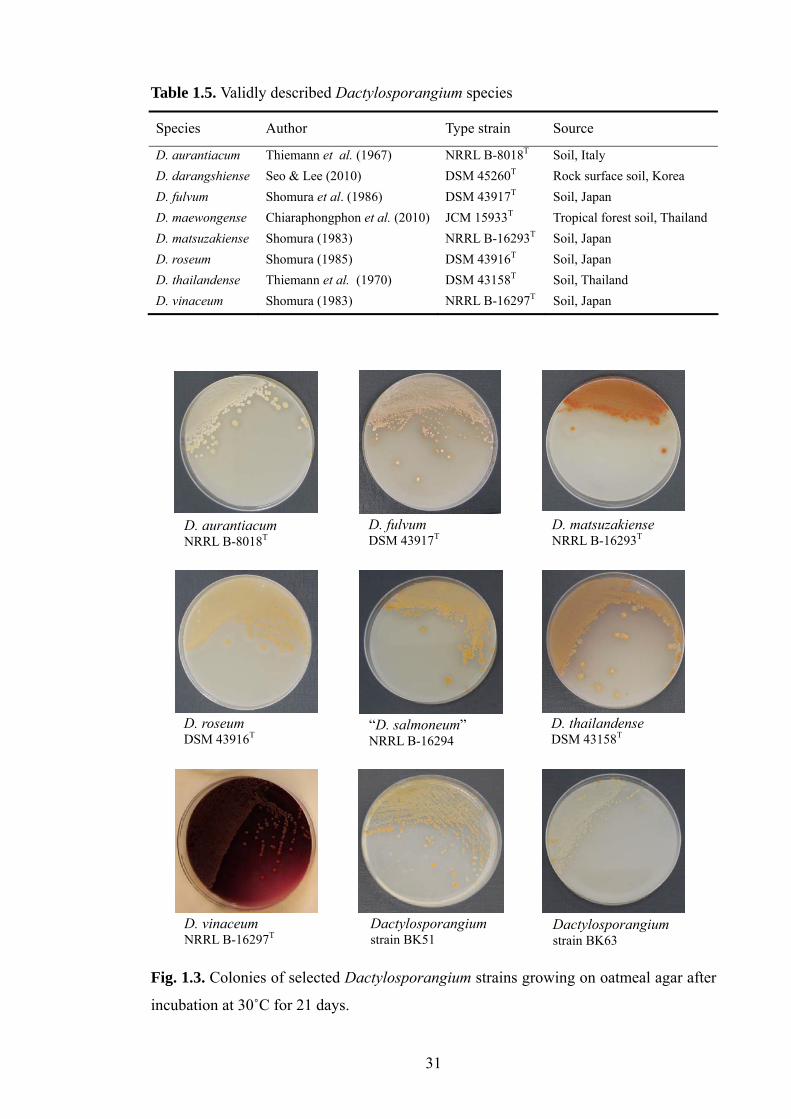
31
Table 1.5. Validly described Dactylosporangium species
Species Author Type strain Source
D. aurantiacum Thiemann et al. (1967) NRRL B-8018T Soil, Italy D. darangshiense Seo & Lee (2010) DSM 45260T Rock surface soil, Korea D. fulvum Shomura et al. (1986) DSM 43917T Soil, Japan D. maewongense Chiaraphongphon et al. (2010) JCM 15933T Tropical forest soil, ThailandD. matsuzakiense Shomura (1983) NRRL B-16293T Soil, Japan D. roseum Shomura (1985) DSM 43916T Soil, Japan D. thailandense Thiemann et al. (1970) DSM 43158T Soil, Thailand D. vinaceum Shomura (1983) NRRL B-16297T Soil, Japan
Fig. 1.3. Colonies of selected Dactylosporangium strains growing on oatmeal agar after
incubation at 30˚C for 21 days.
D. aurantiacum NRRL B-8018T
D. matsuzakiense NRRL B-16293T
D. thailandense DSM 43158T
D. roseum DSM 43916T
D. fulvum DSM 43917T
D. vinaceum NRRL B-16297T
“D. salmoneum” NRRL B-16294
Dactylosporangium strain BK51
Dactylosporangium strain BK63

32
Table 1.6. Chemical and morphological markers found in the members of the genera
classified in the family Micromonosporaceae
Genus Spore vesicles
Spore motility
Isomers of A2pm
whole-cell sugars
Menaqui-none(s) (MK-) a
Phospho-lipid type b
Fatty acid type c
DNA G+C content (mol%)
Actinaurispora – – meso Xyl, Man, Gal 9(H6), 10(H6)
II 3b 72
Actinocatenispora – – meso Ara, Gal, Xyl 9(H4,6) II 3b 72
Actinoplanes + + meso Ara, Xyl 9(H4), 10(H4)
II 2d 72–73
Allocatelliglobosispora – – 3-OH Glu, Rha, Rib, Xyl, Ara, Gal
10(H4,6),9(H4)
II 3b 70
Asanoa – – meso Ara, Gal, Xyl 10(H6,8) II 2d 71–72
Catellatospora – – meso Ara, Gal, Xyl 10(H4,6), 9(H4,6)
II 3b 70-71
Catelliglobosispora – – meso Rha, Man, Xyl, Gal
10(H4) II 3b 70
Catenuloplanes – + L-Lys Xyl 9(H8), 10(H8)
III 2c 71–73
Couchioplanes – + L-Lys Ara, Xyl, Gal 9(H4) II 2c 70–72
Dactylosporangium + + meso Ara, Xyl 9(H4,6,8) II 3b 71–73
Hamadaea – – meso, 3-OH
Xyl, Gal, Man, Rib, Ara, Rha
9(H6) II 3b 70
Jishengella – – meso Xyl, Man, Ara, Rib, Glu
9(H4,6,8), 10(H4)
II 3a 72
Krasilnikovia – – meso Gal, Man, Xyl, Ara, Rib
9(H6,4,8) II 2d 71
Longispora – – meso Ara, Gal, Xyl 10(H4,6) II 2d 70
Luedemannella + – meso Ara, Gal, Man, Rha, Rib, Xyl
9(H2,4,6,8) II 2d 71
Micromonospora – – meso Ara, Xyl 10(H4,6), 9(H4,6)
II 3b 71–73
Phytohabitans – – meso, L-Lys
Gal, Glu, Man, Rib, Xyl
9(H6), 10(H4,6)
II 2d 73
Pilimelia + + meso Ara, Xyl 9(H2,4) II 2d ND
Planosporangium + +/– meso Ara, Xyl 9(H4), 10(H4)
II 3b 71
Plantactinospora – – meso Ara, Gal, Xyl 10(H6,8), 10(H4)
II 2d 70
Polymorphospora – – meso Xyl 10(H4,6), 9(H4,6)
II 2a 71
Pseudosporangium – – meso, 3-OH
Gal, Glu, Rib, Man, Xyl, Ara
9(H6) II 2d 73

33
Table 1.6. (Continued)
Genus Spore vesicles
Spore motility
Isomers of A2pm
whole-cell sugars
Menaqui-none(s) (MK-) a
Phospho-lipid type b
Fatty acid type c
DNA G+C content (mol%)
Rugosimonospora – – 3-OH Ara, Gal, Xyl 9(H6,8) II 2c 72-73
Salinispora – – meso Ara, Gal, Xyl 9(H4) II 3a 70-73
Spirilliplanes – + meso Ara, Xyl 10(H4) II 2d 69
Verrucosispora – – meso Man, Xyl 9(H4) II 2b 70
Virgisporangium + + meso Ara, Gal, Xyl 10(H4,6) II 2d 71
* Data taken from Ørskov (1923), Couch (1950), Kane (1966), Thiemann et al. (1967), Asano &
Kawamoto (1986), Yokota et al. (1993), Rheims et al. (1998), Kudo et al. (1999), Tamura et
al. (1994, 1997, 2001, 2006), Lee & Hah (2002), Matsumoto et al. (2003), Maldonado et al.
(2005), Thawai et al. (2006, 2010), Ara & Kudo (2006, 2007a, b), Ara et al. (2008a, b), Wiese
et al. (2008), Monciardini, et al. (2009), Qin et al. (2009), Lee & Lee (2010), Inahash et al.
(2010) and Xie et al. (2010). Abbreviations: A2pm, diaminopimelic acid; L-Lys, L-lysine; Ara,
arabinose; Gal, galactose; Glu, glucose; Man, mannose; Rha, rhamnose; Rib, ribose; Xyl,
xylose; +, Present; –, absent; ND, not determined.
a, MK-9(H6, H8), notation for a hexa- and octahydrogenated menaquinones with nine isoprene
units. b, Characteristic phospholipids: II, only phosphatidylethanolamine; III, phosphatidylcholine
(with phosphatidylethanolamine, phosphatidylmethylethanolamine and phosphatidylglycerol
variable, phospholipids containing glucosamine absent) (Lechevalier et al. 1977). c, Fatty acid classification after Kroppenstedt (1985). Numbers refer to the type of fatty acid
biosynthetic pathway and letters to the type of fatty acid synthesised. Type 2 encompasses
organisms which have terminally-branched fatty acids, notably, iso- and anteiso-branched
fatty acids, including iso-14/16/18 (Type 2a), iso-15/17 (Type 2b), anteiso-15/17 (Type 2c),
and all of them (Type 2d); Type 3 encompasses organisms which have complex branched
fatty acid types, notably, both 10-methyl-branched (Type 1b) and iso- and/or anteiso-
branched (Type 2) fatty acids, including iso-14/16/18 and 10-methyl-18(Type 3a), iso-15/17
and 10-methyl-17 (Type 3b), iso-14/16/18 and 10-methyl-17 (Type 3c), and iso-14/16/18,
anteiso-15/17 and 10-methyl-18 (Type 3d).
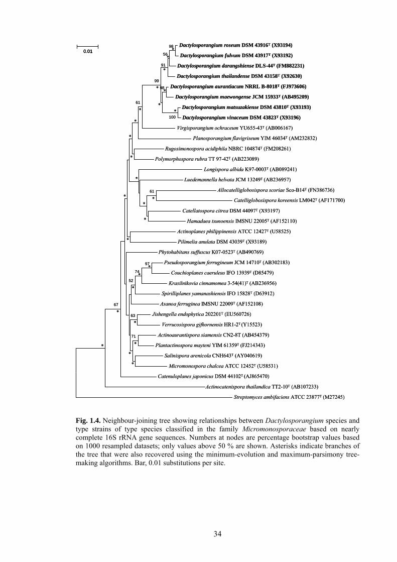
34
Dactylosporangium roseum DSM 43916T (X93194)
Dactylosporangium fulvum DSM 43917T (X93192)
Dactylosporangium darangshiense DLS-44T (FM882231)
Dactylosporangium thailandense DSM 43158T (X92630)
Dactylosporangium aurantiacum NRRL B-8018T (FJ973606)
Dactylosporangium maewongense JCM 15933T (AB495209)
Dactylosporangium matsuzakiense DSM 43810T (X93193)
Dactylosporangium vinaceum DSM 43823T (X93196)
Virgisporangium ochraceum YU655-43T (AB006167)
Planosporangium flavigriseum YIM 46034T (AM232832)
Rugosimonospora acidiphila NBRC 104874T (FM208261)
Polymorphospora rubra TT 97-42T (AB223089)
Longispora albida K97-0003T (AB089241)
Luedemannella helvata JCM 13249T (AB236957)
Allocatelliglobosispora scoriae Sco-B14T (FN386736)
Catelliglobosispora koreensis LM042T (AF171700)
Catellatospora citrea DSM 44097T (X93197)
Hamadaea tsunoensis IMSNU 22005T (AF152110)
Actinoplanes philippinensis ATCC 12427T (U58525)
Pilimelia anulata DSM 43039T (X93189)
Phytohabitans suffuscus K07-0523T (AB490769)
Pseudosporangium ferrugineum JCM 14710T (AB302183)
Couchioplanes caeruleus IFO 13939T (D85479)
Krasilnikovia cinnamomea 3-54(41)T (AB236956)
Spirilliplanes yamanashiensis IFO 15828T (D63912)
Asanoa ferruginea IMSNU 22009T (AF152108)
Jishengella endophytica 202201T (EU560726)
Verrucosispora gifhornensis HR1-2T (Y15523)
Actinoaurantispora siamensis CN2-8T (AB454379)
Plantactinospora mayteni YIM 61359T (FJ214343)
Salinispora arenicola CNH643T (AY040619)
Micromonospora chalcea ATCC 12452T (U58531)
Catenuloplanes japonicus DSM 44102T (AJ865470)
Actinocatenispora thailandica TT2-10T (AB107233)
Streptomyces ambifaciens ATCC 23877T (M27245)
100
98
56
91
98
99
61
97
74
61
71
63
67
52
0.01*
*
*
**
*
*
*
*
*
*
*
**
*
*
**
*
*
*
*
**
*
*
Dactylosporangium roseum DSM 43916T (X93194)
Dactylosporangium fulvum DSM 43917T (X93192)
Dactylosporangium darangshiense DLS-44T (FM882231)
Dactylosporangium thailandense DSM 43158T (X92630)
Dactylosporangium aurantiacum NRRL B-8018T (FJ973606)
Dactylosporangium maewongense JCM 15933T (AB495209)
Dactylosporangium matsuzakiense DSM 43810T (X93193)
Dactylosporangium vinaceum DSM 43823T (X93196)
Virgisporangium ochraceum YU655-43T (AB006167)
Planosporangium flavigriseum YIM 46034T (AM232832)
Rugosimonospora acidiphila NBRC 104874T (FM208261)
Polymorphospora rubra TT 97-42T (AB223089)
Longispora albida K97-0003T (AB089241)
Luedemannella helvata JCM 13249T (AB236957)
Allocatelliglobosispora scoriae Sco-B14T (FN386736)
Catelliglobosispora koreensis LM042T (AF171700)
Catellatospora citrea DSM 44097T (X93197)
Hamadaea tsunoensis IMSNU 22005T (AF152110)
Actinoplanes philippinensis ATCC 12427T (U58525)
Pilimelia anulata DSM 43039T (X93189)
Phytohabitans suffuscus K07-0523T (AB490769)
Pseudosporangium ferrugineum JCM 14710T (AB302183)
Couchioplanes caeruleus IFO 13939T (D85479)
Krasilnikovia cinnamomea 3-54(41)T (AB236956)
Spirilliplanes yamanashiensis IFO 15828T (D63912)
Asanoa ferruginea IMSNU 22009T (AF152108)
Jishengella endophytica 202201T (EU560726)
Verrucosispora gifhornensis HR1-2T (Y15523)
Actinoaurantispora siamensis CN2-8T (AB454379)
Plantactinospora mayteni YIM 61359T (FJ214343)
Salinispora arenicola CNH643T (AY040619)
Micromonospora chalcea ATCC 12452T (U58531)
Catenuloplanes japonicus DSM 44102T (AJ865470)
Actinocatenispora thailandica TT2-10T (AB107233)
Streptomyces ambifaciens ATCC 23877T (M27245)
100
98
56
91
98
99
61
97
74
61
71
63
67
52
0.010.01*
*
*
**
*
*
*
*
*
*
*
**
*
*
**
*
*
*
*
**
*
*
Fig. 1.4. Neighbour-joining tree showing relationships between Dactylosporangium species and type strains of type species classified in the family Micromonosporaceae based on nearly complete 16S rRNA gene sequences. Numbers at nodes are percentage bootstrap values based on 1000 resampled datasets; only values above 50 % are shown. Asterisks indicate branches of the tree that were also recovered using the minimum-evolution and maximum-parsimony tree-making algorithms. Bar, 0.01 substitutions per site.

35
Biotechnological significance of the genus Dactylosporangium. Rare and uncommon
actinomycetes are rich sources of novel secondary metabolites, notably antibiotics
(Lazzarini et al., 2000; Busti et al., 2006). Dactylosporangium strains are a promising
source of novel bioactive compounds of biotechnological interest (Table 1.7) though
members of this genus have rarely featured in screening programmes as they are
difficult to isolate and grow slowly on selective and standard growth media (Hayakawa
et al., 1991a). Indeed, only six species have been assigned to the genus
Dactylosporangium in the past 40 years, hence little is known about either the
taxonomic or the chemical diversity of these organisms.
Table 1.7. Bioactive compounds synthesised by Dactylosporangium strains Organism Metabolite Compound class Reference
D. aurantiacum SF-2185
SF-2185 Azetidine antibiotic Matsumoto et al. (1985)
D. aurantiacum AB 718C-41
Tiacumicins A,B,C,D,E,F, bromo
Macrocyclic antibiotics Theriault et al. (1987); Hochlowski et al. (1997)
D. aurantiacum SANK 61299
A-79197-2 and -3 Di-, tri- saccharides of streptol plant growth inhibitor
Kizuka et al. (2002)
D. fulvum SF-2113 Pyridomycin 12-membered ring compound
Shomura et al. (1986)
D. matsuzakiense SF-2052
Dactimicin Pseudodisaccharide aminoglycoside
Shomura et al. (1980)
D. roseum SF-2186 Orthosomycin Orthosomycin antibiotic group
Shomura et al. (1985)
D. salmoneum ATCC 31224
44161 Polycyclic ether antibiotic Celmer et al. (1978)
D. thailandense G-367
G-367 S1 (2'-N-formylsisomicin, C20H37N5O8)
Aminoglycoside antibiotic
Satoi et al. (1983)
“D. variesporum ” D409-5
Capreomycin Polypeptide antibiotic complex
Tomita et al. (1977)
D. vinaceum SF-2127 Dactimicin Pseudodisaccharide aminoglycoside antibiotic
Shomura et al. (1983)
Dactylosporangium sp. FERM P-8502
AC7230 Polyether antibiotic Yaginuma et al. (1987)
Dactylosporangium sp. ATCC 53693
Dactylocyclines A, B Tetracycline glycoside antibiotic
Wells et al. (1992)
Dactylosporangium sp. RH1
Proline 4-hydroxylase Monomer polypeptide enzyme
Shibasaki et al. (1999)
Dactylosporangium sp. SCC 1695
4a-hydroxy-8-methoxychlortetracycline (Sch 34164)
Tetracycline antibiotic Patel et al. (1987)
Dactylosporangium sp. SF-2908
SF-2809 I, II, III, IV, V, VI
Chymase inhibitor alkaloid anti-allergy
Tani et al. (2004)

36
Dactylosporangium strains known to produce bioactive secondary metabolites
include D. aurantiacum strain AB 718C-41 which produces tiacumicin B (Fidaxomicin,
OPT-80, PAR-101), a novel group of 18-membered macrolide antibiotic discovered at
the Abbott Laboratories, USA (Theriault et al., 1987). Tiacumicin B inhibits RNA
polymerase and shows good activity against pathogenic Gram-positive bacteria and
selected anaerobes, notably Clostridium difficile (Finegold et al., 2004; Biedenbach et
al., 2010). This antibiotic was recently put through phase III clinical trials for the
treatment of Clostridium difficile-associated diarrhoea (Revill et al., 2006; Miller, 2010).
In turn, D. matsuzakiense strain SF-2052 produces dactimicin, a pseudodisaccharide
aminoglycoside antibiotic which was first isolated at Meiji Seika Ltd., Japan (Shomura
et al., 1980). It inhibits Citrobacter diversus, Enterobacter agglomerans, Klebsiella
pneumoniae, Salmonella spp., Serratia marcescens and Yersinia spp., and has a MIC for
90% of strains (MIC90) of less than or equal to 4 μg/ml (Bertelli & Bertelli, 1989; Gu &
Neu, 1989).
Selective isolation of Dactylosporangium strains. Dactylosporangium strains have
been found in small numbers in diverse types of soil, including cultivated field, forest
and pasture soils (Hayakawa et al., 1991a; Vobis, 2006). However, the lack of effective
selective isolation procedures has limited the exploration of members of the genus for
bioprospecting purposes.
Thiemann et al. (1970) recovered 140 dactylosporangiae from soils for the first
time, but did not provide any information how they were isolated. Hayakawa and
Nonomura (1987) isolated dactylosporangiae, microbisporae, micromonosporae and
streptosporangiae on humic acid-vitamin (HV) agar, supplemented with cycloheximide.
Hayakawa and his colleagues (1991a) later developed a more effective method for
isolating dactylosporangiae from heat-preheated soil by using HV agar supplemented
with benzenthonium chloride. However, in practice, the small colonies of the target
organisms are difficult to see, and to isolate from the dark-coloured HV agar plates.
They also devised a chemotactic method which employed collidine and D-xylose as
chemo-attractants and isolated relatively large numbers of Actinoplanes and
Dactylosporangium strains from soil (Hayakawa et al., 1991c), though the identification
of their isolates was mainly dependent on morphological characteristics. Indeed,
dactylosporangiae are difficult to identify in this way as they form indistinct colonies
which can easily be confused with other actinomycetes, such as other genera classified

37
in the family Micromonosporaceae. Consequently, improved methods are needed to
selectively isolate Dactylosporangium strains from natural habitats prior to study their
numbers, roles and taxonomic diversity.
The development of molecular techniques based on precise genotypic information
offers an accurate, rapid, and practical way of evaluating the effectiveness of selective
procedures. The use of taxon-specific oligonucleotide probes provides an effective way
to identify rapidly large numbers of actinomycetes (Mehling et al., 1995; Yoon et al.,
1996; Monciardini et al., 2002) and to detect the presence of target organisms in natural
habitats (Heuer et al., 1997; Monciardini et al., 2002; Blackwood et al., 2005; Kumar et
al., 2007; Mühling et al., 2008). For this purpose, oligonucleotide primers have been
designed for the detection of members of the genera Amycolatopsis (Tan et al., 2006),
Dactylosporangium (Monciardini et al., 2002), Gordonia (Shen & Young, 2005),
Micromonospora (Qiu et al., 2008), Nocardiopsis (Salazar et al., 2002),
Pseudonocardia (Morón et al., 1999) Saccharomonospora (Salazar et al., 2000),
Saccharothrix (Salazar et al., 2002), Streptomonospora (Zhi et al., 2006) and
Streptomyces (Rintala et al., 2001), for the members of Streptomyces violaceusniger 16S
rRNA gene clade (Kumar et al. 2007), and for the families Micromonosporaceae,
Streptomycetaceae, Streptosporangiaceae and Thermomonosporaceae (Monciardini et
al., 2002).
1.7 Antibiotic resistance and self-protection mechanisms of actinomycetes
Antibiotics target many processes that are essential for a cell’s growth, including
cell wall synthesis, DNA duplication, folic acid metabolism, protein synthesis and
transcription (Bush, 2004). The introduction of antibiotics for healthcare was rapidly
accompanied by the appearance of microbial pathogens, which showed resistance to
them. Some of the mechanisms used by pathogenic organisms to counter the effects of
antibiotics are shown in Figure 1.5; some bacteria are intrinsically resistant to certain
antibiotics as they have an impermeable membrane or lack the target of the antibiotic
(Brown & Balkwill, 2009; Allen et al., 2010). Others have multidrug resistance efflux
pumps which excrete antibiotics from the cell into the periplasm, while genetic
mutations can disable the antibiotic-binding sites (Nikaido, 1998; Poole, 2005;
Reynolds & Courvalin, 2005). In addition, inactivation can occur by covalent
modification of the antibiotic, such as that catalysed by acetyltransferases acting on

38
aminoglycoside antibiotics, or by degradation of the antibiotic, such as that catalysed by
β-lactamases acting on β-lactam antibiotics (Bush, 2001; Fischbach & Walsh, 2009).
There are two main mechanisms involved in the development of antibiotic
resistance, namely mutation (Alekshun & Levy, 2007) and acquisition of resistance
genes by horizontal gene transfer (Aminov & Mackie, 2007). Resistance genes can be
transferred between members of distantly related taxonomic groups, including from
Gram-positive to Gram-negative bacteria, through horizontal gene transfer, as
exemplified by the transfer of a kanamycin resistance gene from Streptococcus to
Campylobacter strains (Trieu-Cuot & Courvalin, 1986; Trieu-Cuot et al., 1993; Mindlin
et al., 2006). The origin of antibiotic resistance determinants acquired by horizontal
gene transfer must have resided in the non-pathogenic microbiosphere as human
pathogens were susceptible to antibiotics before the widespread use of antibiotics
(Davies, 1994; Davies, 1997).
Fig. 1.5. Mechanisms of antibiotic resistance in bacteria (taken from Allen et al., 2010)

39
It has been proposed that antibiotic resistance genes originated from the
environmental microbiota, notably antibiotic-producing actinomycetes as these
organisms need resistance mechanisms to protect them against the antibiotic(s) they
produce (Benveniste & Davies, 1973; Roza et al., 1986; Skeggs et al., 1987; Cundliffe,
1995; Mindlin et al., 2006; Hopwood, 2007; Cundliffe & Demain, 2010). Many
resistance mechanisms, including antibiotic inactivation, alteration of permeability
barrier or efflux system, and target modification, which are quite similar to antibiotic
resistance mechanisms, could be involved in conferring self-resistance to the producer
(Cundliffe, 1989; Dairi et al., 1995; Hopwood, 2007; Cundliffe & Demain, 2010). It
seems likely that resistance genes were mobilized through plasmids or transposable
elements to other organisms thereby enabling them to grow in the presence of
antibiotics (Davies, 1997; Poirel et al., 2005; Mindlin et al., 2006).
A well-studied example of horizontal transfer of resistance genes from producers to
pathogens involves the vanHAX operon which encodes enzymes that synthesise
peptidoglycans terminating in D-alanyl-D-lactate instead of D-alanyl-D-alanine (Hong
et al., 2004; Courvalin, 2006). This change decreases the binding affinity of
glycopeptide antibiotics a 1000-fold rendering organisms resistant to teicoplanin and
vancomycin (Reynolds & Courvalin, 2005). The vanHAX operon present in
Enterococcus species has homologues in the glycopeptide-producing actinomycetes,
such as Actinoplanes teichomyceticus ATCC 31131, Amycolotopsis orientalis M43-
05865 and Streptomyces toyocaensis NRRL 15009 (Marshall et al., 1998; Pootoolal et
al., 2002; Serina et al., 2004; Beltrametti et al., 2007). Enterococci and the
glycopeptides producers share 54 to 61% amino acid sequence similarity for VanH, 59
to 63% for VanA, and 61 to 64% for VanX (Marshall et al., 1998), while the gene
orientation of the vanHAX operon is identical in both enterococci and the producers
(Marshall et al., 1998; Courvalin, 2006; Hong et al., 2008).
Bacteria, which show antibiotic resistance, appear to be very widespread in the
environment, as shown by D'Costa et al. (2006). These workers noted that multiple
diverse mechanisms for resistance were associated with soil actinomycetes. They
isolated 480 streptomycetes from agricultural, forest and urban soils and determined
resistance of strains to 21 antibiotics, which represented all major chemical types and
targets of activity. They found multiple mechanisms of resistance amongst the isolates
to both naturally occurring and synthetic antibiotics, and many resistance mechanisms

40
were linked to enzyme inactivation.
Antibiotic resistance cannot just be attributed to the release of antibiotics into the
environment, as bacteria isolated from multiple Siberian permafrost sediments dating
back three million years have been found to be resistant to chloramphenicol, gentamicin,
kanamycin, tetracycline and streptomycin (Mindlin et al., 2008; Petrova et al., 2009),
indicating that indigenous soil bacteria have been under selective pressure to develop
resistance to antibiotics for millions of years. It looks as though actinomycetes are an
important source of new antibiotics, but are also the source of drug resistance.
Consequently, analysing self-resistance mechanism of antibiotic-producing
actinomycetes can help determine the resistance mechanisms of pathogenic
microorganisms that could eventually emerge clinically, and provides strategies that can
be applied in new antibiotic discovery programmes (Hotta et al., 1983, 1992, 1999;
Hotta & Okami, 1996; Wright, 2007, 2010).
Abyssomicins and self-resistance mechanisms. Abyssomicins are effective inhibitors
of para-aminobenzoate (pABA) biosynthesis in some Gram-positive bacteria. The
pABA, which is derived from the key-metabolite of chorismate, is an important
metabolite in the folic acid pathway which is essential for bacterial metabolism (Nichols
et al., 1989; Dosselaere & Vanderleyden, 2001; Quinlivan et al., 2006). Enzymes in
pABA and folic acid biosynthesis are present in many microorganisms, parasites, and
plants, but not in humans, hence this pathway is an interesting target for the
development of novel antibiotics, pesticidal and herbicidal agents (Alun & Jeremy,
2002). Prominent synthetic inhibitors of the folic acid pathway are sulphonamides and
trimethoprim (Huovinen et al., 1995; Sköld, 2001; Brochet et al., 2008).
Abyssomicins, the first known natural inhibitors of the pABA biosynthetic pathway,
are synthesised by Verrucosispora maris AB-18-032, which was isolated from a marine
sediment sample (Riedlinger et al., 2004; Goodfellow et al., 2010b). The abyssomicins
are unique polycyclic polyketide-type 1- antibiotics (Fig. 1.6) in which the main
compound, atrop-abyssomicin C, is active against Gram-positive bacteria, including
Bacillus subtilis and methicillin-resistant Staphylococcus aureus strains (Riedlinger et
al., 2004; Keller et al., 2007a).

41
Fig. 1.6. Structures of abyssomicins isolated from Verrucosispora maris AB-18-032 (taken from Goodfellow & Fiedler, 2010)
All of the abyssomicins mimic the structure of chorismate, the natural substrate for
the PabB subunit of 4-amino-4-deoxychorismate synthase, though only abyssomicin C
and atrop-abyssomicin C bind covalently to PabB by a Michael addition mechanism
(Keller et al., 2007b). These two compounds bind the side chain of Cys263, located in
the proximity of the active site of PabB (Fig. 1.7). However, the resistance mechanism
is not clear hence it is important to determine whether V. maris AB-18-032, like other
antibiotic-producing actinomycetes, can modify the highly conserved abyssomicin
target site to make 4-amino-4-deoxychorismate synthase which is resistant to the drug.
Fig. 1.7. Function of atrop-abyssomicin C as a Michael acceptor. a) Michael addition (pathway A) and dual Michael addition with subsequent rearrangement to an abyssomicin D derivative (pathwayB); b) Proposed reaction with Cys263 based on conversion with S nucleophiles 2-sulfanylethanol and N-acetylcysteine, respectively (taken from Keller et al., 2007).

42
Chapter 2. Materials and Methods
2.1 Source and physico-chemical properties of samples
Source of environmental samples. Twenty-one samples were collected from diverse
geographical locations (Table 2.1). They were sieved, air-dried at room temperature for
5-7 days and stored at 4˚C in plastic containers, apart from the marine sediment samples
which were not dried.
Physico-chemical properties of environmental samples. The pH of the samples were
determined using the procedure described by Reed and Cummings (1945). Two grams
of each sample were added to 18 ml of deionised water, mixed thoroughly, and left for
an hour when the pH was determined using a glass electrode pH meter (Model 320
Mettler-Toledo AG, CH.8603, Schwerzenbach, Switzerland). Each sample was
examined in triplicate and the final pH values recorded as an average of the three
readings. The percentage moisture contents of the triplicated samples were determined
by drying known weights of sediment/soil to constant weight at 105˚C then calculating
the average loss in weight between each set of samples. The dried samples were placed
in a muffle furnace (Carbolite, Sheffield, UK), the temperature raised slowly to 700˚C
and kept constant for 30 minutes to burn off organic matter. After cooling overnight in a
desiccator, the average loss in weight for each set of samples was recorded as the
organic matter content.
2.2 Selective isolation and enumeration
Inoculation of selective media. Two grams of each of the soil samples (Table 2.1)
were resuspended in 18 ml of sterile ¼ strength Ringer’s solution (Oxoid, UK) and
blended in an Ultra-Turrax T25 homogeniser (Junke and Kunkel, Staufen, Germany) for
a minute. The resultant 10-1 suspensions were agitated on a shaker incubator
(Gallenkamp Orbital Incubator, UK) at 150 rpm at room temperature for 30 minutes,
heat pre-treated at 55˚C for 20 minutes in a water bath and cooled at room temperature.
The suspensions were serially diluted down to 10-5 in ¼ strength Ringer’s solution and
triplicate aliquots (100 µl) of the 10-2 to 10-5 dilutions spread, in triplicate, over plates of

43
selective media (Table 2.2) which had been dried for 15 minutes prior to inoculation, as
recommended by Vickers and Williams (1987). Inoculated plates were incubated at
28˚C for 21 days.
Table 2.1. Source and physico-chemical properties of environmental samples
Code Soil/Sediment samples
Location
ANT Antarctic soil Ross Dependency, Antarctica (78°01′S; 163°53′E)
AU96 Arid soil Adelaide, Australia
BK Hay meadow soil Plot 6, Cockle Park Experimental Farm, Northumberland, UK (National Grid reference: NZ 200913)
CA Rhizosphere soil Izmir, Turkey
Ch6 Marine sediment Bahia Tic Toc, Chilean Sea (43°53′ S, 72°87′W)
Ch27 Marine sediment Estero Castro, Chilean Sea (42°30′S, 73°5′W)
CS Chalk soil Ambleside, Cumbria, UK (National Grid reference: NY-357978)
DR Sediment River Derwent, County Durham, UK (National Grid reference: NZ-055490)
ECU Upland soil Riobamba, Ecuador
KHA Agricultural soil Khartoum, Sudan
LS Sediment Shore of Loch Creran, UK (National Grid reference: NM-9124259)
MEG Grassland soil Syros, Greece
MS Rhizosphere soil Kuala Lumpur, Malaysia
NMS Agricultural soil Kuala Lumpur, Malaysia
NOR Marine sediment Østerfjord, Norwegian Sea (60°33′N, 5°19′E)
SAU Arid soil Nairobi, Kenya
SHO Agricultural soil Sholinar, Srinagar, India
TOR Urban soil Torremolinos, Spain
WAD Sand dune soil Borg El-Arab, Egypt
WS Garden soil Samsun, Turkey
ZIZ Arid soil Gazipur district, Bangladesh
Selection and colour grouping of actinomycetes. Organisms growing on the selective
media were putatively identified as actinomycetes by their characteristic colonial
morphology, notably by their ability to form leathery colonies which in many cases
carried aerial hyphae. Two hundred actinomycete colonies were randomly selected from
the isolation plates inoculated with the hay meadow soil suspensions and subcultured
onto oatmeal agar (Shirling & Gottlieb, 1966) plates which were incubated at 28˚C for
21 days. The selected isolates were assigned to groups based on aerial spore mass,

44
colony reverse and diffusible pigment colours which were recorded using National
Bureau of Standards (NBS) Colour Name Chart (Kelly, 1958).
Table 2.2. Media used for the selective isolation of dactylosporangiae
Media Antifungal agents (25 μg/ml)
Antibacterial agents (4 μg/ml)
References
Humic acid-vitamin agar
Cycloheximide Nystatin
Gentamicin or Oxytetracycline
Hayakawa & Nonomura (1987)
Oatmeal agar Cycloheximide Nystatin
Gentamicin or Oxytetracycline
Shirling & Gottlieb (1966)
Raffinose-histidine agar
Cycloheximide Nystatin
Gentamicin or Oxytetracycline
Vickers et al. (1984)
Starch casein agar Cycloheximide Nystatin
Gentamicin or Oxytetracycline
Küster & Williams (1964)
Streptomyces Isolation Medium
Cycloheximide Nystatin
Gentamicin or Oxytetracycline
D'Costa et al.(2006)
Selection and enumeration of presumptive dactylosporangiae. Orange coloured
colonies which lacked aerial hyphae were presumptively assigned to the genus
Dactylosporangium. The numbers of presumptive dactylosporangiae, other filamentous
actinomycetes and bacteria were expressed as the mean number of colony forming units
(cfu’s) per gram dry weight soil.
Maintenance of strains. All of the isolates were grown on oatmeal (Shirling & Gottlieb,
1966) and modified Bennett’s (1949) agars at 28˚C for 3 weeks, and the reference
strains on oatmeal or inorganic salts-starch agar plates under the same conditions (Table
2.3), as described in the relevant chapters. Suspensions of spores and mycelial
fragments were scraped from the oatmeal agar plates and suspended in 1 ml aliquots of
20% (v/v) glycerol in cryo-vials (2 vials per strain) and the resultant preparations kept at
-20˚C for short-term and at -80˚C for long-term storage.
2.3 Molecular taxonomic methods
2.3.1 Genomic DNA extraction
Total genomic DNA was extracted from all of the isolates and reference strains
(Table 2.3), and grown on modified Bennett’s agar (Jones, 1949) for 14 days at 28˚C.
Two or three loopfuls of biomass of each strain were suspended in 200 µl of 1x TE
buffer (pH 8.0; Appendix B), approximately 100 µg of sterile glass beads (≤ 106 μm;
Sigma) added and the resultant preparations disrupted at a speed of 5.0 m/s for 30

45
seconds in a FastPrep® FP120 (Thermo Lab Systems, Waltham, USA). The
preparations were then centrifuged at 13,000 rpm for 2 minutes, each supernatant
transferred to a new tube and 20 µl of RNase (R6148, Sigma) added prior to incubation
at room temperature for 2 minutes. DNA was precipitated from the resultant
preparations by adding 400 µl of cold 100% ethanol and 20 µl of sodium acetate (3M,
pH 5.2), rinsed with 70% ethanol, suspended in 100 µl of 1x TE buffer (pH 8.0), and
quantified using a Nanodrop spectrophotometer (Thermo scientific, Wilmington, USA).
Table 2.3. Isolates and reference strains
Strains Source Tables
Isolates:
200 isolates from hay meadow plot6 soil, Cockle Park, Northumberland, UK Table 3.1, Chapter 3BK-isolates
61 isolates; hay meadow plot 6 soil, Cockle Park Northumberland, UK Table 4.3, Chapter 4
CS-isolates 23 isolates; chalk soil, UK Table 4.3, Chapter 4
DR-isolates 19 isolates; river sediment, County Durham, UK Table 4.3, Chapter 4
ECU-isolates 25 isolates; Upland soil, Ecuador Table 4.3, Chapter 4
LS-isolates 5 isolates; seashore sediment, UK Table 4.3, Chapter 4
MEG-isolates MEG-1, MEG-2; grassland soil, Greece Table 4.3, Chapter 4
MS-isolate MS-1; rhizosphere soil, Malaysia Table 4.3, Chapter 4
NMS-isolates 20 isolates; agricultural soil, Malaysia Table 4.3, Chapter 4
NOR-isolates 15 isolates; marine sediment (616m), Norway Table 4.3, Chapter 4
SHO-isolates 29 isolates; agricultural soil, India Table 4.3, Chapter 4
WAD-isolates WAD-1, WAD-2; sand dune soil, Egypt Table 4.3, Chapter 4
WS-isolates 12 isolates; garden soil, Turkey Table 4.3, Chapter 4
ZIZ-isolates 5 isolates; arid soil, Bangladesh Table 4.3, Chapter 4
Reference strains:
D. aurantiacum NRRL B-8018T; ATCC 23491; DSM 43157; Thiemann D-748; soil, Pagani Gruppo Lepetit, Italy.
Table 5.3, Chapter 5
D. fulvum DSM 43917T; ATCC 43301; NBRC 14381; NRRL B-16292; Meiji Seika Kaisha SF-2113; soil, Chiba Prefecture, Japan.
Table 5.3, Chapter 5
D. matsuzakiense NRRL B-16293T; ATCC 31570; NBRC 14259; Meiji Seika Kaisha SF-2052; Soil, Matsuzaki, Izu Peninsula, Japan. Produces dactimicin.
Table 5.3, Chapter 5

46
Table 2.3. (Continued)
Strains Source Tables
D. roseum DSM 43916T; NRRL B-16295; NBRC 14352; soil,
Shizuoka, Japan. Table 5.3, Chapter 5
“D. salmoneum” NRRL B-16294; DSM 43910; ATCC 31222; NBRC 14103; soil, Japan. produces polycyclic ether antibiotic.
Table 5.3, Chapter 5
D. thailandense DSM 43158T; ATCC 23490, NBRC 12593; Thiemann D-449; soil, Thailand. Degrades natural rubber.
Table 5.3, Chapter 5
D. vinaceum NRRL B-16297T; DSM 43823; ATCC 35207; NBRC 14181; Meiji Seika Kaisha, SF-2127; soil, Japan. Produces dactimicin.
Table 5.3, Chapter 5
“D. variesporum” NRRL B-16296; ATCC 31203, NBRC 14104; soil, Japan. Produces capreomycin.
Table 5.3, Chapter 5
“S. coelicolor” A3(2); K.T. Charter, John Innes Institute, Norwich, UK.
Table 7.2 Chapter 7
S. griseus NRRL B-2682T; ATCC 23345; DSM 40236; NBRC 12875; soil. Produces streptomycin.
Table 7.2 Chapter 7
“S. lividans” TK24; K.T. Charter, John Innes Institute, Norwich, UK.
Table 7.2 Chapter 7
S. venezuelae ATCC 15439; M-2140; soil, Florence, Italy. Produces methymycin.
Table 7.2 Chapter 7
Verrucosispora maris
Isolate AB-18-032; marine sediment sample, Sea of Japan (East Sea).
Chapter 7
* ATCC, American Type Culture Collection Manassas, USA; DSM, Deutsche Sammlung von Mikroorganismen und Zellkulturen, Braunschweig, Germany; NBRC, NITE Biological Resource Center, Chiba, Japan; NRRL, ARS Culture Collection, Northern Regional Research Laboratory, US Department of Agriculture, Peoria, USA; T, Type strain.
2.3.2 Sequencing and analysis of 16S rRNA genes
PCR amplification and sequencing. Amplification of 16S rRNA genes from the
strains (Table 2.3) was carried out using universal bacterial 16S rDNA primers 27F and
1525R (Lane, 1991; Table 2.4) in a T-gradient thermocycler® (Whatman-Biometra,
Göttingen, Germany). All of the reagents for the PCR were purchased from New
England Biolabs, Ipswich, UK. Reactions in a final volume of 50 µl contained 0.2 mM
of each of the four dNTPs, 0.4 µm of primers 27f and 1525r, 1 µl (100 ng) of genomic
DNA, 1.5 mM MgCl2 and 1.25 units Taq DNA polymerase with 1x reaction buffer.
Amplifications were performed under the following conditions: initial denaturation for
5 minutes at 95˚C, 30 cycles of denaturation at 94˚C for 1 minute, annealing at 55˚C for
1 minute, extension at 72˚C for 1 minute, and a final extension step of 10 minutes at
72˚C. The amplified products were analysed by agarose gel electrophoresis in agarose
gels (1.0%, w/v) stained with ethidium bromide. PCR products were purified using a
PCR purification kit (Sigma-Aldrich, UK), according to the manufacturer’s protocol.
The eluted products were stored at -20˚C until required. The sequences of the almost

47
complete length of the 16S rRNA genes were determined by either Macrogen Co.
(Seoul, Korea) or Gene Vision Co. (Newcastle upon Tyne, UK).
Phylogenetic analyses. The chromatogram file in ABI was read in the Chromas
program (Griffith University, Queensland, Australia) and converted into a FASTA
format file. The probable nearest match of each new sequence was determined by
performing a Basic Local Alignment Search Tool (BLAST) search at the National
Center for Biotechnology Information (NCBI; Bethesda, USA) and pairwise sequence
similarities calculated by using a global alignment algorithm, which was implemented at
the EzTaxon web-server (http://www.eztaxon.org; (Chun et al., 2007). The almost
complete 16S rRNA gene sequences were aligned manually using the PHYDIT program
(http://plaza.snu.ac.kr/~jchun/phydit), and ambiguities and other discrepancies resolved
by 16S rRNA secondary structure analysis and by examining the original
chromatographic data.
Phylogenetic trees were inferred using the neighbour-joining (Saitou & Nei,
1987), minimum-evolution (Rzhetsky & Nei, 1992) and maximum-parsimony (Fitch,
1971) tree-making algorithms from the MEGA 3.0 program (Kumar et al., 2004), and
the maximum-likelihood method (Felsenstein, 1981) from the PHYLIP suite of
programs (Felsenstein, 2004). The evolutionary distance model of Jukes & Cantor
(1969) was used to generate evolutionary distance matrices for the neighbour-joining
analysis. The topologies of the resultant trees were evaluated in bootstrap analyses
(Felsenstein, 1985) based on 1000 resamplings of the neighbour-joining dataset using
the CONSENSE and SEQBOOT options from the PHYLIP package.
2.3.3 Molecular fingerprinting
PCR amplification. Repetitive extragenic palindromic PCR (rep-PCR) was carried out
to dereplicate 40 dactylosporangiae strains (Fig. 5.1), as described by Versalovic et al.
(1994), using three bacterial interspersed repetitive sequences, namely BOX elements
(Martin et al., 1992), enterobacterial repetitive intergenic consensus elements
(ERIC;(Hulton et al., 1991) and repetitive extragenic palindromic elements
(REP;(Versalovic et al., 1992); the primers generated the amplification of DNA
fragments of various sizes. Genomic DNA and a negative control were used as template
DNA in a 25 μl PCR mixture with the rep-PCR primers (BOX, ERIC and REP primer
sets; Table 2.4). PCR was performed using the T-gradient Thermocycler® (Whatman-

48
Biometra) under the following conditions; initial DNA denaturation at 95˚C for 5
minutes, 30 cycles of DNA denaturation at 95˚C for 1 minute, annealing at 52˚C (BOX
and ERIC-PCR) or 40˚C (REP-PCR) for 1 minute, extension at 65˚C for 8 minutes, and
a final DNA extension at 65˚C for 16 minutes.
Analysis of DNA fingerprints. Agarose gels were prepared as 1.5% (w/v) gels in 0.5x
TBE buffer supplemented with 0.5 μg/ml ethidium bromide. Mixtures of each DNA
sample (7 μl) and loading dye (3 μl) were added to wells on the gels, and a Gene
Ruler™ 100 bp DNA Ladders Plus (Fermentas Co, York, UK) used as the marker. Gel
electrophoresis was performed at 70 V for 5 hours, and DNA fingerprints visualised and
photographed under UV using a BIO-RAD Fluor-S™ MultiImager (BIO-RAD
laboratories Ltd, Hertfordshire, UK). TIFF images were analysed by using BioNumerics
software (Applied Maths, Kortrijk, Belgium). Three main steps were involved in this
process: (1) definition of lanes and settings, (2) normalisation and designation of bands,
and (3) cluster analysis. Raw data were normalised using a marker lane with clear
banding patterns as the reference profile after conversion of the gel images to the
standard track format. Gel patterns were analysed using a resolution of 1000 and bands
manually assigned with reference to densitometric curves. Similarities between strains
were calculated based on band positions using the Dice coefficient (Dice, 1945), and
clustering achieved using the unweighted pair group method with arithmetic mean
(UPGMA; (Sneath & Sokal, 1973) algorithm. The results of the various analyses were
presented as dendrograms.
Table 2.4. Oligonucleotide primers
Primer Sequence (5’ to 3’) Binding Sitea (5’/3’)
Reference
Bacterial 16S rRNA gene primers:
27F AGAGTTTGATCMTGGCTCAG 8/27 Lane (1991)
1525R AAGGAGGTGWTCCARCC 1544/1525 Lane (1991)
rep-PCR primers:
BOXA1R CTACGGCAAGGCGACGCTGACG - Koeuth et al. (1995)
ERIC1R ATGTAAGCTCCTGGGGATTCAC - Versalovic et al. (1991)
ERIC2 AAGTAAGTGACTGGGGTGAGCG -
REP1R-I IIIICGICGICATCIGGC - Versalovic et al. (1991)
REP2-I ICGICTTATCIGGCCTAC - a Numbering refers to the 16S rRNA gene sequence of Escherichia coli strain rrnB (GenBank accession number: JO1695).

49
2.4 DNA-DNA relatedness experiments
Determination of optimum temperature for renaturation. DNA-DNA relatedness
values between the strains listed in Table 2.5 were determined by using the procedure
described by Gonzalez & Saiz-Jamenez (2005). To determine the thermal denaturation
midpoint of hybrid and reference DNA, purified genomic DNA and mixtures of DNA
from the pairs of strains were denatured and renatured at the optimal temperature for
renaturation (Tor; De Ley et al. 1970). Tor was estimated from mol % G+C contents, as
described by De Ley and his colleagues (1970). The G+C contents of the strains were
determined fluorimetrically by using the following procedure. Each genomic DNA
preparation (2.5 µg) was added to a 0.5 ml tube, the preparation dried in a Speed vac-
DNA 120 (Savant Speed Vac Systems, St. Paul, Minnesota, USA), resuspended in 90 µl
resuspension solution (30% formamide, 0.1x SSC buffer [pH 8.0]) and transferred to
0.2 ml MJ clear tubes (MJ Research Inc, Waltham, USA) suitable for fluorescence
measurements. 10 µl of 1x SYBR Green I (Molecular Probes) was added to each of the
preparations and a thermal ramp run from 25 to 100˚C at 1˚C rise per minute in a PTC-
200 DNA Engine Thermal Cycler (MJ Research Inc). The fluorescence from the tubes
was measured at each step of the thermal ramp and the Tm of each strain calculated from
the minimum value of the slope of the tangent to the melting curve of fluorescence
versus temperature. The mol% G+C content of each strain was calculated using the
equation: mol% G+C = (1.99Tm) - 71.08. The optimal temperature for renaturation was
determined from the G+C values using the equation: Tor = 0.51(mol % G+C) + 47.0, as
described by De Ley et al. (1970).
Estimation of ΔTm between pairs of strains. The generation of genomic DNA hybrids
for estimation of ΔTm values between the pairs of test strains was carried out by adding
each genomic DNA (2.5 µg) from both the reference and the test strains to a 0.5 ml
centrifuge tube; genomic DNA (5.0 µg) from the reference strain to another tube for
control sample. The samples were first dried in a speed vac and the resultant dried
preparations resuspended in 90 µl 0.1x SSC buffer (pH 8.0; Appendix B). The
homologous DNA control and hybrid DNA samples were denatured and rehybridised in
the T-gradient Thermocycler® (Whatman-Biometra) using the following conditions:
99˚C for 10 minutes, Tor for 8 hours followed by progressive steps in which the
temperature was dropped by 10˚C then held for 1 hour until the temperature reached to

50
25˚C. The tubes were then held at 4˚C, 10µl of 1x SYBR Green I was added and the
contents transferred to MJ clear tubes in an PTC -200 DNA Engine Thermal Cycler.
Thermal denaturation was achieved as follows: 25˚C for 15 minutes, and a thermal
ramp from 25 to 100˚C at 0.2˚C per second; fluorescence measurements were taken at
each step of the ramp. The Tm of the homologous and hybrid DNA preparations was
calculated by taking the temperature corresponding to a 50% decrease in fluorescence in
the melting curve of fluorescence versus temperature. ΔTm is the difference between
these two temperatures; differences of 5˚C or more are considered to show that tested
strains belong to different genomic species (Wayne et al., 1987; Rosselló-Mora &
Amann, 2001).
Table 2.5. Dactylosporangium strains examined in the DNA-DNA relatedness
experiments
Test strains Reference strains Isolate BK51 vs: Isolate BK53
Isolate BK69 Isolate BK63 D. aurantiacum NRRL B-8018T D. matsuzakiense NRRL B-16293T D. salmoneum NRRL B-16294T D. vinaceum NRRL B-16297T
Isolate BK63 vs:
D. aurantiacum NRRL B-8018T D. matsuzakiense NRRL B-16293T D. salmoneum NRRL B-16294T D. vinaceum NRRL B-16297T
“D. salmoneum” NRRL B-16294 vs: D. aurantiacum NRRL B-8018T
D. matsuzakiense NRRL B-16293T D. vinaceum NRRL B-16297T
2.5 Phenotypic characterisation
2.5.1 Cultural and morphological characteristics
The tested strains (Table 2.6) were grown on tryptone-yeast extract, yeast extract-
malt extract, oatmeal, inorganic salts-starch, glycerol-asparagine, peptone-yeast extract-
iron and tyrosine agars (ISP media 1-7, respectively; Shirling & Gottlieb, 1966) for 21
days at 28˚C. The resultant colonies were examined by eye to determine substrate
mycelial pigmentation and the colour of any diffusible pigments; colours were recorded
using National Bureau of Standards (NBS) Centroid Colour Charts (Kelly, 1958).

51
Table 2.6. Sources and strain histories of dactylosporangiae examined in the phenotypic
tests
Strains Source
Dactylosporangium sp. BK51, BK63 Plot 6, hay meadow soil, Cockle Park, Northumberland, UK
D. aurantiacum NRRL B-8018T; ATCC 23491; DSM 43157; Thiemann D-748; soil, Pagani Gruppo Lepetit, Italy.
D. fulvum DSM 43917T; NBRC 14381; NRRL B-16292; Meiji Seika Kaisha SF-2113; soil, Chiba Prefecture, Japan.
D. matsuzakiense NRRL B-16293T; ATCC 31570; NBRC 14259; Meiji Seika Kaisha SF-2052; soil, Matsuzaki, Izu Peninsula, Japan. Produces dactimicin.
D. roseum DSM 43916T; NRRL B-16295; NBRC 14352; soil, Shizuoka, Japan.
“D. salmoneum” NRRL B-16294; DSM 43910; ATCC 31222; NBRC 14103; soil, Japan. Produces polycyclic ether antibiotic.
D. thailandense DSM 43158T; ATCC 23490, NBRC 12593; Thiemann D-449; soil, Thailand. Degrades natural rubber.
D. vinaceum NRRL B-16297T; DSM 43823; NBRC 14181; Meiji Seika Kaisha, SF-2127; soil, Japan. Produces dactimicin.
“D. variesporum” NRRL B-16296; ATCC 31203, NBRC 14104; soil, Japan. Produces capreomycin.
Micromorphological properties of the strains were recorded from 3-week-old
oatmeal agar plates using a Nikon Optiphot light microscope fitted with a long working-
distance objective. In addition, the spore arrangement of isolates, BK51 and BK63, and
“D. salmoneum” NRRL B-16294 were determined by scanning electron microscopy
(SEM). Agar plugs, 2 per strain, were prepared from oatmeal agar plates using a sterile
6 mm cork borer, fixed using 2% (v/v) glutaraldehyde in Sorenson’s phosphate buffer in
small bijoux tubes, and the preparations kept at 4˚C overnight. The buffer was then
discarded; the agar plugs dehydrated in an ethanol series (10, 20, 40, 60, 80 and 100%
for 20 minutes) prior to SEM analysis (Electron Microscopy Unit, Newcastle
University). Samples were dried using a Critical Point Dryer (SAMDRYR-780,
Tousimis Research Corporation, USA) and liquid carbon dioxide, mounted on
aluminum stubs coated with gold, using a Gold Sputter (E5100. “Cool” Sputter Coater,
Polaron Equipment Limited, Walford, UK), and examined using a Stereoscan 240
scanning electron microscope (Cambridge Instruments Ltd., Cambridge, UK).

52
2.5.2 Biochemical tests
Test strains and inoculation of test media. The test strains (Table 2.6) were examined
for 120 unit characteristics, as shown in Table 2.7. Most of the tests were carried out in
Replidishes (Sterilin Ltd., Staffordshire, UK) at 28˚C unless stated otherwise, and
results recorded after final readings. Sterile media were aseptically dispensed into each
of the 25 compartments of the Replidishes and individual compartments inoculated with
10 μl aliquots of spore suspensions. To this end, spores from each of the strains were
scraped from oatmeal agar plates, which had been incubated at 28˚C for 21 days, and
transferred to individual 15 ml conical tubes which contained 5 ml of ¼ strength
Ringer’s solution. The resultant spore suspensions were filtered through sterile cotton
wool, centrifuged and resuspended in 2 ml of ¼ strength Ringer’s solutions to give a
turbidity of 5 on the McFarland scale (BioMerieux® Co.). Each inoculum (1 ml) was
pipetted into a sterile compartment in Replidishes and gently agitated to give an even
suspension prior to inoculation. An automatic multipoint inoculator (Denley-Tech;
Denley Instruments Ltd., Sussex, UK) was used to inoculate Replidishes containing
each test media. All of the tests were carried out in duplicate.
Aesculin hydrolysis. This test was carried out using the method of Kutzner (1976) and
results recorded after incubation for 7, 14 and 21 days. Strains with β-glucosidase
activity hydrolyse aesculin to give 6, 7-dihydroxycoumarin which complexes with ferric
ions in the medium to form a brown black melanin-like polymer. Blackening of the test
medium indicated a positive result. It can be difficult to distinguish between a positive
result and the production of dark pigments by some actinomycetes hence all strains
were inoculated onto a negative control (without aesculin) which was compared to the
corresponding aesculin test in order to prevent recording false positive results.
Allantoin hydrolysis. The hydrolysis of allantoin (0.33%, w/v) was detected using the
basal medium of Gordon et al. (1974) supplemented with phenol red as the pH indicator.
After incubation for 14 days, inoculated Replidishes were examined for the
development of an alkaline reaction which was indicated by a colour change of the
medium from orange to pink red. A positive test result, that is, the production of a pink-
red colour, indicates the presence of two hydrolytic enzymes, one of which hydrolyses
allantoin to allantoic acid and the other which catalyses the formation of urea and
glycoxalate.
53
Table 2.7. Unit characters
A. Biochemical tests: Aesculin hydrolysis Allantoin hydrolysis Arbutin hydrolysis Catalase production Hydrogen sulphide
production* Nitrate reduction* Urease production*
B. Degradation tests (%,
w/v): Adenine (0.4) Casein (1) Cellulose (1.0) Chitin (0.2) DNA (0.2) Elastin (0.3) Gelatin (0.4) Guanine (0.05) Hypoxanthine (0.4) RNA (0.3) Starch (1.0) Trybutyrin (0.1% v/v) L-Tyrosine (0.5) Tween 20 (1.0% v/v) Tween 40 (1.0% v/v) Tween 60 (1.0% v/v) Tween 80 (1.0% v/v) Uric acid (0.5) Xanthine (0.4) Xylan (0.4)
C. Nutritional tests:
Growth on sole carbon sources At 1.0 %, w/v: Monosaccharides: Hexoses: D-Fructose D-Galactose D-Glucose D-Mannose
L –Sorbose D- Xylose Pentoses: Amygdalin L-Arabinose L-Fucose D-Furanose L-Rhamnose D-Ribose Disaccharides: D-Cellobiose α- Lactose D-Maltose D-Melibiose D-Sucrose D-Trehalose Tri & tetrasaccharides: D (+) Melezitose D (+) Raffinose Polysaccharides: Dextrin Glycogen Inulin Pectin Sugar alcohols: Adonitol D-Arabitol Dulcitol meso-Inositol D-Mannitol D-Sorbitol Glycosides: Aesculin Arbutin Salicin
At 0.1%, w/v: Citric acid L(+) Lactic acid Malic acid
Oxalic acid Propionic acid Pyruvic acid Succinic acid L (+) Tartaric acid Urea Uric acid
D. Tolerance tests: Growth at: pH 4.0, 5.0, 9.0 and 10.0 4˚C, 10˚C, 15˚C, 20˚C, 25˚C, 30˚C, 37˚C and 42˚C Growth in presence of (%, w/v): Sodium chloride (1.5, 3.0, 5.0 and 7.0) Lysozyme (0.05) Resistance to antibiotics (µg/ml): Ampicillin (4): Cephaloridine (2) Chloramphenicol (8) Ciprofloxacin (2) Clindamycin (8) Gentamicin (8) Kanamycin (8) Lincomycin (8) Novobiocin (8) Oxytetracycline (16) Penicillin-G (2) Rifampicin (16) Streptomycin (4) Tetracycline (8) Tylosin (8) Vancomycin (2)
*, tests carried out in sterilised Bijoux tubes. The remaining tests were carried out in Replidishes.

54
H2OHN NH
HN NH2
O
O
O
HN
H2N
O
CO2H
HN
NH2
O
H2O
H2N NH2
O
+
OH
O
O
H
Allantoin Allantoic acid Urea Glycoxalate
Arbutin hydrolysis. The hydrolysis of arbutin (0.1%, w/v), a hydroquinone-α-
glucopyranoside, was detected using the basal medium described by Williams et al.
(1983). The hydrolysis of arbutin is catalysed by the hydrolase enzyme, β-glucosidase,
resulting in the production of glucose and a hydroquinone, the latter complexes with the
iron in the medium to form a black-brown melanin-like polymer. Blacking of the
medium after 5, 10 or 15 days was recorded as a positive result.
Catalase production. The presence of catalase was detected by adding a few drops of
20% (v/v) hydrogen peroxide solution to 7 day old cultures grown on modified
Bennett’s agar (Jones, 1949). The production of oxygen bubbles from the reduction of
hydrogen peroxide was recorded as a positive reaction.
Hydrogen sulphide production. The ability of the test strains to produce hydrogen
sulphide was determined following the method described by Küster & Williams (1964).
Tubes of nitrate broth (Appendix A) were inoculated and strips of sterile lead acetate
paper hooked over their necks. Hydrogen sulphide converts lead acetate to lead sulphide
resulting in blackening of the lead acetate paper. The blackening of lead acetate strips
after incubation for 7 days was recorded positive.
Nitrate reduction. This test was performed to detect the presence of nitrate and nitrite
reductases. Tubes containing 3 ml nitrate broth (0.1%, w/v; (Gordon & Mihm, 1962)
were inoculated and incubated for 14 days when a few drops of reagents A and B
(Appendix B) were added. The sulfanilic acid in reagent A reacts with nitrite to form a
diazonium salt which forms a stable red dye in the presence of α-naphthylamine in
reagent B. The development of a red colour on addition of reagent B indicated the
presence of nitrite and was recorded as positive for nitrate reductase activity. In the
absence of a colour change traces of zinc dust were added to the broths. Zinc ions
catalyse the same reaction as nitrate reductase hence when nitrate is still present the
addition of zinc reduces it to nitrite with the formation of a characteristic red colour;
such results were recorded as nitrate and nitrite reductase negative. The continued
55
absence of any colour change following the addition of zinc indicates that nitrate has
been reduced to gaseous nitrogen; such reactions were recorded positive for nitrate and
nitrite reductase.
NO3- + 2 H+ + 2 e NO2
- + H2O N2
Nitrate Nitrite Nitrogen gas
Urease production. The strains were tested for their ability to hydrolyse urea using the
basal medium (Appendix A) of Gordon et al. (1974) supplemented with urea (1.76 %,
w/v). Inoculated Replidishes were incubation for 14 days; and then examined for the
development of a pink-red colour which was recorded as a positive result.
NH2CONH2 + 2 H2O 2NH3 + CO2 + H2O
Urea Ammonia
2.5.3 Degradation tests
Adenine, casein, cellulose, elastin, guanine, hypoxanthine, L-tyrosine, uric acid,
xanthine and xylan were incorporated into modified Bennett’s agar (Jones, 1949) with
care taken to ensure an even distribution of the insoluble compounds; each compound
was sterilised by tyndalisation before adding to molten modified Bennett’s agar.
Inoculated plates were read after 7, 14 and 21 days. In all cases, disappearance of the
substrate from under and around the area of growth was recorded as a positive result.
Adenine, guanine, hypoxanthine and xanthine are nitrogenous purine bases involved in
the biosynthesis of nucleic acids, L-tyrosine is an aromatic amino acid and elastin is a
protein found in animal connective tissue. The degradation of uric acid and xylan
indicate the production of uricase and xylanase enzymes, respectively.
Casein. The degradation of casein was detected in modified Bennett’s agar (Jones,
1949) supplemented with skim milk powder (10 g per litre). Inoculated Replidishes
were incubated for 15 days when the presence of clear zones from around and under
areas of growth indicated a positive result. Similarly chitinolytic activity was observed
on colloidal chitin agar (Hsu & Lockwood, 1975) after 14, 21 and 28 days and positive
results recorded when a clear zone was detected around colonies.
Nitrate reductase Nitrite reductase
Urease

56
Cellulose. Cellulose is composed of β-(1-4) linked glucose subunits.
Carboxymethylcellulose plates inoculated with the test strains were incubated for 14
days then flooded with Congo red solution (0.1%, w/v) for 15 minutes at room
temperature. Undigested cellulose stained a pinky red when excess reagent was
removed; a pale orange to straw colour zone around areas of growth indicated the
breakdown of cellulose, such results were recorded positive.
DNA and RNA. The degradation of DNA was detected using Bacto DNase test agar
(Difco; Appendix A) which contains DNA (0.2%, w/v). Ribonucleic acid breakdown
was examined in tryptose agar (Goodfellow et al., 1979) supplemented with RNA
(0.3%, w/v). In each case, inoculated 7-day-old plates were flooded with 1 N
hydrochloric acid which causes nucleic acids to precipitate as a fibrous mass. Positive
reactions were indicated by the presence of clear zones from under and around the
growth of the tested strains.
Gelatin. The degradation of gelatin was detected using modified Bennett’s agar (Jones,
1949) supplemented with gelatin (0.4%, w/v). Inoculated plates were incubated for 7
days then flooded with acidic mercuric chloride solution (Appendix B), the latter acts as
a denaturing agent complexing with gelatin and causing it to precipitate. Extracellular
proteases hydrolyse gelatin into small peptides and amino acids which do not react with
mercuric chloride, positive reactions were indicated by the presence of clear zones from
under and around the growth of the tested strains.
Starch. Starch, the main polysaccharide in plants, is composed of α-D-glucopyranose
sub-units in two different structural configurations; amylose, an α-(1-4) linked polymer,
and amylopectin, an α-(1-4) linked molecule with α-(1-6) branches. This test was
carried out to detect the production of extracellular α-amylases which degrade starch.
The test was performed by using modified Bennett’s agar (Jones, 1949) supplemented
with soluble starch (10 g/ litre). Inoculated plates were incubated for 15 days prior to
flooding with Lugol’s iodine (Appendix B); iodine complexes with left handed, α-
helical, amylose molecules resulting in the formation of a dark blue starch-iodine
complex. Extracellular α-amylases show endo-glycosidase activity and hence are able to
hydrolyse starch molecules by random attack at points distant from the chain ends to
form short polysaccharide chains (dextrins) and simple sugars which are unable to

57
complex with iodine. Positive results are indicated by the formation of clear zones from
around the growth area following the addition of Lugol’s iodine.
Tributyrin. The breakdown of glycerol tributyrate was carried out using trybutyrin agar
(Appendix A; Sigma T3688). Inoculated plates were read after 7 and 14 days. Positive
results were recorded when clear zones were observed from under and around colonies.
Tweens. Tweens 20, 40, 60 and 80 are a homologous series of water soluble, high
molecular weight fatty acid esters of a polyoxyalkaline derivative of sorbitan which
differ only in fatty acid components. Tweens 20, 40, 60 and 80 contain mono-laurate,
palmitic, stearic and oleic acids, respectively and can be used to detect the production of
specific esterases. Esterases produced by tested strains diffuse into the medium and
hydrolyse ester linkages releasing free fatty acids which combine with calcium ions
(Ca2+) present in the medium to form insoluble calcium salts which precipitate out as
white crystals characteristic for each of the Tweens. The basal medium of Sierra (1957)
supplemented with individual Tweens (10 ml/litre), was inoculated, incubated and
examined for the characteristic precipitate which indicated a positive response.
2.5.4 Nutritional tests
The ability of test strains to metabolise carbon compounds (Table 2.7) as sole
sources of carbon for energy and growth was examined using carbon utilisation agar
(ISP medium 9; Appendix A; Shirling & Gottlieb, 1966) as the basal medium. Solutions
of each carbon source were sterilised by steaming at 101˚C for 30 minutes on three
consecutive days then added to the basal medium to give the required concentration.
Media dispensed into Replidishes were inoculated, incubated and examined after 7, 14
and 21 days. The test strains were also inoculated onto the basal media (the negative
control) and onto the basal medium supplemented with 1%, glucose (the positive
control). A positive result was recorded when growth on the test medium was equal or
greater that on the positive control, but negative when growth on the test plate was
equal to or less than that on the negative control plate.
2.5.5 Tolerance tests
Temperature. The test strains were examined for their ability to grow on modified
Bennett’s agar (Jones, 1949) at 4˚C, 10˚C, 20˚C, 25˚C, 30˚C, 37˚C and 42˚C. Inoculated
Replidishes were incubated and read weekly for 6 weeks at 4˚C and 10˚C, and for 3

58
weeks at the other temperatures. In all cases, visible growth was recorded as a positive
result. The Replidishes incubated at 37˚C and 42˚C were placed into plastic bags in
order to prevent the inoculated medium from drying out.
pH. The ability of the test strains to grow over a range of pH regimes (Table 2.7) was
examined on modified Bennett’s agar adjusted to the appropriate pH using 0.1 M
solutions of potassium dihydrogen phosphate (KH2PO4) and dipotassium hydrogen
phosphate (K2HPO4). Growth was recorded as a positive result after incubation for up to
21 days.
Sodium chloride and lysozyme. The test strains were examined for their ability to
grow on modified Bennett’s agar (Jones, 1949) supplemented with sodium chloride at
1.5%, 3%, 5% and 7% (w/v) and lysozyme at 0.05% (v/v). Inoculated plates were read
after 7, 14 and 21 days when the growth of strains was compared to that on control
plates without the supplements; positive reactions were recorded when growth on the
test plates was greater than that on the control plate and a negative one when growth
was less or the same as on the control plate.
Resistance to antibiotics. The test strains were examined for their ability to grow in the
presence of different antibiotics at various concentrations (Table 2.7). All of the
antibiotic solutions were filter sterilised then added to sterile cooled modified Bennett’s
agar (Jones, 1949) to give the required concentrations before dispensing into
Replidishes; control plates lacking antibiotic were also inoculated. Inoculated plates
were incubated for up to 21 days. Organisms were recorded as resistant (+) where
growth on the test plate was greater than or equal to that on the control plate and
sensitive (-) when growth was less than that on the control plate.
2.6 Chemotaxonomic tests
Detection of diaminopimelic acid isomers. Test strains (Table 2.6) were grown on
modified Bennett’s agar (Jones, 1949) for 14 days at 28˚C. The isomeric forms of
diaminopimelic acid (A2pm) were determined by thin-layer-chromatography (TLC) of
whole-organism hydrolysates on cellulose acetate sheets after Staneck & Roberts
(1974). A loopful of biomass of each isolate was harvested, placed into a 2 ml cryotube
which contained 500 μl of 6 N HCl, mixed and heated at 100˚C in an oven for 4 hours.

59
The resultant whole-organism hydrolysates were centrifuged at 6000 rpm for 4 minutes,
and the supernatants collected and evaporated to dryness at 100˚C for 2 to 3 hours in a
heating block held in a fume cupboard. Once most of the acid had evaporated, 1ml of
distilled water was added to each preparation prior to evaporating at 100˚C. Finally, 100
μl of distilled water was added to each preparation to produce the extract needed for
TLC.
The isomers of A2pm were separated by one-dimensional TLC using glass backed
cellulose TLC plates (20×20 cm; No.5716. Merck, Darmstadt, Germany). An aliquot (1
μl × 2) of each hydrolysate was applied as a spot along a line 2 cm from the base of the
TLC plate. A standard solution (0.01 M) of A2pm (Sigma), which contained a mixture
of DL-, LL- and meso-A2pm isomers, was prepared and 1 μl applied to each TLC plate
on either side of the test samples. The plates were developed in glass tanks saturated
with a solvent (methanol: water: 6 N HCl: pyridine; 80:26:4:10; v/v/v/v) for 4 hours or
until the solvent front was approximately 5 cm from the top of the plate. Plates were air-
dried, sprayed with freshly prepared ninhydrin solution (0.2%, w/v dissolved in
acetone) and heated at 100˚C for 3 minutes in an oven. The isomers of A2pm appeared
as dark purple to brown spots that had a lower retention factor (Rf) value than amino
acids, the latter yielded blue to purple coloured spots. The A2pm isomers of the test
strains were identified by comparison with the standard mixture. The Rf values of the
A2pm isomers run in the order: 3-hydroxy-A2pm, meso-A2pm, DD-A2pm and LL-A2pm
(from lowest to highest).
Detection of mycolic acids. The alkaline methanolysis procedure, modified from
Minnikin (1975), was used to examine the isolates (Table 2.6) for the presence of
mycolic acids; Nocardia brasiliensis N318T was used as the control. Two loopfuls of
wet biomass per strain were suspended in 1 ml of 5% (v/v) aqueous tetrabutyl-
ammonium hydroxide (diluted from a 40% commercial solution; Sigma) held in 2 ml
vials which contained 100 μg glass beads (<106 μm diameter; Sigma). The samples
were homogenised for 20 seconds (FastPrep FP120 Cell Disruptor, speed 5 m/s),
incubated at 100˚C for 4 hours and cooled to room temperature prior to centrifugation at
4000 rpm for 5 minutes. The supernatants were transferred to clean tubes which
contained a solution of dichloromethane (1 ml) and iodomethane (25 μl) and the
resultant preparations shaken on a tumble shaker (Model TM1 Tumbler, speed No.5) for
30 minutes. The preparations were centrifuged (2000 rpm, 3 minutes), the upper

60
aqueous layers discarded, and the lower organic layers, which contained the methyl
ester derivatives, were evaporated to dryness under a stream of nitrogen gas at 37˚C.
Petroleum ether (50 μl) was added to reconstitute each sample for TLC analysis.
The mixtures of fatty acid methyl esters (FAME) and mycolic acid methyl esters
(MAME) were separated by one-dimensional TLC using aluminium-backed silica gel
TLC sheets (10×20 cm; No.5554, silica gel 60F254, Merck, Darmstadt, Germany). The
samples (3 μl) were applied in 1 μl aliquots onto a line 1cm from the base of each sheet
and the TLC plates developed in saturated glass tanks containing petroleum ether-
acetone (95:5, v/v). The plates were air-dried when the solvent front approached the top
of the plates; following air-drying the chromatograms were developed a second time in
the same direction. The resultant chromatograms were sprayed with a 5% (w/v) solution
of ethanolic molybdophosphoric acid (diluted from a 10% commercial solution; Sigma
P-1518) and immediately dried in an oven at 180˚C for 5 minutes. The positions of the
separated FAMEs and MAMEs were revealed as dark blue/black spots on a pale green
background. The presence or absence of mycolic acids in the test strains was determined
by comparison with the reference strain.
Determination of whole-organism sugar patterns. Whole-organism sugar patterns
were determined according to the procedure described by Schaal (1983). Freeze-dried
biomass (ca. 25 mg) of each isolate (Table 2.6) was hydrolysed with 1.5 ml of 1N
H2SO4 in a sealed 2 ml vial held at 100˚C for 3 hours, the hydrolysate cooled to room
temperature, neutralised (pH 5.0-5.5) by adding barium hydroxide (ca. 0.5 g) and
shaken several times. The resultant preparation and the remaining Ba(OH)2 were
centrifuged at 6000 rpm for 5 minutes and the supernatant carefully removed and
evaporated to dryness on a hot block at 40˚C. The residue was reconstituted with 0.1 ml
distilled water and 0.25 ml pyridine, thoroughly mixed and the pyridine phase separated
from the aqueous phase by centrifugation at 6000 rpm for 5 minutes. The upper pyridine
layer, which contained the purified sugar extract, was transferred to a small glass vial
and concentrated under a stream of nitrogen at room temperature to give a final volume
of 0.3 ml.
The concentrated pyridine extracts (3 μl) were spotted in 1 μl aliquots onto a line
1 cm from the base of cellulose TLC aluminium sheets (10×20 cm; No.5552, Merck)
and the sugars separated by ascending, one-dimensional TLC using n-butanol/water/
61
pyridine/toluene (10:6:6:1, v/v/v/v). Chromatograms were developed twice with
intermediate air drying, and the sugars visualised by spraying chromatograms with an
aniline-phthalate reagent (aniline, 2 ml; phthalic acid, 3.25 g in 100 ml water-saturated
n-butanol), air drying and heating at 100˚C for 4 minutes when the hexoses, pentoses
and 6-deoxy sugars gave brown, red and grey-green spots, respectively. The Rf values
of the sugars increase in the order: galactose, glucose, mannose, arabinose, xylose,
ribose, rhamnose and 6-deoxytalose. Mixtures of the sugars (arabinose, galactose,
glucose, mannose, ribose; 1%, w/v in pyridine) were used as standards. The sugar
patterns of the strains were identified by comparison with the standards.
Determination of muramic acid residues in the peptidoglycan. The N-acyl type of
muramic acid in the cell wall peptidoglycan of the strains was determined using a
modification of the glycolate test (Uchida & Seino, 1997). Dried biomass (ca. 10 mg) of
each strain (Table 2.6) was hydrolysed with 500 μl of 6 N HCl at 100˚C in an oven for 2
hours and allowed to cool to room temperature before adding 1 ml of water-saturated
diethyl ether. The resultant preparations were mixed on a tumble shaker (Model TM1
Tumbler) for 30 minutes, vortexed for two minutes and centrifuged at 5000 rpm for 2
minutes. The extracts of the upper layers were discarded to remove any interfering
compounds from the glycolate fraction, and the extraction procedure repeated on the
original hydrolysates using a further 1 ml of water-saturated diethyl ether. The upper
layers of the samples were transferred to clean microcentrifuge tubes, using Pasteur
pipettes, treated with 5 μl of 0.1 N sodium hydroxide, and the preparations inverted
several times prior to evaporating to dryness at room temperature in a fume hood.
The residues were redissolved in 25 μl of sterile distilled water and 500 μl of
freshly prepared DON reagent (0.02% w/v, 2,7-dihydroxynaphthalene dissolved in
concentrated sulphuric acid). The DON reagent was prepared the day before use, stored
overnight at 4˚C and was only considered stable if the colour had changed from yellow
to colourlessness. The preparations were heated at 100˚C for 20 minutes, cooled to
room temperature and vortexed briefly then allowed to stand for 10 minutes and the
results recorded. The test strains were examined for N-acetylated muramic acid acyl
groups (positive controls: Amycolatopsis orientalis KCTC 9412T and
Saccharopolyspora rectivirgula F1T) and N-glycolated acyl groups (positive controls:
Gordonia bronchialis N654T and Nocardia brasiliensis 318T), a negative control
lacking biomass was also used. Organisms characterised by the presence of N-

62
glycolated acyl groups produce a deep red to purple colour reaction whereas those
which contain N-acetylated acyl groups remain clear or pale yellow. The colour
reactions for the tested strains were compared with those of the control tubes.
Cellular fatty acid analysis. Cellular fatty acids (FAME) of the test strains (Table 2.6)
were analyzed by gas-liquid chromatography using the Microbial Identification System
(MIS; Sasser, 1990). The fatty acids were saponified, methylated, and extracted from
whole-organisms according to the MIS operation manual (Sasser, 1990). Biomass
(50mg) harvested from tryptic soy broth was added to 1ml of 30% NaOH/methanol
(1:1, v/v), vortexed then heated at 100˚C for 5 minutes. The resultant preparations were
mixed, heated at 100˚C for 25 minutes and allowed to cool at room temperature. Each
reaction mixture was then added to 2 ml of 6 N HCl/methanol (3.25: 2.75, v/v), mixed,
heated at 80˚C for 10 minutes and cooled quickly in a tray of cold tap water.
Fatty acid methyl esters were extracted from the acidic aqueous phase with 1.25
ml of hexane/tert-butylmethylether (1:1, v/v), transferred into a clean vial and analyzed
using a model HP-5890II plus gas chromatograph (Hewlett-Packard Co.) equipped with
a HP-Ultra 2, phenyl methyl silicone fused silica capillary column (25m × 0.2mm), an
auto injector and a flame ionization detector (FID); the injector and detector
temperatures were at 250˚C and 300˚C, respectively. The temperature was increased
from 170˚C to 270˚C at a rate of 5˚C /minute using hydrogen as the carrier gas.
Identification of the FAMEs was achieved by using the Sherlock system which
compares the retention times of peaks with calibration standards (HP 19298-60500).
Menaquinone analysis. Menaquinones were extracted from the strains (Table 2.6) by
using the small-scale integrated procedure of Collins et al. (1985). Freeze-dried biomass
(100mg) of each strain was treated with 2 ml of 0.3% aqueous NaCl/methanol (1:10,
v/v) and 2 ml of petroleum ether, mixed on a tube rotator for 30 minutes, and the upper
layer transferred to a clean vial. The lower layer was treated with a further 1ml of
petroleum ether, mixed for 30 minutes, and the upper layer added to the previous one.
The combined extracts were transferred to small glass vials and evaporated to dryness
by using nitrogen and stored at - 20˚C until required.
The extracted menaquinones were resuspended in 200 μl of isopropanol and
analysed using a high performance liquid chromatography (HPLC; LC-10AS, Shimadzu
63
Co.) fitted with a reverse-phase C18 column (250 × 4.0 mm, 5 μm particle size; RP-18-
Lichrosorb column; Capital Analytical, UK). Methanol/isopropanol (2:1, v/v) was used
as the isocratic mobile phase with a flow rate of 1 ml/min at ambient temperature;
aliquots of each sample (10 μl) were injected into the HPLC. The menaquinones were
detected at a wavelength of 270 nm using a UV detector (SPD-10A, Shimadzu Co.), and
the resultant chromatograms integrated using a C-R6A Chromatopac integrator
(Shimadzu Co.). The menaquinones were identified by comparing their retention times
with those of reference samples extracted from Streptomyces indiaensis JCM 3053T
which has a well known menaquinone profile (Kudo & Seino, 1987)
Polar lipid analysis. Polar lipids were extracted from the biomass remaining after the
extraction of menaquinones (Collins et al., 1985). Each sample of cell debris was heated
in a boiling water bath for 5 minutes, cooled to room temperature, and 2.3 ml of
chloroform/methanol/0.3% aqueous NaCl (9:10:3, v/v/v) added. The resultant samples
were mixed on a tube rotator for an hour, each supernatant was transferred to a clean
tube to which 0.75 ml of chloroform/methanol/0.3% aqueous NaCl (5:10:4, v/v/v) was
added prior to mixing for 30 minutes. Each of the resultant preparations was
centrifuged; the supernatants combined with the corresponding previous one, 1.3 ml
chloroform and 1.3 ml of 0.3% aqueous NaCl added and the preparations mixed
thoroughly prior to centrifugation. Each of lower layers was transferred to a clean vial,
evaporated to dryness in a nitrogen stream and stored at –20˚C.
The lipid extracts were dissolved in 50 μl of chloroform/methanol (2:1) and 10 μl
of the resultant samples applied to the corners of five 10 x 10 cm silica-gels TLC plates
(No. 1.05721, Merck Co.). Thin-layer-chromatography was carried out using
chloroform/methanol/water (65:65:4, v/v/v) in the first direction and chloroform/acetic
acid/methanol/water (80:15:12:4, v/v/v/v) in the second one. The resultant spots were
characterized using differential stains after Komagata & Suzuki (1987). The first plate
was sprayed with 5% (w/v) phosphomolybdic acid in ethanol (Sigma P-1518, Sigma
Co.) and charred at 120˚C for 10 minutes to detect all of the lipids, the second one
sprayed with ninhydrin (Sigma N-0507) and heated at 100˚C for 10 minutes to detect
amino-lipids, and then cooled prior to spraying with Dittmer’s reagent (Molybdenum
Blue, Sigma M-3389) to detect phospholipids. The third plate was sprayed with α-
naphtholsulfuric acid and heated at 100˚C for 10 minutes to detect glycolipids, and the
fourth plate sprayed with Dragendorff reagent (Sigma D-7518) to detect choline-

64
containing lipids. Finally, the lipids on each of the TLC plates were identified by
comparing their motilities with those of authentic lipids (Sigma PH-9) applied to the
fifth plate.
2.7 Antibiotic resistance profiles
Forty four strains taken to represent the colour-groups were screened against 41
antibiotics (Table 2.8) which were mainly purchased from Sigma-Aldrich; ciprofloxacin
and polymixin B were from Fluka Biochemika, and nalidixic acid from Duchefa
Biochemie. Spores of each isolate were scrapped from oatmeal agar plates, which had
been incubated 28˚C for 21 days, suspended in ¼ strength Ringer’s solution, filtered
through sterile cotton wool, centrifuged at 3000 rpm for 15 minutes, and resuspended in
¼ strength Ringer’s solutions to give a turbidity of 5 on the McFarland scale
(BioMerieux® Co.). Each inoculum (1 ml) was pipetted into a sterile compartment in
Replidishes and gently agitated to give an even suspension prior to inoculation.
Table 2.8. Antibiotics used in the antibiotic profiling tests (20 μg/ml)
Amikacin Doxycycline Polymixin B
Amoxicillin Erythromycin Penicillin G
Ampicillin Fosfomycin Rifampicin
Apramycin Fusidic acid Streptomycin
Bacitracin Gentamicin Sulfadiazine
Carbenicillin Kanamycin Sulfamethoxazole
Cefoxitin Lincomycin Tetracycline
Cephalexin Minocycline Tobramycin
Cephradine Nalidixic acid Trimethoprim
Cephaloridine Neomycin Tylosin tartrate
Chloramphenicol Novobiocin Vancomycin
Chlortetracycline Oleandomycin
Clarithromycin Oxacillin
Ciprofloxacin Oxytetracycline
Clindamycin Paromycin
An automatic multipoint inoculator (Denley-Tech; Denley Instruments Ltd.,
Sussex, UK) was used to inoculate Replidishes containing modified Bennett’s agar

65
(Jones, 1949) supplemented with 20 μg/ml of the appropriate antibiotic; control plates
lacking antibiotics were also inoculated. The inoculated plates were incubated at 28˚C
for 14 days. All of the tests were carried out in duplicate. Strains growing on modified
Bennett’s agar supplemented with an antibiotic were recorded as resistant (1) and those
failing to grow as susceptible (0). The resultant data were examined using the simple
matching coefficient (SSM ;(Sokal & Michener, 1958), which includes both positive and
negative similarities, and clustering achieved using the unweighted pair group
methods with arithmetic averages (UPGMA) algorithm (Sneath & Sokal, 1973), using
NTSYS software (Rohlf, 1998); the results were presented in a dendrogram.
2.8 Detection of dactylosporangial diversity using a culture-independent approach
Extraction of community DNA. Community DNA was extracted from 21
environmental samples (Table 2.1) using a method slightly modified from that of
Griffiths et al. (2000). Briefly, each environmental sample (0.5 g wet weight) was added
to a 1.5 ml screw-capped tube which contained 0.5 ml of hexadecyltrimethyl-
ammonium bromide (CTAB) extraction buffer and 0.5 ml of a mixture of phenol-
chloroform-isoamyl alcohol (25:24:1, v/v) and 0.3 g of acid-washed sterile glass beads
(≤ 106 μm; Sigma). The preparations were lysed twice at 5.5 m/s for 30 seconds using a
ribolyser (Fastprep® FP220A Thermo Instrument; Qbiogene, France), cooled on ice for
2 minutes and the DNA containing aqueous phases separated by centrifugation (16,000
x g) for 5 minutes. Each supernatant was transferred to a fresh 1.5 ml tube and the
phenol removed by adding an equal volume of chloroform-isoamyl alcohol (24:1, v/v)
followed by centrifugation at (16,000x g) for 5 minutes. The supernatants were
transferred to fresh 1.5 ml Eppendorf tubes and the nucleic acids precipitated by adding
1 ml of 30% (w/v) polyethelene glycol dissolved in 1.6 M NaCl (PEG). The resultant
preparations were briefly vortexed, incubated at -80˚C for an hour, centrifuged at
maximum speed for 5 minutes, the DNA pellets washed with 500 µl of 70% (v/v) ice-
cold ethanol, air-dried and redissolved in 50 µl of TE buffer (pH 8.0). The DNA
preparations were checked for purity by agarose-gel electrophoresis and the pure
extracted DNA preparations stored at -20˚C.
Nested PCR for detection of 16S rRNA genes. A nested PCR approach was used to
detect dactylosporangial 16S rRNA genes in each of the community DNA preparations.
Bacterial 16S rRNA genes were amplified from each of the preparations by using

66
universal primers 27F and 1525R (Table 2.4), as described by Tan et al. (2006). The
resultant primary PCR products were diluted tenfold as templates for the secondary
PCR reactions. Amplification of 16S rRNA genes of members of the family
Micromonosporaceae was achieved using the family-specific PCR primers M2F and
A3R (Table 2.9) and the presence of dactylosporangiae by using the genus-specific PCR
primers D3F and D2R (Table 2.9), as described by Monciardini et al. (2002). The
nested PCR reaction mixture, prepared in a final reaction volume of 50 µl, contained 2.5
µl of each forward and reverse primer, 5 µl of 10x reaction buffer (Bioline), 2.5 µl of
DMSO, 0.8 µl of 12.5 mM dNTPs (Bioline), 1.5 µl of 50 mM MgCl2 and 0.5 µl Taq
DNA polymerase (Bioline) and 1 µl of template DNA. The PCR reactions were carried
out in the PCR thermocycler (Whatman Biometra TgradientTM) together with positive
and negative controls.
Table 2.9. PCR primers used to detect the presence of Dactylosporanium and
Micromonosporaceae clones in environmental samples
Primer Sequence(5’-3’) Target genes* Size of PCR product (bp) Reference
M2F SAGAAGAAGCGCCGGCC
A3R CCAGCCCCACCTTCGAC
Family Micromonosporaceae-specific 16S rRNA
900 – 1000
Monciardini et al. (2002)
D3F GCGGCTTGTTGCGTCAG
D2R CCGCTGGCAACATCGAACA
Genus Dactylosporangium-specific 16S rRNA
500 – 600
Monciardini et al.(2002)
Cloning, sequencing and phylogenetic analysis of amplified PCR products. Cloning
of the purified PCR products of Dactylosporangium-specific 16S rRNA genes amplified
from the community DNA samples was performed using a StrataClone PCR Cloning
Kit (Strategene, UK). Ligations were achieved by mixing 1.5 µl of cloning buffer, 1 µl
of each PCR product (5-50 ng) and 0.5 µl StrataClone Vector. The resultant
preparations were incubated at room temperature for 5 minutes, placed on ice and the
transformations allowed to proceed by adding 1 µl of ligation mixture to thawed
StrataClone SoloPack competent cells. The resultant preparations were mixed gently,
placed on ice for 20 minutes, heated at 42˚C for 45 seconds and incubated on ice for 2
minutes. Pre-warmed Luria–Bertani (LB;(Miller, 1972) broth (250 µl) was added to
each tube prior to incubation at 37˚C in a shaker incubator (Gallenkamp Orbital
Incubator, UK) for an hour at 160 rpm and centrifugation for 1 minute at 13,000 x g.
The supernatants were discarded and the pellets redissolved in 100 µl of LB medium,

67
and the transformation mixtures (100 µl) plated onto pre-warmed LB-ampicillin plates
which had been inoculated with 40 µl of 2% X-gal and incubated overnight at 37˚C.
Selected white colonies were streaked, using sterilised toothpicks onto LB-
ampicillin plates, incubated overnight at 37˚C and transformants screened by using the
colony-PCR technique with M13 forward and reverse primers (Hofmann & Brian,
1991). The purified colony PCR products were sequenced, and the partial 16S rRNA
gene sequences of the clones aligned manually against corresponding sequences of the
type strains of Dactylosporangium species retrieved from DDBJ/EMBL/GenBank
databases using the pairwise alignment option. Phylogenetic trees were inferred using
the neighbour-joining (Saitou & Nei, 1987), minimum-evolution (Rzhetsky & Nei,
1992) and maximum-parsimony (Fitch, 1971) tree-making algorithms from the MEGA
3.0 program (Kumar et al., 2004).
2.9 Antimicrobial potential and activity of isolates
Screening for non-ribosomal peptide and type I polyketide synthase genes. Fifty
two representative Dactylosporangium isolates (Table 4.4) were screened for the
presence of non-ribosomal peptide synthetase (NRPS) and type I polyketide synthase
(PKS) genes using NRPS and PKS primers (Table 2.10), as recommended by Ayuso-
Sacido & Genilloud (2005) and Courtois et al (2003), respectively. The amplification
reactions were performed in a final reaction volumes of 25 µl, which contained 1 µl of
0.4 µM of either NRPS or PKS specific primers, 2.5 µl of 10x reaction buffer, 1 µl of
DMSO, 1.6 µl of 2.5 mM dNTPs (Bioline), 1.5 µl of 50 mM MgCl2 , 0.25 µl BioTaq
DNA polymerase (Bioline), 15.15 µl sterile distilled water and 1 µl of extracted DNA
template. The PCR reaction temperature profile was initial denaturation at 95˚C for 5
minutes, 35 cycles of denaturation at 95˚C for 30 seconds, annealing at 57˚C (PKS
specific primers) or 61˚C (NRPS specific primers) for 30 seconds, extension at 72˚C for
1 minute with a final extension step at 72˚C for 10 minutes. The taxonomic
relationships of the tested strains was established by constructing a 16S rRNA gene tree
using the UPGMA algorithm from the MEGA 3.0 program (Kumar et al., 2004) .

68
Table 2.10. PCR primers used to detect PKS and NRPS genes
Primer Sequence(5’-3’) Target genes* Size of PCR product (bp)
Reference
K1F TSAAGTCSAACATCGGBCA
M6R CGCAGGTTSCSGTACCAGTA PKS-I 1200
– 1400 Ayuso- Sacido et al. (2005)
A3F GCSTACSYSATSTACACSTCSGG A7R SASGTCVCCSGTSCGGTAS
NRPS 700 – 800
Ayuso- Sacido et al. (2005)
* NRPS, non-ribosomal peptide synthetase; PKS-1, type-I polyketide synthase.
Evaluation of antimicrobial activity. The antimicrobial activities of the 52
representative isolates (Table 4.4) were examined in vitro against a panel of
microorganisms using a standard agar diffusion assay for the detection of antibacterial
and antifungal activity (Barry & Thornsberry, 1991). The bacterial and yeast strains
were grown on LB and YM media (1% yeast extract, 0.5% polypeptone, 1% glucose
and 0.3% malt extract, pH 5.5), respectively at 30˚C for 2 days prior to seeding the test
media. Agar plugs from each of isolates grown on oatmeal agar at 28˚C for 21 days
were transferred to MH medium (Mueller–Hinton medium; Difco) plates which had
been seeded with Bacillus subtilis DSM 10T, Escherichia coli DSM 5698, Kocuria
rhizophila ATCC 9341, Pseudomonas fluorescens DSM 50090T and Staphylococcus
aureus DSM 20231T, or onto the YM medium inoculated with Candida albicans Tu 164
and Saccharomyces cerevisiae 6A. The resultant plates were incubated at 30˚C and the
diameters of any inhibition zones were measured after 24 and 48 hours.
2.10 Self-resistance of Verrucosispora maris AB-18-032 to atrop-abyssomicin C
Antibiotic. A freeze-dried atrop-abyssomicin C sample obtained from Professor H-P
Fiedler (University of Tübingen, Germany) was dissolved in 100% methanol (1 mg/ml).
Cultivation of Bacterial strains The atrop-abyssomicin C-producing strain, V. maris
AB-18-032, and S. griseus NRRL B-2682T were grown at 28˚C for 3 days either on
inorganic salts-starch agar (ISP medium 4;(Shirling & Gottlieb, 1966) or on tryptic soy
broth (Oxoid). Abyssomicin-resistant exconjugants were grown on ISP 4 agar,
supplemented with chloramphenicol (25 μg/ml), at 28˚C for 3 days. Escherichia coli
DH5α and ET12567/pUB307 were cultured on Luria–Bertani (LB) broth (Miller, 1972)
at 37˚C for 24 hours (Hanahan, 1983; Kieser et al., 2000).

69
Sensitivity of Streptomyces strains to atrop-abyssomicin C. The sensitivity of,
“Streptomyces coelicolor” A3(2), Streptomyces griseus NRRL B-2682T, “S. lividans”
TK24 and S. venezuelae ATCC 15439 to atrop-abyssomicin C was assayed in a
minimal medium (glucose 0.5%, tri-sodium citrate·2H2O 0.05%, KH2PO4 0.3%,
K2HPO4 0.7%, MgSO4·7H2O 0.01% and (NH4)2SO4 0.1%), as recommended by
Riedlinger et al. (2004). Filter paper disks soaked in various concentrations of
antibiotics (5 μg, 10 μg, 20 μg, 40 μg, 60 μg and 80 μg) were placed on each of the
plates of minimal agar which were then inoculated with each of the Streptomyces strains.
The resultant agar plates were incubated at 28˚C for 3 days and examined for the
presence of any clear zones.
Conjugation of P1-derived artificial chromosomal library of Verrucosispora maris
AB-18-032. A P1-derived artificial chromosome (PAC) library of genomic DNA of V.
maris AB-18-032 was custom made by Bio S&T Co. (Montreal, Canada). An
intergeneric conjugation standard protocol was carried out to introduce the PAC library
of genomic DNA to the atrop-abyssomicin C sensitive strain, S. griseus NRRL B-2682T,
as described by Kieser et al. (2000). A culture of the donor Escherichia coli strain
ET12567/pUZ307 containing pESAC13 (Sosio et al., 2000) was grown in LB broth in
the presence of kanamycin (25 μg/ml) to an OD600 of 0.2–0.6. The resultant cells were
washed twice with an equal volume of LB broth to remove the antibiotic and then
suspended in 0.1 volumes of LB broth. Spores (104-106) of the recipient strain, S.
griseus NRRL B-2682T, were suspended in 450μl of 2 × Yeast-Tryptone broth (yeast
extract 1%, tryptone 1.6%, NaCl 0.5%) and incubated at 50˚C for 10 minutes. The spore
solution was allowed to cool at room temperature prior to the addition of 0.5 ml of the E.
coli donor cells (1.25 × 10⁶-108 cells). The resultant preparations were mixed for 10
minutes at room temperature and 100 μl of the serial dilutions used to spread on
Mannitol soya flour (MS) plates (Kieser et al. 2000) supplemented with 10 mM MgCl2.
Spore pellet from the remaining master dilution was recovered by centrifugation
(13,000 rpm, 1 minute), resuspended in 100 μl of LB broth, and plated on MS plate. The
conjugation plates were incubated at 28˚C for 16-18 hours, then overlaid with 1.5ml
sterilised water containing kanamycin (50 μg/ml), and the resultant preparations were
incubated at 28˚C for 3 days.
Selection of resistant conjugants. The selection of atrop-abyssomicin C resistant
exconjugants was achieved using a double layer method (Kropinski et al., 2009). The

70
Base plates contained 20 ml of minimal medium, amended with 1.5% agar. A spore
suspension of S. griseus exconjugants (50 μl) mixed with 3 ml of minimal broth
containing 0.8% soft agar supplemented with kanamycin (25 μg/ml) and atrop-
abyssomicin C (50 μg/ml) was poured over basal agar plates. After incubation at 30˚C
for 48 hours, resistant exconjugants were selected and transferred to inorganic salts-
starch agar supplemented with kanamycin (25 μg/ml).
PCR primers. Four primers (Table 2.11) designed using Primer3 software (Rozen &
Skaletsky, 2000) were used to detect putative abyssomicin resistance genes present in
the exconjugants. Three primer pairs, AS, ICS and PAB, targeted chorismate converting
enzymes, namely anthranilate synthase, isochorismate synthase and 4-amino-4-
deoxychorismate synthase, respectively, and the remaining one primer, abyD, was
designed to detect a putative abyssomicin transporter gene located in the abyssomicin
biosynthetic gene cluster. DNA sequences of the genes were obtained from whole
genome data of V. maris AB 18-032 by using a next-generation sequencing system (454
Life Sciences, Roche)(Jem Stach, personal communication); the primers were supplied
by Eurogentec Co. (Seraing, Belgium).
Table 2.11. PCR primers designed for screening putative resistance genes of
Verrucosispora maris AB-18-032
Primer name Sequence (5’-3’) Target gene
Length of fragment (bp)
abyDf ATCCTGCAGAACCTGACCTG
abyDr CAGGAACAACAGCGAGAACA
Abyssomicin synthetic gene cluster
400
ASf AGACCACCTTCACCGAACTG
ASr GCTCTCCTCGATTGTCACCT
Anthranilate synthase
950
ICSf CGAGAACATCGACAAGGTCA
ICSr AAGTCCTGGTTGTTGCCATC
Isochorismatase 750
PABf GCAATCCAGGACTTCCTCAC
PABr GTGTCTGGTTGCGGATCAT
ADC synthase 1100
Isolation of genomic DNA. The selected abyssomicin-resistant exconjugants were
inoculated into 10 ml TSB broth supplemented with kanamycin (25 μg/ml). The
resultant preparation was incubated at 30˚C for 48 hours, the cells harvested by
centrifugation (5,000 rpm × 20 minutes), resuspended in 200 μl of TEG buffer (50 mM
glucose, 25 mM Tris-HCl, 10 mM EDTA pH 8.0) containing 100 μg of lysozyme and
71
20 μg of RNaseA, and incubated at 37˚C for 30 minutes, 400 μl of 0.2 M NaOH, 10%
(w/v) SDS were added and the mixture left at room temperature for 5 minutes prior to
the addition of 300 μl of 3M potassium acetate (pH 5.2). The preparation was then
vortexed, left on ice for 10 minutes, microcentrifuged for 10 minutes, the supernatant
extracted with an equal volume of phenol : chloroform (1:1, v/v), and nucleic acids
precipitated by the addition of an equal volume of isopropanol. The nucleic acids were
harvested by centrifugation, washed in 70% (v/v) ethanol, dried, and dissolved in sterile
water (Kieser et al., 2000).
PCR screening of putative resistance genes. The genomic DNA preparations of S.
griseus NRRL B-2682T and V. maris AB-18-032 were used as the negative and positive
control samples, respectively in the PCR screening reaction. The standard PCR mixture
(25 μl) contained 1.5 U of Phusion DNA polymerase (Finnzymes, Espoo, Finland) in
the recommended buffer; 2.5 mM MgCl2, 0.6 μM of each of the forward and reverse
primers, 0.2 μM of each deoxynucleoside triphosphate (dATP, dGTP, dCTP, and dTTP),
and 1 to 2 μl of DNA template (ca. 0.2 μg of genomic bacterial DNA). The PCR was
performed using a GeneAmp PCR system 9600 thermocycler (PE Applied Biosystems,
Foster City, USA) under the following conditions: initial hotstart at 98˚C for 3 minutes;
30 cycles of 98˚C for 20 seconds, 60˚C for 30 seconds, and 72˚C for 45 seconds; with a
final extension at 72˚C for 10 minutes. The presence of amplified PCR products was
confirmed by using a 1.5% agarose gel, followed by UV visualization after ethidium
bromide staining.

72
Chapter 3. Comparative study of antibiotic resistance profiles and
taxonomy of representative soil actinomycetes
3.1 Abstract
Two hundred colonies of filamentous actinomycetes randomly selected from
starch-casein agar plates seeded with soil suspensions from a hay meadow soil were
assigned to colour-groups based on aerial spore mass, substrate mycelial and diffusible
pigments. Forty-four representatives of the colour-groups were clustered and
compared based on antibiotic resistance profile and 16S rRNA sequence data. On
average, each isolate showed resistance to 19 out of 41 antibiotics tested. All of the
isolates were resistant to amoxicillin, fosfomycin and trimethoprim and most to
nalidixic acid, oleandomycin, sulfadiazine and sulfamethoxazole. Most of the isolates
were closely related to type strains of Streptomyces species though some were closely
related to authentic Actinomadura, Dactylosporangium, Micromonospora and
Streptosporangium strains. Partial congruence found between corresponding groups
based on the antibiotic resistance profile and 16S rRNA sequence data, was reflected by
a relatively low Pearson correlation coefficient based on an overall comparison between
the two datasets. In contrast, complete congruence was observed between 16S rRNA
and vanHAX gene sequences of nine vancomycin-resistant isolates. This study provides
compelling evidence that filamentous actinomycetes in soil are a rich reservoir of
resistance determinants which may spread into broader microbial communities, and
hence could promote resistance of pathogenic microorganisms to clinically significant
antibiotics.
3.2 Introduction
Antibiotic resistance genes probably originated from soil bacteria, notably from
antibiotic-producing filamentous actinomycetes (Benveniste & Davies, 1973; Pang et
al., 1994; Davies, 1997; Marshall et al., 1998; Mindlin et al., 2006; Woo et al., 2006;
Wright, 2010) hence soil may serve as a reservoir of known and novel antibiotic
resistance genes (Riesenfeld et al., 2004; D'Costa et al., 2006; Dantas et al., 2008; Allen
et al., 2010; Wright, 2010). This problem was addressed by D’Costa et al. (2006) who
screened a large number of soil streptomycetes against 21 antibiotics, including natural
73
products, semi-synthetic derivatives of antibiotics, and completely synthetic molecules,
and found that they showed a remarkably level of resistance against both established
and recently introduced clinically significant antibiotics.
Actinomycetes are an integral part of the indigenous soil microflora (Goodfellow
& Williams, 1983; Ul-Hassan & Wellington, 2009; Goodfellow et al., 2010) and are
well known as the primary source of commercially significant antibiotics (Demain,
1998; Strohl, 2004; Bérdy, 2005; Goodfellow & Fiedler, 2010). The search for new
metabolites from actinomycetes has been bedevilled by the view that antibiotic
production is strain specific and highly variable among species (Strohl, 2004) and by
the proposition that phylogenetically distant strains can synthesise identical metabolites
(Metsa-Ketela et al., 2002; Muramatsu et al., 2005; Blodgett et al., 2010). However,
improvements in actinomycete systematics have underpinned the previously held
concept that taxonomic diversity can be used as a surrogate for chemical diversity,
especially at the species level (Ward & Goodfellow, 2004; Jensen, 2010). The coupling
of taxonomic and chemical diversity in actinomycetes underpins the importance of
screening novel actinomycetes in drug discovering programmes (Bull et al., 2000; Busti
et al., 2006; Goodfellow & Fiedler, 2010). A comparison of the full genomic sequences
of the type strains of Salinispora arenicola and Salinispora tropica helps explain the
ability of these species to synthesise core sets of species-specific secondary metabolites
(Penn et al., 2009; Jensen, 2010). These findings are in line with reports that secondary
metabolite profiles of filamentous fungi are species-specific (Larsen et al., 2005;
Frisvad et al., 2008).
Many actinomycetes synthesise more than one antibiotic and show resistance to
multiple ones (Fujisawa & Weisblum, 1981; Phillips et al., 1994; Hotta & Okami, 1996;
Hotta, 1999; D'Costa et al., 2006; Mindlin et al., 2006). Antibiotic biosynthetic genes
and corresponding resistance genes may be genetically related through coevolution
(Pootoolal et al., 2002; Cundliffe & Demain, 2010; Laskaris et al., 2010) or horizontal
gene transfer (Davies, 1997; Wiener et al., 2002; Wright, 2010). This link between
resistance profile and secondary metabolite production means that information derived
from studies on antibiotic resistant patterns of representatives of filamentous
actinomycete taxa can be used to design media for selective isolation of specific
members from complex actinomycete communities in natural habitats (Yamashita et al.,
1985; Bibikova et al., 1989; Phillips et al., 1994; Hotta & Okami, 1996; Hotta, 1999;

74
Goodfellow, 2010). Antibiotic resistance data of specific actinomycete taxa have been
used for taxonomic purposes (Goodfellow & Orchard, 1974; De Boer et al., 1990;
Kageyama et al., 2004) and for formulating novel selective isolation media (Hayakawa,
2008; Goodfellow, 2010).
Vancomycin is one of the few effective drugs available to treat infections caused
by methicillin-resistant Staphylococcus aureus (MRSA) and enterococci (Bush, 2004).
It inhibits cell wall biosynthesis by binding to the D-alanyl-D-alanine (D-Ala-D-Ala)
terminus of lipid-attached peptidoglycan precursors on the outside of the cytoplasmic
membrane (Courvalin, 2006). This interaction blocks the formation of mature
peptidoglycan, principally by denying transpeptidase access to its substrate thereby
preventing the formation of peptide crosslinks between polysaccharide strands which
give cell walls their rigidity (Hong et al., 2004). Resistance in clinically significant
bacteria, such as vancomycin-resistant enterococci (VRE), is due to the biosynthesis of
a peptidoglycan terminating in D-alanine-D-lactate (D-Ala-D-Lac) rather than D-
alanine-D-alanine (D-Ala-D-Ala), a process which leads to a low binding affinity to
vancomycin. This remodelling of cell wall precursors requires three enzymes: VanH,
which converts pyruvate into D-lactate; VanA, a D-Ala-D-Lac ligase; and VanX, a D-
Ala-D-Ala dipeptidase that cleaves any residual circulating D-Ala-D-Ala dipeptide
thereby ensuring that peptidoglycan precursors terminate uniformly in D-Ala-D-Lac
(Walsh et al., 1996; Healy et al., 2000). These enzymes are encoded by a cluster of
three genes, vanH-vanA-vanX, which can be readily identified by PCR analysis in
resistant bacteria (Marshall et al., 1998).
The vanHAX gene cluster is conserved in significant pathogens (e.g. VRE) and in
glycopeptide-producing actinomycetes (Marshall et al., 1998; Pootoolal et al., 2002)
and can also be found in the genomes of environmental bacteria which do not produce
glycopeptides, such as Desulfitobacterium hafniense Y51 (Kalan et al., 2009),
Paenibacillus apiarius PA-B2B (Guardabassi et al., 2004; 2005) and “Streptomyces
coelicolor” A3(2) (Hong et al., 2004). D’Costa et al. (2006) found that the 1% of
actinomycete isolates were resistant to vancomycin and contained the vanHAX gene
cluster. It seems likely, therefore, that phylogenies based on vanH-vanA-vanX
sequences may mirror those derived from 16S rRNA gene sequences in vancomycin-
resistant filamentous actinomycetes.

75
The present study was designed to determine whether a relationship exists between
taxonomic diversity and corresponding antibiotic resistance patterns of filamentous
actinomycetes isolated from a hay meadow soil which had not been exposed to
antibiotics or fertilisers (Shiel & Rimmer, 1984; Hopkins et al., 2009). To this end, the
antibiotic resistance patterns of 44 representative actinomycetes were compared with the
position of these organisms in 16S rRNA actinomycetes gene trees. In addition, a
comparison was made between phylogenies based on 16S rRNA gene and VanHAX
gene cluster sequences of 9 vancomycin-resistant streptomycetes. The resultant
genotypic and phenotypic data provide significant evidences that soil actinomycetes are
a rich reservoir of resistance determinants and relatively low correlation exists between
the resistance profiles and taxonomic position of the tested strains.
3.3 Materials and methods
Sampling site. Soil samples were collected from Palace Leas hay meadow plot 6 at
Cockle Park Experimental Farm, Northumberland, UK (National Grid Reference NZ
200913) in September 2005. The samples (100 g) were taken from the top 5cm of soil in
the centre of the plot, using a sterile spatula, and placed into plastic bags. The samples
were sieved, air-dried in sterile petri-dishes at room temperature for 7 days and mixed to
obtain a composite sample which was stored at 4˚C.
Physico-chemical properties of the composite soil sample. The bulk pH of the
composite soil sample was determined following the procedure described by Reed and
Cummings (1945). The sample was examined in triplicate and the final pH recorded as
an average of the three readings. The triplicate samples (ca. 1g) were dried to constant
weight at 105˚C and the moisture content recorded as the percentage loss of weight of
the three readings. The dried samples were placed in a muffle furnace (Carbolite,
Sheffield, England, UK) and the temperature raised slowly to 700˚C and kept constant
for 30 minutes to burn off organic matter prior to cooling overnight and reweighing.
The average weight loss percentage of the triplicate samples was recorded as the
organic matter content.
Selective isolation and enumeration of presumptive actinomycetes. Two grams of
the composite soil sample were resuspended in 18 ml of sterile ¼ strength Ringer’s
solution (Oxoid, UK) and blended in an Ultra-Turrax T25 homogeniser (Junke and

76
Kunkel, Staufen, Germany) for a minute. The resultant 10-1 suspension was shaken on a
shaker incubator (Gallenkamp Orbital Incubator, Loughborough, UK) at 150 rpm at
room temperature for 30 minutes, heat pre-treated at 55˚C for 20 minutes in a water bath,
and cooled at room temperature. The soil suspension was serially diluted in ¼ strength
Ringer’s solution down to 10-5 and the triplicate aliquots (100 µl) of each dilustion
spread over starch-casein agar plates (Küster & Williams, 1964), supplemented with
cycloheximide and nystatin (each at 25 μg/ml), which had been dried for 15 minutes
prior to inoculation, as recommended by Vickers and Williams (1987). The inoculated
plates were incubated at 28˚C for 21 days when organisms putatively identified as
filamentous actinomycetes were distinguished by their characteristic colonial
morphology, notably by their ability to form leathery colonies which usually carried
abundant aerial hyphae. The numbers of presumptive actinomycetes and bacterial
colonies were counted and the results expressed as the number of colony forming units
(cfu) per gram dry weight of soil.
Selection, maintenance and colour-grouping. Two hundred colonies of filamentous
actinomycetes were randomly selected from the starch-casein isolation plates and
subcultured onto oatmeal agar (Shirling & Gottlieb, 1966) plates which were incubated
at 28˚C for 21 days. The resultant plates were examined by eye to determine aerial spore
mass colour, substrate mycelial pigmentation and the colours of any diffusible pigments
using National Bureau of Standards (NBS) Colour Name Charts (Kelly, 1958). The
isolates were assigned to multi-membered and single-membered colour-groups based on
pigments produced on oatmeal agar (Table 3.1), and were maintained on oatmeal agar
slopes and as suspensions of spores and hyphal fragments in glycerol (20%, v/v) at -
20˚C.
Antibiotic resistance profiling. Forty four isolates representing the multimembered
and some of the single-membered colour-groups were screened against 41 antibiotics
which were purchased from Sigma-Aldrich, apart from ciprofloxacin and polymixin B,
which were bought from Fluka Biochemika, and nalidixic acid which was obtained
from Duchefa Biochemie (Table 3.2). Each antibiotic was sterilised by filtration and the
working concentration adjusted to 20 μg/ml. Spores of each of the isolates were scraped
from oatmeal agar plates and suspended in ¼ strength Ringer’s solution after incubation
at 28˚C for 21 days. The resultant spore suspensions were filtered through sterile cotton
wool, centrifuged at 3000 rpm for 15 minutes and resuspended in ¼ strength Ringer’s

77
solutions to give a turbidity equivalent to McFarland no. 5 standard (BioMerieux). The
inocula (1 ml) were pipetted into sterile compartments of Replidishes (Bibby-Sterlin
Ltd; Stone, UK) and gently agitated to give an even suspension prior to inoculation. An
automatic multipoint inoculator (Denley-Tech; Denley Instruments Ltd., Sussex, UK)
was used to inoculate Replidishes containing modified Bennett’s agar (Jones, 1949)
supplemented with the appropriate antibiotics; control plates lacking antibiotics were
also inoculated and incubated at 28˚C for 14 days; all of the tests were carried out in
duplicate. Strains growing on modified Bennett’s agar supplemented with the antibiotics
were scored as resistant (1) and those failing to grow as susceptible (0). The resultant
data were examined using the simple matching coefficient (SSM ;(Sokal & Michener,
1958), which includes both positive and negative similarities, and clustering achieved
using the unweighted pair group method with arithmetic averages (UPGMA) algorithm
(Sneath & Sokal, 1973), using NTSYS software (Rohlf, 1998), and the results presented
in a dendrogram.
Phylogenetic analyses of 16S rRNA gene sequences. The phylogenetic positions of
the 44 representative isolates were determined following 16S rRNA gene sequence
analyses. Extraction of genomic DNA, PCR amplification and direct sequencing of the
purified PCR products were carried out as described by Antony-Babu et al. (2008). The
resultant, almost complete, 16S rRNA gene sequences of the isolates were examined
using the EzTaxon server (http://www.EzTaxon.org) (Chun et al., 2007) to determine
their nearest validly described taxonomic neighbour. Phylogenetic trees of isolates
provisionally assigned to specific genera were inferred using the UPGMA tree-making
algorithm taken from the MEGA 3.0 program (Kumar et al., 2004); the evolutionary
distance model of Jukes & Cantor (1969) was used to generate evolutionary distance
matrices (Sneath & Sokal, 1973).
Comparison of antibiotic resistance and 16S rRNA similarity data. The distance
values acquired from the antibiotic resistance profiles and corresponding 16S rRNA
gene sequences were converted to percentage similarity values using PHYDIT (Chun,
1995) and EXCEL (Microsoft Co. USA) software. The overall correlation between the
resultant datasets was determined using the SAS program (version 9.2, SAS institute,
USA) and the correlation factor calculated using the Pearson correlation coefficient
(Rodgers & Nicewander, 1988). The resultant correlation values were presented as a
plot using the EXCEL program.

78
Comparison of vanHAX gene cluster and 16S rRNA gene sequences. Nine out of the
200 strains (isolates BK14-BK22) that are resistant to vancomycin were selected in
order to compare phylogenies based on their 16S rRNA gene and vanH-vanA-vanX gene
sequences. PCR amplification of the genes in the vanHAX gene cluster were performed,
as described by D’Costa et al. (2006), and specific PCR primers (forward primer, 5’-
TCGG(A/G)GTG(A/G)TCGG(G/A)AC(G/A)GG-3’; reverse primer, 5’-
CA(C/G/A)AG(G/C/A)AGCAG(C/G/A)CCGAA(G/C)CC-3’) used to amplify the genes in the
vanHAX gene cluster. Purified PCR products were cloned into a pCR2.1 TOPO cloning
vector (Invitrogen, UK) and transformed into Escherichia coli TOP electrocompetent
cells using a TOPO Cloining kit (Invitrogen, UK). Plasmid DNA from colonies with the
correct insert was extracted using a Miniprep Plasmid DNA extraction kit (QIAGEN,
UK), and extracted constructs confirmed by digestion of plasmid DNA with EcoR1 and
sequenced. 16S rRNA gene sequence analyses of nine isolates were carried out as
described by Antony-Babu et al. (2008).
Phylogenetic trees based on the vanHAX gene cluster sequences (ca. 1780
nucleotides) and on the corresponding 16S rRNA gene sequences (ca. 1450 nucletides)
were generated by using the neighbour-joining algorithm (Saitou & Nei, 1987); the
Jukes & Cantor (1969) model was used to calculate evolutionary distance matrices. The
topologies of the resultant phylogenetic trees were evaluated by bootstrap analyses
(Felsenstein, 1985) based on 1000 resamplings of the neighbour-joining dataset.
3.4 Results
Composite soil sample: actinomycete and bacterial counts and physico-chemical
properties. The physico-chemical properties of the composite soil sample were 5.4 ±
0.2 for pH, 4.0 ± 0.1 (%) for moisture content and 9.8 ± 0.3 (%) for organic matter
content. The numbers of presumptive actinomycetes and total bacterial counts were 1.4
× 106 and 5.3 × 10⁶ cfu/ gram dry weight composite soil, respectively.
Antibiotic resistance profiles of dereplicated isolates. The 200 randomly chosen
actinomycetes were assigned to 17 major (5-37 isolates), 4 minor (2-4 isolates) and 5
single-membered colour-groups (Table 3.1). The percentages of the 44 dereplicated
isolates resistant to each of the 41 antibiotics at a concentration of 20 μg/ml are shown

79
in Table 3.2. Identical antibiotic resistance profiles were obtained with the duplicated
isolates. On average, each isolate showed resistance to 19 out of 41 antibiotics tested.
All of the isolates were resistant to amoxicillin, fosfomycin and trimethoprim and
sensitive to apramycin and minocycline. In addition, 80% or more were resistant to
nalidixic acid, oleandomycin, oxacillin, sulfadiazine and sulfomethoxazole, and over
90% sensitive to amikacin, doxycycline, gentamicin, kanamycin, neomycin,
paromomycin, tobramycin and vancomycin. The antibiotic resistance patterns of
individual isolates are shown in Figure 3.1. Nearly 40% of the isolates were resistant to
between 21 and 25 of the antibiotics. The isolate showing the most pronounced
resistance, strain BK21, was resistant to 32 antibiotics. In contrast, isolate BK66 was
resistant to only 4 of the antibiotics. It can be seen from Figure 3.2(a) that the isolates
were assigned to 12 groups at a cut-off value of 70% in the SSM, UPGMA analysis.
Table 3.1. Assignment of filamentous actinomycetes to multi- and single-membered
colour-groups
Colour- group
Aerial spore mass colour
Reverse colour
Colour of diffusible pigment
Number of strains
Strainsa (BK- )
Multimembered groups 1
Dark gray
Black
Brown
49 1, 2, 3, 4, 5, 6, 7, 8, 9, 10, 11, 12, 13, 14, 15, 16, 17, 18, 19, 20, 21, 22, 23, 24, 25, 26, 27, 28, 29, 30, 31, 32, 33, 34, 35, 36, 37, 38, 39, 40, 41, 42, 43, 44, 45, 46, 47, 48, 49
2 Gray Gray Brown 13 89, 90, 91, 92, 93, 94, 95, 96, 97, 98, 99, 100, 101
3 Dark brown Black Light brown
12 102, 103, 104, 105, 106, 107, 108, 109, 110, 111, 112, 113
4 Grayish brown
Brown Light brown
8 160, 161, 162, 163, 164, 165, 166, 167
5 Gray Brown None 5 186, 187, 188, 189, 190
6 White Dark brown
Brown 5 114, 115, 116, 117, 118
7 White Brown None 4 153, 154, 155, 178

80
Table 3.1. (Continued)
Colour- group
Aerial spore mass colour
Reverse colour
Colour of diffusible pigment
Number of strains
Strainsa (BK- )
8 White Purple Brown 4 156, 157, 158, 159
9 Light green
Brown Brown 5 119, 123, 124, 125, 126
10 Light yellow
Light brown
Light brown
6 127, 128, 129, 130, 131, 132,
11 Yellow Light brown
Light purple
6 133, 134, 135, 136, 137, 138
12 Light gray Light yellow
Light green
3 120, 121, 122
13 Light gray Light yellow
Yellowish brown
8 168, 169, 171, 172, 173, 174, 175, 176
14 Light gray Gray Light yellow
5 192, 193, 194, 195, 196
15 Light gray Yellow Yellow 8 79, 82, 80, 81, 83, 180, 181, 182
16 Light gray Brown Brown 4 183, 184, 185, 191
17 Light gray Greyish-yellow
Light green
6 84, 85, 86, 88, 87, 179
18 White Light yellow
None 6 139, 140, 141, 142, 143, 144
19 White Light yellow
Light yellow
8 145, 146, 147, 148, 149, 150, 151, 152
20 Light orange
Orange None 9 50, 51, 52, 53, 54, 55, 56, 57, 63
21 Dark orange
Orange Yellow 21 58, 60, 59, 61, 62, 64, 65, 66, 67, 68, 69, 70, 71, 72, 73, 74, 75, 76, 78, 77, 177
Single-membered groups
22 White Black Yellow 1 170
23 White Dark orange
Orange 1 197
24 Light yellow
Orange Yellow 1 198
25 Light yellow
Yellow None 1 199
26 Gray Light brown
None 1 200
a The code numbers of the representative strains included in the 16S rRNA sequencing and antibiotic resistance profiling studies are given in bold and underlined.

81
`
AmikacinAmoxicillinAmpicillin
Apramycin Bacitracin
Carbenicillin Cefoxitin
CephalexinCephradine
CephaloridineChloramphenicolChlortetracycline
ClarithromycinCiprofloxacinClindamycinDoxycycline
ErythromycinFosfomycinFusidic acidGentamicinKanamycin LincomycinMinocycline
Nalidixic acidNeomycin
NovobiocinOleandomycin
OxacillinOxytetracycline
Paromycin Polymixin BPenicillin G Rifampicin
Streptomycin Sulfadiazine
SulfamethoxazoleTetracycline Tobramycin
Trimethoprim Tylosin
Vancomycin
Isolates (BK-) A
ntib
iotic
s (2
0mg/
L)
13 21 28 49 51 53 57 58 61 62 63 65 66 101 108 109 115 119 120 122 125 128 129 134 138 139 143 147 148 157 160 161 166 168 170 177 178 179 180 185 189 190 192 199
Fig. 3.1. Heat map showing the antibiotic-resistance profiles of the 44 representative filamentous actinomycetes ( , Resistant; , Susceptible )

82
BK-13 166 101 49 170 115
128 157 178 108 109 119 125
129138 160 199 143 139 134 28 190 189 161 192 120 180 122 147 148 58 65 61 168 185 179 21 51
53 5763 177 62 66
Similarity 20 40 60 80 100
Nucleotide substitutions (x100)
420
28491081092119015713101166160161170189115178125134129199119128138120180122179192168185666217758616551535763139143147148
(a) (b)
Cut-off point (70%)
1
2
3
4
5
6
78
9
1011 12
BK-Fig.3.2. Comparison of the (a) antibiotic
resistance profiles of the 44 representative filamentous isolates based on a SSM and UPGMA analysis and (b) a corresponding analysis of 16S rRNA genes based on a UPGMA analysis
Streptosporangium clade
Dactylosporangium clade
Micromonospora clade
Streptomyces subclade 3
Actinomadura clade
Streptomyces subclade 2
Streptomyces subclade 1
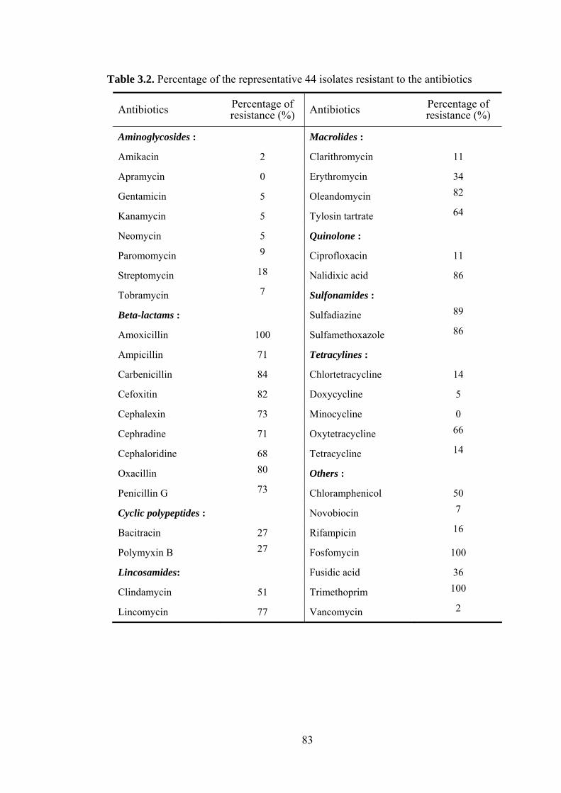
83
Table 3.2. Percentage of the representative 44 isolates resistant to the antibiotics
Antibiotics Percentage of resistance (%) Antibiotics Percentage of
resistance (%)
Aminoglycosides : Macrolides :
Amikacin 2 Clarithromycin 11
Apramycin 0 Erythromycin 34
Gentamicin 5 Oleandomycin 82
Kanamycin 5 Tylosin tartrate 64
Neomycin 5 Quinolone :
Paromomycin 9 Ciprofloxacin 11
Streptomycin 18 Nalidixic acid 86
Tobramycin 7 Sulfonamides :
Beta-lactams : Sulfadiazine 89
Amoxicillin 100 Sulfamethoxazole 86
Ampicillin 71 Tetracylines :
Carbenicillin 84 Chlortetracycline 14
Cefoxitin 82 Doxycycline 5
Cephalexin 73 Minocycline 0
Cephradine 71 Oxytetracycline 66
Cephaloridine 68 Tetracycline 14
Oxacillin 80 Others :
Penicillin G 73 Chloramphenicol 50
Cyclic polypeptides : Novobiocin 7
Bacitracin 27 Rifampicin 16
Polymyxin B 27 Fosfomycin 100
Lincosamides: Fusidic acid 36
Clindamycin 51 Trimethoprim 100
Lincomycin 77 Vancomycin 2

84
Phylogenetic analyses. Almost complete 16S rRNA gene sequences were obtained for
the 44 representative filamentous actinomycetes. The percentage similarities and
number of nucleotide differences found between each isolate and its nearest
phylogenetic neighbour are shown in Table 3.3. Most of the isolates shared their highest
16S rRNA similarity with type strains of Streptomyces species, the remainder with
validly described Actinomadura, Dactylosporangium, Micromonospora and
Streptosporangium species. It can be seen from Figure 3.2(b) that the isolates can be
assigned to five 16S rRNA gene clades which correspond to the genera Actinomadura,
Dactylosporangium, Micromonospora, Streptomyces and Streptosporangium.
Isolates BK147 and BK148 were most closely related to the type strains of A.
bangladeshensis (98.5% similarity, 22 nt difference at 1431 locations), and isolates
BK51, BK53, BK57 and BK63 to D. aurantiacum DSM 43157T, sharing similarities
with the latter within the range 98.7 to 99.2 %, values equivalent to 12 to 18 nt
difference at 1434 locations. Similarly, isolates BK62, BK66 and BK177 were most
closely related to the type strain of M. matsumotoense showing similarities with this
organism within the range 98.8 to 99.7, values corresponding to between 5 and 17 nt
differences at 1443 locations. In contrast, isolates BK58, BK61 and BK65 were most
closely related to the type strain of M. saelicesensis (99.7% similarity, 4 nt differences
at 1433 locations), M. echinaurantiaca (99.1% similarity, 13 nt differences at 1437
locations) and M. chaiyaphumensis (99.3% similarity, 10 nt differences at 1422
locations), respectively. Isolate BK143 had an identical 16S rRNA gene sequence with
S. album DSM 43023T; the corresponding score between isolate BK143 and this strain
was 98.8 %, a value equivalent to 17 nt differences at 1423 locations. All of these
isolates were recovered in clades corresponding to these generic assignments (Fig. 3.2b).
The 30 Streptomyces isolates shared 16S rRNA gene similarities of 98.4–100%
with their closest phylogenetic neighbours (Table 3.3). These organisms can be assigned
to three subclades (Fig. 3.2b). Six of the subclade 1 isolates were most closely related to
S. sanglieri NBRC 100784T (99.5-100% similarity) and 5 isolates to S. drozdowiczii
NBRC 101007T (99.4-99.5% similarity). Isolate BK21 shared an identical 16S rRNA
gene sequence with the type strain of S. sanglieri. In addition, isolates BK115 and BK
178 have identical 16S rRNA gene sequences; they were most closely related to S.
gelaticus NRRL B-2928T, sharing a 98.9% similarity with the latter (16 nt differences at
1435 sites).
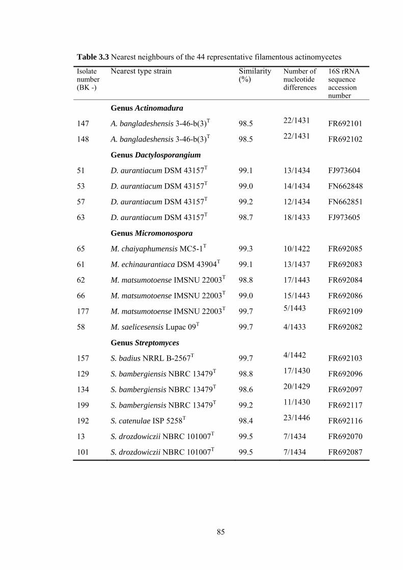
85
Table 3.3 Nearest neighbours of the 44 representative filamentous actinomycetes
Isolate number (BK -)
Nearest type strain Similarity (%)
Number of nucleotide differences
16S rRNA sequence accession number
Genus Actinomadura
147 A. bangladeshensis 3-46-b(3)T 98.5 22/1431 FR692101
148 A. bangladeshensis 3-46-b(3)T 98.5 22/1431 FR692102
Genus Dactylosporangium
51 D. aurantiacum DSM 43157T 99.1 13/1434 FJ973604
53 D. aurantiacum DSM 43157T 99.0 14/1434 FN662848
57 D. aurantiacum DSM 43157T 99.2 12/1434 FN662851
63 D. aurantiacum DSM 43157T 98.7 18/1433 FJ973605
Genus Micromonospora
65 M. chaiyaphumensis MC5-1T 99.3 10/1422 FR692085
61 M. echinaurantiaca DSM 43904T 99.1 13/1437 FR692083
62 M. matsumotoense IMSNU 22003T 98.8 17/1443 FR692084
66 M. matsumotoense IMSNU 22003T 99.0 15/1443 FR692086
177 M. matsumotoense IMSNU 22003T 99.7 5/1443 FR692109
58 M. saelicesensis Lupac 09T 99.7 4/1433 FR692082
Genus Streptomyces
157 S. badius NRRL B-2567T 99.7 4/1442 FR692103
129 S. bambergiensis NBRC 13479T 98.8 17/1430 FR692096
134 S. bambergiensis NBRC 13479T 98.6 20/1429 FR692097
199 S. bambergiensis NBRC 13479T 99.2 11/1430 FR692117
192 S. catenulae ISP 5258T 98.4 23/1446 FR692116
13 S. drozdowiczii NBRC 101007T 99.5 7/1434 FR692070
101 S. drozdowiczii NBRC 101007T 99.5 7/1434 FR692087

86
Table 3.3 (Continued)
Isolate number (BK -)
Nearest type strain Similarity (%)
Number of nucleotide difference
16S rRNA sequence accession number
160 S. drozdowiczii NBRC 101007T 99.4 9/1434 FR692104
161 S. drozdowiczii NBRC 101007T 99.4 9/1434 FR692105
166 S. drozdowiczii NBRC 101007T 99.4 8/1434 FR692106
178 S. gelaticus NRRL B-2928T 98.9 16/1435 FR692110
115 S. gelaticus NRRL B-2928T 98.9 16/1435 FR692090
119 S. hirsutus NBRC 12786T 98.6 20/1437 FR692091
128 S. hirsutus NBRC 12786T 98.4 23/1435 FR692095
138 S. hirsutus NBRC 12786T 98.7 21/1435 FR692098
122 S. lydicus NBRC 13058T 99.8 3/1435 FR692093
179 S. lydicus NBRC 13058T 99.2 11/1435 FR692111
170 S. mirabilis NBRC 13450T 99.7 4/1433 FR692108
120 S. nigrescens NBRC 12894T 99.6 7/1439 FR692092
168 S. paucisporeus NBRC 102072T 98.8 17/1402 FR692107
125 S. prasinus NBRC 12810T 98.8 18/1430 FR692094
189 S. olivochromogenes NBRC 3178T 99.9 2/1432 FR692114
21 S. sanglieri NBRC 100784T 100 0/1434 FR692078
28 S. sanglieri NBRC 100784T 99.6 6/1434 FR692080
49 S. sanglieri NBRC 100784T 99.5 7/1434 FR692081
108 S. sanglieri NBRC 100784T 99.6 6/1434 FR692088
109 S. sanglieri NBRC 100784T 99.6 6/1434 FR692089
180 S. sioyaensis NRRL B-5408T 99.5 7/1438 FR692112
185 S. sporoclivatus NBRC 100767T 99.9 2/1435 FR692113
190 S. sanglieri NBRC 100784T 99.9 1/1434 FR692115
Genus Streptosporangium
139 S. album DSM 43023T 100 0/1424 FR692099
143 S. album DSM 43023T 98.8 17/1423 FR692100

87
The remaining subclade 1 strains, isolates BK157, BK170 and BK189, were most
closely related to the type strains of S. badius (99.1% similarity), S. mirabilis (99.7%
similarity) and S. olivochromogenes (99.9%), respectively. The seven subclade 2 strains,
isolates BK119, BK125, BK138, BK129, BK134, BK138 and BK199, were most
closely related to the type strains of S. hirsutus (98.6% similarity), S. prasinus (98.8%
similarity), S. hirsutus (98.7% similarity), S. bambergiensis (98.8% similarity), S.
bambergiensis (98.6% similarity), S. hirsutus (98.7% similarity) and S. bambergiensis
(99.2% similarity), respectively. Two of the subclade 3 strains, isolates BK122 and
BK179, shared their highest 16S rRNA gene similarity to Streptomyces lydicus NBRC
13058T (99.8 and 99.2%, respectively). The remaining strains in this subclade, isolates
BK120, BK168, BK180 and BK185, were most closely related to the type strains of S.
nigrescens (99.6% similarity) S. paucisporeus (98.8% similarity), S. sioyaensis (99.5%
similarity) and S. sporoclivatus (99.9% similarity), respectively.
Comparison of antibiotic resistance and 16S rRNA gene sequence data. Some of the
isolates were assigned to corresponding groups in the analyses based on the antibiotic
resistance profiles and 16S rRNA gene sequence data, notably the isolates which were
most closely related to bona fide members of the genera Actinomadura,
Dactylosporangium, and Streptosporangium (Fig. 3.2). In contrast, the isolates closely
related to the Micromonospora strains were assigned to two multimembered and one
single-membered cluster based on their antibiotic resistance profiles; one of the
multimembered clusters was composed of isolates BK62 and BK66, which are
phylogenetically close to M. matsumotoense IMSNU 22003T. It is also interesting that
the putative Actinomadura, Dactylosporangium, Micromonospora and
Streptosporangium isolates belonged to colour-groups 19, 20, 21 and 18, respectively.
All but 4 of the 30 isolates found to be closely related to Streptomyces type strains
were recovered in three aggregate groups defined at the 70% similarity level in the
dendrogram based on the antibiotic resistance data (Fig. 3.2a). The most pronounced
congruence was found between isolates which shared high 16S rRNA similarity values,
as exemplified by isolates BK13, BK101 and BK166, isolates BK108 and BK109,
isolates BK125 and BK129, isolates BK120 and BK180, and isolates BK168 and
BK185. There was little evidence of a correlation between the membership of colour-
groups and taxa defined in the 16S rRNA gene sequence and antibiotic-resistance

88
pattern analyses though isolates BK108 and BK109 clustered together in all three
analyses.
The partial congruence found between corresponding groups in the dendrograms
(Fig. 3.3) was reflected in the relatively low value of Pearson correlation coefficient,
0.543 (p=0.001 < 0.01) in the overall comparison between the two sets of data presented
as similarity values. It can be seen from Figure 3.3 that pairs of isolates with 16S rRNA
gene similarities between 96 and 100% showed relatively high resistance profile
similarities of between 50 and 100%. In contrast, pairs of isolates with 16S rRNA gene
similarities between 88 and 92% showed a wide-range resistance profile similarities,
namely from 25 to 92% .
Fig. 3.3. Correlation of 16S rRNA gene similarities and antibiotic resistance profiles of
the 44 isolates.
Comparison of phylogenies based on vanHAX gene cluster and 16S rRNA gene
sequences. All 9 isolates which were resistant to vancomycin shared identical 16S
rRNA gene sequences (ca. 1434 bp) with one another and with the type strain of S.
sanglieri. The sequences of the genes comprising the vanHAX gene cluster (ca. 1780
bp) were also identical. It can be seen from Figure 3.4 that the vancomycin-resistant
strains formed a distinct branch in each of the phylogenetic trees.
20
40
60
80
100
88 90 92 94 96 98 100
Res
ista
nce
prof
ile si
mila
rity
(
S SM
, %
)
16S rRNA gene similarity (%)

89
Fig. 3.4. Position of the vancomycin-resistant isolates in phylogenetic trees based on (a) VanHAX gene cluster and (b) 16S rRNA gene sequence data
Act
inob
acte
ria
Firm
icut
es
Isolate BK17
Isolate BK14
Isolate BK15
Isolate BK21
Isolate BK18
Isolate BK16
Isolate BK19
Isolate BK20
Isolate BK22
Streptomyces coelicolor A3(2)(NC003888)
Streptomyces toyocaensis NRRL 15009 (U82965)
Amycolatopsis coloradensis DSM 44225 (DQ246438)
Amycolatopsis orientalis C329.2 (AF060799)
Actinoplanes teichomyceticus ATCC 31131(AJ605139)
Rhodococcus globerulus RG-S1F (AY648697)
Enterococcus faecium N97-330 (AF175293)
Ruminococcus gauvreauii CCRI-16110 (EF508033)
Enterococcus gallinarum N04-0414 (DQ172830)
Clostridium lavalense CCRI-9842 (AY772783)
Enterococcus faecalis V583 (EFU35369)
Staphylococcus aureus pLW043 (AE017171)
Bacillus circulans VR0709 (Y15704, Y15705, Y15708)
Paenibacillus apiarius PA-B2B (DQ018711)
Paenibacillus thiaminolyticus PT-2B1(DQ018710)
100
100
98
52
51
100
10088
73
91
100
0.05
(a)
Actinoplanes teichomyceticus DSM 43866T (AJ865472)
Ruminococcus gauvreauii CCRI-16110T (EF529620)
Streptomyces coelicolor A3(2) (NC003888)
Amycolatopsis orientalis IMSNU 20058T(AJ400711)
Amycolatopsis coloradensis NRRL 3218T (AF051341)
Rhodococcus globerulus NCIMB 12315T (X81931)
Enterococcus faecium DSM 20477T(AJ276355)
Enterococcus faecalis JCM5803T (AB012212)
Enterococcus gallinarum LMG 13129T (AF039900)
Staphylococcus aureus ATCC 12600T (D83357)
Bacillus circulans IAM12462T (D78312)
Paenibacillus thiaminolyticus IFO15656T (AB073197)
Paenibacillus apiarius DSM5581T (AB073201)
Clostridium lavalense CCRI-9842T (EF564277) 100
99
94
99
Isolate BK22 Isolate BK18
Isolate BK21
Isolate BK17
Isolate BK15
Isolate BK16
Isolate BK20
Isolate BK14
Isolate BK19
88
100
100
54
99
100
96
84
0.02
(b)

90
3.6 Discussion
The present study confirms and extends the pioneering work of D’Costa et al.
(2006) who found that large numbers of streptomycetes isolated from several soils grew
in the presence of one or more of a panel of 21 antibiotics. On average, the organisms
were resistant to 7 or 8 of the antibiotics. In this investigation, 44 out of 200
dereplicated actinomycetes, notably streptomycetes, were resistant to 18 or 19
antibiotics which included representatives of 41 structural types. Isolates BK21, which
had a 16S rRNA gene sequence identical to that of the type strain of S. sanglieri, was
resistant to 32 of the antibiotics. All of the isolates were resistant to amoxicillin,
fosfomycin and trimethoprim and a further 35 isolates to nalidixic acid, oleandomycin,
oxacillin, sulfadizine and sulfamethoxazole. The isolates were more resistant to
antibiotics, which targeted the folic acid pathway, such as sulphonamides and
trimethoprim (86~100%), and cell wall biosynthesis, such as β-lactam antibiotics
(68~100%). In contrast, less resistance was shown to aminoglycosides (0~18%) and
tetracyclines (0~14%) apart from oxytetracyline (66%). These data provide further
compelling evidence that soil actinomycetes are a rich reservoir of resistance
determinants which may be spread into broader microbial communities, and hence
could promote antibiotic resistance amongst pathogenic bacteria (Riesenfeld et al.,
2004; D'Costa et al., 2006; Dantas et al., 2008; Laskaris et al., 2010; Wright, 2010).
All of the dereplicated filamentous actinomycetes were isolated from a hay
meadow soil which had not be exposed to either antibiotics or fertilizers (Shiel &
Rimmer, 1984; Hopkins et al., 2009) This result provide further evidence that
substantial populations of antibiotic-resistant bacteria are present in such habitats even
though direct evidence for the production of antibiotics in soil is limited (Anukool et al.,
2004). However, there is convincing evidence for the coexistence and coevolution of
antibiotic resistance and biosynthetic genes in soil streptomycetes (Laskaris et al., 2010).
The representative set of isolates for the antibiotic resistance studies were selected
from the colour-groups as it is known that streptomycete group based on aerial spore
mass, substrate mycelial and diffusible pigment colours are predictive; strains taken to
represent such groups key out to novel and established species (Goodfellow & Haynes,
1984; Williams & Vickers, 1988; Atalan et al., 2000; Antony-Babu et al., 2008) or to
groups of closely related species (Sembiring et al., 2000; Goodfellow et al., 2007). It is
91
encouraging that the isolates found to be closely related to established members of the
genera Actinomadura, Dactylosporangium, Micromonospora and Streptosporangium
were recovered in corresponding colour-groups. In contrast, isolates found to be closely
related to the type strains of S. drozdowiczii and S. sanglieri belonged to more than one
colour-group, a result which can be attributed to the fact these organisms produce gray
to grayish brown, and brown to light brown aerial spores and diffusible pigments,
respectively on oatmeal agar. This deduction needs to be tested using the computer-
based system introduced by Antony-Babu et al. (2010), which highlights relationship
between colour-groups and corresponding rep-PCR data of alkaliphilic streptomycetes.
It is not clear from the present study that the extent to which a relationship exists
between the antibiotic resistance profiles and the taxonomic positions of filamentous
actinomycetes. However, the relatively low correlation coefficient (0.543) found
between the corresponding datasets may be more apparent than real due to the small
number and taxonomic diversity of dereplicated filamentous actinomycetes which
featured in this study. This interpretation is supported by the observations that greater
correlation was found between the corresponding datasets for isolates found to share
high 16S rRNA gene similarities. Indeed, this interpretation is strongly endowed given
the complete congruence found between the phylogenetic trees based on the analyses of
the 16S rRNA gene and vanH-vanA-vanX gene sequences of the vancomycin-resistant
strains identified as Streptomyces sanglieri.

92
Chapter 4. Detection, selective isolation and characterisation of
Dactylosporangium strains from diverse environmental
samples
4.1 Abastract
A culture-independent, nested PCR procedure based on genus-specific
oligonucleotide primers detected the presence of members of the genus
Dactylosporangium in 14 out of 21 diverse environmental samples. Clones generated
from the 14 positive environmental samples formed distinct phyletic lines in the
Dactylosporangium 16S rRNA gene tree. Dactylosporangium strains isolated from 7 of
these samples using a medium designated to be selective for members of the genus
Dactylosporangium, namely Streptomyces Isolation Medium supplemented with
gentamicin and antifungal antibiotics. One hundred and two out of 219 representative
presumptive dactylosporangiae were considered as authentic members of the genus
Dactylosporangium as they gave PCR amplification products using the genus-specific
primers and had chemical features typical of dactylosporangiae. Representatives of the
Dactylosporangium isolates formed distinctive phyletic lines in the 16S rRNA
dactylosporangial gene tree, contained the non-ribosomal peptide or type-I polyketide
synthase genes and inhibited the growth of Bacillus subtilis, Kocuria rhizophila or
Staphylococcus aureus strains. It is evident from these results that the genus
Dactylosporangium is underspeciated, widely distributed in natural habitats and is a
potentially rich source of novel secondary metabolites.
4.2 Introduction
There is an urgent need to find new drugs, especially antibiotics, to control the
spread of antibiotic resistant pathogens (Payne et al., 2007; Spellberg et al., 2008;
Fischbach & Walsh, 2009) and to treat life-threatening diseases such as cancer (Olano et
al., 2009). Amongst prokaryotes, filamentous actinomycetes, notably streptomycetes,
remain a unique source of new antibiotics (Bérdy, 2005; Newman & Cragg, 2007;
Goodfellow & Fiedler, 2010) and this is likely to continue as full genome sequences of
model actinomycete strains contain over 20 natural product biosynthetic gene clusters
for the production of known or predicted secondary metabolites, as exemplified by

93
studies on Amycolatopsis mediterranei U32 (Zhao et al., 2010), Saccharopolyspora
erythraea NRRL 23338 (Oliynyk et al., 2007), Salinispora tropica CNB 440 (Udwary
et al., 2007), Streptomyces avermitilis (Ikeda et al., 2003) and “Streptomyces
coelicolor” A3(2) (Bentley et al., 2002). In contrast, few, if any, such gene clusters have
been detected in whole genomes of other prokaryotes (Goodfellow & Fiedler, 2010).
It is becoming increasingly difficult to discover new antibiotics from common
actinomycetes as screening such organisms tends to lead to the costly rediscovery of
known secondary metabolites (Busti et al., 2006; Lam, 2007). This problem can be
addressed by using standard procedures for the selective isolation of novel
actinomycetes from poorly studied habitats (Bredholdt et al., 2007; Hong et al., 2009;
Okoro et al., 2009), by applying novel procedures for the selective isolation and
characterisation of uncommon and rare actinomycetes (Suzuki et al., 2001a, b; Tan et
al., 2006; Gontang et al., 2007) and by detecting the presence of novel, rare and target
actinomycetes in environmental samples using culture-independent methods prior to the
application of selective isolation procedures (Stach et al., 2003; Maldonado et al., 2005).
These strategies have lead to the isolation of novel actinomycetes with the capacity to
produce diverse new bioactive compounds (Fiedler et al., 2005; Jensen et al., 2005,
2007; Goodfellow & Fiedler, 2010),
Effective taxon-specific strategies are required for the selective isolation of
neglected filamentous actinomycetes, not least for the isolation of members of the genus
Dactylosporangium which encompasses strains known to produce anti-allergic drugs
(Tani et al., 2004), antibiotics, such as dactimicin (Shomura et al., 1980) and
tiacumicins (Theriault et al., 1987), and a plant growth inhibitor (Kizuka et al., 2002).
The genus Dactylosporangium encompasses aerobic, filamentous actinomycetes which
form motile spores in spore vesicles borne on short sporangiophores on substrate
hyphae and have hydroxy and/or meso- diaminopimelic acids in whole-organism
hydrolysates, but lack mycolic acids (Vobis, 1989; Kim et al., 2010). Dactylosporangiae
have been isolated from a range of terrestrial habitats (Hayakawa et al., 1991a;
Hayakawa, 2008) but have rarely featured in pharmacological screening programmes as
they are difficult to isolate in large numbers and grow slowly.
The present study was designed to establish the extent of dactylosporangial
taxonomic diversity in diverse environmental samples using a culture-independent
94
procedure prior to developing a reliable strategy for the selective isolation and
characterisation of dactylosporangiae known to be present in environmental samples. In
addition, representative cultivable isolates were examined to determine their
phylogenetic diversity and whether they contained non-ribosomal peptide and type-I
polyketide synthase genes, and to evaluate their ability to inhibit the growth of a panel
of pathogenic microorganisms.
4.3 Materials and methods
Sources and physico-chemical properties of environmental samples. Twenty-one
environmental samples from diverse geographical locations (Table 4.1) were sieved, air-
dried at room temperature for 5-7 days and stored at 4˚C in plastic containers, apart
from the marine sediment samples which were not dried. The pH of the samples was
determined using the procedure described by Reed and Cummings (1945). Two grams
of each sample were added to 18 ml of deionised water, mixed thoroughly, and left for
an hour when the pH were determined using a glass electrode pH meter (Model 320
Mettler- Toledo AG, CH.8603, Schwerzenbach, Switzerland). Each sample was
examined in triplicate and the final pH values recorded as an average of the three
readings. The percentage moisture content of the triplicated samples was determined by
drying known weights of sediment/soil to constant weight at 105˚C then calculating the
average loss in weight between each set of samples. The dried samples were placed in a
muffle furnace (Carbolite, Sheffield, UK) and the temperature raised slowly to 700˚C
and kept constant for 30 minutes to burn off organic matter. After cooling overnight in a
desiccator, the average loss in weight for each set of samples was recorded as the
organic matter content.
Extraction of community DNA. Community DNA was extracted from all 21
environmental samples using a method slightly modified from that of Griffiths et al.
(2000). Briefly, each sample (0.5 g wet weight) was added to a 1.5 ml screw-capped
tube which contained 0.5 ml of hexadecyltrimethylammonium bromide (CTAB)
extraction buffer and 0.5 ml of a mixture of phenol-chloroform-isoamyl alcohol
(25:24:1, v/v) and 0.3 g of acid-washed sterile glass beads (≤ 106 μm; Sigma). The
preparations were lysed twice at 5.5 m/s for 30 seconds using a ribolyser (Fastprep®
FP220A Thermo Instrument; Qbiogene, France), cooled on ice for 2 minutes and the
DNA containing aqueous phases separated by centrifugation (16,000 x g) for 5 minutes.

95
Table 4.1. Sources and physico-chemical properties of environmental samples
Code Soil/Sediment samples Location pH
Moisture content
(%)
Organic matter content
(%)
ANT Antarctic soil Ross Dependency, Antarctica (78°01′S; 163°53′E)
7.3 0.3 0.4
AU96 Arid soil Adelaide, Australia 5.5 0.2 1.7
BK Hay meadow soil Plot 6, Cockle Park Experimental Farm, Northumberland, UK (National Grid reference: NZ 200913)
5.5 4.0 9.5
CA Rhizosphere soil Izmir, Turkey 7.4 1.0 8.2
Ch6 Marine sediment Bahia Tic Toc, Chilean Sea (43°53′ S, 72°87′W) 7.8 19.6 2.5
Ch27 Marine sediment Estero Castro, Chilean Sea (42°30´S, 73°5´W) 7.9 20.6 3.0
CS Chalk soil Ambleside, Cumbria, UK (National Grid reference: NY-357978) 7.2 2.4 3.2
DR River sediment River Derwent, County Durham, UK (National Grid reference: NZ-055490) 6.9 5.0 4.5
ECU Upland soil Riobamba, Ecuador 6.7 2.5 5.6
KHA Agricultural soil Khartoum, Sudan 5.8 0.2 6.3
LS Sediment Shore of Loch Creran, UK (National Grid reference: NM-9124259) 7.7 25.0 4.5
MEG Grassland soil Syros, Greece 7.3 0.4 2.5
MS Rhizosphere soil Kuala Lumpur, Malaysia 7.4 1.2 4.1
NMS Agricultural soil Kuala Lumpur, Malaysia 5.7 0.3 7.1
NOR Marine sediment Østerfjord, Norwegian Sea (60°33′N, 5°19′E) 8.7 21.2 4.2
SAU Arid soil Nairobi, Kenya 5.9 1.0 2.0
SHO Agricultural soil Sholinar, Srinagar, India 5.5 0.7 6.1
TOR Urban soil Torremolinos, Spain 6.5 4.0 3.5
WAD Sand dune soil Borg El-Arab, Egypt 7.8 0.8 1.0
WS Garden soil Samsun, Turkey 6.7 2.8 11.3
ZIZ Arid soil Gazipur district, Bangladesh 6.5 3.5 4.0

96
Each supernatant was transferred to a fresh 1.5 ml tube and phenol removed by adding
an equal volume of chloroform-isoamyl alcohol (24:1, v/v) followed by centrifugation
(16000 x g) for 5 minutes. Each supernatant was transferred to a fresh 1.5 ml eppendorf
tube and the nucleic acids precipitated by adding 1 ml of 30% (w/v) polyethylene glycol
(PEG) dissolved in 1.6 M NaCl. The resultant preparations were briefly vortexed,
incubated at -80˚C for an hour, centrifuged at maximum speed for 5 minutes, the DNA
pellets washed with 500 µl of 70% (v/v) ice-cold ethanol, air-dried and redissolved in
50 µl of TE buffer (pH 8.0). The DNA preparations were checked for purity by agarose-
gel electrophoresis and the purified extracted DNA preparations stored at -20˚C.
Nested PCR for detection of 16S rRNA dactylosporangial genes. A nested PCR
approach was used to detect dactylosporangial 16S rRNA genes in each of the
community DNA preparations. Bacterial 16S rRNA genes were amplified from these
preparations by using universal primers 27F and 1525R (Table 2), as described by Tan
et al.(2006). The resultant primary PCR products were diluted tenfold as templates for
the secondary PCR reactions. Amplification of the 16S rRNA genes specific for
members of the family Micromonosporaceae was performed using the PCR primers
M2F and A3R (Table 4.2) and the presence of dactylosporangiae sought using genus-
specific PCR primers D3F and D2R (Table 4.2), as described by Monciardini et al.
(2002). The nested PCR reaction mixture, prepared in a final reaction volume of 50 µl,
contained 2.5 µl of each forward and reverse primer, 5 µl of 10x reaction buffer
(Bioline), 2.5 µl of DMSO, 0.8 µl of 12.5 mM dNTPs (Bioline), 1.5 µl of 50 mM
MgCl2 and 0.5 µl Taq DNA polymerase (Bioline) and 1 µl of template DNA. The PCR
reactions were carried out in a PCR thermocycler (Whatman Biometra TgradientTM,
Germany) together with positive and negative controls.
Cloning, sequencing and phylogenetic analysis of amplified PCR products. Cloning
of the purified PCR products of Dactylosporangium-specific 16S rRNA genes amplified
from community DNA samples was performed using a StrataClone PCR Cloning Kit
(Strategene, UK). Ligations were achieved by mixing 1.5 µl of cloning buffer, 1 µl of
each PCR product (5-50 ng) and 0.5 µl StrataClone Vector and the resultant
preparations incubated at room temperature for 5 minutes then placed on ice, the
transformations were performed by adding 1µl of ligation mixture to thawed
StrataClone SoloPack competent cells. The resultant preparations were mixed gently,
placed on ice for 20 minutes, heated at 42˚C for 45 seconds and incubated on ice for 2

97
minutes. Pre-warmed Luria-Bertani (LB; (Miller, 1972) broth (250 µl) was added to
each tube prior to incubation at 37˚C in a shaker incubator (Gallenkamp Orbital
Incubator, UK) for an hour at 160 rpm and centrifugation for 1 minute at 13,000 x g.
The supernatants were discarded and the pellets redissolved in 100 µl of LB medium,
and the transformation mixtures (100 µl) plated onto pre-warmed LB-ampicillin plates
which had been inoculated with 40 µl of 2% X-gal and incubated overnight at 37˚C.
Randomly selected white colonies were streaked, using sterilised toothpicks, onto LB-
ampicillin plates, incubated overnight at 37˚C, transformants screened by using the
colony-PCR technique with M13 forward and reverse primers (Hofmann & Brian,
1991), and purified colony PCR products sequenced. The partial 16S rRNA gene
sequences of the clones were aligned manually against corresponding sequences of the
type strains of Dactylosporangium species retrieved from DDBJ/EMBL/GenBank
databases using the pairwise alignment option. Phylogenetic trees were inferred using
the neighbour-joining (Saitou & Nei, 1987), minimum-evolution (Rzhetsky & Nei,
1992) and maximum-parsimony (Fitch, 1971) tree-making algorithms from the MEGA
3.0 program (Kumar et al., 2004).
Table 4.2. PCR primers
Primer Sequence(5’-3’) Target genes* Size of PCR product (bp) References
27F AGAGTTTGATCMTGGCTCAG
1525R AAGGAGGTGWTCCARCC
Universal 16S rRNA
1400 – 1500
Lane (1991)
M2F SAGAAGAAGCGCCGGCC
A3R CCAGCCCCACCTTCGAC
Family Micromonosporaceae-specific 16S rRNA
900 – 1000
Monciardini et al. (2002)
D3F GCGGCTTGTTGCGTCAG
D2R CCGCTGGCAACATCGAACA
Genus Dactylosporangium-specific 16S rRNA
500 – 600
Monciardini et al.(2002)
K1F TSAAGTCSAACATCGGBCA
M6R CGCAGGTTSCSGTACCAGTA
PKS-I
1200 – 1400
Ayuso- Sacido et al. (2005)
A3F GCSTACSYSATSTACACSTCSGG
A7R SASGTCVCCSGTSCGGTAS
NRPS
700 – 800
Ayuso- Sacido et al. (2005)
* NRPS, non-ribosomal peptide synthetase; PKS-1, type- I polyketide synthase.

98
Selective isolation and enumeration of dactylosporangiae. A pilot experiment was
carried out to determine whether representatives of seven validly described
Dactylosporangium species (D. aurantiacum NRRL B-8018T, D. fulvum DSM 43917T,
D. matsuzakiense NRRL B-16293T, D. roseum DSM 43916T, D. salmoneum NRRL B-
16294T, D. thailandense DSM 43158T and D. vinaceum NRRL B16297T) grew on five
media designed to be selective for members of the genus Dactylosporangium; namely,
humic acid-vitamin agar (Hayakawa & Nonomura, 1987), oatmeal agar (Shirling &
Gottlieb, 1966), raffinose-histidine agar (Vickers et al., 1984), starch casein agar
(Küster & Williams, 1964) and Streptomyces Isolation Medium (D'Costa et al., 2006)
supplemented with antifungal antibiotics and either gentamicin or oxytetracycline. The
putative selective media were also evaluated for their effectiveness in isolating members
of the genus Dactylosporangium from a soil sample taken from Palace Leas hay
meadow plot 6 at Cockle Park Experimental Farm, Northumberland (Atalan et al.,
2000), using the procedure described below.
Two grams of the soil sample were suspended in 18 ml of ¼ strength Ringer’s
solution (Oxoid), mixed by vortexing (Vortex Genie 2 Model G.560E, USA) and
blended for a minute using an ULTRA TUREX ®-Antried T 25 Blender. The resultant
suspension was shaken in a Gallenkamp Orbital Incubator at 150 rpm for 30 minutes at
room temperature, pre-treated at 55˚C for 20 minutes in a water bath, and cooled at
room temperature prior to the preparation of a tenfold serial dilution in ¼ Ringer’s
solution. Aliquots (100 µl) of the 10-2 to 10-4 dilutions were spread, in triplicate, over
plates of the five selective media supplemented with antifungal antibiotics (25 μg/ml of
both cycloheximide and nystatin) and an antibacterial antibiotic (4 μg/ml of either
gentamicin or oxytetracycline), and the plates incubated at 28˚C for 21 days; in all cases
the isolation plates were dried for 15 minutes prior to inoculation, as recommended by
Vickers et al. (1984). Orange coloured colonies which lacked aerial hyphae were
presumptively assigned to the genus Dactylosporangium. The numbers of presumptive
dactylosporangiae, other filamentous actinomycetes and bacteria were expressed as the
mean number of colony forming units (cfu’s) per gram dry weight soil. The
Streptomyces Isolation Medium supplemented with cycloheximide, nystatin and
gentamicin supported the growth of all of the Dactylosporangium type strains and the
highest number of putative dactylosporangiae isolated from soil suspensions prepared
from Palace Leas hay meadow, plot 6. In light of these results, the 13 environmental

99
samples found to contain dactylosporangiae in the culture-independent experiments
(Table 4.3) were examined using the isolation procedure described above.
Selection, purification and maintenance of dactylosporangiae. Over 200
presumptive Dactylosporangium strains (Table 4.3) representing different colonial
morphologies were taken from the selective isolation plates, using sterile toothpicks,
and transferred onto oatmeal agar plates which were incubated at 28˚C for up to 21 days.
The isolates were checked for purity by streaking onto another set of oatmeal plates
which were incubated as before. Biomass from each of the purified isolates was scraped
from the oatmeal agar plates and suspended in 1 ml glycerol tube (20%, v/v). The
resultant glycerol stocks were preserved at -20˚C and served as a source of inoculum.
16S rRNA gene analysis and chemical characterisation of Dactylosporangium
isolates. The 219 isolates were tested for their ability to produce the
Dactylosporangium-specific PCR amplification product. Total genomic DNA was
extracted from the isolates using Gen EluteTM Bacterial Genomic Kits (Sigma-Aldrich
Co.), following the manufacturer’s protocol, stored at -20˚C, and PCR reactions
performed with the genus-specific primers, as described by Monciardini et al. (2002).
The 102 isolates which gave the genus-specific amplification product (Table 4.4) were
the subject of 16S rRNA gene sequencing studies using the universal primers, 27F and
1525R (Table 4.2), as described by Tan et al. (2006). Phylogenetic analyses were
carried out on the resultant sequences using the tree making algorithms mentioned
earlier.
The 102 isolates were also examined for the presence of diaminopimelic acid
(A2pm) isomers in whole-organism hydrolysates by using the procedure described by
Staneck & Roberts (1974), albeit with the minor modifications introduced by Tan et al.
(2006). Briefly, two loopfuls of wet biomass of each isolate grown on oatmeal agar for
21 days at 28˚C were suspended in 500 μl of 6 N HCl containing 100 μg sterile glass
beads (≤ 106 μm; Sigma), homogenised at 5.0 m/s for 20 seconds using the ribolyser
(Fastprep® FP220A Instrument), incubated at 100˚C for 4 hours and centrifuged at
13,000 rpm for 5 minutes. The resultant supernatants were transferred to fresh tubes,
evaporated to dryness on a hotblock (90-100˚C), and the hydrolysates reconstituted with
distilled water (100 μl) prior to thin-layer-chromatographic (TLC) analysis. The isolates

100
were also examined for the presence of mycolic acids by using the alkaline
methanolysis procedure described by Minnikin et al. (1980).
Table 4.3. Numbers of presumptive and confirmed Dactylosporangium strains isolated from environmental samples*
Code Soil/Sediment samples Presumptive isolates
Confirmed isolates
BK Hay meadow soil Plot 6, UK 61 54
CA Rhizosphere soil, Turkey 0 0
CS Chalk soil, UK 23 19
DR River sediment, UK 19 4
ECU Upland soil, Ecuador 25 1
LS Seashore sediment, UK 5 0
MEG Grassland soil, Greece 2 0
MS Rhizosphere soil, Malaysia 1 0
NMS Agricultural soil, Malaysia 20 11
NOR Marine sediment, Norway 15 0
SHO Agricultural soil, India 29 6
WAD Sand dune soil, Egypt 2 0
WS Garden soil, Turkey 12 7
ZIZ Arid soil, Bangladesh 5 0
Total 219 102
* The remaining samples, ANT, AU96, Ch6, Ch27, KHA, SAU and TOR, were not examined using the selective isolation procedure as community DNA extracted from them gave negative reactions using the genus-specific PCR primers. In contrast, Micromonosporaceae strains were detected in all of the samples.
Non-ribosomal peptide and type I polyketide synthase genes. Fifty two
representative Dactylosporangium isolates (Table 4.4) were screened for the presence of
non-ribosomal peptide (NRPS) and type I polyketide synthase (PKS) genes using NRPS
and PKS primers, as recommended by Ayuso-Sacido & Genilloud (2005) and Courtois
et al. (2003), respectively. The amplification reactions were performed in a final
reaction volume of 25 µl, which contained 1 µl of 0.4 µM of either NRPS or PKS
specific primers (Table 4.2), 2.5 µl of 10x reaction buffer, 1 µl of DMSO, 1.6 µl of 2.5
mM dNTPs (Bioline), 1.5 µl of 50 mM MgCl2 , 0.25 µl BioTaq DNA polymerase

101
(Bioline), 15.15 µl sterile distilled water and 1 µl of extracted DNA template. The PCR
reaction temperature profile was initial denaturation at 95˚C for 5 minutes, 35 cycles of
denaturation at 95˚C for 30 seconds, annealing at 57˚C (PKS specific primers) or 61˚C
(NRPS specific primers) for 30 seconds and extension at 72˚C for 1 minute with an
extension step at 72˚C for 10 minutes. The taxonomic relationships of the strains were
established by constructing a 16S rRNA gene tree using the unweighted-pair-group
method with arithmetic mean (UPGMA; (Sneath & Sokal, 1973) algorithm from the
MEGA 3.0 program (Kumar et al., 2004) .
Table 4.4. Dactylosporangium strains isolated from the environmental samples
Code Soil/Sediment samples Isolation Media† Isolates
HV-GM BK-5, BK-10, BK-11
HV-OTC BK-12, BK-14 OA-GM BK-13, BK-15, BK-16 OA-OTC BK-6, BK-43, BK-65 RH-GM BK-19, BK-31, BK-56
RH-OTC BK-26, BK-27, BK-61 SCA-GM BK-29, BK-36, BK-37, BK-45, BK-46 BK-64,
BK-66, BK-67 SCA-OTC BK-17, BK-68, BK-71, BK-72, BK-91, BK-92 SIM-GM BK-18, BK-22, BK-41, BK-42, BK-49, BK-50,
BK-53, BK-60, BK-62, BK-77, BK-78, BK-93, BK-94, BK-97
BK Hay meadow soil, UK
SIM-OTC BK-32, BK-33, BK-43, BK-54, BK-57, BK-66, BK-73, BK-76, BK-79
CS Chalk soil, UK SIM+GM CS-1, CS-2, CS-3, CS-4, CS-5, CS-7, CS-8, CS-9, CS-10, CS-13, CS-14, CS-15, CS-16, CS-17, CS-19, CS-20, CS-21, CS-23, CS-24
DR River sediment, UK SIM-GM DR-11, DR-12, DR-16, DR-20
ECU Agricultural soil, Ecuador SIM-GM ECU-2
NMS Agricultural soil, Malaysia SIM-GM NMS-1, NMS-2, NMS-3, NMS-6, NMS-7, NMS-9, NMS-10, NMS-11, NMS-12, NMS-14, NMS-18
SHO Agricultural soil, India SIM-GM SHO-9, SHO-10, SHO-11, SHO-12, SHO-13, SHO-22
WS Garden soil, Turkey SIM-GM WS-1, WS-2, WS-3, WS-5, WS-6, WS-7, WS-10
* Isolates given in bold were included in the 16S rRNA sequencing studies. All of the isolates from the
samples CS, DR, ECU, NMS, SHO and WS, and 4 isolates from the BK sample (BK-51, BK-53, BK-57 and BK-63) were examined for NRPS and PKS genes and included in the antibiotic assays. The selective isolation media were supplemented with antifungal (25 μg/ml of both cycloheximide and nystatin), and antibacterial antibiotics (4 μg/ml of either gentamicin or oxytetracycline).
† Media; HV, Humic acid-Vitamin agar; OA, Oatmeal agar; RH, Raffinose-Histidine agar; SCA, Starch
Casein agar; SIM, Streptomyces Isolation Medium. Antibiotics; GM, gentamicin; OTC, oxytetracycline.

102
Evaluation of antimicrobial activity. The antimicrobial activities of 52 representative
isolates (Table 4.4) were examined in vitro against a panel of microorganisms using a
standard agar diffusion assay for the detection of antibacterial and antifungal activity
(Barry & Thornsberry, 1991). The bacterial and yeast strains were grown on LB (Miller,
1972) and YM media (1% yeast extract, 0.5% polypeptone, 1% glucose and 0.3% malt
extract, pH 5.5), respectively at 30˚C for 2 days prior to seeding the test media. Agar
plugs from each of the isolates, grown on oatmeal agar at 28˚C for 21 days, were
transferred to MH medium (Mueller–Hinton medium; Difco) plates which had been
seeded with Bacillus subtilis DSM 10, Escherichia coli DSM 5698, Kocuria rhizophila
ATCC 9341, Pseudomonas fluorescens DSM 50090 and Staphylococcus aureus DSM
20231, or onto the YM medium inoculated with Candida albicans Tu 164 and
Saccharomyces cerevisiae 6A, and the resultant plates incubated at 30˚C. The diameters
of any inhibition zones were measured after 24 and 48 hours.
4.4 Results
Culture-independent Dactylosporangium diversity. Dactylosporangiae were detected
in 14 out of the 21 environmental samples and members of the family
Micromonosporaceae in all of them (Table 4.3). Purified PCR products from the 14
environmental samples were cloned and sequenced. A BLAST search of the sequences
of the 26 clones from the hay meadow plot 6 sample and 1-4 clones from the other
samples showed that they belonged to the 16S rRNA Dactylosporangium gene tree (Fig.
4.1) and shared similarities with the Dactylosporangium type strains within the range of
95.6-99.9 %, values corresponding to 1-31 nucleotide differences over 549 locations. It
is evident from Figure 4.1 that the clones can be assigned to three clades, the taxonomic
integrity of which was supported by all of the tree-making algorithms and high
bootstrap values.
The largest group, clade I, contained 41 clones (60 % of total) which were assigned
to 4 multimembered and one single-membered subclade (Fig. 4.1). The largest taxon,
subclade 1, contained 17 clones from 6 environmental samples together with the type
strains of D. matsuzakines, D. salmoneum and D. vinaceum. The latter formed a distinct
group together with individual clones from agricultural (NMS), arid (ZIZ), grassland
(MEG) and rhizosphere soils (MS); this taxon was supported by all of the tree-making
algorithms and a 64 % bootstrap value.

103
Clone MS-12 (FN652979)Clone NMS34 (FN652995)
D. matsuzakiense DSM 43810T (X93193)D. vinaceum DSM 43823T (X93196)
D. salmoneum NRRL B-16294T (FJ973607)Clone MEG-18 (FN652985)Clone ZIZ-39 (FN652998)
Clone MEG-17 (FN652984)Clone MEG-19 (FN652986)
Clone DR-21(FN652987)Clone DR-24 (FN652990)
Clone DR-22 (FN652988)Clone DR-23 (FN652989)
Clone WS-15 (FN652982)Clone BK-3-3 (FN652943)Clone BK-3-10 (FN652950)
Clone BK-3-9 (FN652949)Clone WS-13 (FN652980)
Clone BK-5-1 (FN652953)Clone WS-14 (FN652981)
Clone LS-8 (FN652975)Clone NOR-43 (FN653002)
Clone NOR-44 (FN653003)Clone BK-5-9 (FN652956)
D. aurantiacum NRRL B-8018T(FJ973606)Clone BK-6-15 (FN652967)
Clone BK-5-17 (FN652958)D. luteum BK51T(FJ973604)
Clone BK-5-5 (FN652954)Clone BK-3-8 (FN652948)Clone BK-6-2 (FN652960)Clone NOR-42 (FN653001)
D. luridum BK63T (FJ973605)Clone BK-6-5 (FN652962)
Clone BK-6-9 (FN652963)Clone LS-5 (FN652972)
Clone BK-3-2 (FN652942)Clone BK-5-10 (FN652957)
Clone ZIZ-40 (FN652999)Clone ZIZ-64 (FN653009)
Clone LS-6 (FN652973)Clone NOR-41 (FN653000)
Clone WAD-56 (FN653006)Clone NOR-62 (FN653008)
Clone WAD-51 (FN653005)Clone WAD-59 (FN653007)Clone LS-7 (FN652974)
D. maewongense MW2-25T (AB495209)Clone BK-3-4 (FN652944)Clone BK-5-7 (FN652955)Clone BK-3-5 (FN652945)Clone BK-3-7 (FN652947
Clone BK-3-15 (FN652952)Clone BK-3-13 (FN652951)
Clone BK-6-3 (FN652961)Clone BK-6-13 (FN652965)
Clone BK-6-14 (FN652966)Clone BK-3-6 (FN652946)
Clone BK-6-1 (FN652959)Clone ECU-2 (FN652964)
D. roseum DSM43916T(X93194)D. thailandense DSM43158T(X92630)
Clone MS-10 (FN652977)Clone NMS-35 (FN652996)Clone NMS-36 (FN652997)
Clone MS-9 (FN652976)Clone MS-11(FN652978)
D. fulvum DSM 43917T(X93192)Clone WS-16 (FN652983) Clone SHO-29 (FN652991)
Clone CA-3 (FN652970)Clone CA-2 (FN652969)
Clone SHO-32 (FN652994)Clone CA-1 (FN652968)Clone CA-4 (FN652971)Clone SHO-30 (FN652992)
Clone SHO-31(FN652993)Clone CS-46 (FN653004)
Virgisporangium aurantiacum YU438-5T(AB006169)Micromonospora chalcea ATCC12452T(U58531)
99
10086
99
58
57
87
81
96
98
65
64
83
86
64
66
57
80
0.01
**
*
*
*
**
*
**
I
II
III
*
*
*
*
*
*
4549
49
1
3
4
5
6
7
8
2
*
CladeMultimembered
SubcladeClone MS-12 (FN652979)Clone NMS34 (FN652995)
D. matsuzakiense DSM 43810T (X93193)D. vinaceum DSM 43823T (X93196)
D. salmoneum NRRL B-16294T (FJ973607)Clone MEG-18 (FN652985)Clone ZIZ-39 (FN652998)
Clone MEG-17 (FN652984)Clone MEG-19 (FN652986)
Clone DR-21(FN652987)Clone DR-24 (FN652990)
Clone DR-22 (FN652988)Clone DR-23 (FN652989)
Clone WS-15 (FN652982)Clone BK-3-3 (FN652943)Clone BK-3-10 (FN652950)
Clone BK-3-9 (FN652949)Clone WS-13 (FN652980)
Clone BK-5-1 (FN652953)Clone WS-14 (FN652981)
Clone LS-8 (FN652975)Clone NOR-43 (FN653002)
Clone NOR-44 (FN653003)Clone BK-5-9 (FN652956)
D. aurantiacum NRRL B-8018T(FJ973606)Clone BK-6-15 (FN652967)
Clone BK-5-17 (FN652958)D. luteum BK51T(FJ973604)
Clone BK-5-5 (FN652954)Clone BK-3-8 (FN652948)Clone BK-6-2 (FN652960)Clone NOR-42 (FN653001)
D. luridum BK63T (FJ973605)Clone BK-6-5 (FN652962)
Clone BK-6-9 (FN652963)Clone LS-5 (FN652972)
Clone BK-3-2 (FN652942)Clone BK-5-10 (FN652957)
Clone ZIZ-40 (FN652999)Clone ZIZ-64 (FN653009)
Clone LS-6 (FN652973)Clone NOR-41 (FN653000)
Clone WAD-56 (FN653006)Clone NOR-62 (FN653008)
Clone WAD-51 (FN653005)Clone WAD-59 (FN653007)Clone LS-7 (FN652974)
D. maewongense MW2-25T (AB495209)Clone BK-3-4 (FN652944)Clone BK-5-7 (FN652955)Clone BK-3-5 (FN652945)Clone BK-3-7 (FN652947
Clone BK-3-15 (FN652952)Clone BK-3-13 (FN652951)
Clone BK-6-3 (FN652961)Clone BK-6-13 (FN652965)
Clone BK-6-14 (FN652966)Clone BK-3-6 (FN652946)
Clone BK-6-1 (FN652959)Clone ECU-2 (FN652964)
D. roseum DSM43916T(X93194)D. thailandense DSM43158T(X92630)
Clone MS-10 (FN652977)Clone NMS-35 (FN652996)Clone NMS-36 (FN652997)
Clone MS-9 (FN652976)Clone MS-11(FN652978)
D. fulvum DSM 43917T(X93192)Clone WS-16 (FN652983) Clone SHO-29 (FN652991)
Clone CA-3 (FN652970)Clone CA-2 (FN652969)
Clone SHO-32 (FN652994)Clone CA-1 (FN652968)Clone CA-4 (FN652971)Clone SHO-30 (FN652992)
Clone SHO-31(FN652993)Clone CS-46 (FN653004)
Virgisporangium aurantiacum YU438-5T(AB006169)Micromonospora chalcea ATCC12452T(U58531)
99
10086
99
58
57
87
81
96
98
65
64
83
86
64
66
57
80
0.010.01
**
*
*
*
**
*
**
I
II
III
*
*
*
*
*
*
4549
49
1
3
4
5
6
7
8
2
*
CladeMultimembered
Subclade
Fig. 4.1. Neighbour-joining tree based on about 545 bp of 16S rRNA gene sequences showing relationships between clones generated from 14 environmental samples using nested PCR amplification. Asterisks indicate branches of the tree that were also recovered using the minimum-evolution and maximum-parsimony tree-making algorithms. Numbers at the nodes are bootstrap values based on 1,000 resamplings of the neighbour-joining dataset; only values above 50% are shown. Bar, 0.01 substitutions per nucleotide. The codes showing the source of the clones are explained in Table 4.3

104
Four of the13 remaining subclade 1 clones were from hay meadow plot 6 soil (BK
clones), a further 6 clones from this soil formed subclade 3 together with the type strains
of D. aurantiacum, D. luridum and D. luteum and a clone from the marine sediment
sample (NOR) taken from the Norwegian Sea. The remaining clones were assigned to
subclades 2, 3 and 5. Three pairs of clones recovered in subclade 4 formed groups
supported by all of the tree-making algorithms and by high bootstrap values (Fig. 4.1);
one of these pairs comprised 2 out of the 3 clones from the Bangladeshian soil sample
(ZIZ).
Clade II contained 17 clones (25% of total) which were assigned to three
multimembered subclades, the integrity of which was supported by all of the tree-
making algorithms and by high bootstrap values (Fig. 4.1). The type strain of D.
maewongense was included in, but only loosely associated, with clade II. Subclade 5
contained 6 clones from hay meadow plot 6 (BK) and subclade 6 another 6 clones from
this source and a single clone from a upland soil (ECU). Subclade 7 encompassed
clones from the Malaysian soil samples (MS and NMS) and the type strains of D.
fulvum, D. roseum and D. thailandense. The third clade contained remaining 10 clones
(16%) of total, these were assigned to one single-membered and one multimembered
subclade; the latter, subclade 8, contained clones from agricultural (SHO), chalk (CS)
and rhizosphere (CA) soils.
Selective isolation of presumptive dactylosporangiae. All of the Dactylosporangium
type strains grew well on humic acid-vitamin (HV), oatmeal (OA), raffinose-histidine
(RH), starch-casein (SCA) and Streptomyces Isolation Medium (SIM) agars
supplemented with the antifungal antibiotics and with either gentamicin or
oxytetracycline. They formed compact, tough colonies with slightly wrinkled surfaces
which lacked aerial hyphae. The substrate mycelia were pale orange or yellow on the
OA, SCA and SIM agar plates, but red or brown coloured on the corresponding HV and
RH plates. Presumptive dactylosporangial colonies were detected on all of the selective
media inoculated with suspensions prepared from hay meadow plot 6 soil (Fig. 4.2).
The highest count, 8.3×10⁴ cfu/g dry weight soil, was recorded on SIM agar
supplemented with gentamicin; these isolates accounted for 56 % of the total number of
colonies growing on the isolation plates.

105
Fig. 4.2. Average number of colonies (104 cfu/g dry weight soil) of presumptive dactylosporangiae (black bar, █), other actinomycetes (grey bar, █) and other bacteria (white bar, □) detected on the five selective isolation media supplemented with antibacterial and antifungal compounds. Error bars show standard deviations.
Media; HV, Humic acid-Vitamin agar; OA, Oatmeal agar; RH, Raffinose-
Histidine agar; SCA, Starch Casein agar; SIM, Streptomyces Isolation Medium.
(A) Selective isolation media supplemented with 4 μg/ml of gentamicin, and 25 μg/ml of both cycloheximide and nystatin.
(B) Selective isolation media supplemented with 4 μg/ml of oxytetracycline, and 25 μg/ml of both cycloheximide and nystatin.
0
2
4
6
8
10
12
14
16
HV OA RH SCA SIM
No.
of c
olon
ies
(x
104 C
FU/g
soil)
0
2
4
6
8
10
12
14
16
HV OA RH SCA SIM
No.
of c
olon
ies
(x
104 C
FU/g
soil)

106
Relatively high numbers of presumptive dactylosporangiae, 3.5×10⁴ cfu/g dry
weight soil, were detected on the corresponding isolation plates supplemented with
oxytetracyline, these isolates accounted for 38 % of the total number of colonies. In
contrast, relatively few presumptive dactylosporangiae , 0.2-2.0×10⁴ cfu/g dry weight
soil, grew on the HV, OA or RH plates, these isolates accounted for between 3-15 % of
the total number of colonies.
In general, lower numbers of presumptive dactylosporangiae were seen on the
selective isolation plates supplemented with oxytetracycline; none grew on the RH
plates. Consequently, the Streptomyces Isolation Medium supplemented with
gentamicin and the antifungal antibiotics was used to try and isolate dactylosporangiae
from the 14 environmental samples found to contain members of this taxon in the
culture-independent study. Dactylosporangiae were isolated from all but one of these
samples, the exception was the rhizosphere soil from Turkey, though, in general, fewer
presumptive dactylosporangiae were detected compared with those growing on isolation
media inoculated with suspensions from the hay meadow soil.
Characterisation of presumptive Dactylosporangium strains. Two hundred and
nineteen presumptive dactylosporangiae, selected from the isolation plates inoculated
with suspensions of the 14 environmental samples known to contain members of the
genus (Table 4.3), were transferred to oatmeal agar plates which were incubated at 28˚C
for 21 days. Nearly 50% of the isolates (102 out of 219) from 7 of the environmental
samples gave the characteristic PCR amplification product with the genus-specific
primers and hence were considered to be bona fide members of this taxon. Whole-cell
hydrolysates of these isolates contained 3-hydroxy and/or meso-A2pm and lacked
mycolic acids, properties consistent with their assignment to the genus
Dactylosporangium.
Most of the isolates were taken from media inoculated with suspensions prepared
from the agricultural (NMS), chalk (CS), garden (SHO) and hay meadow (BK) soils, the
remainder from the upland soil (ECU) and the river sediment (DR). When almost
complete 16S rRNA gene sequences (about 1405 nucleotides) of the 63 isolates taken to
represent different colony types, environmental samples and isolation media (Table 4.4)
were compared with representatives of the other genera classified in the family
Micromonosporaceae, they were found to belong to the genus Dactylosporangium (data
107
not shown). These isolates were distributed throughout the 16S rRNA
Dactylosporangium gene tree (Fig. 4.3), and shared similarity values within the range of
97.1% and 100% with the Dactylosporangium type strains.
The isolates were assigned to 4 multimembered and one single-membered clade
defined at a 16S rRNA gene sequence similarity level of 99.0% (Fig. 4.3), a value
which corresponds to 14 nucleotide differences over 1396 locations. It can be seen from
the Figure 4.3 that 12 subclades representing separate phyletic lines can be recognised
though the bootstrap values of 30% is low. The taxonomic status of the largest group,
clade I, was supported by all of the tree-making algorithms. This taxon contained 27 out
of the 63 isolates (43% of total) and 2 Dactylosporangium type strains, these organisms
were recovered in three multimembered and one single-membered subclade. Subclade 1
contained 5 isolates from the hay meadow plot (BK) and 2 from the river sediment (DR).
Subclade 2 encompassed an additional 5 isolates from the hay meadow soil, three of
which, isolates BK-15, BK-92 and BK-93, had identical 16S rRNA gene sequences with
the type strain of D. luteum; the status of this taxon was supported by all of the tree-
making algorithms and by a bootstrap value of 61%. Subclade 3, the largest group,
contained 13 out of the 18 strains isolated from the chalk soil (CS). The type strain of D.
luridum formed a single-membered subclade at the periphery of clade I.
The clade II contained 19 isolates (30 % of total) and two Dactylosporangium type
strains which were assigned to four multimembered and one single-membered subclade.
The taxonomic status of all of the subclades was supported by all of the tree-making
algorithms and in three cases by high bootstrap values (Fig. 4.3). The type strain of D.
maewongense formed a single-membered taxon which was loosely associated with
subclade 4, the latter contained 7 strains isolated from three of the environmental
samples. Subclade 5 was composed of two isolates from the river sediment (DR) and
subclade 6 of an isolate from the upland soil (ECU) and three strains from the chalk soil
(CS). The final taxon, subclade 7, encompassed all six isolates from the Indian
agricultural soil (SHO). These isolates shared 16S rRNA gene similarities of 99.7 to
99.9% with the type strain of D. aurantiacum, values which corresponded to between 1
and 4 nucleotide differences at 1397 locations.

108
Isolate DR-11 (FN662878)Isolate DR-12 (FN662879)
Isolate BK-57 (FN662851)Isolate BK-56 (FN662850)Isolate BK-97 (FN662858)Isolate BK-61 (FN662852)Isolate BK-66 (FN662853)Isolate BK-67 (FN662854)Isolate BK-54 (FN662849)
Isolate BK-93 (FN662857)Isolate BK-92 (FN662856)Isolate BK-15 (FN662846)D. luteum BK-51T (FJ973604)
Isolate BK-53 (FN662848)Isolate BK-14 (FN662845)Isolate CS-21 (FN662875)
Isolate CS-13 (FN662868)Isolate CS-14 (FN662869)Isolate CS-1 (FN662859)Isolate CS-9 (FN662866)
Isolate CS-3 (FN662861)Isolate CS-23 (FN662876)Isolate CS-15 (FN662870)Isolate CS-24 (FN662877)Isolate CS-16 (FN662871)Isolate CS-19 (FN662873)
Isolate CS-7 (FN662864)Isolate CS-20 (FN662874)
D. luridum BK-63T (FJ973605)Isolate CS-2 (FN662860)
Isolate CS-5 (FN662863)Isolate BK-10 (FN662844)
Isolate BK-43 (FN662847)Isolate BK-68 (FN662855)
Isolate WS-1 (FN662900)Isolate CS-17 (FN662872)D. maewongense MW2-25T (AB495209)
Isolate DR-16 (FN662880)Isolate DR-20 (FN662881)
Isolate ECU-2 (FN662882)Isolate CS-4 (FN662862)Isolate CS-10 (FN662867)Isolate CS-8 (FN662865)D. aurantiacum NRRL B-8018T (FJ973606)Isolate SHO-12 (FN662897)Isolate SHO-10 (FN662895)Isolate SHO-9 (FN662894)
Isolate SHO-13 (FN662898)Isolate SHO-11 (FN662896)
Isolate SHO-22 (FN662899)Isolate WS-7 (FN662905)Isolate WS-6 (FN662904)Isolate WS-3 (FN662902)Isolate NMS-3 (FN662885)Isolate WS-5 (FN662903)
Isolate WS-10 (FN662906)Isolate NMS-6 (FN662886)Isolate NMS-18 (FN662893)
D. matsuzakiense DSM 43810T (X93193)D.vinaceum DSM 43823T (X93196)
D.salmoneum NRRL B-16294T (FJ973607)Isolate WS-2 (FN662901)Isolate NMS-10 (FN662889)
D.thailandense DSM43158T(X92630)D.fulvum DSM 43917T(X93192)
D.roseum DSM43916T (X93194)Isolate NMS-9 (FN662888)Isolate NMS-2 (FN662884)
Isolate NMS-7 (FN662887)Isolate NMS-11 (FN662890)
Isolate NMS-14 (FN662892)Isolate NMS-1 (FN662883)Isolate NMS-12 (FN662891)
Virgisporangium aurantiacum YU438-5T (AB006169)Micromonospora chalcea ATCC12452T (U58531)
9095
54
60
98
6989
55
64
89
72
62
64
68
96
84
99
53
86
70
79
52
63
78
61
0.01
*
*
*
**
*
*
* ***
*
*
*
*
*
**
*
**
*
**
I
II
IV
1
2
3
4
8
12
5
6
7
11
30
30
40
43
*
**
*
III
CladeMultimembered
Subclade
910
Isolate DR-11 (FN662878)Isolate DR-12 (FN662879)
Isolate BK-57 (FN662851)Isolate BK-56 (FN662850)Isolate BK-97 (FN662858)Isolate BK-61 (FN662852)Isolate BK-66 (FN662853)Isolate BK-67 (FN662854)Isolate BK-54 (FN662849)
Isolate BK-93 (FN662857)Isolate BK-92 (FN662856)Isolate BK-15 (FN662846)D. luteum BK-51T (FJ973604)
Isolate BK-53 (FN662848)Isolate BK-14 (FN662845)Isolate CS-21 (FN662875)
Isolate CS-13 (FN662868)Isolate CS-14 (FN662869)Isolate CS-1 (FN662859)Isolate CS-9 (FN662866)
Isolate CS-3 (FN662861)Isolate CS-23 (FN662876)Isolate CS-15 (FN662870)Isolate CS-24 (FN662877)Isolate CS-16 (FN662871)Isolate CS-19 (FN662873)
Isolate CS-7 (FN662864)Isolate CS-20 (FN662874)
D. luridum BK-63T (FJ973605)Isolate CS-2 (FN662860)
Isolate CS-5 (FN662863)Isolate BK-10 (FN662844)
Isolate BK-43 (FN662847)Isolate BK-68 (FN662855)
Isolate WS-1 (FN662900)Isolate CS-17 (FN662872)D. maewongense MW2-25T (AB495209)
Isolate DR-16 (FN662880)Isolate DR-20 (FN662881)
Isolate ECU-2 (FN662882)Isolate CS-4 (FN662862)Isolate CS-10 (FN662867)Isolate CS-8 (FN662865)D. aurantiacum NRRL B-8018T (FJ973606)Isolate SHO-12 (FN662897)Isolate SHO-10 (FN662895)Isolate SHO-9 (FN662894)
Isolate SHO-13 (FN662898)Isolate SHO-11 (FN662896)
Isolate SHO-22 (FN662899)Isolate WS-7 (FN662905)Isolate WS-6 (FN662904)Isolate WS-3 (FN662902)Isolate NMS-3 (FN662885)Isolate WS-5 (FN662903)
Isolate WS-10 (FN662906)Isolate NMS-6 (FN662886)Isolate NMS-18 (FN662893)
D. matsuzakiense DSM 43810T (X93193)D.vinaceum DSM 43823T (X93196)
D.salmoneum NRRL B-16294T (FJ973607)Isolate WS-2 (FN662901)Isolate NMS-10 (FN662889)
D.thailandense DSM43158T(X92630)D.fulvum DSM 43917T(X93192)
D.roseum DSM43916T (X93194)Isolate NMS-9 (FN662888)Isolate NMS-2 (FN662884)
Isolate NMS-7 (FN662887)Isolate NMS-11 (FN662890)
Isolate NMS-14 (FN662892)Isolate NMS-1 (FN662883)Isolate NMS-12 (FN662891)
Virgisporangium aurantiacum YU438-5T (AB006169)Micromonospora chalcea ATCC12452T (U58531)
9095
54
60
98
6989
55
64
89
72
62
64
68
96
84
99
53
86
70
79
52
63
78
61
0.010.01
*
*
*
**
*
*
* ***
*
*
*
*
*
**
*
**
*
**
I
II
IV
1
2
3
4
8
12
5
6
7
11
30
30
40
43
*
**
*
III
CladeMultimembered
Subclade
910
Fig. 4.3. Neighbour-joining tree based on almost complete 16S rRNA gene sequences
showing relationships between the isolates and between them and the type strains of Dactylosporangium species. Asterisks indicate branches of the tree that were also recovered using the minimum-evolution and maximum-parsimony tree-making algorithms. Numbers at the nodes are bootstrap values based on 1,000 resamplings of the neighbour-joining dateset; only values at or above 30% are shown. Bar, 0.01 substitutions per nucleotide. The codes showing the source of the isolates are explained in Table 4.3.

109
The 9 isolates and 3 Dactylosporangium type strains assigned to clade III formed a
well circumscribed taxon (Fig. 4.3). These organisms were assigned to three
multimembered and two single-memebered subclades, the stratus of which was
supported by all of the tree-making algorithms and by high bootstrap values. Subclade 8
encompassed 5 isolates from the Turkish garden soil (WS) and a single isolate from the
Malaysian agricultural soil (NMS); two isolates from this latter source constituted
subclade 9. Subclade 10 was contained the type strains of D. matsuzakiense and D.
vinaceum, these strains shared a 16S rRNA gene similarity of 99.6%, a value equivalent
to 6 nucleotide differences at 1396 locations. Two organisms, the type strain of D.
salmoneum and an isolate from the Turkish garden soil (isolate WS-2), formed single-
membered subclades at the periphery of clade III.
Seven of the 11 Malaysian soil isolates (NMS) and 3 Dactylosporangium type
strains were assigned to two multimembered and two single-membered subclades in
clade IV. Subclade 11 contained the type strains of D. fulvum and D. roseum, and
isolate NMS-9. The type strains shared a 16S rRNA gene similarity of 99.7%, a value
equivalent to 4 nucleotide differences at 1396 locations. Isolate NMS-9 was closely
related to D. fulvum; these organisms shared a similarity of 99.5%, a value
corresponding to 7 nucleotide differences at 1396 locations. The final multimembered
taxon, subclade 12, contained five isolates which shared 16S rRNA gene similarities
within the range 99.1 to 99.4% with the closest type strain, D. thailandense, these
values are equivalent to 9-13 nucleotide differences at 1388 locations. The two
remaining organisms, the type strains of D. thailandense and isolate NMS-2, formed
single-membered subclades. Isolate NMS-10, which formed the single-membered clade,
was recovered at the periphery of clade IV. This strain was most closely related to D.
thailandense DSM 43158T, these organisms shared a similarity of 99.1%, a value
equivalent to 13 nucleotide differences at 1388 locations.
Antimicrobial activity and detection of the PKS and NRPS genes. It can be seen
from Figure 4.4 that 24 out of the 52 representative Dactylosporangium isolates
inhibited the growth of one or more of the 7 strains included in the agar diffusion assays.
Eleven of these isolates inhibited the growth of B. subtilis DSM 10, 22 that of K.
rhizophila ATCC 9341, and 6 S. aureus DSM 20231.

110
0.0000.0040.008
++--+-
++---
+++--------+-+--+--------++++-
+++-+
+++++-+
++++-
+++-
K. rhizophilaATCC 9341
S. aureusDSM 20231
B. subtilisDSM 10PKS-INRPS
----NMS-12----NMS-1
-+-+NMS-7
-+-+NMS-11
----NMS-14
-+++NMS-9
----WS-10
Isolate Code
Detection of genea Activity againstb:
CS-19 + + - -CS-15 + + ++ ++CS-20 + + - -CS-23 + + - -CS-16 + + - -CS-13 - + - +BK-57 + - - -DR-11 + + - -CS-3 - + + +CS-9 + + ++ -CS-7 - - + +DR-12 - + - -BK-15 + - - -BK-53 + - - -BK-68 - - - -CS-21 + + - -CS-1 + + + -CS-14 + + ++ +CS-24 + + - -SHO-10 + - - -SHO-12 + + - -SHO-9 + + - -SHO-13 + - - -SHO-11 - - - -SHO-22 + - - -CS-8 + + - -CS-4 + + - -ECU-2 - + + -CS-17 + - - -DR-16 - - - -DR-20 + - - -CS-2 + - - -CS-5 + + - +WS-1 + + - -WS-2 + + - -CS-10 + + - -NMS-6 - + - -NMS-18 + - - -WS-7 + - - -
WS-6 + - - -WS-5 + + - -NMS-3 - + - -WS-3 - - - -NMS-2 - + + -NMS-10 - + - -
Fig. 4.4. Antibiotic activity and detection of NRPS/PKS genes in representative
Dactylosporangium isolates compared with their position in a 16S rRNA gene tree based on almost complete sequences. a: +, present; -, absent. b: Estimated by measuring the diameter of the zone of growth inhibition.; +, ++ and +++, weak, moderate and strong activity, respectively; -, no activity

111
Four of the strains isolated from the chalk soil, namely isolates CS-3, CS-7, CS-14
and CS-15, showed activity against all three of these strains. In contrast, none of the
isolates from the Indian agricultural soil (SHO) and few from the Turkish garden soil
(WS) showed any antimicrobial activity. None of the isolates inhibited the growth of C.
albicans Tu 164, E. coli DSM 5698, P. fluorescens DSM 50090 or S. cerevisiae 6A.
Thirty five of the 52 representative Dactylosporangium isolates contained NRPS
genes and 29 PKS-I genes (Fig. 4.4). Twenty one of these isolates (40% of the total)
contained both sets of genes. However, there was no clear correlation between the
distribution of NRPS and PKS-I genes and the capacity of isolates to show antibacterial
activity. Thus, while isolates CS-14, CS-15 and NMS-9 contained both sets of genes
and showed antibacterial activity, other isolates, such as CS-7 and NMS-12, exhibited
antibacterial activity even though NRPS and PKS-I genes were not detected.
4.5 Discussion
The primary aim of this study was to determine the distribution and taxonomic
diversity of members of the genus Dactylosporangium in diverse natural habitats using
culture-independent and culture-dependent procedures. A nested PCR approach was
used to detect the presence of dactylosporangial 16S rRNA genes in community DNA
extracted from 27 diverse environmental samples. Bands of the expected size were
generated from all of the samples using the universal and Micromonosporaceae-specific
primer sets. The final round of PCR, using the Dactylosporangium-specific primers of
Monciardini et al. (2002), showed that members of this genus were present in 14 of the
environmental samples. These results provide further evidence that nested PCR primers
provide on effective way of detecting specific components of actinobacterial
communites present in natural habitats (Rheims & Stackebrandt, 1999; Mincer et al.,
2005; Shen et al., 2007; Xin et al., 2008; Lefebvre et al., 2009; Sun et al., 2010).
Phylogenetic analysis of the small clone library generated from 14 community
DNA preparations using the genus-specific primers showed that all of the phylotypes
fell within the Dactylosporangium 16S rRNA gene tree. Most of clones showed
between 95 and 99% 16S rRNA sequence similarity to their closest phylogenetic
neighbour though a few shared identical, or almost identical sequences to the type
strains of either D. luridum or D. luteum. Consequently, it seems highly likely that most

112
of the clones represent novel species, as formally described members of the clade with
16S rRNA gene similarities above 99.4% have DNA:DNA relatedness values below
70% (Kim et al., 2010), that is, under the cut-off point recommended for delineation of
bacterial species (Wayne et al., 1987). The clones were distributed throughout the
dactylosporangial gene tree indicating that the genus-specific primers of Monciardini et
al. (2002) can be used to detect unknown members of the clade; this diversity is likely
to be genuine as care was taken in the culture-independent studies to detect chimeras.
The results of the culture-independent study indicate that taxonomically diverse
members of the genus Dactylosporangium are present in diverse natural habitats. The
clones generated from community DNA extracted from the hay meadow soil (BK)
showed a particularly high degree of taxonomic diversity. However, there was little
evidence that clones from specific habitats clustered together though this may reflect the
small size of the clone library. Nevertheless, it has been shown for the first time that
taxonomically diverse representatives of the Dactylosporangium 16S rRNA gene clade
are present in freshwater and marine habitats; these findings are of both ecological and
industrial interest, not least because actinomycetes from marine sediments are proving
to be an especially rich source of novel secondary metabolites, notably antibiotics (Bull
& Stach, 2007; Baltz, 2008; Olano et al., 2009; Goodfellow & Fiedler, 2010; Jensen,
2010).
The Dactylosporangium type strains formed characteristic orange or brown
colonies, which lacked aerial hyphae, on all of the media designed to selectively isolate
dactylosporangiae from natural habitats. Similar characteristic colonies were detected
on all of the putative selective media following inoculation with suspensions prepared
from the hay meadow plot 6 soil and subsequent incubation at 28˚C for 21 days. The
highest counts of presumptive dactylosporangial were recorded on plates of
Streptomyces Isolation Medium supplemented with gentamicin and the antifungal
antibiotics hence this medium was chosen for the subsequent isolation experiments.
Presumptive dactylosporangiae were isolated from 13 of the 14 environmental
samples found to contain members of the genus Dactylosporangium in the culture-
independent studies though counts of these organisms were fewer than those found on
plates inoculated with suspensions of hay meadow plot 6 soil. Counts from this latter
source compare favourably with those based on more rigorous procedures
113
recommended for the selective isolation of Dactylosporangium and Actinoplanes
(Hayakawa et al., 1991b; Hayakawa, 2008). The current procedure has several
advantages over the earlier ones notably that the presumptive dactylosporangiae are
easy to recognise and account for a high proportion of colonies growing on the isolation
plates.
The generic assignment of 102 out of the 211 isolates taken from the selective
isolation plates (48.3%) and provisionally assigned to the genus Dactylosporangium
was underpinned when it was shown that they produced a characteristic amplification
product of the expected size with the genus specific primers D3F and D2R (Monciardini
et al., 2002). All of these isolates formed stable orange or brown coloured mycelia
devoid of aerial hyphae, produced whole-organism hydrolysates rich in hydroxyl and/or
meso-diaminopimelic acid, but lacked mycolic acids, properties consistent with their
assignment to the genus Dactylosporangium (Vobis, 1989, 2006). The genetic location
of 63 of these isolates was confirmed by the 16S rRNA gene sequencing studies.
The 63 representative isolates formed a phylogenetically diverse group within the
16S rRNA Dactylosporangium gene tree. The type strains of the two most closely
related species, D. fulvum and D. roseum, share a 16S rRNA gene similarity of 99.7%,
which corresponds to 4 nucleotide differences at 1396 locations, while those of D.
luridum and D. aurantiacum share a 16S rRNA gene similarity of 99.4% (7 nucleotide
differences at 1435 sites) and have a DNA:DNA relatedness value of 53% (Kim et al.,
2010), a value well below the recommend cut-off point for assigning strains to the same
genomic species (Wayne et al., 1987). Clearly on these measures, the isolates from the
chalk (CS) and hay meadow (BK) soils can be assigned to, or are very closely related to
D. luteum though they do fall into different subclades in clade I. In contrast, most of the
remaining isolates (57% of the total) fall into new centres of taxonomic variation which
can be equated with presumptive novel species. There is also additional evidence that
isolates from specific habitats tend to cluster together, as exemplified by isolates from
the two agricultural soils (NMS, SHO). The isolation of 3 strains from the River
Derwent sediment sample (DR) is in line with results from the culture-independent
study.
It can be concluded from this study that the genus Dactylosporangium is grossly
underspeciated as many presumptively novel dactylosporangial species were detected in
114
diverse environmental samples using both culture-dependent and culture-independent
approaches. It is particularly encouraging that the isolation and characterisation strategy
proved to be effective as rare and presumptively novel dactylosporangiae were isolated
from diverse soil samples. These findings provide further evidence that innovative
isolation procedures are needed if members of specific actinomycete taxa are to be
isolated from natural habitats known, from culture-independent studies, to contain them
(Goodfellow, 2010; Goodfellow & Fiedler, 2010). Such studies are particularly relevant
with respect to commercially significant actinomycete taxa as they facilitate the
judicious selection of isolates for low throughout pharmacological screens (Bull et al.,
2000; Goodfellow & Fiedler, 2010). Results from the present study also suggest that
Dactylosporangium strains may be a rich source of novel secondary metabolites as
representative strains contained both NRPS and type-I PKS genes and showed evidence
of antibacterial activity.
It is also evident from this study that improved selective isolation procedures
together with the identification of phylotypes provides an effective way of tackling the
great plate count anomaly (Staley & Konopka, 1985), whereby only a small and skewed
fraction of prokaryotic diversity present in natural habitats is detected using traditional
cultural and characterisation procedures. Indeed, both innovative culture-dependent and
culture-independent methods are needed to unravel the full extend of prokaryotic
diversity in natural habitats, not least because each of these approaches is flawed by
biases which can distort the richness and structure of microbial communities in nature
(Hugenholtz et al., 1998; Bull et al., 2000; Mincer et al., 2005; Xin et al., 2008; Hirsch
et al., 2010).
It is also becoming clearer that rapid and accurate procedures are required to
characterise and formally describe the many kinds of actinomycetes which grow on
selective isolation plates, a point highlighted in both the present investigation and in
comparable studies on amycolatopsis (Tan et al., 2006), gordoniae (Goodfellow et al.,
1998), rhodococci (Colquhoun et al., 1998) and alkaliphilic streptomycetes (Antony-
Babu & Goodfellow, 2008).

115
Chapter 5. Three new species of Dactylosporangium isolated from soil:
Dactylosporangium luridum sp. nov., Dactylosporangium
luteum sp. nov. and Dactylosporangium salmoneum sp. nov.,
nom. rev.
5.1 Abstract
Forty strains isolated from soil taken from a hay meadow were assigned to the
genus Dactylosporangium on the basis of colonial properties. 16S rRNA gene
sequencing showed that the isolates formed a group that was most closely related to the
type strain of Dactylosporangium aurantiacum, but well separated from other
Dactylosporangium type strains and from “Dactylosporangium salmoneum” NRRL B-
16294. Twelve out of thirteen representative isolates had identical 16S rRNA gene
sequences and formed a subclade that was distinct from corresponding phyletic lines
composed of the remaining isolate, strain BK63T, the “Dactylosporangium salmoneum”
strain and each of the Dactylosporangium type strains. DNA:DNA relatedness data
showed that representatives of the multimembered 16S rRNA subclade, isolate BK63T
and the “Dactylosporangium salmoneum” subclade formed distinct genomic species; all
of these organisms had chemotaxonomic and morphological properties consistent with
their classification in the genus Dactylosporangium. They were also distinguished from
one another and from the Dactylosporangium type strains using a range of phenotypic
properties. The combined genotypic and phenotypic data showed that isolate BK63T,
isolates BK51T, BK53 and BK69, and strain NRRL B-16294 should be classified in the
genus Dactylosporangium as novel species. The names proposed for these taxa are
Dactylosporangium luridum sp. nov., Dactylosporangium luteum sp. nov. and
Dactylosporangium salmoneum sp. nov. ; the respective type strains of these species are
BK63T(= DSM 45324T = KACC 20933T = NRRL B-24775T), BK51T (= DSM 45323T =
KACC 20899T = NRRL B-24774T) and NRRL B-16294T (= DSM 43910T).
5.2 Introduction
The genus Dactylosporangium was proposed by Thiemann et al. (1967) to
accommodate aerobic, filamentous actinomycetes which released motile spores formed
116
in sporangia borne on short sporangiosphores carried on substrate mycelia. At the time
of writing, the genus comprises six recognised species: Dactylosporangium
aurantiacum (the type species; Thiemann et al., 1967), D. fulvum (Shomura et al., 1986),
D. matsuzakiense (Shomura et al., 1980), D. roseum (Shomura et al., 1985), D.
thailandense (Thiemann et al., 1967) and D. vinaceum (Shomura et al., 1983); two
additional taxa, “D. salmoneum” and “D. variesporum”, have been cited as species
incertae sedis (Vobis, 1989). Dactylosporangiae form a distinct phyletic branch in the
Micromonosporaceae 16S rRNA gene tree (Koch et al., 1996; Thawai et al., 2008), and
can be distinguished from other genera classified in the family Micromonosporaceae by
using a combination of chemotaxonomic, morphological and phylogenetic data (Vobis,
2006; Ara et al., 2008). In contrast, members of Dactylosporangium species show
minimal differences in chemical, morphological and physiological properties and are
distinguished mainly on the basis of diffusible pigment and substrate mycelial colours
(Vobis, 1989, 2006).
The primary aim of the present investigation was to determine the taxonomic status
of a group of filamentous soil actinomycetes which had colonial and morphological
properties characteristic of members of the genus Dactylosporangium. To this end, the
isolates were compared with the type strains of Dactylosporangium species and with “D.
salmoneum” NRRL B-16294 in a polyphasic taxonomic study. The resultant genotypic
and phenotypic data showed that the isolates represented two novel Dactylosporangium
species, namely Dactylosporangium luridum sp. nov. and Dactylosporangium luteum sp.
nov. It is also proposed that “D. salmoneum” NRRL B-16294 be recognized as a novel
species.
5.3 Materials and methods
Selective isolation, purification, maintenance of dactylosporangiae. Filamentous
actinomycetes were isolated on plates of Streptomyces Isolation Medium (per litre
distilled water: 0.4 g casein, 1.0 g starch, 0.1 g CaCO3, 0.2 g KH2PO4, 0.5 g KNO3, 0.1
g MgSO4, 15 g agar), supplemented with cycloheximide (20 μg ml-1) and
oxytetracycline (20 μg ml-1), following inoculation with suspensions of soil taken from
Palace Leas meadow hay plot 6 (Atalan et al., 2000) at Cockle Park Experimental Farm,
Northumberland, UK (National Grid Reference NZ 200913). Forty isolates which
produced orange coloured substrate mycelia characteristic of Dactylosporangium strains

117
were purified and maintained on oatmeal agar (ISP medium 3; Shirling & Gottlieb,
1966) at room temperature and as suspensions of mycelial fragments in glycerol (20%,
v/v) at -20˚C.
DNA extraction, molecular fingerprinting and phylogenetic analyses. DNA
extracted from the 40 isolates was screened using BOX, ERIC and REP-PCR primers
(Versalovic et al., 1991, 1994). The resultant band patterns were analysed using
Pearson’s correlation coefficient and clustering achieved by the UPGMA algorithm
using BIONUMERICS software (Applied Maths) and the data presented as a consensus
dendrogram . Thirteen strains were taken to represent the taxonomic variation shown by
the isolates based on their rep-PCR profiles. These isolates, together with the
Dactylosporangium type strains and “D. salmoneum” NRRL B-16194, were grown in
shake flasks of modified Bennett’s broth (Jones, 1949) at 28˚C for 14 days at 150 rpm
to obtain biomass for the molecular systematic studies. The cultures were checked for
purity and harvested by centrifugation. The biomass preparations were washed in
NaCl/EDTA buffer (0.1M EDTA, pH 8.0, 0.1M NaCl) and stored at -20˚C until needed.
The phylogenetic positions of the strains were determined by 16S rRNA gene
sequence analysis. Chromosomal DNA, PCR and direct sequencing of the purified
products were carried out as described by Tan et al. (2006). The resultant, almost
complete, 16S rRNA gene sequences (1430-1439 nucleotides) were aligned manually,
using the jPHYDIT program (Jeon et al., 2005) against corresponding sequences of
representatives of genera classified in the family Micromonosporaceae, retrieved from
the GenBank database. Phylogenetic trees were inferred using the neighbour-joining
(Saitou & Nei, 1987) and maximum-parsimony (Fitch, 1971) tree-making algorithms
from the MEGA version 3 program (Kumar et al., 2004), and the maximum-likelihood
method (Felsenstein, 1981) from the PHYLIP suite of programs (Felsenstein, 1993).
The evolutionary distance model of Jukes & Cantor (1969) was used to generate an
evolutionary distance matrix for the neighbour-joining algorithm. The topologies of the
resultant trees were evaluated in a bootstrap analysis (Felsenstein, 1985) based on 1000
resamplings of the neighbour-joining dataset using the CONSENSE and SEQBOOT
options from the PHYLIP package.
Partial RNA polymerase β−subunit (rpoB) gene sequences, generated for four
representative isolates, strains BK51T, BK53, BK63T and BK69, “D. salmoneum”

118
NRRL B-16294 and the Dactylosporangium type strains were compared with
corresponding genes of members of genera classified in the family
Micromonosporaceae, drawn from GenBank, by using the same tree-making algorithms
as above, but with “Streptomyces coelicolor” A3(2) as the outgroup.
Determination of DNA:DNA relatedness and DNA G+C contents. DNA:DNA
relatedness studies were carried out to determine the finer taxonomic relationships
between isolates BK51T, BK63T and “D. salmoneum” NRRL B-16294 and their closest
phylogenetic neighbours. The levels of DNA:DNA relatedness between the tested
strains was established by measuring the divergence between the thermal denaturation
midpoints of homologous and heterologous DNA (ΔTm) following the procedure
developed by Gonzalez & Saiz-Jimenez (2005). Confidence can be placed in the
resultant data as comparable results have been obtained when the same strains have
featured in thermal denaturation and spectrophotometric studies (Jurado et al. 2005;
Goodfellow et al 2007). ΔTm values of 5.0 and 6.0˚C correspond to DNA:DNA
relatedness levels of 60 and 70%, respectively (Rosselló-Mora & Amann, 2001). DNA
G+C contents were determined for all of the strains included in the DNA:DNA
experiments using the procedure described by Gonzalez & Saiz-Jimenez (2005).
Chemotaxonomical analysis. Biomass for the chemotaxonomic studies was prepared
by growing isolates BK51T and BK63T, “D. salmoneum” NRRL B-16294 and the
Dactylosporangium type strains in modified Bennett’s broth at 150 rpm for 21 days at
28°C; cells were harvested by centrifugation, washed in distilled water, re-centrifuged
and freeze-dried. Standard procedures were used to extract and analyse the isomers of
diaminopimelic acid (A2pm; Staneck & Roberts, 1974), isoprenoid quinones (Collins,
1994), muramic acid type (Uchida et al., 1999), polar lipids (Minnikin et al., 1984) and
whole-organism sugars (Schaal, 1985), using appropriate controls. Cellular fatty acids
were extracted, methylated and analyzed by GC using the standard Sherlock MIDI
(Microbial Identification) system (Sasser, 1990) and mycolic acids sought by using the
TLC procedure introduced by Minnikin et al. (1975).
Biochemical, cultural and morphological studies. Isolates BK51T, BK63T and “D.
salmoneum” NRRL B-16294 were grown on tryptone-yeast extract, yeast extract-malt
extract, oatmeal, inorganic salts-starch, glycerol-asparagine, peptone-yeast extract-iron
and tyrosine agars (ISP media 1-7, respectively; Shirling and Gottlieb, 1966) for 21

119
days at 28°C. Colonies growing on these media were examined by eye to determine
substrate mycelial pigmentation and the colour of any diffusible pigments; colours
were recorded using National Bureau of Standards (NBS) Colour Name Charts (Kelly,
1958). The peptone- yeast extract- iron and tyrosine agar plates were examined to
determine whether the strains produced melanin pigments. The strains were also
examined to establish whether they grew on Luria-Bertani (LB), nutrient agar (NA) and
trypticase soy agar (TSA) (Sambrook et al. 1989) after incubation at 28°C for 3 weeks.
Isolates BK51T and BK63T together with “D. salmoneum” NRRL B-16294 were
examined for micromorphological properties on three week-old inorganic salts-starch
and oatmeal agar plates by using a Nikon Optiphot light microscope fitted with a long-
working-distance objective. In addition, gold coated, dehydrated preparations from the
oatmeal agar plates were examined using a Cambridge Stereoscan 240 scanning
electron microscopy following the procedure described by O’Donnell et al. (1993).
Preparations from the oatmeal agar plates were flooded with sterile water and left for 60
minutes then examined for the presence of motile spores using a light microscope.
Isolate BK51T, BK53, BK63T and BK69, “D. salmoneum” NRRL B-16294 and the
Dactylosporangium type strains were examined for a broad range of phenotypic
properties using media and methods described by Goodfellow et al.(1979).
5.4 Results and discussion
Molecular fingerprinting. It is evident that all but one of the isolates can be assigned
to a homogeneous cluster that was well separated from all of the marker strains (Fig.
5.1). The remaining isolate, strain BK63T, formed a distinct branch in the dendrogram,
as did “D. salmoneum” NRRL B-16294 and each of Dactylosporangium the marker
strains.

120
10090807060504030 BOX ERIC REP
D. roseum DSM 43916T
D. fulvum DSM 43917T
Isolate BK63T
YIM 45824D. thailandense DSM 43158T
YIM 45828“D. salmoneum” NRRL B-16294D. matsuzakiense NRRL B-16293T
D. vinaceum NRRL B-16297T
“D. variesporum” NRRL B-16296Isolate BK50Isolate OT7-7Isolate OT6-10Isolate OT7-4Isolate OT6-2Isolate OT7-8Isolate OT6-3Isolate OT7-3Isolate OT5-10Isolate OT7-5Isolate BK51T
Isolate OT4-7Isolate OT4-6Isolate OT2-1Isolate OT2-9Isolate OT2-3Isolate BK52Isolate BK53Isolate BK54Isolate OT5-6Isolate BK55Isolate OT8-2Isolate OT8-3Isolate OT8-6Isolate OT8-8Isolate OT8-9Isolate BK56Isolate OT7-6Isolate BK57Isolate OT4-4Isolate OT7-2Isolate OT5-8Isolate OT3-10Isolate OT4-4Isolate OT8-1Isolate BK60Isolate BK67Isolate BK68Isolate BK69D. aurantiacum NRRL B-8018T
Name and strain codes10090807060504030 BOX ERIC REPBOX ERIC REP
D. roseum DSM 43916T
D. fulvum DSM 43917T
Isolate BK63T
YIM 45824D. thailandense DSM 43158T
YIM 45828“D. salmoneum” NRRL B-16294D. matsuzakiense NRRL B-16293T
D. vinaceum NRRL B-16297T
“D. variesporum” NRRL B-16296Isolate BK50Isolate OT7-7Isolate OT6-10Isolate OT7-4Isolate OT6-2Isolate OT7-8Isolate OT6-3Isolate OT7-3Isolate OT5-10Isolate OT7-5Isolate BK51T
Isolate OT4-7Isolate OT4-6Isolate OT2-1Isolate OT2-9Isolate OT2-3Isolate BK52Isolate BK53Isolate BK54Isolate OT5-6Isolate BK55Isolate OT8-2Isolate OT8-3Isolate OT8-6Isolate OT8-8Isolate OT8-9Isolate BK56Isolate OT7-6Isolate BK57Isolate OT4-4Isolate OT7-2Isolate OT5-8Isolate OT3-10Isolate OT4-4Isolate OT8-1Isolate BK60Isolate BK67Isolate BK68Isolate BK69D. aurantiacum NRRL B-8018T
Name and strain codes
Fig. 5.1. A consensus dendrogram showing relationships between the isolates and reference Dactylosporangium strains based on similarity values derived by using Pearson’s correlation coefficient and the UPGMA algorithm in an analysis of rep-PCR fingerprints generated by using BOX, ERIC and REP-PCR primers. Isolates given in bold were included in the 16S rRNA gene sequencing studies.

121
Phylogenetic analysis of 16S rRNA and rpoB genes. In the 16S rRNA
Micromonosporaceae gene tree, the isolates, “D. salmoneum” NRRL B-16294 and the
Dactylosporangium type strains formed a well delineated clade (Fig. 5.2). The members
of the taxon were most closely related to representatives of the genus Virgisporangium,
a relationship which was supported by all of the tree-making algorithms (Fig. 5.3). All
but one of the isolates had identical 16S rRNA gene sequences and formed a distinct
subclade together with the type strain of D. aurantiacum; they shared a similarity of
99.4% with the latter, a value that corresponded to 9 nucleotide differences over 1435
locations. Relatively high 16S rRNA gene similarities were shown with the type strains
of D. fulvum (98.1%), D. matsuzakiense (98.3%), D. roseum (98.2%), D. thailandense
(98.5%) and D. vinaceum (98.2%), as well as with the “D. salmoneum” strain (98.2%).
The remaining isolate, strain BK63T, also shared its highest affinity with D.
aurantiacum NRRL B-8018T; these organisms had a similarity of 98.9%, a value
equivalent to 16 nucleotide differences across 1436 sites. The isolates together with the
type strain of D. aurantiacum formed a subclade that was supported by all of the tree-
making algorithms.
A second 16S rRNA subclade contained “D. salmoneum” NRRL B-16294 and the
type strains of D. matsuzakiense and D. vinaceum. The close relationship between this
taxon, the D. matsuzakiense subclade, and the D. aurantiacum subclade is underpinned
by all of the tree-making algorithms. “D. salmoneum” NRRL B-16294 and the type
strains of D. matsuzakiense and D. vinaceum shared similarities of 98.9% and 98.8%,
respectively, values that corresponded to 16 and 17 nucleotide differences over 1432
locations.
D. matsuzakiense DSM 43810T and D. vinaceum DSM 43823T showed a 16S
rRNA gene similarity of 99.6%, a value equivalent to 6 nucleotide differences over
1468 locations. D. fulvum DSM 43917T and D. roseum DSM 43916T, the two most
closely related type strains showed a similarity of 99.7%, this is equivalent to 5
nucleotide differences across 1471 sites. These strains, together with D. thailandense
DSM 43158T, formed a well delineated taxon, the D. thailandense subclade, the
integrity of which was underscored by all of the tree-making algorithms and by a
bootstrap value of 94%.

122
Isolate BK50Isolate BK51(FJ973604)Isoalte BK52Isolate BK53Isolate BK54Isolate BK55Isolate BK56Isolate BK57Isolate BK60Isolate BK67Isolate BK68Isolate BK69Dactylosporangium aurantiacum NRRL B-8018T(FJ973606)Isolate BK63(FJ973605)
“Dactylosporangium salmoneum” NRRL B-16294(FJ973607)Dactylosporangium matsuzakiense DSM 43810T(X93193)
Dactylosporangium vinaceum DSM 43823T(X93196)Dactylosporangium thailandense DSM 43158T(X92630)
Dactylosporangium roseum DSM 43916T(X93194)Dactylosporangium fulvum DSM 43917T(X93192)
Virgisporangium ochraceum YU655-43T(AB006167)Virgisporangium aurantiacum YU438-5T(AB006169)
Luedemannella helvata JCM 13249T(AB236957)Polymorphospora rubra TT 97-42T(AB223089)
Longispora albida K97-0003T(AB089241)Catellatospora citrea DSM 44097T(X93197)
Catelliglobosispora koreensis LM 042T(AF171700)Hamadaea tsunoensis IMSNU 22005T(AF152110)
Asanoa ferruginea IMSNU 22009T(AF152108)Spirilliplanes yamanashiensis IFO 15828T(D63912)
Krasilnikovia cinnamomea 3-54(41)T(AB236956)Couchioplanes caeruleus subsp.caeruleus NBRC 13939T(D85479)
Pseudosporangium ferrugineum JCM 14710T(AB302183)Couchioplanes caeruleus subsp.azureus NBRC 13993T(D85478)Actinoplanes philippinensis ATCC 12427T(U58525)Planosporangium flavigriseum YIM 46034T(AM232832)
Micromonospora chalcea ATCC 12452T(U58531)Salinispora arenicola CNH-643T(AY040619)
Pilimelia anulata DSM 43039T(X93189)Catenuloplanes japonicus DSM 44102T(AJ865470)
Verrucosispora gifhornensis HR1-2T(Y15523)Actinocatenispora thailandica TT2-10T(AB107233)
Streptomyces ambofaciens ATCC 23877T(M27245)
100
100*
42*
99*
98*
96*98*
81*
75*
74*
49*
97*
58*
86*
77*
60
59
56
*
*
86
0.01Isolate BK50Isolate BK51(FJ973604)Isoalte BK52Isolate BK53Isolate BK54Isolate BK55Isolate BK56Isolate BK57Isolate BK60Isolate BK67Isolate BK68Isolate BK69Dactylosporangium aurantiacum NRRL B-8018T(FJ973606)Isolate BK63(FJ973605)
“Dactylosporangium salmoneum” NRRL B-16294(FJ973607)Dactylosporangium matsuzakiense DSM 43810T(X93193)
Dactylosporangium vinaceum DSM 43823T(X93196)Dactylosporangium thailandense DSM 43158T(X92630)
Dactylosporangium roseum DSM 43916T(X93194)Dactylosporangium fulvum DSM 43917T(X93192)
Virgisporangium ochraceum YU655-43T(AB006167)Virgisporangium aurantiacum YU438-5T(AB006169)
Luedemannella helvata JCM 13249T(AB236957)Polymorphospora rubra TT 97-42T(AB223089)
Longispora albida K97-0003T(AB089241)Catellatospora citrea DSM 44097T(X93197)
Catelliglobosispora koreensis LM 042T(AF171700)Hamadaea tsunoensis IMSNU 22005T(AF152110)
Asanoa ferruginea IMSNU 22009T(AF152108)Spirilliplanes yamanashiensis IFO 15828T(D63912)
Krasilnikovia cinnamomea 3-54(41)T(AB236956)Couchioplanes caeruleus subsp.caeruleus NBRC 13939T(D85479)
Pseudosporangium ferrugineum JCM 14710T(AB302183)Couchioplanes caeruleus subsp.azureus NBRC 13993T(D85478)Actinoplanes philippinensis ATCC 12427T(U58525)Planosporangium flavigriseum YIM 46034T(AM232832)
Micromonospora chalcea ATCC 12452T(U58531)Salinispora arenicola CNH-643T(AY040619)
Pilimelia anulata DSM 43039T(X93189)Catenuloplanes japonicus DSM 44102T(AJ865470)
Verrucosispora gifhornensis HR1-2T(Y15523)Actinocatenispora thailandica TT2-10T(AB107233)
Streptomyces ambofaciens ATCC 23877T(M27245)
100
100*
42*
99*
98*
96*98*
81*
75*
74*
49*
97*
58*
86*
77*
60
59
56
*
*
86
0.01
Fig. 5.2. Neighbour-joining tree based on almost-complete 16S rRNA gene sequences showing relationships between the isolates, “D. salmoneum” NRRL B-16294, the type strains of recognised Dactylosporangium species and members of genera classified in the family Micromonosporaceae. Asterisks indicate branches of the tree that were also recovered using the maximum-likelihood and maximum-parsimony tree-making algorithms. Numbers at the nodes indicate the levels of bootstrap support (%), only values above 50% are shown. Bar, 0.01 substitutions per site.

123
Isolate BK50Isolate BK51T(FJ973604)Isolate BK52Isolate BK53Isolate BK54Isolate BK55Isolate BK56Isolate BK57Isolate BK60Isolate BK67Isolate BK68Isolate BK69
Dactylosporangium aurantiacum NRRL B-8018T(FJ973606)
D. luridum BK63T(FJ973605)“D. salmoneum” NRRL B-16294(FJ973607)
D.matsuzakiense DSM 43810T(X93193)
D.vinaceum DSM 43823T(X93196)
D.thailandense DSM 43158T(X92630)
D.roseum DSM 43916T(X93194)
D.fulvum DSM 43917T(X93192)
Virgisporangium ochraceum YU655-43T(AB006167)
Virgisporangium aurantiacum YU438-5T(AB006169)
Micromonospora chalcea ATCC 12452T(U58531)
100*
100*
100*
97*
94*
88*
85*
64*
60*
99*
0.01
*
D. luteum
Isolate BK50Isolate BK51T(FJ973604)Isolate BK52Isolate BK53Isolate BK54Isolate BK55Isolate BK56Isolate BK57Isolate BK60Isolate BK67Isolate BK68Isolate BK69
Dactylosporangium aurantiacum NRRL B-8018T(FJ973606)
D. luridum BK63T(FJ973605)“D. salmoneum” NRRL B-16294(FJ973607)
D.matsuzakiense DSM 43810T(X93193)
D.vinaceum DSM 43823T(X93196)
D.thailandense DSM 43158T(X92630)
D.roseum DSM 43916T(X93194)
D.fulvum DSM 43917T(X93192)
Virgisporangium ochraceum YU655-43T(AB006167)
Virgisporangium aurantiacum YU438-5T(AB006169)
Micromonospora chalcea ATCC 12452T(U58531)
100*
100*
100*
97*
94*
88*
85*
64*
60*
99*
0.010.01
*
D. luteum
Fig. 5.3. Neighbour-joining tree based on almost-complete 16S rRNA gene sequences showing relationships between strains BK51T and BK63T, “D. salmoneum” NRRL B-16294 and the type strains of Dactylosporangium and Virgisporangium species. The root position of the neighbour-joining tree was obtained using Micromonospora chalcea ATCC 12452T as the outgroup. The same tree-making algorithms and criteria were used as detailed in Figure 5.2.
Phylogenetic analyses based on partial RNA polymerase β−subunit (rpoB) gene
sequences have provided valuable data in polyphasic studies designed to clarify
relationships within and between genera of actinomycetes (Kim et al., 1999, 2004;
Goodfellow et al., 2007). Isolates BK51T, BK53 and BK69 had identical rpoB
sequences and formed a subclade with D. aurantiacum NRRL B-8018T, the integrity of
this taxon was underpinned by all of the tree-making algorithms and by a 95%
bootstrap value. The remaining isolate, strain BK63T, formed a distinct single
membered subclade at the periphery of the Dactylosporangium rpoB gene tree. The “D.
salmoneum” strain also formed a distinct subclade that was associated with D. roseum
DSM 43917T. These results are in good agreement with those from the 16S rRNA
analyses as they show that the isolates fall into one distinct multimembered and one
single membered clade though discrepancies are apparent between the topologies of the
two trees (see Figures. 5.3 and 5.4).

124
0.01Isolate BK51T(FJ973612)Isolate BK53Isolate BK69
Dactylosporangium aurantiacum NRRL B-8018T(FJ973615)
D.thailandense DSM43158T(FJ973610)
D.matsuzakiense NRRL B-16293T(FJ973614)
D.vinaceum NRRL B-16297T(FJ973616)
“D.salmoneum” NRRL B-16294(FJ973613)
D.roseum DSM 43916T(FJ973609)
D.fulvum DSM 43917T(FJ973608)
D. luridum BK63T(FJ973611)
Micromonospora echinospora ATCC 15836T(AY280788)
Salinispora arenicola CNS-205(CP000850)
Salinispora tropica CNB-440T(CP000667)
Streptomyces coelicolor A3(2) (AL939121)
87*
99*
89*
91*
95*
69*
55**
*
D. luteum0.010.01
Isolate BK51T(FJ973612)Isolate BK53Isolate BK69
Dactylosporangium aurantiacum NRRL B-8018T(FJ973615)
D.thailandense DSM43158T(FJ973610)
D.matsuzakiense NRRL B-16293T(FJ973614)
D.vinaceum NRRL B-16297T(FJ973616)
“D.salmoneum” NRRL B-16294(FJ973613)
D.roseum DSM 43916T(FJ973609)
D.fulvum DSM 43917T(FJ973608)
D. luridum BK63T(FJ973611)
Micromonospora echinospora ATCC 15836T(AY280788)
Salinispora arenicola CNS-205(CP000850)
Salinispora tropica CNB-440T(CP000667)
Streptomyces coelicolor A3(2) (AL939121)
87*
99*
89*
91*
95*
69*
55**
*
D. luteum
Fig. 5.4. Neighbour-joining tree based on partial rpoB gene sequences showing relationships between strains BK51T and BK63T, “D. salmoneum” NRRL B-16294, the type strains of recognised Dactylosporangium speices and members of genera classified in the family Micromonosporaceae. The same tree making algorithms and criteria were used as cited in Figure 5.2. DNA:DNA relatedness. Isolate BK51T showed very low ΔTm values (0.2-1.6°C) with
isolates BK53 and BK69 (Table 5.1), values well below the cut-off point recommended
for the delineation of genomic species (ΔTm > 5.0°C; Wayne et al. 1987). In contrast,
isolate BK51T shows ΔTm levels well above this cut-off point with isolate BK63T, “D.
salmoneum” NRRL B-16294 and the type strains of D. aurantiacum, D. matsuzakiense
and D. vinaceum. Isolate BK63T also exhibits relatively high ΔTm values (5.8-6.2°C)
with the type strains of related Dactylosporangium species and with “D. salmoneum”
NRRL B-16294, indicating that it belongs to a distinct genomic species. “D.
salmoneum” NRRL B-16294 can also be assigned to a new genomic species as it
exhibits high ΔTm values (5.8-7.5°C) with its nearest neighbours, namely D.
matsuzakiense NRRL B-16293T and D. vinaceum NRRL B-16297T. The strains had
DNA G+C contents within the range 70-74 mol % (see Table 5.4).

125
Table 5.1. Levels of DNA-DNA relatedness between isolates BK51T, BK63 T and “D.
salmoneum” NRRL B-16294, and their closest phylogenetic neighbours based on Tm
values
Strains Tm (°C)
DNA-DNA relatedness (%)
Nucleotide base differences/ total number of 16S
rRNA nucleotides
Isolate BK51T vs: Isolate BK53
Isolate BK69 Isolate BK63T D. aurantiacum NRRL B-8018T D. matsuzakiense NRRL B-16293T D. salmoneum NRRL B-16294T D. vinaceum NRRL B-16297T
Isolate BK63 vs:
D. aurantiacum NRRL B-8018T D. matsuzakiense NRRL B-16293T “D. salmoneum” NRRL B-16294 D. vinaceum NRRL B-16297T
“D. salmoneum” NRRL B-16294 vs:
D. aurantiacum NRRL B-8018T
D. matsuzakiense NRRL B-16293T
D. vinaceum NRRL B-16297T
1.6 0.2 7.2 7.2 7.6 5.8 9.5
6.2 5.8 6.0 5.8
7.5 6.0 6.5
82 89 53 53 51 61 42
59 61 60 61
52 60 57
0/1444 0/1444 11/1439 9/1435 24/1430 21/1431 26/1430
16/1436 22/1432 18/1433 23/1432
23/1407 16/1468 17/1476
Chemotaxonomical properties. All of the organisms contained mixtures of 3-hydroxy
and meso-diaminopimelic acid, N-glycolylmuramic acid, hexahydrogenated and
octahydrogenated menaquinones with nine isoprene units (MK-9[H6] and MK-9[H8]) as
predominant menaquinones, and arabinose, galactose, glucose, mannose and xylose in
whole-organism hydrolysates, mycolic acids were not found. The isolates contained 13-
methyltetradecanoic acid (iso-C15:0) and 14-methylpentadecanoic acid (iso-C16:0) as
predominant fatty acids with varying kinds and proportions of minor compounds, and
diphosphatidylglycerol, phosphatidylethanolamine, phosphatidylinositol and
phosphatidylglycerol as major polar lipids (phospholipid pattern type 2 sensu
Lechevalier et al., 1977) with a discontinous distribution of phosphatidylinositol
mannoside, an unknown aminolipid and an unknown phospholipid (Table 5.2). These
results are in good agreement with those from previous studies (Kroppenstedt, 1985;
Vobis, 2006).

126
Table 5.2. Chemotaxonomic properties of strains BK51T and BK63T, “D. salmoneum”
NRRL B-16294 and the type strains of recognised Dactylosporangium species
Strains: 1, BK51T; 2, BK63T; 3, D. aurantiacum NRRL B-8018T, 4, D. fulvum DSM 43917T; 5, D. matsuzakiense NRRL B-16293T; 6, D. roseum DSM 43916T; 7, “D. salmoneum” NRRL B-16294; 8, D. thailandense DSM 43158T; 9, D. vinaceum NRRL B-16297T. All data were acquired in the present study. -, Not detected.
1 2 3 4 5 6 7 8 9
Fatty acids (% of total)
iso-C14:0 3.3 4.0 4.9 2.8 1.5 1.2 1.4 1.9 6.8
anteiso-C15:0 8.1 9.7 3.5 4.2 3.4 2.2 4.7 4.0 10.0
iso-C15:0 38.7 45.0 27.1 32.0 18.4 27.8 33.5 30.1 17.5
C16:0 2.0 0.7 1.6 4.6 1.1 3.3 0.4 0.5 -
iso-C16:0 21.1 13.9 42.3 25.6 43.0 32.9 31.3 36.7 54.6
iso-C16:1 G 2.2 2.0 0.3 - 7.9 - 3.0 7.1 -
C17:0 2.1 5.8 1.4 1.8 1.1 - 0.3 0.9 2.4
10-methyl C17:0 2.1 4.0 1.8 0.6 2.7 4.8 1.2 1.8 -
anteiso-C17:0 5.8 3.4 4.1 5.7 7.2 4.7 9.1 6.5 8.8
iso-C17:0 4.0 2.0 4.3 5.1 3.5 4.4 7.0 4.0 -
C17:1 w8c 1.9 5.4 0.5 - 2.1 - 0.2 0.6 -
C18:0 1.4 0.2 1.6 5.7 1.1 2.4 0.5 0.4 -
C18:1 w9c 2.7 0.4 2.4 2.1 1.9 5.4 0.4 0.4 -
Menaquinones (% of total)
MK-9 (H2) - 15 - - - - - - -
MK-9 (H4) 10 25 10 - - - 14 - -
MK-9 (H6) 23 50 20 28 10 15 47 10 16
MK-9 (H8) 66 5 69 71 90 82 36 88 80
Major polar lipids*
DPG, PE, PG, PI, PL
DPG, PE, PG, PI, PL
DPG, PE, PG,
PI, PIM, PL
DPG, PE, PG,
PI
DPG, PE, PG, PI, PL
DPG, PE, PG,
PI
DPG, PE, PG, PI, PL
DPG, PE, PG,
PI, PIM, PL
DPG, PE, PG, PI, PL
*DPG, diphosphatidylglycerol; PE, phosphatidylethanolamine; PG, phosphatidylglycerol; PI, phosphatidylinositol; PIM, phosphatidylinositol mannosides; PL, unknown phospholipid.

127
Biochemical, cultural and morphological properties. All of the strains grew well on
inorganic salts-starch, oatmeal and tyrosine agar plates producing a range of substrate
mycelial colours but relatively few diffusible pigments (Table 5.3). The type strains of
D. roseum, D. thailandense and D. vinaceum together with “D. salmoneum” NRRL B-
16294 grew well on all of the media except glycerol-asparagine agar. In contrast,
isolates BK51T and BK63T grew poorly on the glycerol-asparagine, peptone-yeast
extract-iron, tryptone-yeast extract and yeast extract-malt extract agar plates. In general,
few of the organisms produced soluble pigments on the agar media though D. vinaceum
NRRL B-16297T formed soluble pigments on all of these media. It is also interesting
that the two isolates can be distinguished by the colour of the substrate mycelia they
produced on the various media.
All of the strains formed irregular branched substrate hyphae (0.3-0.7 μm in
diameter), which penetrated the agar. Similarly, all three strains formed sporangia which
released motile spores. Motile spores were formed in finger-like, short, narrow
sporangia produced directly on substrate mycelium (Fig. 5.5). They also formed globose
bodies with smooth surfaces at the top of short sporophores on substrate hyphae.
Fig. 5.3. Scanning electron micrographs of isolate BK51T grown on ISP 2 medium for 2 weeks (a), isolate BK63T grown on ISP 7 medium for 2 weeks (b) and strain “D. salmoneum” NRRL B-16294 grown on ISP 7 medium for 8 weeks (c). Bar, 1.0 μm.
All of the strains were positive for catalase, degraded aesculin and xylan, used
glycogen, D(-)-glucose and sucrose as sole carbon sources, and were sensitive to
novobiocin (8 μg ml-1) and rifampicin (16 μg ml-1). All were negative for nitrite
reduction, did not degrade adenine, cellulose, chitin, guanine, hypoxanthine, pectin,
tributyrin, Tween 20, uric acid or xanthine, did not use L(+)- lactic acid, L(-)-sorbose,
oxalic acid or D(-)-ribose as sole carbon sources and did not grow in the presence of 3%
NaCl (w/v) or 0.05% lysozyme (Table 5. 4).

128
Table 5.3. Growth and cultural characteristics of the strains, BK51T and BK63T, “D. salmoneum” NRRL B-16294 and the type strains of
recognised Dactylosporangium species
Strains: 1, BK51T; 2, BK63T; 3, D. aurantiacum NRRL B-8018T; 4, D. fulvum DSM 43917T ; 5, D. matsuzakiense NRRL B-16293T; 6, D. roseum DSM 43916T; 7, “D. salmoneum” NRRL B-16294; 8, D. thailandense DSM 43158T; 9, D. vinaceum NRRL B-16297T. Data were obtained in this study.
Culture medium 1 2 3 4 5 6 7 8 9
Glycerol-asparagine agar : Growth Poor Poor Poor Poor Poor Poor Moderate Moderate Poor Substrate mycelium Orange Light yellow Light yellow Light yellow Orange Light yellow Light yellow Light yellow Orange Soluble pigment None None None None None None None None Light brown
Inorganic salts-starch agar : Growth Abundant Moderate Abundant Abundant Abundant Abundant Abundant Abundant Abundant Substrate mycelium Orange Light yellow Light yellow Brown Reddish brown Light yellow Light orange Light brown Deep purple Soluble pigment None None None Light brown None None None Light brown Black purple
Oatmeal agar : Growth Abundant Moderate Abundant Abundant Abundant Abundant Abundant Abundant Abundant Substrate mycelium Orange Light yellow Orange Yellow Reddish orange Orange Deep orange Light brown Purple Soluble pigment None None None Light brown None None None Light brown Black purple
Peptone-yeast extract-iron agar : Growth Poor Poor Poor Poor Poor Moderate Moderate Moderate Moderate Substrate mycelium Orange Light yellow Yellow Yellow Orange Orange Yellow Orange Reddish orangeSoluble pigment None None None None None None None None Brown
Tryptone-yeast extract agar : Growth Poor Poor Moderate Moderate Moderate Moderate Abundant Abundant Moderate Substrate mycelium Orange Light yellow Orange Light brown Orange Orange Yellow Orange Reddish brownSoluble pigment None None None None None None None None Brown
Tyrosine agar : Growth Moderate Moderate Moderate Moderate Moderate Moderate Moderate Moderate Moderate Substrate mycelium Orange Light yellow Orange Reddish yellow Light brown Light yellow Light brown Yellow Reddish orangeSoluble pigment Light brown None Light brown None Light brown None None None Light brown
Yeast extract-malt extract agar : Growth Poor Poor Moderate Abundant Moderate Moderate Abundant Abundant Moderate Substrate mycelium Orange Light yellow Orange Light brown Orange Orange Yellow Orange Reddish brownSoluble pigment None None None None None None None Light yellow Light brown

129
Table 5.4. Differential phenotypic characteristics of strains BK51T and BK63T, “D.
salmoneum” NRRL B-16294 and the type strains of recognized Dactylosporangium species.
Strains: 1, BK51T (identical results for strains BK53 and BK69); 2, BK63T; 3, D. aurantiacum NRRL B-8018T; 4, D. fulvum DSM 43917T; 5, D. matsuzakiense NRRL B-16293T; 6, D. roseum DSM 43916T; 7, “D. salmoneum” NRRL B-16294; 8, D. thailandense DSM 43158T; 9, D. vinaceum NRRL B16297T. +, positive; –, negative. All data were obtained in the present study.
Characteristics 1 2 3 4 5 6 7 8 9
Biochemical tests : Allantoin hydrolysis - - - - - + - + - Coagulation and peptonization of milk - - - - - - - - + Hydrogen sulphide production - + + + + + + + + Nitrate reduction + + + - - + + - - Urease production - - + + - - - + +
Degradation tests : Arbutin + - + + + + + + + Casein - - - - - + + - + DNA - - + + - - + - - Elastin - - + - - + - - - Gelatin - - + + - + + + + RNA - - - - - - + - - Starch + + - + + - + + + Tween 40 - - - + + + + + + Tween 60 - + + + + + + + + Tween 80 - - - + + - + + -
Sole carbon sources (1.0%, w/v) : Adonitol + - - + - - + - - L(+)-Arabinose + - - - - - + - - D(+)-Arabitol + - - - - - - - - D(+)-Cellobiose + - + + - + + + - Dextrin + - - + - - + - - D(-)-Fructose + - - - - - + + - D(+)-Galactose + - - + - - + + - Glycerol + - + + - - + - - Myo-inositol - - - - - - + - - Inulin + - + + - + + + + D(+)-Lactose + - + - - - + + - D(+)-Maltose + - - - - + + + - D(+)-Mannitol + - - + - - + + - D(+)-Mannose + - - - - + + + - D(+)-Melibiose + - + - - - + - - α-Methyl D-glucoside + - + + - - + + - D(+)-Raffinose + - + + - + + + - L(-)-Rhamnose + - + - - - + + - Salicin - - + + - - + - - Starch + + + + + - + + + D(+)-Trehalose + + + + - - + + - D(+)-Xylose + + + + - - + + -

130
Table 5.4 (Continued).
Sole carbon sources (0.1%, w/v) : Citric acid + - - - - - + + - L(+)-Lactic acid + - - + - - - + - Malic acid + - - + - + + + - Propionic acid + - - + - + + - - Pyruvic acid + - + + + + + + - L(+)-Tartaric acid + - - + - - + + -
Growth at : 15°C + - + - + - - + - 37°C - - + + + + + + - pH4.0 - + - - + + + + - pH10.0 + - - + - - + + -
Production of melanin pigments on tyrosine agar
+ + + - - - - - +
Growth in presence of :
NaCl 1.5%, w/v + + - - + - + - -
Resistance to antibiotics (μg ml-1) : Ampicillin (4) - - - - + - + + + Cephaloridine (2) - - - - + - + + - Chloramphenicol (8) - - - + - - - + - Ciprofloxacin (2) - - + + + + + + - Clindamycin (8) + - + + + - + + - Gentamicin (8) + - + + + - + + + Kanamycin (8) - - - - + - + - + Lincomycin (8) + - + + + - + + + Oxytetracycline (8) - - + + + - + + - Penicillin G (2) + - - - + + + + + Streptomycin (4) - - - + - - - - - Tetracycline (8) - - + + - - - - - Tylosin (8) - - + + + - + + - Vancomycin (2) - - - - + - - + +
DNA G+C content (mol%) 74 70 73 71 73 73 73 71 73
Isolates BK51T, BK53 and BK69 had identical phenotypic profiles which served to
distinguish them from isolate BK63T, “D. salmoneum” NRRL B-16294 and the type
strains of recognised Dactylosporangium species. Indeed, the three isolates were the
only strains to use D(+)-arabitol as a sole carbon source and to be unable to produce
H2S or degrade Tween 80. Similarly, unlike most of the other strains they used adonitol,
L(+)-arabinose, dextrin, D(-)-fructose and D(+)-melibiose as sole carbon sources, but
were unable to hydrolyse gelatin or grow at 37°C. Isolate BK63T and “D. salmoneum”
NRRL B-16294 were also readily separated from one another and from the
Dactylosporangium type strains, notably by the inability of strain BK63T to metabolise
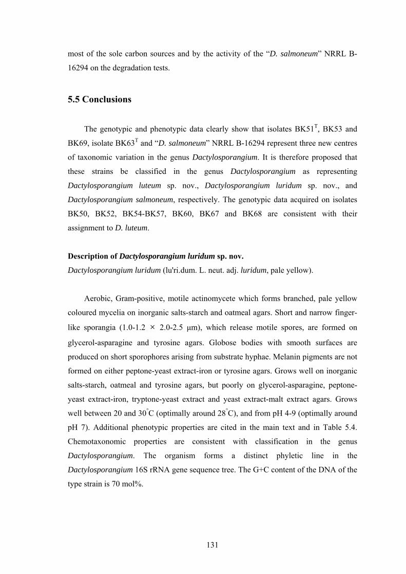
131
most of the sole carbon sources and by the activity of the “D. salmoneum” NRRL B-
16294 on the degradation tests.
5.5 Conclusions
The genotypic and phenotypic data clearly show that isolates BK51T, BK53 and
BK69, isolate BK63T and “D. salmoneum” NRRL B-16294 represent three new centres
of taxonomic variation in the genus Dactylosporangium. It is therefore proposed that
these strains be classified in the genus Dactylosporangium as representing
Dactylosporangium luteum sp. nov., Dactylosporangium luridum sp. nov., and
Dactylosporangium salmoneum, respectively. The genotypic data acquired on isolates
BK50, BK52, BK54-BK57, BK60, BK67 and BK68 are consistent with their
assignment to D. luteum.
Description of Dactylosporangium luridum sp. nov.
Dactylosporangium luridum (lu'ri.dum. L. neut. adj. luridum, pale yellow).
Aerobic, Gram-positive, motile actinomycete which forms branched, pale yellow
coloured mycelia on inorganic salts-starch and oatmeal agars. Short and narrow finger-
like sporangia (1.0-1.2 × 2.0-2.5 μm), which release motile spores, are formed on
glycerol-asparagine and tyrosine agars. Globose bodies with smooth surfaces are
produced on short sporophores arising from substrate hyphae. Melanin pigments are not
formed on either peptone-yeast extract-iron or tyrosine agars. Grows well on inorganic
salts-starch, oatmeal and tyrosine agars, but poorly on glycerol-asparagine, peptone-
yeast extract-iron, tryptone-yeast extract and yeast extract-malt extract agars. Grows
well between 20 and 30°C (optimally around 28°C), and from pH 4-9 (optimally around
pH 7). Additional phenotypic properties are cited in the main text and in Table 5.4.
Chemotaxonomic properties are consistent with classification in the genus
Dactylosporangium. The organism forms a distinct phyletic line in the
Dactylosporangium 16S rRNA gene sequence tree. The G+C content of the DNA of the
type strain is 70 mol%.

132
The type strain, BK63T (= DSM 45324T =KACC 20933T = NRRL B-24775T), was
isolated from a soil sample taken from Palace Leas hay meadow plot 6 at Cockle Park
Experimental Farm, Northumberland, UK.
Description of Dactylosporangium luteum sp. nov.
Dactylosporangium luteum (lu'te.um. L. neut. adj. luteum, orange-yellow, flame-
coloured).
Aerobic, Gram-positive, motile actinomycetes which forms branched, orange-
yellow coloured mycelia on oatmeal and inorganic salts-starch agars. Short and narrow
finger-like sporangia (1.0-1.2 × 2.0-2.5 μm), which release motile spores, are formed
on glycerol-asparagine, tyrosine and yeast extract-malt extract agars. Globose bodies
with smooth surfaces are produced on short sporophores arising from substrate hyphae.
Melanin pigments are not formed on either peptone-yeast extract-iron or tyrosine agars.
Grows well on inorganic salts-starch, oatmeal and tyrosine agars, but poorly on
glycerol-asparagine, peptone-yeast extract-iron, tryptone-yeast extract and yeast
extract-malt extract agars. Grows well between 15 and 30°C (optimally around 28°C),
and from pH 5-10 (optimally around pH 7). Additional phenotypic properties are cited
in the main text and in Table 5.4. Chemotaxonomic properties are consistent with
classification in the genus Dactylosporangium. The organism forms a distinct phyletic
line in the Dactylosporangium 16S rRNA gene sequence tree. The G+C content of the
DNA of the type strain is 74 mol%.
The type strain, BK51T (= DSM 45323T= KACC 20899T = NRRL B-24774T) , was
isolated from a soil sample taken from Palace Leas hay meadow plot 6 at Cockle Park
Experimental Farm, Northumberland, UK.
Description of Dactylosporangium salmoneum sp. nov.
Dactylosporangium salmoneum (sal.mo.ne'um. L. n. salmo -onis, salmon; L. adj. suff. -
eus -a -um, suffix used with various meanings; N.L. neut. adj. salmoneum, salmon-
coloured).
The description is based on data from the present study and from Celmer et al.
(1978). Aerobic, Gram-positive, motile actinomycete that forms branched, orange

133
coloured mycelia on inorganic salts-starch and oatmeal agars. Numerous sporangia are
formed on calcium malate plates. Dactyloform sporangia are enlarged slightly towards
the apex (1.5 × 5.5 μm) and contain 3 to 4 spores, which are mainly elliptical (1.1-1.6 ×
2.2-2.7 μm) and are motile. Globose bodies with smooth surfaces are formed at the top
of short sporophores on substrate hyphae. Melanin pigments are not produced on either
peptone-yeast extract-iron or tyrosine agars. Good growth is shown on glycerol-
asparagine, inorganic salts-starch, oatmeal, peptone-yeast extract-iron, tryptone-yeast
extract, tyrosine and yeast extract-malt extract agars; grows poorly on Luria-Bertani,
nutrient and trypticase soy agars. Grows well between 20 and 37 °C (optimally around
28°C), and at pH 4-10 (optimally around pH 7). Additional phenotypic properties are
cited in the main text and in Table 5.4. Chemotaxonomic properties are consistent with
classification in the genus Dactylosporangium. The organism forms a distinct phyletic
line in the Dactylosporangium 16S rRNA gene sequence tree. The G+C content of the
DNA of the type strain is 73 mol%.
The type strain, NRRL B-16294T (=ATCC 31222T =DSM 43910T = JCM 3272T =
NBRC 14103T), was isolated from soil in Japan.
134
Chapter 6. Reclassification of “Dactylosporangium variesporum” as
Saccharothrix variisporea corrig. (ex Tomita et al. 1977) sp.
nov., nom. rev.
6.1 Abstract
In the course of a polyphasic study it was observed that “Dactylosporangium
variesporum” NRRL B-16296 is misclassified in the genus Dactylosporangium as it
exhibits properties consistent with its assignment to the genus Saccharothrix.
Phylogenetic analyses based on 16S rRNA gene sequences show that the strain falls
within the evolutionary radiation of the genus Saccharothrix, a result which is supported
by corresponding chemotaxonomic and morphological markers. The strain is
phylogenetically most closely, albeit loosely, related to Saccharothrix espanaensis, but
can be readily distinguished from this and other validly described Saccharothrix species
by using a range of phenotypic properties. The combined genotypic and phenotypic data
demonstrate conclusively that this strain should be classified as a new species in the
genus Saccharothrix for which the name Saccharothrix variisporea sp. nov. is proposed.
The type strain is NRRL B-16296T(= ATCC 31203T = DSM 43911T = JCM 3273T =
NBRC 14104T).
6.2 Introduction
“Dactylosporangium variesporum” sp. nov. was proposed by Tomita et al. (1977)
to accommodate a strain isolated from a soil sample collected in India which they
considered to have chemical and morphological properties consistent with its
assignment to the genus Dactylosporangium. The microorganism, strain D409-5,
produces capreomycin and was reported by Tomita and his colleagues to form finger-
shaped sporangia, singly, in pairs or in clusters on substrate hyphae, to contain meso-
diaminopimelic acid (meso–A2pm) in the cell wall peptidoglycan, and galactose,
mannose and rhamnose in whole-cell hydrolysates. Uchida & Seino (1997), however,
questioned the placement of this strain in the genus Dactylosporangium as they were
unable to observe sporangia and found that the peptidoglycan contained N-glycolated as
opposed to N-acetylated muramic acid moieties.

135
When “D. variesporum” NRRL B-16296 was included in a study designed to
clarify the taxonomy of invalidly described Dactylosporangium species, it was found to
have properties consistent with its assignment to the genus Saccharothrix. In the present
investigation, strain NRRL B-16296 (= strain D409-5) was the subject of a polyphasic
study which showed that it formed a novel Saccharothrix species for which the name
Saccharothrix variisporea sp. nov. is proposed.
6.3 Materials and methods
Cultivation and maintenance of the strains. “D. variesporum” NRRL B-16296 and
the equivalent strain DSM 43911 were maintained at -20°C as glycerol suspensions
(20%, v/v). Biomass for 16S rRNA gene sequence analyses of these strains and for the
chemotaxonomic analyses of strain NRRL B-16296 was prepared in modified Bennett’s
broth (Jones, 1949) at 150 rpm in shake flasks for 21 days at 28°C. Cells were checked
for purity and harvested by centrifugation. Biomass for chemotaxonomic analyses of the
strain was washed twice in distilled water and freeze-dried. Cells collected for DNA
isolation and sequencing of 16S rRNA genes were washed in NaCl/EDTA buffer (0.1M
EDTA, 0.1M NaCl, pH8.0) and stored at -20°C.
Molecular sequencing and phylogenetic analysis. Preparation of genomic DNA,
amplification and direct sequencing of the purified PCR products of 16S rRNA genes
prepared from strains NRRL B-16296 and DSM 43911 were performed after Tan et al.
(2006). The resultant, almost complete, 16S rRNA gene sequences (1458 nucleotides)
were aligned manually, using the jPHYDIT program (Jeon et al., 2005), against
corresponding sequences of the type strains of Saccharothrix species retrieved from the
GenBank database. Phylogenetic trees were inferred using the neighbour-joining
(Saitou & Nei, 1987), minimum-evolution and maximum-parsimony algorithms
(Takahashi & Nei, 2000) from the MEGA 3.1 program (Kumar et al., 2004). The
evolutionary distance model of Jukes & Cantor (1969) was used to generate an
evolutionary distance matrix for the neighbour-joining algorithm. The topologies of the
resultant trees were evaluated by bootstrap analysis (Felsenstein, 1985) based on 1000
resamplings of the neighbour-joining dataset. The root position of the neighbour-joining
tree was obtained using Pseudonocardia thermophila IMSNU 20112T as the outgroup.

136
Chemotaxonomical analyses. Chemotaxonomic studies were undertaken to confirm
that strain NRRL B-16296T exhibited a chemical profile which supported its
classification in the genus Saccharothrix. Standard procedures were used to extract and
analyse the isomers of diaminopimelic acid (Staneck & Roberts, 1974), isoprenoid
quinones (Collins, 1994), muramic acid type (Uchida et al., 1999), polar lipids
(Minnikin et al., 1984) and whole-organism sugars (Schaal, 1985), using appropriate
controls. Cellular fatty acids were extracted, methylated and analyzed by GC using the
standard Sherlock MIDI (Microbial Identificatin) system (Sasser, 1990) and mycolic
acids sought using the TLC procedure described by Minnikin et al. (1975).
Biochemical, cultural and morphological analysis. Strain NRRL B-16296T was
examined for a broad range of phenotypic properties using standard media and methods
described by Goodfellow et al. (1979). The gross morphological properties of the strain
were observed on plates of tryptone-yeast extract, yeast extract-malt extract, oatmeal,
inorganic salts-starch, glycerol-asparagine, peptone-yeast extract-iron and tyrosine agar
plates (ISP media 1-7, respectively; Shirling and Gottlieb, 1966) which were incubated
for 21 days at 28°C. Morphological characteristics were observed by examining gold-
coated, dehydrated preparations from a 14-day-old culture of the strain grown on
inorganic salts-starch agar at 28°C, using a Cambridge Stereoscan 240 scanning electron
microscope and the procedure described by O'Donnell et al. (1993).
6.4 Results and discussion
Phylogenetic analysis of 16S rRNA gene sequences. Identical 16S rRNA gene
sequences were obtained for “D. variesporum” NRRL B-16296 and DSM 43911 hence
only the sequence for strain NRRL B-16296 was deposited in GenBank (GQ917213)
and is considered further. It can be seen in Figure 6.1 that this strain falls within the
evolutionary radiation of the genus Saccharothrix based on the phylogenetic analysis of
16S rRNA genes. A subclade supported by all of the tree-making algorithms and by a
bootstrap value of 61% is formed by this strain and the type strains of S. espanaensis, S.
mutabilis subsp. capreolus and S. mutabilis subsp. mutabilis.
Strain NRRL B-16296T and S. mutabilis subsp. capreolus NRRL 2773T are
reported to produce the antibiotic capreomycin though NRRL B-16296T was found to
be most closely related to S. espanaensis NRRL 15764T; the two strains shared a 16S

137
rRNA gene similarity of 98.9 %, a value which corresponded to 16 nucleotides
differences at 1446 locations. The corresponding values between strain ‘NRRL B-
16296T’ with the type strains of S. mutabilis subsp. capreolus and S. mutabilis subsp.
mutabilis were 98.5% (21 nucleotides differences at 1425 locations) and 98.8% (18
nucleotides differences at 1430 locations), respectively. The 16S rRNA gene sequence
similarities between strain NRRL B-16296T and the type strains of the remaining
Saccharothrix species ranged from 97.5% (with S. violaceirubra) to 98.5% (with S.
australiensis). The 16S rRNA of strain NRRL B-16296T contains the unique
oligonucletide signature characteristic for members of the genus Saccharothrix (Labeda
& Kroppenstedt, 2007).
S. mutabilis subsp. capreolus DSM 40225T (X76965)
S. mutabilis subsp. mutabilis DSM 43853T (X76966)
S. espanaensis NRRL B-15764T (AF114807)
NRRL B-16296 (GQ917213 )
S. texasensis NRRL B-16134T (AF114814)
S. violaceirubra NBRC 102064T (AB284261)
S. australiensis NRRLB-11239T (AF114803)
S. algeriensis NRRL B-24137T (AY054972)
S. xinjiangensis NBRC 101911T (AB381939)
S. coeruleofusca NRRL B-16115T (AF114805)
S. syringae NRRL B-16468T (AF114812)
S. longispora NRRL B-16116T (AF114809)
Lechevalieria flava NRRL B-16131T (AF114808)
Lechevalieria aerocolonigenes NRRL B-3298T (AF114804)
Pseudonocardia thermophila IMSNU 20112T (AJ252830)
99*
69*
100*
94*
61*
51*
87*
0.01
*
*
**
S. mutabilis subsp. capreolus DSM 40225T (X76965)
S. mutabilis subsp. mutabilis DSM 43853T (X76966)
S. espanaensis NRRL B-15764T (AF114807)
NRRL B-16296 (GQ917213 )
S. texasensis NRRL B-16134T (AF114814)
S. violaceirubra NBRC 102064T (AB284261)
S. australiensis NRRLB-11239T (AF114803)
S. algeriensis NRRL B-24137T (AY054972)
S. xinjiangensis NBRC 101911T (AB381939)
S. coeruleofusca NRRL B-16115T (AF114805)
S. syringae NRRL B-16468T (AF114812)
S. longispora NRRL B-16116T (AF114809)
Lechevalieria flava NRRL B-16131T (AF114808)
Lechevalieria aerocolonigenes NRRL B-3298T (AF114804)
Pseudonocardia thermophila IMSNU 20112T (AJ252830)
99*
69*
100*
94*
61*
51*
87*
0.010.01
*
*
**
Fig. 6.1. Neighbour-joining tree based on almost complete 16S rRNA gene sequences showing the position of Saccharothrix variisporea NRRL B-16296T in the Saccharothrix gene tree. Numbers on branches indicate percentage bootstrap values from 1000 replicates (only values > 50% are shown). Asterisks indicate branches that were also recovered in the minimum-evolution and maximum-parsimony trees. Bar, 0.01 substitutions per nucleotide position.
Determination of G+C content (mol%) and DNA–DNA relatedness. The DNA G+C
composition of strain NRRL B-16296T was determined by using the procedure
described by Gonzalez & Saiz-Jimenez (2005) and found to be 74 mol %. DNA–DNA
relatedness studies were not carried out between strain NRRL B-16296T and its closest
phylogenetic neighbours as type strains of Saccharothrix species with similar 16S rRNA

138
gene sequences have DNA–DNA hybridization values well below the 70% cut-off point
recommended by Wayne et al. (1987) for the circumscription of strains that belong to
the same genomic species. Thus, the type strains of S. espanaensis and S. mutabilis,
have a 16S rRNA gene similarity of 99.0%, but a DNA-DNA relatedness value of 0 %
(Labeda & Lechevalier, 1989) whereas the type strains of S. syringae and S.
xinjiangensis share a 16S rRNA gene similarity of 99.1 % but have a DNA–DNA
relatedness value of only 50 % (Hu et al., 2004). Similarly, S. algeriensis NRRL B-
24137T and S. australiensis NRRL 11239T have a 16S rRNA gene similarity of 98.8 %
and a DNA–DNA relatedness of 55.9 % (Zitouni et al., 2004).
Chemotaxonomic properties. The strain contained meso-diaminopimelic acid (wall
chemotype Ⅲ sensu Lechevalier & Lechevalier 1970), N-acetylated muramic acid
moieties, galactose, mannose and rhamnose as diagnostic whole-organism sugars,
tetrahydrogenated menaquinones with nine isoprene units (MK-9[H4]) as the
predominant menaquinone, and diphosphatidylglycerol, phosphatidylethanolamine,
phosphatidylinositol and phosphatidylglycerol as major polar lipids (phospholipid
pattern type Ⅱ sensu; Lechevalier et al. 1977) together with a trace of
phosphatidylinositol mannoside and single unknown aminolipid and phopholipid
components. The predominant fatty acids were 13-methyltetradecanoic (iso-C15:0), 14-
methylpentadecanoic (iso-C16:0) and 14-methylhexadecanoic (anteiso-C17:0) acids;
mycolic acids were not present. All of these properties are in line with the classification
of the strain in the genus Saccharothrix (Labeda et al. 1984; Otoguro et al., 2009).
Biochemical, cultural and morphological properties. It can be seen from Table 6.1
that strain B-16296T can be clearly distinguished from the type strains of Saccharothrix
species, including its nearest neighbours, S. espanaensis NRRL 15764T and the type
strains of the two subspecies of S. mutabilis, by using a combination of phenotypic
properties. Strain B-16296T, unlike the type strain of S. espanaensis, grows at 45°C and
degrades starch and tyrosine, and utilizes sugars such as L(+)-arabinose, dextrin, myo-
inositol, D(+)-lactose, D(+)-melibiose, D(+)-raffinose, and D(+)-xylose as sole sources
of carbon for energy and growth. Additional phenotypic properties of the strain are
given in Table 1 and in the species description.

139
Table 6.1. Phenotypic properties which separate strain NRRL B-16296T from the type
strains of Saccharothrix species.
Taxa: 1, strain NRRL B-16296T; 2, S. algeriensis NRRL B-24137T; 3, S. australiensis NRRL 11239T; 4,
S. coeruleofusca DSM 43679T; 5, S. espanaensis NRRL 15764T; 6, S. longispora DSM 43749T; 7, S.
mutabilis subsp. capreolus DSM 40225T; 8, S. mutabilis subsp. mutabilis NRRL B-16077T; 9, S. syringae
DSM 43886T; 10, S. texasensis NRRL B-16134T; 11, S. violaceirubra KCTC 19326T; 12, S. xinjiangensis
NBRC 101911T. Data for the reference strains were taken from Labeda (2002), Zitouni et al. (2004), Hu
et al. (2004) and Otoguro et al. (2009). +, Positive; –, negative; ND, not determined; w, weakly positive;
v, variable.
Characteristic 1 2 3 4 5 6 7 8 9 10 11 12
Decomposition of : Adenine – – – – – + – – – – – ND
Hypoxanthine + – – – + – + + – + – ND
Starch + – – + – + + + + + – +
Tyrosine + + + + – + – + + + + +
Nitrate reduction – + + – w + – + – + + –
Assimilation of :
Citrate – + – – v + + + – – – –
Lactate – ND w – + + – + – + – –
Malate + ND + – + + + + + + – ND
Utilization of :
Arabinose + – – + – + + + + + – +
Dextrin + – + + – + + + + + – ND
Inositol + – – – – – + + – + – ND
Lactose + – – + – + – + + + – +
Melibiose + – – – – – + + + + – +
α-Methyl-D-glucoside + – – + – – – + – + – +
Raffinose + – – + – – – + + – – +
Rhamnose + – – + – + – – + + – +
Salicin + ND – + – ND + + w + – ND
Sorbitol – – + + – – – – – – – –
Sucrose + – – + + + – + + + + +
D-Xylose + – – + v + + + + + – +
Growth at :
4 % NaCl (w/v) + + + + + + + – + – – –
5 % NaCl (w/v) – – – + – + + – + – – –
37°C + + + + + + + + + + – +
45°C w + + + – – + + + – – +
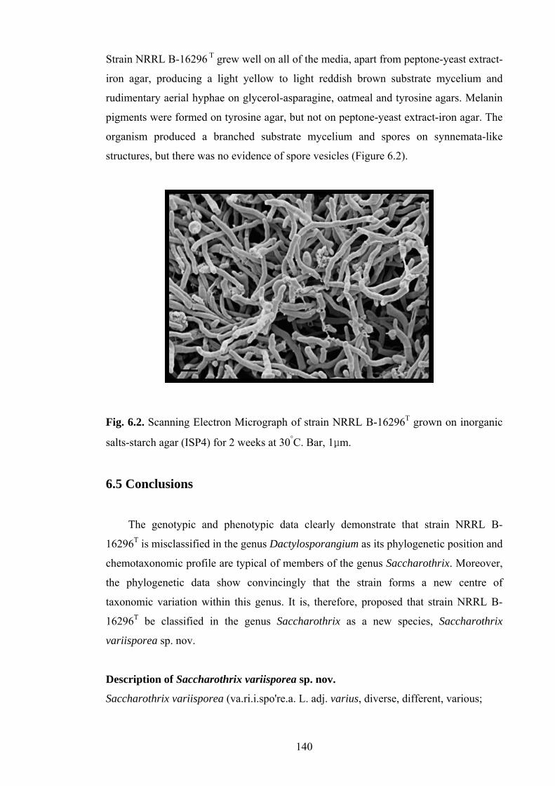
140
Strain NRRL B-16296 T grew well on all of the media, apart from peptone-yeast extract-
iron agar, producing a light yellow to light reddish brown substrate mycelium and
rudimentary aerial hyphae on glycerol-asparagine, oatmeal and tyrosine agars. Melanin
pigments were formed on tyrosine agar, but not on peptone-yeast extract-iron agar. The
organism produced a branched substrate mycelium and spores on synnemata-like
structures, but there was no evidence of spore vesicles (Figure 6.2).
Fig. 6.2. Scanning Electron Micrograph of strain NRRL B-16296T grown on inorganic
salts-starch agar (ISP4) for 2 weeks at 30°C. Bar, 1μm.
6.5 Conclusions
The genotypic and phenotypic data clearly demonstrate that strain NRRL B-
16296T is misclassified in the genus Dactylosporangium as its phylogenetic position and
chemotaxonomic profile are typical of members of the genus Saccharothrix. Moreover,
the phylogenetic data show convincingly that the strain forms a new centre of
taxonomic variation within this genus. It is, therefore, proposed that strain NRRL B-
16296T be classified in the genus Saccharothrix as a new species, Saccharothrix
variisporea sp. nov.
Description of Saccharothrix variisporea sp. nov.
Saccharothrix variisporea (va.ri.i.spo're.a. L. adj. varius, diverse, different, various;

141
Gr. n. spora, a seed, in microbiology, a spore; L. suff. eus, -a, -um, suffix with various
meanings, but in general, made of or belongs to; N.L. fem. adj. variisporea, with
different spores).
The description is based on data taken from the present study and from the earlier
work of Tomita et al. (1977). Aerobic, Gram-positive actinomycete which forms
branched, light orange coloured mycelia on inorganic salts-starch and oatmeal agars.
Grows well between 30 and 37°C, from pH 4 to 10, and optimally around pH 7. Positive
for aesculin and allantoin hydrolysis, catalase, coagulation and peptonization of milk,
and for hydrogen sulfide and urease production, but is negative for nitrate reduction.
Degrades arbutin, casein, DNA, elastin, gelatin, Tweens 40, 60 and 80, uric acid, and
xylan, but not cellulose, chitin, guanine, pectin, RNA, tributyrin, Tween 20 or xanthine.
D(+)-cellobiose, D(+)-galactose, D(-)-glucose, glycerol, glycogen, inulin, mannitol,
D(+)-maltose, D(+)-mannose, D(-)-ribose, starch and D(+)-trehalose (at 1.0%, w/v), and
L(+)-tartaric acid, L(-)-malic acid, propionic acid, pyruvic acid and uric acid (at 0.1%,
w/v) are used as sole carbon sources, but not adonitol, D(+)-arabitol, dulcitol, D(-)-
sorbitol, L(-)-sorbose or xylitol (at 1.0%, w/v) or L(+)-lactic acid, oxalic acid, or urea
(at 0.1%, w/v). Resistant (μg ml-1) to ampicillin (4), cephaloridine (2), clindamycin (8),
gentamicin (8), kanamycin (8), lincomycin (8), oxytetracycline (8), penicillin G (2),
tylosin (8) and vancomycin (2), but is sensitive to ciprofloxacin (2), chloramphenicol
(8), novobiocin (8), rifampicin (16), streptomycin (4) and tetracycline (8). Growth
occurs in the presence of 4% (w/v) NaCl and 0.05% lysozyme, but not in the presence
of 5% NaCl. Additional phenotypic properties are shown in Table 1 and in the main text.
The predominant fatty acids are iso-C16:0 (39.2%), anteiso-C17:0 (11.9%), iso-C15:0
(9.9%), 9-methyl C16:0 (8.4%), iso-C17:0 (6.9%), iso-C16:1H (6.7%), C17:1ω6c (5.3%),
C16:1 2-OH (1.8%), cis 9-C16:1 (1.7%), anteiso-C15:0 (1.3%), cis 9-C17:1 (1.1%) and
anteiso-C17:1C (1.0%). The G+C content of the type strain is 74 mol%.
The type strain, NRRL B-16296T (=ATCC 31203T =DSM 43911T =JCM 3273T =NBRC
14104T), was isolated from a soil sample from India.

142
Chapter 7. Search for a self-resistance mechanism of Verrucosispora
maris AB-18-032 to atrop-abyssomicin C
7.1. Abstract
P1-derived artificial chromosome (PAC) library of genomic DNA of the
Verrucosispora maris AB-18-032 was introduced into an abyssomicin-sensitive strain,
Streptomyces griseus NRRL B-2682T (MIC 5-10 μg/ml) by conjugation. Five mutants
which showed resistance to atrop-abyssomicin C were isolated. Exconjugants were
screened by PCR reactions for putative abyssomicin resistance genes (abyD,
isochorsmatase, pabB and trpE) identified from the abyssomicin biosynthetic gene
cluster (abyD) and the whole genome of V. maris AB-18-032 (isochorismatase, pabB
and trpE), respectively. All of the exconjugants were negative for the putative resistance
genes, indicating that the atrop-abyssomicin C resistance shown by the mutants is not
conferred by the putative resistance genes from V. maris AB-18-032 hence the
resistance observed in the exconjugants is probably conferred by mutations in the target
protein (PabB) or through decreased uptake or increased efflux of atrop-abyssomicin C.
7.2. Introduction
Global antibiotic resistance is becoming an increasing public health problem with
the emergence of multi-drug resistant bacterial pathogens, such as methicillin-resistant
Staphylococcus aureus (MRSA) and vancomycin-resistant enterococci (VRE)
(Spellberg et al., 2008). Every new generation of older antibiotics and new compounds
approved eventually face the emergence of resistance (Wright, 2010). Actinomycetes
produce two-thirds of the known natural products, notably antibiotics (Bérdy, 2005),
and have evolved self-resistant mechanisms to protect themselves from the toxicity of
these molecules, thereby making them ‘hot spots’ for studying the evolution of
antibiotic resistance (Cundliffe, 1989; Davies, 1997; D'Costa et al., 2006; Mindlin et al.,
2006; Hopwood, 2007; Wright, 2010). It has been suggested that a number of
mechanisms, including target modification, antibiotic inactivation, permeability barrier
alterations or an efflux system could be involved in conferring the self-resistance to the
producer (Mindlin et al., 2006; Hopwood, 2007; Cundliffe & Demain, 2010). Analysis

143
of the molecular basis of resistance in antibiotic-producing actinomycetes can help
determine the resistance mechanisms of pathogenic organisms which could eventually
emerge clinically, and provides strategies which can be applied in new antibiotic
discovery programmes (Hotta et al., 1983, 1992, 1999; Hotta & Okami, 1996; Wright,
2007, 2010). Therefore, if the self-resistance mechanisms of newly discovered
antibiotics are elucidated, it could be possible to identify and screen the resistance
determinants from environmental samples, environmental isolates and clinical
pathogens (Guardabassi et al., 2004, 2005; Guardabassi & Agerso, 2006; D'Costa et al.,
2007; Wright, 2010).
New drug compounds have been sought from actinomyetes isolated from extreme
habitats and sources of which target new biosynthetic processes (Bull & Stach, 2007).
Atrop-abyssomicin C, the first known natural inhibitor of the para-aminobenzoate
(pABA) biosynthesis pathway, is produced by Verrucosispora maris AB-18-032, a
strain isolated from a marine sediment sample (Riedlinger et al., 2004). It is a
polycyclic polyketide-type antibiotic which shows antibiotic activity against Gram-
positive bacteria, including B. subtilis and methicillin-resistant Staphylococcus aureus.
This compound is the main component of the abyssomicin complex, which also
includes abyssomicin B, C, D, G and H (Keller et al., 2007a).
Recently, the mode of action of atrop-abyssomicin C has been elucidated. Keller
et al. (2007b) showed that the highly conserved PabB subunit of 4-amino-4-
deoxychorismate (ADC) synthase from Bacillus subtilis is the molecular target of
atrop-abyssomicin C. This compound acts as a covalent binder to the side chain of
Cys263, located in the proximity of the active site of PabB. However, the resistance
mechanism still remains unclear although it has been suggested that resistance may be
through the modification of target (PabB) or the presence of a PabB homologue that
may be able to complement inhibition of the PabB enzyme (Keller et al., 2007b). It is
important, therefore, to see whether the strain, Verrucosispora maris AB-18-032, like
other antibiotic-producing actinomycetes, can modify or complement the abyssomicin
target site to make ADC synthase resistant to the compound. Recently, the whole
genome of the strain, Verrucosispora maris AB-18-032, has been partially sequenced
and the putative resistance genes have been identified (Jem Stach, personal
communication). This work is designed to elucidate the role and identification of
putative resistance genes which are responsible for self-resistance. To this end, a P1-

144
derived artificial chromosome (PAC) library of genomic DNA of strain V. maris AB-18-
02 was introduced into Streptomyces griseus NRRL B-2682T, a strain known to be
susceptible to atrop-abyssomicin C by conjugation. Resistant exconjugants were
isolated and screened for the presence of putative abyssomicin resistance genes. The
results indicate that resistance in the exconjugants is conferred either by mutation within
the S. griseus strain or by cloning of unidentified resistance genes from V. maris AB-18-
032.
7.3. Materials and methods
The cultivation of test strains, selection of atrop-abyssomicin C sensitive strains,
conjugation of genomic DNA library, selection of exconjugants, PCR primer design,
extraction of genomic DNA from exconjugants, and PCR screening of putative
resistance genes, were carried out using procedures described earlier in section 2.10.
7.4. Results
Sensitivity of Streptomyces strains to abyssomicin. The sensitivity of, “S. coelicolor”
A3(2), S. griseus NRRL B-2682T, “S. lividans” TK24 and S. venezuelae ATCC 15439 to
abyssomicin was tested by agar plate diffusion assay using minimal media (Table 7.1).
All of four stains were sensitive to abyssomicin at ≥10 μg with “S. coelicolor” A3(2), “S.
lividans” TK24 and S. venezuelae ATCC 15439 showing similar sensitivities to
abyssomicin. The remaining strain S. griseus NRRL B-2682T showed higher sensitivity
and was chosen for further studies.
Table 7.1. Antibacterial activity of atrop-abyssomicin C, determined by the agar plate
diffusion assay (inhibition zone in mm)
Concentration “S. coelicolor” A3(2)
S. griseus NRRL B-2682T
“S. lividans” TK24
S. venezuelae ATCC 15439
5 μg 0 0 0 0 10 μg 1 3 2 2 20 μg 5 10 5 5 40 μg 10 20 10 10 60 μg 15 25 15 15 80 μg 20 30 20 20

145
Selection of resistant exconjugants. Streptomyces griseus NRRL B-2682T was
conjugated with a genomic DNA library of V. maris AB-18-032 in plasmid pESAC13.
Seven exconjugants were initially selected as they grew on agar plates containing 40
μg/ml abyssomicin. The parental S. griseus was totally inhibited with 40 μg/ml atrop-
abyssomicin C under similar conditions. When spores prepared from the resistant clones
were allowed to grow again in the presence of atrop-abyssomicin C, all of five clones,
SG-19, SG-26, SG-27, SG032 and SG-33, showed resistance to abyssomicin, as well as
conserving the kanamycin resistance marker of the pESAC13 vector (Fig. 7.1).
Three exconjugants, SG-19, SG-26 and SG-32 were particularly resistant to atrop-
abyssomicin C showing very reduced inhibition zones. In contrast, two exconjugants,
SG-27 and SG-33, were moderately resistant to atrop-abyssomicin C showing larger
inhibition zones than the other exconjugants. A control culture of S. griseus strain
carrying the empty pESAC13 did not show any resistance to the drug in the absence of
kanamycin.
Fig. 7.1. Sensitivity of 5 exconjugants and the wild type strain of S. griseus to 40 μg
(disk) of atrop-abyssomicin C after culturing at 30˚C for 48 hours. (a), wild type strain;
(b), exconjugants SG-19; (c), SG-26; (d), SG-27; (e), SG-32; (f), SG-33.
Putative resistant gene screening. Four PCR primers designed from target genes
thought to be responsible for self-resistance in the V. maris AB-18-032, were used to

146
screen the exconjugants. The primers were tested on the genomic DNA of both V. maris
AB-18-032 and S. griseus NRRL B-2682T, as the positive and negative controls,
respectively. The primers were found to be specific to the target genes of the strain. PCR
amplified product was not observed when these primers were tested on the five
abyssomicin-resistant exconjugants.
Two PCR primers designed from the abyD gene of the biosynthetic gene cluster
and pabB gene of the 4-amino-4-deoxychorismate synthase gene cluster, showed weak
bands. However, the amplified PCR products were different to the expected size and
were also observed in the negative control DNA (Fig. 7.2).
(a)
(b) (c) (d) Fig. 7.2. Agarose gel electrophoresis of PCR amplifications derived from PCR reactions
using target gene primers (Table 2.11). (a), abyD; (b), AS; (c), ICS; (d), PAB.
Lane: 1, SG-19; 2, SG-26; 3, SG-27; 4, SG-32; 5, SG-33; 6, V. maris AB-18-032; 7, S.
griseus NRRL B-2682T; M, molecular marker (100bp ladder; MBI Fermentas, UK)

147
7.5 Discussion
The para-aminobenzoic acid (p-ABA) derived from the key-metabolite of
chorismate, is an important metabolite in the folic acid pathway which is essential to
bacterial metabolism (Nichols et al., 1989). The enzymes of the pABA and folic acid
biosynthetic pathway are conserved in many microorganisms, parasites and plants, but
not in humans hence this pathway is an interesting target for the development of novel
antibiotics, pesticidal and herbicidal agents (Alun & Jeremy, 2002). Sulfonamides and
trimethoprim are prominent synthetic inhibitors of the folic acid pathway (Huovinen et
al., 1995). The natural antibiotic, atrop-abyssomicin C is a very efficient inhibitor of the
pABA) biosynthesis in the Gram-positive bacteria.
A selection of chorismate-utilising enzymes, including 4-amino-4-
deoxychorismate (ADC) synthase, anthranilate synthase and isochorismate synthase
have been considered to be evolutionarily related on the basis of sequence homology,
structural similarity, and a unified reaction mechanism (He et al., 2004). In all three
enzymatic reactions, the C4 hydroxy group of chorismate is replaced by the addition of
a nucleophile at the C2 position (Fig. 7.3). pABA biosynthesis from chorismate requires
two enzymes: ADC synthase, which converts chorismate and glutamine into ADC and
glutamate, and ADC lyase which catalyses an elimination reaction of ADC to produce
pABA. ADC synthase is a heterodimer composed of two dissimilar subunits, PabA and
PabB. PabA functions as a glutamine amidotransferase that hydrolyses glutamine (Gln)
to glutamate (Glu) and NH4+. PabB catalyses the formation of 4-amino-4-
deoxychorismate from chorismate and ammonia provided by the amidotransferase of
PabA (He & Toney, 2006).
It is evident from Table 7.1 that atrop-abyssomicin C showed a strong inhibitory
activity against the four Streptomyces strains. The minimal inhibitory concentration
(MIC) of atrop-abyssomicin C against these strains was 5-10 μg. The inhibitory activity
of the antibiotic was significantly stronger when the test organism, S. griseus NRRL B-
2682T was grown in minimal media. These results indicate that the S. griseus with the
highest susceptibility to the atrop-abyssomicin, is the most appropriate strain to
investigate the mechanism of self-resistance of V. maris AB-18-032 .

148
Fig. 7.3. Three chorismate-utilizing enzymes of E. coli sharing a common reaction
mechanism: anthranilate synthase (AS), 4-amino-4-deoxychorismate synthase (ADCS),
and isochorismate synthase (IS) (adapted from He et al., 2004) .
A number of studies have shown that biosynthetic and resistance genes of antibiotic-
producing actinomycetes are genetically related due to the mechanism of self-protection
(Cundliffe, 1989; Pootoolal et al., 2002; Mindlin et al., 2006; Hopwood, 2007). It was
for this reason that five of the mutants were selected for PCR screening of the target
gene, which might be involved in self-resistance to abyssomicin. The three primers were
used to detect the isochorismatase, pabB and trpE(G) genes coding for three chorismate
utilising enzymes, IS, ADCS and AS, respectively (Table 2.11). The remaining primer
was used to detect the abyD gene which was located in the atrop-abyssomicin C
biosynthetic gene cluster. However, none of these genes were detected by PCR
screening of the exconjugants indicating that the resistance mechanism working in the
mutants of S. griseus does not involve any of these genes hence further work is needed
to identify the mechanism(s) of resistance in the exconjugants.
Sequencing of the pabB gene of the exconjugants will reveal whether or not
mutatons in the target protein confer resistance to atrop-abyssomicin C in Streptomyces
strains. If this were not the case, then determination of the genes transferred from V.
maris AB-18-032 to S. griseus NRRL B-2682T might allow identification of the atrop-
abyssomicin C resistance genes. Cloning of genomic DNA from the exconjugants back

149
into atrop-abyssomicin C sensitive S. griseus strains would enable any mutations that
confer resistance to abyssomicin, which were not the result of pabB mutations, to be
identified. Once identified, these resistance genes could be used to detect the resistance
in either environmental isolates or clinical pathogens, where atrop-abyssomicin C was
to be used clinically.

150
Chapter 8. General discussion and perspectives for future work
8.1. General discussion
The importance of filamentous actinomycetes in biotechnology is a function of
their remarkable metabolic diversity, their long association with natural habitats and the
needs of human beings (Goodfellow & O'Donnell, 1989; Bull et al., 2000; Ul-Hassan &
Wellington, 2009; Goodfellow & Fiedler, 2010). However, the overall value of these
organisms to humankind is difficult to measure as they exhibit both beneficial and
detrimental roles, notably in healthcare. Thus, on one hand, they are the richest source
of clinically significant antibiotics (Strohl, 2004; Bérdy, 2005; Goodfellow & Fiedler,
2010), but on the other hand, they are the likely source of antibiotic-resistance genes
present in pathogenic microorganisms (Davies, 1997; D'Costa et al., 2006; Mindlin et
al., 2006; Woo et al., 2006; Wright, 2010). The battle between filamentous
actinomycetes as friends and foes was the underlying theme of this project which was
designed (a) to establish whether a correlation exists between antibiotic resistance
patterns and the taxonomy of dereplicated strains of filamentous actinomycetes isolated
from a hay meadow soil, (b) to determine the taxonomic diversity and biotechnological
potential of members of the genus Dactylosporangium, and (c) to try and establish the
mechanism(s) which protects Verrucosispora maris strain AB-18-032 against the potent
antibiotic it produces, namely atrop-abyssomicin C. Most of objectives were achieved
to a greater or lesser extent.
An important indicator of antibiotic production in soil is the widespread occurrence
of antibiotic-resistance amongst indigenous soil bacteria, especially streptomycetes
(D'Costa et al., 2006; Laskaris et al., 2010; Wright, 2010). In the present investigation, a
small, but reasonably representative, set of filamentous actinomycetes, notably
streptomycetes, showed an astonishing capacity to grow in the presence of a panel of
antibiotics. Indeed, on average, these organisms showed resistance to 19 of the 41 tested
antibiotics. These results support the view that filamentous actinomycetes present in soil
are a reservoir of resistant determinants which can be spread into the broader microbial
community (D'Costa et al., 2007; Wright, 2007, 2010).
The 16S rRNA gene sequencing studies showed that most of the dereplicated
filamentous actinomycetes belonged to the genus Streptomyces though some were

151
phylogenetically close to authentic Actinomadura, Dactylosporangium,
Micromonospora and Streptosporangium strains. The weak correlation found between
the antibiotic resistance patterns and the taxonomy of the dereplicated strains may
reflect that small and heterogeneous nature of the complement of strains as a higher
correlation was found with isolates which showed high levels of 16S rRNA gene
similarity. This somewhat generous interpretation of the data is supported by the
discovery that all of the vancomycin-resistant isolates keyed out as Streptomyces
sanglieri (Manfio et al., 2003).
Improved selective isolation and characterisation procedures show that filamentous
actinomycetes once thought to be rare in natural habitats are widely distributed and may
be integral parts of microbial communities, as shown by studies on the genera
Amycolatopsis (Tan et al., 2006), Micromonospora (Qiu et al., 2008), nocardiae
(Orchard et al., 1977; Maldonado et al., 2000), Planobispora (Suzuki et al., 2001),
Rhodococcus (Colquhoun et al., 1998) and Salinispora (Mincer et al., 2005; Jensen &
Mafnas, 2006). Effective taxon-specific isolation procedures are needed for the selective
isolation and characterisation of members of most filamentous actinomycete taxa of
commercial interest (Goodfellow & Fiedler, 2010), not least the genus
Dactylosporangium, which encompass strains known to produce antibiotics (Hayakawa
et al., 1991; 2000; Hayakawa, 2008).
One of the major achievements of the project was the discovery that Streptomyces
Isolation Medium (SIM) supplemented with gentamicin and antifungal antibiotics
supported the growth of relatively large numbers of presumptive novel
Dactylosporangium species. The novelty of selected isolates was confirmed in a
polyphasic study which led to the formal recognition of two species,
Dactylosporangium luteum and Dactylosporangium luridum. A third species,
Dactylosporangium salmoneum was proposed for an organism which had no formal
nomenclatural standing. The application of the new selective isolation procedure
showed that Dactylosporangium strains are widely distributed in natural habitats,
notably soil. Preliminary studies also showed that novel members of the genus may
prove to be a source of new antibiotics as some isolates contained non-ribosomal
peptide and type-I polyketide synthase genes and inhibited the growth of Gram-positive
bacteria.
152
A key aim of the present study was to gain an insight into the distribution and
taxonomic diversity of members of the genus Dactylosporangium in a number of
environmental samples using culture-independent and culture-dependent procedures.
Although it was not the intention to estimate the full extent of dactylosporangial
diversity in the environmental samples, an unexpectedly high degree of taxonomic
variation was achieved in a small clone library. It was also interesting that the range of
diversity detected using the two approaches was comparable indicating that the genus-
specific primers of Monciardini et al. (2002) can be used to detect new centres of
taxonomic diversity in dactylosporangial gene tree. This means that the primer set
together with other appropriate molecular techniques, such as denaturing gradient gel
electrophoresis (DGGE), single-strand conformational polymorphism (SSCP) and
terminal restriction fragment length polymorphism (T-RFLP), could be used to
prescreen environmental samples before selective isolation studies. Such a twin-track
strategy would allow broader ecological issues to be addressed, such as unraveling the
geographical distribution, abundance, diversity and roles of Dactylosporangium species
in diverse habitats, as well as providing novel biological material for pharmaceutical
screens.
The twin-track strategy outlined above is labour intensive which may account for
the fact that it has rarely been used in the search for novel secondary metabolites,
notably antibiotics. However, studies along these lines have shown that members of the
obligate marine actinomycete Salinispora are a rich source of novel antibiotics (Jensen
et al., 2005; Jensen et al., 2007; Udwary et al., 2007), including the anticancer drug,
salinsporamide which is currently in clinical trials (Fenical et al., 2009). Indeed, the
species-specificity of Salinispora biosynthetic pathways (Jensen et al., 2007) strongly
supports the view of a coupling between taxonomic and chemical diversity, as has been
inferred from studies on Amycolatopsis regifaucium (Tan et al., 2007), Streptomyces
clavuligerus (Ward & Goodfellow, 2004) and Stretomyces violaceusniger-like strains
(Goodfellow et al., 2007).
It was disappointing that the search for a mechanism whereby the Verrucosispora
maris AB-18-032 strain protects itself against atrop- abyssomcin C was not successful
though the work is only a preliminary study. Further studies using the resistant
exconjugants would be required to find the responsible gene or protein located in the
cell and to obtain advance insight into the real mechanism(s) involved in resistance.

153
8.2 Perspectives for future work
Ecology:
• Selective isolation and identification of representatives of common Streptomyces
present in plot 6 of the hay meadow soil to establish whether a strain correlation
exists between their position in the 16S rRNA gene tree and ability to grow in
the presence of antibiotics.
• Comparison of Streptomyces Isolation medium (SIM), supplemented with
gentamicin and antifungal antibiotics agents, with other media recommended for
the selective isolation of members of the genus Dactylosporangium.
• Determination of the full extent of dactylosporangial diversity in natural habitats
using appropriate molecular procedures (e.g., PCR-DGGE, PCR-SSCP and
Pyrosequencing method).
• Biogeographical studies on the selected Dactylosporangium species to
determine whether they show site specific endemism.
• Ecophysiological studies on representatives of common Dactylosporangium
species to foster an understanding of their role in natural habitats.
Systematics:
• Formal description of additional Dactylosporangium species following
polyphasic studies on presumptively novel strains isolated in this study.
• Establishment of minimal standards for the recognition of new
Dactylosporangium species.
• Characterisation of representatives of the putative Dactylosporangium isolates
which gave negative results against the genus-specific primers.
• Formal description of the putatively novel Actinomadura, Micromonospora,
Streptomyces and Streptosporangium strains included in the 16S rRNA gene
sequencing studies.
Bioprospecting:
• Screening of metabolites from representatives of dactylosporangiae for novel
antibacterial, antifungal, antitumor and nematocidal activity.
• Identification of the compounds giving interesting hits in the antibacterial assays
using appropriate chemical procedures.

154
• Application of genome mining and genome scanning methods to representatives
of the Dactylosporangium strains to detect cryptic biosynthetic gene clusters that
can be activated by genetic manipulation and/or non-standard fermentation
conditions.
• Whole-genome sequencing of representative dactylosporangiae to establish their
potential to produce novel antibiotics.
• Establishment of the degree of coupling between the taxonomic and chemical
diversity of representatives of Dactylosporangium species.
Resistance:
• Search for other putative resistance genes transported to exconjugants which are
resistant to atrop-abyssomicin C.
• Investigation of genetic mechanism whereby Verrucosispora maris strain AB18-
032 is protected and regulated against atrop-abyssomicin C.
• Screening of resistance determinant of atrop-abyssomicin C in other bacteria,
including environmental isolates and environmental samples.

155
References Adiri, R. S., Gophna, U. & Ron, E. Z. (2003). Multilocus sequence typing (MLST) of Escherichia coli O78 strains. FEMS Microbiol Lett 222, 199-203. Alam, M. T., Merlo, M. E., Takano, E. & Breitling, R. (2010). Genome-based phylogenetic analysis of Streptomyces and its relatives. Mol Phylogenet Evol 54, 763-772. Alderson, G., Ritchie, D. A., Cappellano, C., Cool, R. H., Ivanova, N. M., Huddleston, A. S., Flaxman, C. S., Kristufek, V. & Lounes, A. (1993). Physiology and genetics of antibiotic production and resistance. Res Microbiol 144, 665-672. Alekshun, M. N. & Levy, S. B. (2007). Molecular mechanisms of antibacterial multidrug resistance. Cell 128, 1037-1050. Allen, H. K., Donato, J., Wang, H. H., Cloud-Hansen, K. A., Davies, J. & Handelsman, J. (2010). Call of the wild: Antibiotic resistance genes in natural environments. Nat Rev Microbiol 8, 251-259. Alun, B. & Jeremy, P. D. (2002). The folic acid biosynthesis pathway in bacteria: evaluation of potential for antibacterial drug discovery. Bioessays 24, 637-648. Amann, R. I., Ludwig, W. & Schleifer, K.-H. (1995). Phylogenetic identification and in situ detection of individual microbial cells without cultivation. Microbiol Rev 59, 143-169. Aminov, R. I. & Mackie, R. I. (2007). Evolution and ecology of antibiotic resistance genes. FEMS Microbiol Lett 271, 147-161. Antony-Babu, S. & Goodfellow, M. (2008). Biosystematics of alkaliphilic streptomycetes isolated from seven locations across a beach and dune sand system. Antonie van Leeuwenhoek 94, 581-591. Antony-Babu, S., Stach, J. E. M. & Goodfellow, M. (2008). Genetic and phenotypic evidence for Streptomyces griseus ecovars isolated from a beach and dune sand system. Antonie van Leeuwenhoek 94, 63-74. Antony-Babu, S., Stach, J. & Goodfellow, M. (2010). Computer-assisted numerical analysis of colour-group data for dereplication of streptomycetes for bioprospecting and ecological purposes. Antonie van Leeuwenhoek 97, 231-239. Anukool, U., Gaze, W. H. & Wellington, E. M. H. (2004). In situ monitoring of streptothricin production by Streptomyces rochei F20 in soil and rhizosphere. Appl Environ Microbiol 70, 5222-5228. Ara, I. & Kudo, T. (2006). Three novel species of the genus Catellatospora, Catellatospora chokoriensis sp. nov., Catellatospora coxensis sp. nov. and Catellatospora bangladeshensis sp. nov., and transfer of Catellatospora citrea subsp. methionotrophica Asano and Kawamoto 1988 to Catellatospora methionotrophica sp. nov., comb. nov. Int J Syst Evol Microbiol 56, 393-400.

156
Ara, I. & Kudo, T. (2007). Luedemannella gen. nov., a new member of the family Micromonosporaceae and description of Luedemannella helvata sp. nov. and Luedemannella flava sp. nov. J Gen Appl Microbiol 53, 39-51. Ara, I. & Kudo, T. (2007). Krasilnikovia gen. nov., a new member of the family Micromonosporaceae and description of Krasilnikovia cinnamonea sp. nov. Actinomycetologica 21, 1-10. Ara, I., Bakir, M. A. & Kudo, T. (2008). Transfer of Catellatospora koreensis Lee et al. 2000 as Catelliglobosispora koreensis gen. nov., comb. nov. and Catellatospora tsunoense Asano et al. 1989 as Hamadaea tsunoensis gen. nov., comb. nov., and emended description of the genus Catellatospora Asano and Kawamoto 1986 emend. Lee and Hah 2002. Int J Syst Evol Microbiol 58, 1950-1960. Ara, I., Matsumoto, A., Bakir, M. A., Kudo, T., Omura, S. & Takahashi, Y. (2008). Pseudosporangium ferrugineum gen. nov., sp. nov., a new member of the family Micromonosporaceae. Int J Syst Evol Microbiol 58, 1644-1652. Asano, K. & Kawamoto, I. (1986). Catellatospora, a new genus of the Actinomycetales. Int J Syst Bacteriol 36, 512-517. Atalan, E., Manfio, G. P., Ward, A. C., Kroppenstedt, R. M. & Goodfellow, M. (2000). Biosystematic studies on novel streptomycetes from soil. Antonie van Leeuwenhoek 77, 337-353. Athalye, M., Lacey, J. & Goodfellow, M. (1981). Selective isolation and enumeration of actinomycetes using rifampicin. J Appl Bacteriol 51, 289-297. Ayuso, A., Clark, D., González, I., Salazar, O., Anderson, A. & Genilloud, O. (2005). A novel actinomycete strain de-replication approach based on the diversity of polyketide synthase and nonribosomal peptide synthetase biosynthetic pathways. Appl Microbiol Biotechnol 67, 795-806. Ayuso-Sacido, A. & Genilloud, O. (2005). New PCR primers for the screening of NRPS and PKS-I systems in actinomycetes: Detection and distribution of these biosynthetic gene sequences in major taxonomic groups. Microb Ecol 49, 10-24. Baltz, R. H. (2008). Renaissance in antibacterial discovery from actinomycetes. Curr Opin Pharmacol 8, 557-563. Barry, A. L. & Thornsberry, C. (1991). Susceptibility tests: diffusion test procedures. In Manual of Clinical Microbiology, pp. 1117-1125. Edited by A. Balows, W. J. J. Hausler, K. L. Herrmann, H. D. Isenberg & H. J. Shadomy. Washington, D.C: American Society for Microbiology. Beltrametti, F., Consolandi, A., Carrano, L., Bagatin, F., Rossi, R., Leoni, L., Zennaro, E., Selva, E. & Marinelli, F. (2007). Resistance to glycopeptide antibiotics in the teicoplanin producer is mediated by van gene homologue expression directing the synthesis of a modified cell wall peptidoglycan. Antimicrob Agents Chemother 51, 1135-1141.

157
Benson, D. A., Karsch-Mizrachi, I., Lipman, D. J., Ostell, J. & Sayers, E. W. (2009). GenBank. Nucleic Acids Res 37, D26-D31. Bentley, S. D., Chater, K. F., Cerdeno-Tarraga, A.-M., Challis, G. L., Thomson, N. R., James, K. D., Harris, D. E., Quail, M. A., Kieser, H., Harper, D., Bateman, A., Brown, S., Chandra, G., Chen, C. W., Collins, M., Cronin, A., Fraser, A., Goble, A., Hidalgo, J., Hornsby, T., Howarth, S., Huang, C.-H., Kieser, T., Larke, L., Murphy, L., Oliver, K., O'Neil, S., Rabbinowitsch, E., Rajandream, M.-A., Rutherford, K., Rutter, S., Seeger, K., Saunders, D., Sharp, S., Squares, R., Squares, S., Taylor, K., Warren, T., Wietzorrek, A., Woodward, J., Barrell, B. G., Parkhill, J. & Hopwood, D. A. (2002). Complete genome sequence of the model actinomycete Streptomyces coelicolor A3(2). Nature 417, 141-147. Benveniste, R. & Davies, J. (1973). Aminoglycoside antibiotic-inactivating enzymes in actinomycetes similar to those present in clinical isolates of antibiotic-resistant bacteria. Proc Natl Acad Sci U S A 70, 2276-2280. Bérdy, J. (1995). Are actinomycetes exhausted as a source of secondary metabolites? Biotechnologia 7, 13–34. Bérdy, J. (2005). Bioactive microbial metabolites: a personal view. J Antibiot (Tokyo) 58, 1-26. Bergmann, S., Schumann, J., Scherlach, K., Lange, C., Brakhage, A. A. & Hertweck, C. (2007). Genomics-driven discovery of PKS-NRPS hybrid metabolites from Aspergillus nidulans. Nat Chem Biol 3, 213-217. Bertelli, A. & Bertelli, A. A. E. (1989). Dactimicin - a most interesting member of a new class of aminoglycosides. Drugs Exp Clin Res 15, 105-107. Bibikova, M. V., Ivanitskaia, L. P., Singal, E. M. & Vostrov, S. N. (1989). Grouping of Micromonospora based on their cultural-morphologic features, antibiotic-sensitivity and formation of antibiotics. Mickrobiologiya 58, 635-641. Biedenbach, D. J., Ross, J. E., Putnam, S. D. & Jones, R. N. (2010). In vitro activity of Fidaxomicin (OPT-80) tested against contemporary clinical isolates of Staphylococcus spp. and Enterococcus spp. Antimicrob Agents Chemother 54, 2273-2275. Bister, B., Bischoff, D., Ströbele, M., Riedlinger, J., Reicke, A., Wolter, F., Bull, A. T., Zähner, H., Fiedler, H. P. & Süssmuth, R. D. (2004). Abyssomicin C-A polycyclic antibiotic from a marine Verrucosispora strain as an inhibitor of the p-aminobenzoic acid/tetrahydrofolate biosynthesis pathway. Angew Chem Int Ed 43, 2574-2576. Blackwood, C. B., Oaks, A. & Buyer, J. S. (2005). Phylum- and class-specific PCR primers for general microbial community analysis. Appl Environ Microbiol 71, 6193-6198. Blodgett, J. A. V., Oh, D.-C., Cao, S., Currie, C. R., Kolter, R. & Clardy, J. (2010). Common biosynthetic origins for polycyclic tetramate macrolactams from phylogenetically diverse bacteria. Proc Natl Acad Sci U S A 107, 11692-11697.

158
Bochner, B. R. (2003). New technologies to assess genotype-phenotype relationships. Nat Rev Genet 4, 309-314. Bochner, B. R., Giovannetti, L. & Viti, C. (2008). Important discoveries from analysing bacterial phenotypes. Mol Microbiol 70, 274-280. Bredholdt, H., Galatenko, O. A., Engelhardt, K., Fjaervik, E., Terekhova, L. P. & Zotchev, S. B. (2007). Rare actinomycete bacteria from the shallow water sediments of the Trondheim fjord, Norway: isolation, diversity and biological activity. Environ Microbiol 9, 2756-2764. Brocchieri, L. (2001). Phylogenetic inferences from molecular sequences: review and critique. Theor Popul Biol 59, 27-40. Brochet, M., Couve, E., Zouine, M., Poyart, C. & Glaser, P. (2008). A naturally occurring gene amplification leading to sulfonamide and trimethoprim resistance in Streptococcus agalactiae. J Bacteriol 190, 672-680. Brown, M. & Balkwill, D. (2009). Antibiotic resistance in bacteria isolated from the deep terrestrial subsurface. Microb Ecol 57, 484-493. Bulina, T. I., Alferova, I. V. & Terekhova, L. P. (1997). A new method for the isolation of actinomycetes with the use of microwave irradiation of soil samples. Mikrobiologiya 66, 278-282. Bull, A. T., Ward, A. C. & Goodfellow, M. (2000). Search and discovery strategies for biotechnology: the paradigm shift. Microbiol Mol Biol Rev 64, 573-606. Bull, A. T., Stach, J. E. M., Ward, A. C. & Goodfellow, M. (2005). Marine actinobacteria: perspectives, challenges, future directions. Antonie van Leeuwenhoek 87, 65-79. Bull, A. T. & Stach, J. E. M. (2007). Marine actinobacteria: new opportunities for natural product search and discovery. Trends Microbiol 15, 491-499. Bush, K. (2001). New β-lactamases in gram-negative bacteria: Diversity and impact on the selection of antimicrobial therapy. Clin Infect Dis 32, 1085-1089. Bush, K. (2004). What do we mean by antibiotic resistance? Expert Rev Anti Infect Ther 2, 165-167. Bush, K. (2004). Vancomycin-resistant Staphylococcus aureus in the clinic: Not quite Armageddon. Clin Infect Dis 38, 1056-1057. Busti, E., Monciardini, P., Cavaletti, L., Bamonte, R., Lazzarini, A., Sosio, M. & Donadio, S. (2006). Antibiotic-producing ability by representatives of a newly discovered lineage of actinomycetes. Microbiology 152, 675-683. Castro, D., Pujalte, M. J., Lopez-Cortes, L., Garay, E. & Borrego, J. J. (2002). Vibrios isolated from the cultured manila clam (Ruditapes philippinarum): Numerical taxonomy and antibacterial activities. J Appl Microbiol 93, 438-447.

159
Celmer, W. D., Cullen, W. P., Moppett, C. E., Routien, J. B., Jefferson, M. T., Shibakawa, R. & Tone, J. (1978). Polycyclic ether antibiotic produced by new species of Dactylosporangium. US Patent 4, 081532. Chiaraphongphon, S., Suriyachadkun, C., Tamura, T. & Thawai, C. (2010). Dactylosporangium maewongense sp nov., isolated from soil. Int J Syst Evol Microbiol 60, 1200-1205. Chun, J. (1995).Computer-assisted Classification and Identification of Actinomycetes. Ph.D. thesis. Newcastle upon Tyne, UK: University of Newcastle Chun, J. & Goodfellow, M. (1995). A phylogenetic analysis of the genus Nocardia with 16S rRNA gene sequences. Int J Syst Bacteriol 45, 240-245. Chun, J., Lee, J. H., Jung, Y., Kim, M., Kim, S., Kim, B. K. & Lim, Y. W. (2007). EzTaxon: a web-based tool for the identification of prokaryotes based on 16S ribosomal RNA gene sequences. Int J Syst Evol Microbiol 57, 2259-2261. Clemons, P. A. (2004). Complex phenotypic assays in high-throughput screening. Curr Opin Chem Biol 8, 334-338. Collins, M. D., Goodfellow, M., Minnikin, D. E. & Alderson, G. (1985). Menaquinone composition of mycolic acid-containing actinomycetes and some sporoactinomycetes. J Appl Bacteriol 58, 77-86. Collins, M. D. (1994). Isoprenoid quinones. In Chemical Methods in Prokaryotic Systematics, pp. 265-309. Edited by M. Goodfellow & A. G. O'Donnell. Chichester: Wiley. Colquhoun, J. A., Mexson, J., Goodfellow, M., Ward, A. C., Horikoshi, K. & Bull, A. T. (1998). Novel rhodococci and other mycolate actinomycetes from the deep sea. Antonie van Leeuwenhoek 74, 27-40. Colwell, R. R. (1970). Polyphasic taxonomy of bacteria. In Culture Collections of Microorganisms: Proceedings of the International Conference on Culture Collections, pp. 421-436. Edited by H. Iizuka & T. Hasegawa. Tokyo: University Tokyo Press. Couch, J. N. (1950). Actinoplanes, a new genus of the Actinomycetales. J Elisha Mitchell Sci Soc 66, 87-92. Courtois, S., Cappellano, C. M., Ball, M., Francou, F.-X., Normand, P., Helynck, G., Martinez, A., Kolvek, S. J., Hopke, J., Osburne, M. S., August, P. R., Nalin, R., Guerineau, M., Jeannin, P., Simonet, P. & Pernodet, J.-L. (2003). Recombinant environmental libraries provide access to microbial diversity for drug discovery from natural products. Appl Environ Microbiol 69, 49-55. Courvalin, P. (2006). Vancomycin resistance in gram-positive cocci. Clin Infect Dis 42, S25-S34. Cross, T. (1982). Actinomycetes: a continuing source of new metabolites. Dev Ind Microbiol 33, 1-18.

160
Cundliffe, E. (1989). How antibiotic-producing organisms avoid suicide. Annu Rev Microbiol 43, 207-233. Cundliffe, E. (1995). Analysis of two capreomycin-resistance determinants from Streptomyces capreolus and characterization of the action of their products. Gene 167, 121-126. Cundliffe, E. & Demain, A. L. (2010). Avoidance of suicide in antibiotic-producing microbes. J Ind Microbiol Biotechnol 37, 643-672. Dairi, T., Aisaka, K., Katsumata, R. & Hasegawa, M. (1995). A self-defense gene homologous to tetracycline effluxing gene essential for antibiotic production in Streptomyces aureofaciens. Biosci Biotechnol Biochem 59, 1835-1841. Dantas, G., Sommer, M. O. A., Oluwasegun, R. D. & Church, G. M. (2008). Bacteria subsisting on antibiotics. Science 320, 100-103. Davies, J. (1994). Inactivation of antibiotics and the dissemination of resistance genes. Science 264, 375-382. Davies, J. E. (1997). Origins, acquisition and dissemination of antibiotic resistance determinants. CIBA Foundation Symposia, 15-35. D'Costa, V. M., McGrann, K. M., Hughes, D. W. & Wright, G. D. (2006). Sampling the antibiotic resistome. Science 311, 374-377. D'Costa, V. M., Griffiths, E. & Wright, G. D. (2007). Expanding the soil antibiotic resistome: exploring environmental diversity. Curr Opin Microbiol 10, 481-489. De Boer, L., Dijkhuizen, L., Grobben, G., Goodfellow, M., Stackebrandt, E., Parlett, J. H., Whitehead, D. & Witt, D. (1990). Amycolatopsis methanolica sp. nov., a facultatively methylotrophic actinomycete. Int J Syst Bacteriol 40, 194-204. De Ley, J., Cattoir, H. & Reynaerts, A. (1970). The quantitative measurement of DNA hybridization from renaturation rates. Eur J Biochem 12, 133-142. De Vos, P., Garrity, G. M., Jones, D., Krieg, N. R., Ludwig, W., Rainey, F. A., Schleifer, K. H. & Whitman, W. B. (2009). Bergey's Manual of Systematic Bacteriology, 2nd edn. New York: Springer. Demain, A. L. (1998). Microbial natural products: alive and well in 1998. Nat Biotechnol 16, 3-4. Dhote, V., Gupta, S. & Reynolds, K. A. (2008). An O-phosphotransferase catalyzes phosphorylation of hygromycin A in the antibiotic-producing organism Streptomyces hygroscopicus. Antimicrob Agents Chemother 52, 3580-3588. Dice, L. R. (1945). Measures of the amount of ecological association between species. Ecology 26, 297-302. Donadio, S., Monciardini, P. & Sosio, M. (2007). Polyketide synthases and
161
nonribosomal peptide synthetases: the emerging view from bacterial genomics. Nat Prod Rep 24, 1073-1109. Dosselaere, F. & Vanderleyden, J. (2001). A metabolic node in action: Chorismate-utilizing enzymes in microorganisms. Crit Rev Microbiol 27, 75-131. Duangmal, K., Ward, A. C. & Goodfellow, M. (2005). Selective isolation of members of the Streptomyces violaceoruber clade from soil. FEMS Microbiol Lett 245, 321-327. Dunbar, J., Ticknor, L. O. & Kuske, C. R. (2000). Assessment of microbial diversity in four Southwestern United States soils by 16S rRNA gene terminal restriction fragment analysis. Appl Environ Microbiol 66, 2943-2950. Dunbar, J., Barns, S. M., Ticknor, L. O. & Kuske, C. R. (2002). Empirical and theoretical bacterial diversity in four Arizona soils. Appl Environ Microbiol 68, 3035-3045. Enright, M. C. & Spratt, B. G. (1999). Multilocus sequence typing. Trends Microbiol 7, 482-487. Ezaki, T., Hashimoto, Y. & Yabuuchi, E. (1989). Fluorometric deoxyribonucleic acid-deoxyribonucleic acid hybridization in microdilution wells as an alternative to membrane filter hybridization in which radioisotopes are used to determine genetic relatedness among bacterial strains. Int J Syst Bacteriol 39, 224-229. Feinstein, L. M., Woo, J. S. & Blackwood, C. B. (2009). Assessment of bias associated with incomplete extraction of microbial DNA from soil. Appl Environ Microbiol 75, 5428-5433. Felsenstein, J. (1981). Evolutionary trees from DNA sequences: a maximum likelihood approach. J Mol Evol 17, 368-376. Felsenstein, J. (1985). Confidence limits on phylogenies: An approach using the bootstrap. Evolution 39, 783-791. Felsenstein, J. (1993).PHYLIP (Phylogeny Inference Package), version 3.5c. Seattle, USA.: Department of Genetics, University of Washington. Felsenstein, J. (2004).PHYLIP (Phylogeny Inference Package), version 3.6. Seattle, USA: Department of Genome Science, University of Washington. Fenical, W., Jensen, P. R., Palladino, M. A., Lam, K. S., Lloyd, G. K. & Potts, B. C. (2009). Discovery and development of the anticancer agent salinosporamide A (NPI-0052). Bioorg Med Chem 17, 2175-2180. Fiedler, H.-P., Bruntner, C., Bull, A. T., Ward, A. C., Goodfellow, M., Potterat, O., Puder, C. & Mihm, G. (2005). Marine actinomycetes as a source of novel secondary metabolites. Antonie van Leeuwenhoek 87, 37-42. Fierer, N. & Jackson, R. B. (2006). The diversity and biogeography of soil bacterial communities. Proc Natl Acad Sci U S A 103, 626-631.

162
Finegold, S. A., Molitoris, D., Vaisanen, M. L., Song, Y. L., Liu, C. X. & Bolanos, M. (2004). In vitro activities of OPT-80 and comparator drugs against intestinal bacteria. Antimicrob Agents Chemother 48, 4898-4902. Fischbach, M. A. & Walsh, C. T. (2009). Antibiotics for emerging pathogens. Science 325, 1089-1093. Fitch, W. M. & Margoliash, E. (1967). Construction of phylogenetic trees. Science 155, 279-284. Fitch, W. M. (1971). Toward defining the course of evolution: minimum change for a specific tree topology. Syst Zool 20, 406-416. Fitzpatrick, K. A., Kersh, G. J. & Massung, R. F. (2010). Practical method for extraction of PCR-Quality DNA from environmental soil samples. Appl Environ Microbiol 76, 4571-4573. Fox, G. E., Wisotzkey, J. D. & Jurtshuk, J. P. (1992). How close is close: 16S rRNA sequence identity may not be sufficient to guarantee species identity. Int J Syst Bacteriol 42, 166-170. Frisvad, J. C., Andersen, B. & Thrane, U. (2008). The use of secondary metabolite profiling in chemotaxonomy of filamentous fungi. Mycol Res 112, 231-240. Fry, N. R., Warwick, S., Saunders, N. A. & Embley, T. M. (1991). The use of 16S ribosomal RNA analyses to investigate the phylogeny of the family Legionellaceae. J Gen Microbiol 137, 1215-1222. Fujisawa, Y. & Weisblum, B. (1981). A family of r-determinants in Streptomyces spp. that specifies inducible resistance to macrolide, lincosamide, and streptogramin type B antibiotics. J Bacteriol 146, 621-631. Gevers, D., Cohan, F. M., Lawrence, J. G., Sprat, B. G., Coeyne, T., Feil, E. J., Stackebrandt, E., Van de Peer, Y., Vandamme, P., Thompson, F. L. & Swings, J. (2005). Opinion: Re-evaluating prokaryotic species. Nat Rev Microbiol 3, 733 - 739. Gevers, D., Dawyndt, P., Vandamme, P., Willems, A., Vancanneyt, M., Swings, J. & De Vos, P. (2006). Stepping stones towards a new prokaryotic taxonomy. Philos Trans R Soc, B 361, 1911-1916. Gillis, M., Vandamme, P., De Vos, P., Swings, J. & Kersters, K. (2005). Polyphasic taxonomy. In Bergey's Manual of Systematic Bacteriology, pp. 43-48. Edited by D. J. Brenner, N. R. Krieg, J. T. Staley & G. M. Garrity. New York: Springer. Ginolhac, A., Jarrin, C., Robe, P., Perriere, G., Vogel, T. M., Simonet, P. & Nalin, R. (2005). Type I polyketide synthases may have evolved through horizontal gene transfer. J Mol Evol 60, 716-725. Gontang, E. A., Fenical, W. & Jensen, P. R. (2007). Phylogenetic diversity of Gram-positive bacteria cultured from marine sediments. Appl Environ Microbiol 73, 3272-3282.

163
Gontang, E. A., Gaudêncio, S. P., Fenical, W. & Jensen, P. R. (2010). Sequence-based analysis of secondary-metabolite biosynthesis in marine actinobacteria. Appl Environ Microbiol 76, 2487-2499. Gonzalez, J. M. & Saiz-Jimenez, C. (2005). A simple fluorimetric method for the estimation of DNA-DNA relatedness between closely related microorganisms by thermal denaturation temperatures. Extremophiles 9, 75-79. Goodfellow, M. & Orchard, V. A. (1974). Antibiotic sensitivity of some nocardioform bacteria and its value as a criterion for taxonomy. J Gen Microbiol 83, 375-387. Goodfellow, M., Alderson, G. & Lacey, J. (1979). Numerical taxonomy of Actinomadura and related actinomycetes. J Gen Microbiol 112, 95-111. Goodfellow, M., Minnikin, D. E., Todd, C., Alderson, G., Minnikin, S. M. & Collins, M. D. (1982). Numerical and chemical classification of Nocardia amarae. J Gen Microbiol 128, 1283-1297. Goodfellow, M., Weaver, C. R. & Minnikin, D. E. (1982). Numerical classification of some rhodococci, corynebacteria and related organisms. J Gen Microbiol 128, 731-745. Goodfellow, M. & Williams, S. T. (1983). Ecology of actinomycetes. Annu Rev Microbiol 37, 189-216. Goodfellow, M. & Haynes, J. A. (1984). Actinomycetes in marine sediments. In Biological, Biochemical and Biomedical Aspects of Actinomycetes, pp. 453–472. Edited by L. Ortiz-Ortiz, L. F. Bojalil & V. Yakoleff. New York: Academic Press. Goodfellow, M. & Williams, E. (1986). New strategies for the selective isolation of industrially important bacteria. Biotechnol Genet Eng Rev 4, 213-262. Goodfellow, M., Lacey, J., Athalye, M., Embley, T. M. & Bowen, T. (1989). Saccharopolyspora gregorii and Saccharopolyspora hordei: two new actinomycete species from fodder. J Gen Microbiol 135, 2125-2139. Goodfellow, M. & O'Donnell, A. G. (1989). Search and discovery of industrially-significant actinomycetes. In Microbial Products: New Approach, pp. 343-383. Edited by S. Baumberg, I. S. Hunter & P. M. Rhodes. Cambridge, UK: Cambridge University Press. Goodfellow, M., Stanton, L. J., Simpson, K. E. & Minnikin, D. E. (1990). Numerical and chemical classification of Actinoplanes and some related actinomycetes. J Gen Microbiol 136, 19-36. Goodfellow, M., Zakrzewska-Czerwinska, J., Thomas, E. G., Mordarski, M., Ward, A. C. & James, A. L. (1991). Polyphasic taxonomic study of the genera Gordona and Tsukamurella including the description of Tsukamurella wratislaviensis sp. nov. Zbl Bakt 275, 162-178. Goodfellow, M. (1992). The family Nocardiaceae. In The Prokaryotes, pp. 1188-1213. Edited by A. Balows, H. G. Trueper, M. Dworkin, W. Harder & K. H. Schleifer. New York: Springer-Verlag.

164
Goodfellow, M. & O'Donnell, A. G. (1994). Modern Microbial Methods. Chemical Methods in Prokaryotic Systematics. Chichester, UK: Wiley & Sons. Goodfellow, M., Manfio, G. P. & Chun, J. (1997). Towards a practical species concept for cultivable bacteria. In The Units of Biodiversity - Species in Practice, pp. 25-59. Edited by M. F. Claridge, H. A. Dawah & M. R. Wilson. London: Chapman and Hall. Goodfellow, M., Alderson, G. & Chun, J. (1998). Rhodococcal systematics: problems and developments. Antonie van Leeuwenhoek 74, 1-18. Goodfellow, M. & Maldonado, L. A. (2006). The families Dietziaceae, Gordoniaceae, Nocardiaceae and Tsukamurellaceae. In The Prokaryotes, pp. 843-888. Edited by M. Dworkin, S. Falkow, K. H. Schleifer & E. Stackebrandt. New York: Springer. Goodfellow, M., Kumar, Y., Labeda, D. P. & Sembiring, L. (2007). The Streptomyces violaceusniger clade: a home for streptomycetes with rugose ornamented spores. Antonie van Leeuwenhoek 92, 173-199. Goodfellow, M. (2010). Selective isolation of actinobacteria. In Manual of Industrial Microbiology and Biotechnology, pp. 13-27. Edited by R. H. Baltz, Demain A. L., Davies, J. E. Washington, DC: ASM Press. Goodfellow, M. & Fiedler, H. P. (2010). A guide to successful bioprospecting: informed by actinobacterial systematics. Antonie van Leeuwenhoek 98, 119-142. Goodfellow, M., Kämpfer, P., Busse, H. J., Trujillo, M., Suzuki, K.-E., Ludwig, W. & Whitman, W. B. (2010). Bergey's Manual of Systematic Bacteriology. Volume 3: The Actinobacteria, 2nd edn. New York: Springer. Goodfellow, M., Maldonado, L. A., Jones, A. L., Mexson, J., Fiedler, H.-P., Stach, J. E. M. & Bull, A. T. (2010). Verrucosispora maris sp. nov., a novel actinomycete isolated from a marine sediment which produces abyssomicins. Int J Syst Evol Microbiol, In press. Gordon, R. E. & Mihm, J. M. (1962). The type species of the genus Nocardia. J Gen Microbiol 27, 1-10. Gordon, R. E., Barnett, D. A., Handerhan, J. E. & Pang, C. H. (1974). Nocardia coeliaca, Nocardia autotrophica and the nocardin strain. Int J Syst Bacteriol 24, 54-63. Gregory, P. H. & Lacey, M. E. (1963). Mycological examination of dust from mouldy hay associated with farmer's lung disease. J Gen Microbiol 30, 75-88. Griffiths, R. I., Whiteley, A. S., O'Donnell, A. G. & Bailey, M. J. (2000). Rapid method for coextraction of DNA and RNA from natural environments for analysis of ribosomal DNA- and rRNA-based microbial community composition. Appl Environ Microbiol 66, 5488-5491. Grundmann, H., Hori, S., Enright, M. C., Webster, C., Tami, A., Feil, E. J. & Pitt, T. (2002). Determining the genetic structure of the natural population of Staphylococcus

165
aureus: A comparison of multilocus sequence typing with pulsed-field gel electrophoresis, randomly amplified polymorphic DNA analysis, and phage typing. J Clin Microbiol 40, 4544-4546. Gu, J. W. & Neu, H. C. (1989). In vitro activity of dactimicin, a novel pseudodisaccharide aminoglycoside, compared with activities of other aminoglycosides. Antimicrob Agents Chemother 33, 1998-2003. Guardabassi, L., Christensen, H., Hasman, H. & Dalsgaard, A. (2004). Members of the genera Paenibacillus and Rhodococcus harbor genes homologous to enterococcal glycopeptide resistance genes vanA and vanB. Antimicrob Agents Chemother 48, 4915-4918. Guardabassi, L., Perichon, B., van Heijenoort, J., Blanot, D. & Courvalin, P. (2005). Glycopeptide resistance vanA operons in Paenibacillus strains isolated from soil. Antimicrob Agents Chemother 49, 4227-4233. Guardabassi, L. & Agerso, Y. (2006). Genes homologous to glycopeptide resistance vanA are widespread in soil microbial communities. FEMS Microbiol Lett 259, 221-225. Guo, Y., Zheng, W., Rong, X. & Huang, Y. (2008). A multilocus phylogeny of the Streptomyces griseus 16S rRNA gene clade: use of multilocus sequence analysis for streptomycete systematics. Int J Syst Evol Microbiol 58, 149-159. Gupta, R. S. (2009). Protein signatures (molecular synapomorphies) that are distinctive characteristics of the major cyanobacterial clades. Int J Syst Evol Microbiol 59, 2510-2526. Hahn, M. W. (2009). Description of seven candidate species affiliated with the phylum Actinobacteria, representing planktonic freshwater bacteria. Int J Syst Evol Microbiol 59, 112-117. Hanahan, D. (1983). Studies on transformation of Escherichia coli with plasmids. J Mol Biol 166, 557-580. Handelsman, J. (2004). Metagenomics: Application of genomics to uncultured microorganisms. Microbiol Mol Biol Rev 68, 669-685. Hanka, L. J. & Schaadt, R. D. (1988). Methods for isolation of streptoverticillia from soil. J Antibiot (Tokyo) 41, 576-578. Harayama, S. & Kasai, H. (2006). Bacterial phylogeny reconstruction from molecular sequences. In Molecular Identification, Systematics and Population Structure of Prokaryotes, pp. 23-50. Edited by E. Stackebrandt. Berlin: Springer-Verlag. Hayakawa, M. & Nonomura, H. (1987). Efficacy of atificial humic-acid as a selective nutrient in HV agar used for the isolation of soil actinomycetes. J Ferment Technol 65, 609-616. Hayakawa, M. & Nonomura, H. (1987). Humic acid-vitamin agar, a new medium for the selective isolation of soil actinomycetes. J Ferment Technol 65, 501-509.

166
Hayakawa, M., Ishizawa, K. & Nonomura, H. (1988). Distribution of rare actinomycetes in Japanese soils. J Ferment Technol 66, 367-373. Hayakawa, M., Kajiura, T. & Nonomura, H. (1991). New methods for the highly selective isolation of Streptosporangium and Dactylosporangium from soil. J Ferment Bioeng 72, 327-333. Hayakawa, M., Sadakata, T., Kajiura, T. & Nonomura, H. (1991). New methods for the highly selective isolation of Micromonospora and Microbispora from soil. J Ferment Bioeng 72, 320-326. Hayakawa, M., Tamura, T. & Nonomura, H. (1991). Selective isolation of Actinoplanes and Dactylosporangium from soil by using gamma-collidine as the chemoattractant. J Ferment Bioeng 72, 426-432. Hayakawa, M., Momose, Y., Kajiura, T., Yamazaki, T., Tamura, T., Hatano, K. & Nonomura, H. (1995). A selective isolation method for Actinomadura viridis in soil. J Ferment Bioeng 79, 287-289. Hayakawa, M., Takeuchi, T. & Yamazaki, T. (1996). Combined use of trimethoprim with nalidixic acid for the selective isolation and enumeration of actinomycetes from soil. Actinomycetologica 10, 80-90. Hayakawa, M., Otoguro, M., Takeuchi, T., Yamazaki, T. & Iimura, Y. (2000). Application of a method incorporating differential centrifugation for selective isolation of motile actinomycetes in soil and plant litter. Antonie van Leeuwenhoek 78, 171-185. Hayakawa, M. (2008). Studies on the isolation and distribution of rare actinomycetes in soil. Actinomycetologica 22, 12-19. He, Z., Stigers Lavoie, K. D., Bartlett, P. A. & Toney, M. D. (2004). Conservation of mechanism in three chorismate-utilizing enzymes. J Am Chem Soc 126, 2378-2385. He, Z. & Toney, M. D. (2006). Direct detection and kinetic analysis of covalent intermediate formation in the 4-amino-4-deoxychorismate synthase catalyzed reaction. Biochemistry 45, 5019-5028. Head, I. M., Saunders, J. R. & Pickup, R. W. (1998). Microbial evolution, diversity, and ecology: A decade of ribosomal RNA analysis of uncultivated microorganisms. Microb Ecol 35, 1-21. Healy, V. L., Lessard, I. A. D., Roper, D. I., Knox, J. R. & Walsh, C. T. (2000). Vancomycin resistance in enterococci: reprogramming of the D-Ala-D-Ala ligases in bacterial peptidoglycan biosynthesis. Chem Biol 7, R109-R119. Heuer, H., Krsek, M., Baker, P., Smalla, K. & Wellington, E. M. H. (1997). Analysis of actinomycete communities by specific amplification of genes encoding 16S rRNA and gel-electrophoretic separation in denaturing gradients. Appl Environ Microbiol 63, 3233-3241. Hirsch, P. R., Mauchline, T. H. & Clark, I. M. (2010). Culture-independent

167
molecular techniques for soil microbial ecology. Soil Biol Biochem 42, 878-887. Hochlowski, J. E., Jackson, M., Rasmussen, R. R., Buko, A. M., Clement, J. J., Whittern, D. N. & McAlpine, J. B. (1997). Production of brominated tiacumicin derivatives. J Antibiot (Tokyo) 50, 201-205. Hofmann, M. A. & Brian, D. A. (1991). Sequencing PCR DNA amplified directly from a bacterial colony. Biotechniques 11, 30-31. Hong, H. J., Hutchings, M. I., Neu, J. M., Wright, G. D., Paget, M. S. B. & Buttner, M. J. (2004). Characterization of an inducible vancomycin resistance system in Streptomyces coelicolor reveals a novel gene (vanK) required for drug resistance. Mol Microbiol 52, 1107-1121. Hong, H. J., Hutchings, M. I. & Buttner, M. J. (2008). Vancomycin resistance VanS/VanR two-component systems. Adv Exp Med Biol 631, 200-213. Hong, K., Gao, A. H., Xie, Q. Y., Gao, H., Zhuang, L., Lin, H. P., Yu, H. P., Li, J., Yao, X. S., Goodfellow, M. & Ruan, J. S. (2009). Actinomycetes for marine drug discovery isolated from mangrove soils and plants in China. Mar Drugs 7, 24-44. Hopkins, D. W., Macnaughton, S. J. & O'Donnell, A. G. (1991). A dispersion and differential centrifugation technique for representatively sampling microorganisms from soil. Soil Biol Biochem 23, 217-225. Hopkins, D. W., Waite, I. S., McNicol, J. W., Poulton, P. R., Macdonald, A. J. & O'Donnell, A. G. (2009). Soil organic carbon contents in long-term experimental grassland plots in the UK (Palace Leas and Park Grass) have not changed consistently in recent decades. Glob Chang Biol 15, 1739-1754. Hopwood, D. A. (2007). How do antibiotic-producing bacteria ensure their self-resistance before antibiotic biosynthesis incapacitates them? Mol Microbiol 63, 937-940. Hotta, K., Takahashi, A., Okami, Y. & Umezawa, H. (1983). Relationship between antibiotic resistance and antibiotic productivity in actinomycetes which produce aminoglycoside antibiotics. J Antibiot (Tokyo) 36, 1789-1791. Hotta, K., Ishikawa, J., Ogata, T. & Mizuno, S. (1992). Secondary aminoglycoside resistance in aminoglycoside-producing strains of Streptomyces. Gene 115, 113-117. Hotta, K. & Okami, Y. (1996). Diversity in aminoglycoside antibiotic resistance of actinomycetes and its exploitation in the search for novel antibiotics. J Ind Microbiol Biotechnol 17, 352-358. Hotta, K. (1999). Characterization and application of multiple aminoglycoside resistance of actinomycetes. Actinomycetologica 13, 49-57. Hsu, S. C. & Lockwood, J. L. (1975). Powdered chitin agar as a selective medium for enumeration of actinomycetes in water and soil. Appl Microbiol 29, 422-426. Hu, Y. T., Zhou, P. J., Zhou, Y. G., Liu, Z. H. & Liu, S. J. (2004). Saccharothrix
168
xinjiangensis sp. nov., a pyrene-degrading actinomycete isolated from Tianchi Lake, Xinjiang, China. Int J Syst Evol Microbiol 54, 2091-2094. Hugenholtz, P., Goebel, B. M. & Pace, N. R. (1998). Impact of culture-independent studies on the emerging phylogenetic view of bacterial diversity. J Bacteriol 180, 4765-4774. Hulton, C. S. J., Higgins, C. F. & Sharp, P. M. (1991). ERIC sequences: a novel family of repetitive elements in the genomes of Escherichia coli, Salmonella typhimurium and other enterobacteria. Mol Microbiol 5, 825-834. Huovinen, P., Sundstrom, L., Swedberg, G. & Skold, O. (1995). Trimethoprim and sulfonamide resistance. Antimicrob Agents Chemother 39, 279-289. Hwang, Y. B., Lee, M. Y., Park, H. J., Han, K. & Kim, E. S. (2007). Isolation of putative polyene-producing actinomycetes strains via PCR-based genome screening for polyene-specific hydroxylase genes. Process Biochem 42, 102-107. Ikeda, H., Ishikawa, J., Hanamoto, A., Shinose, M., Kikuchi, H., Shiba, T., Sakaki, Y., Hattori, M. & Omura, S. (2003). Complete genome sequence and comparative analysis of the industrial microorganism Streptomyces avermitilis. Nat Biotechnol 21, 526-531. Inahashi, Y., Matsumoto, A., Danbara, H., Omura, S. & Takahashi, Y. (2010). Phytohabitans suffuscus gen. nov., sp. nov., a novel actinomycete of the family Micromonosporaceae isolated from a plant root. Int J Syst Evol Microbiol, In press. Jaccard (1908). Nouvelles recherches sur la distribution florale. Bull Soc Vaudoise Sci Nat 44, 223-270. Jahnke, K. D. (1994). A modified method of quantitative colorimetric DNA-DNA hybridization on membrane filters for bacterial identification. J Microbiol Methods 20, 273-288. Jenke-Kodama, H., Sandmann, A., Muller, R. & Dittmann, E. (2005). Evolutionary implications of bacterial polyketide synthases. Mol Biol Evol 22, 2027. Jensen, H. L. (1930). Actinomycetes in Danish soils. Soil Sci 30, 59-77. Jensen, P. R., Mincer, T. J., Williams, P. G. & Fenical, W. (2005). Marine actinomycete diversity and natural product discovery. Antonie van Leeuwenhoek 87, 43-48. Jensen, P. R. & Mafnas, C. (2006). Biogeography of the marine actinomycete Salinispora. Environ Microbiol 8, 1881-1888. Jensen, P. R., Williams, P. G., Oh, D.-C., Zeigler, L. & Fenical, W. (2007). Species-specific secondary metabolite production in marine actinomycetes of the genus Salinispora. Appl Environ Microbiol 73, 1146-1152. Jensen, P. R. (2010). Linking species concepts to natural product discovery in the post-genomic era. J Ind Microbiol Biotechnol 37, 219-224.

169
Jeon, Y. S., Chung, H. W., Park, S., Hur, I., Lee, J. H. & Chun, J. (2005). jPHYDIT: a JAVA-based integrated environment for molecular phylogeny of ribosomal RNA sequences. Bioinformatics 21, 3171-3173. Jin, Z. H., Lin, J. P., Xu, Z. N. & Cen, P. L. (2002). Improvement of industry-applied rifamycin B-producing strain, Amycolatopsis mediterranei, by rational screening. J Gen Appl Microbiol 48, 329-334. Jones, K. L. (1949). Fresh isolates of actinomycetes in which the presence of sporogenous aerial mycelia is a fluctuating characterstic. J Bacteriol 57, 141-145. Jukes, T. H. & Cantor, C. R. (1969). Evolution of protein molecules. In Mammalian Protein Metabolism, pp. 21-132. Edited by H. N. Munro. New York, N.Y.: Academic Press. Jung, H. M., Jeya, M., Kim, S. Y., Moon, H. J., Kumar Singh, R., Zhang, Y. W. & Lee, J. K. (2009). Biosynthesis, biotechnological production, and application of teicoplanin: Current state and perspectives. Appl Microbiol Biotechnol 84, 417-428. Jurado, V., Laiz, L., Gonzalez, J. M., Hernandez-Marine, M., Valens, M. & Saiz-Jimenez, C. (2005). Phyllobacterium catacumbae sp nov., a member of the order 'Rhizobiales' isolated from Roman catacombs. Int J Syst Evol Microbiol 55, 1487-1490. Kageyama, A., Yazawa, K., Nishimura, K. & Mikami, Y. (2004). Nocardia inohanensis sp. nov., Nocardia yamanashiensis sp. nov. and Nocardia niigatensis sp. nov., isolated from clinical specimens. Int J Syst Evol Microbiol 54, 563-569. Kalan, L., Ebert, S., Kelly, T. & Wright, G. D. (2009). Noncanonical vancomycin resistance cluster from Desulfitobacterium hafniense Y51. Antimicrob Agents Chemother 53, 2841-2845. Kämpfer, P., Kroppenstedt, R. M. & Dott, W. (1991). A numerical classification of the genera Streptomyces and Streptoverticillium using miniaturized physiological tests. J Gen Microbiol 137, 1831-1891. Kane, W. D. (1966). A new genus of Actinoplanaceae, Pilimelia, with a description of two species, Pilimelia terevasa and Pilimelia anulata. J Elisha Mitchell Sci Soc 82, 220-230. Kang, C.-H., Nam, Y.-D., Chung, W.-H., Quan, Z.-X., Park, Y.-H., Park, S.-J., Desmone, R., Wan, X.-F. & Rhee, S.-K. (2007). Relationship between genome similarity and DNA-DNA hybridization among closely related bacteria. J Ind Microbiol Biotechnol 17, 945-951. Kanz, C., Aldebert, P., Althorpe, N., Baker, W., Baldwin, A., Bates, K., Browne, P., van den Broek, A., Castro, M., Cochrane, G., Duggan, K., Eberhardt, R., Faruque, N., Gamble, J., Diez, F. G., Harte, N., Kulikova, T., Lin, Q., Lombard, V., Lopez, R., Mancuso, R., McHale, M., Nardone, F., Silventoinen, V., Sobhany, S., Stoehr, P., Tuli, M. A., Tzouvara, K., Vaughan, R., Wu, D., Zhu, W. M. & Apweiler, R. (2005). The EMBL nucleotide sequence database. Nucleic Acids Res 33, D29-D33.

170
Keller, S., Nicholson, G., Drahl, C., Sorensen, E., Fiedler, H. P. & Süssmuth, R. D. (2007). Abyssomicins G and H and atrop-abyssomicin C from the marine Verrucosispora strain AB-18-032. J Antibiot (Tokyo) 60, 391-394. Keller, S., Schadt, H. S., Ortel, I. & Süssmuth, R. D. (2007). Action of atrop-abyssomicin C as an inhibitor of 4-amino-4-deoxychorismate synthase PabB. Angew Chem Int Ed 46, 8284-8286. Kelly, K. L. (1958). Centroid notations for the revised ISCC-NBS color name blocks. J Res Nat Bureau Standards USA 61, 472. Kieser, T., Bibb, M. J., Buttner, M. J., Chater, K. F. & Hopwood, D. A. (2000). Practical Streptomyces Genetics, 2nd edn. Norwich, UK: The John Innes Institute. Kim, B. J., Lee, S. H., Lyu, M. A., Kim, S. J., Bai, G. H., Kim, S. J., Chae, G. T., Kim, E. C., Cha, C. Y. & Kook, Y. H. (1999). Identification of mycobacterial species by comparative sequence analysis of the RNA polymerase gene (rpoB). J Clin Microbiol 37, 1714-1720. Kim, S.-B., Lonsdale, J., Seong, C.-N. & Goodfellow, M. (2003). Streptacidiphilus gen. nov., acidophilic actinomycetes with wall chemotype I and emendation of the family Streptomycetaceae (Waksman and Henrici (1943)AL) emended. Rainey et al. 1997. Antonie van Leeuwenhoek 83, 107-116. Kim, B. J., Kim, C. J., Chun, J., Koh, Y. H., Lee, S. H., Hyun, J. W., Cha, C. Y. & Kook, Y. H. (2004). Phylogenetic analysis of the genera Streptomyces and Kitasatospora based on partial RNA polymerase beta-subunit gene (rpoB) sequences. Int J Syst Evol Microbiol 54, 593-598. Kim, K. R., Kim, T. J. & Suh, J. W. (2008). The gene cluster for spectinomycin biosynthesis and the aminoglycoside-resistance function of spcM in Streptomyces spectabilis. Curr Microbiol 57, 371-374. Kim, B. Y., Stach, J. E. M., Weon, H. Y., Kwon, S. W., Labeda, D. P. & Goodfellow, M. (2010). Dactylosporangium luridum sp. nov., Dactylosporangium luteum sp. nov. and Dactylosporangium salmoneum sp. nov, nom. rev., isolated from soil. Int J Syst Evol Microbiol 60, 1813 - 1823. Kirk, J. L., Beaudette, L. A., Hart, M., Moutoglis, P., Klironomos, J. N., Lee, H. & Trevors, J. T. (2004). Methods of studying soil microbial diversity. J Microbiol Methods 58, 169-188. Kizuka, M., Enokita, R., Shibata, K., Okamoto, Y., Inoue, Y. & Okazaki, T. (2002). Studies on actinomycetes isolated from plant leaves-new plant growth inhibitors A-79197-2 and-3 from Dacthylosporangium aurantiacum SANK 61299. Actinomycetologica 16, 14-16. Koch, C., Kroppenstedt, R. M., Rainey, F. A. & Stackebrandt, E. (1996). 16S ribosomal DNA analysis of the genera Micromonospora, Actinoplanes, Catellatospora, Catenuloplanes, Couchioplanes, Dactylosporangium, and Pilimelia and emendation of the family Micromonosporaceae. Int J Syst Bacteriol 46, 765-768.
171
Koeuth, T., Versalovic, J. & Lupski, J. R. (1995). Differential subsequence conservation of interspersed repetitive Streptococcus pneumoniae BOX elements in diverse bacteria. Genome Res 5, 408-418. Komagata, K. & Suzuki, K.-I. (1987). Lipid and cell wall analysis in bacterial systematics. Methods Microbiol 19, 161-206. Koonin, E. V. (2009). Darwinian evolution in the light of genomics. Nucleic Acids Res 37, 1011-1034. Krieg, N. R. (2005). Identification of prokaryotes. In Bergey's Manual of Systematic Bacteriology, pp. 33-38. Edited by D. J. Brenner, N. R. Krieg, J. T. Staley & G. M. Garrity. New York: Springer. Kropinski, A., Mazzocco, A., Waddell, T., Lingohr, E., Johnson, R., Clokie, M. & Kropinski, A. (2009). Enumeration of bacteriophages by double agar overlay plaque assay. Methods Mol Biol 501, 69-76. Kroppenstedt, R. M. (1985). Fatty acid and menaquinone analysis of actinomycetes and related organisms. In Chemical Methods in Bacterial Systematics (Society for Applied Bacteriology Technical Series vol 20), pp. 173-199. Edited by M. Goodfellow & D. E. Minnikin. New York: Academic Press. Kroppenstedt, R. M. & Goodfellow, M. (2006). The Family Thermomonosporaceae: Actinocorallia, Actinomadura, Spirillospora and Thermomonospora. In The prokaryotes, pp. 682-724. Edited by M. Dworkin, S. Falkow, K. H. Schleifer & E. Stackebrandt. New York: Springer. Kudo, T. & Seino, A. (1987). Transfer of Streptosporangium indianense Gupta 1965 to the genus Streptomyces as Streptomyces indiaensis (Gupta 1965) comb. nov. Int J Syst Bacteriol 37, 241-244. Kudo, T., Nakajima, Y. & Suzuki, K. I. (1999). Catenuloplanes crispus (Petrolini et al. 1993) comb. nov.: Incorporation of the genus Planopolyspora petrolini 1993 into the genus Catenuloplanes Yokota et al. 1993 with an amended description of the genus Catenuloplanes. Int J Syst Bacteriol 49, 1853-1860. Kumar, S., Tamura, K. & Nei, M. (2004). MEGA3: integrated software for molecular evolutionary genetics analysis and sequence alignment. Brief Bioinform 5, 150-163. Kumar, Y., Aiemsum-ang, P., Ward, A. C. & Goodfellow, M. (2007). Diversity and geographical distribution of members of the Streptomyces violaceusniger 16S rRNA gene clade by clade-specific PCR primers. FEMS Microbiol Ecol 62, 54-63. Kumar, Y. & Goodfellow, M. (2008). Five new members of the Streptomyces violaceusniger 16S rRNA gene clade: Streptomyces castelarensis sp. nov., comb. nov., Streptomyces himastatinicus sp. nov., Streptomyces mordarskii sp. nov., Streptomyces rapamycinicus sp. nov. and Streptomyces ruanii sp. nov. Int J Syst Evol Microbiol 58, 1369-1378. Kurtböke, D. I. (2003). Use of bacteriophage for the selective isolation of rare actinomycetes. In Selective Isolation of Rare Actinomycetes, pp. 10-54. Edited by I.

172
Kurtböke. Nambour, Queensland, Australia: Queensland Complete Printing Services. Küster, E. & Williams, S. T. (1964). Selection of media for isolation of streptomycetes. Nature 202, 928-929. Kutzner, H. J. (1976).Methoden zur Untersuchung von Streptomyceten und eingen anderen Actinomyceten. Darmstadt: Teilsammlung Darmstadt am Institut fur Microbiologi de Technischen Hochschule. Labeda, D. P., Testa, R. T., Lechevalier, M. P. & Lechevalier, H. A. (1984). Saccharothrix: a new genus of the Actinomycetales related to Nocardiopsis. Int J Syst Bacteriol 34, 426-431. Labeda, D. P. & Lechevalier, M. P. (1989). Amendment of the genus Saccharothrix Labeda et al. 1984 and descriptions of Saccharothrix espanaensis sp. nov., Saccharothrix cryophilis sp. nov., and Saccharothrix mutabilis comb. nov. Int J Syst Bacteriol 39, 420-423. Labeda, D. P. & Shearer, M. C. (1990). Isolation of actinomycetes for biotechnological applications. In Isolation of Biotechnological Organisms from Nature, pp. 1-19. Edited by D. P. Labeda. London: McGraw-Hill. Labeda, D. P. (2002). The family Actinosynnemataceae. In The Prokaryotes, pp. 654-668. Edited by M. Dworkin, S. Falkow, E. Rosenberg, K. Schleifer & E. Stackebrandt. New York: Springer. Labeda, D. P. & Kroppenstedt, R. M. (2005). Stackebrandtia nassauensis gen. nov., sp. nov. and emended description of the family Glycomycetaceae. Int J Syst Evol Microbiol 55, 1687-1691. Labeda, D. P. & Kroppenstedt, R. M. (2007). Proposal of Umezawaea gen. nov., a new genus of the Actinosynnemataceae related to Saccharothrix, and transfer of Saccharothrix tangerinus Kinoshita et al. 2000 as Umezawaea tangerina gen. nov., comb. nov. Int J Syst Evol Microbiol 57, 2758-2761. Lam, K. S. (2007). New aspects of natural products in drug discovery. Trends Microbiol 15, 279-289. Lane, D. J. (1991). 16S/23S rRNA sequencing. In Nucleic Acid Techniques in Bacterial Systematics, pp. 115-148. Edited by E. Stackebrandt & M. Goodfellow. Chichester: John Wiley & Sons. Lanoot, B., Vancanneyt, M., Cleenwerck, I., Wang, L., Li, W., Liu, Z. & Swings, J. (2002). The search for synonyms among streptomycetes by using SDS-PAGE of whole-cell proteins. Emendation of the species Streptomyces aurantiacus, Streptomyces cacaoi subsp. cacaoi, Streptomyces caeruleus and Streptomyces violaceus. Int J Syst Evol Microbiol 52, 823-829. Lanoot, B., Vancanneyt, M., Hoste, B., Vandemeulebroecke, K., Cnockaert, M. C., Dawyndt, P., Liu, Z., Huang, Y. & Swings, J. (2005). Grouping of streptomycetes using 16S-ITS RFLP fingerprinting. Res Microbiol 156, 755-762.
173
Lapage, S. P., Sneath, P. H. A., Lessel, E. F., Skerman, V. B. D., Seeliger, H. P. R. & Clark, W. A. (1975). International Code of Nomenclature of Bacteria. Washington, DC: ASM Press. Lapage, S. P., Sneath, P. H. A., Lessel, E. F., Skerman, V. B. D., Seeliger, H. P. R. & Clark, W. A. (1992). International Code of Nomenclature of Bacteria (1990 Revision). Bacteriological Code. Washington, DC: ASM Press. Larsen, T. O., Smedsgaard, J., Nielsen, K. F., Hansen, M. E. & Frisvad, J. C. (2005). Phenotypic taxonomy and metabolite profiling in microbial drug discovery. Nat Prod Rep 22, 672-695. Laskaris, P., Tolba, S., Calvo-Bado, L. & Wellington, L. (2010). Coevolution of antibiotic production and counter-resistance in soil bacteria. Environ Microbiol 12, 783-796. Lazzarini, A., Cavaletti, L., Toppo, G. & Marinelli, F. (2000). Rare genera of actinomycetes as potential producers of new antibiotics. Antonie van Leeuwenhoek 78, 399-405. Lechevalier, M. P. & Lechevalier, H. A. (1970). Chemical composition as a criterion in the classification of aerobic actinomycetes. Int J Syst Bacteriol 20, 435-443. Lechevalier, M. P., De Bièvre, C. & Lechevalier, H. A. (1977). Chemotaxonomy of aerobic actinomycetes: phospholipid composition. Biochem Syst Ecol 5, 249-260. Lechevalier, M. P. (1981). Ecological associations involving actinomycetes. Zentralbl Bakteriol Mrkrobiol Hyg Abt 1 Suppl 11, 159-166. Lee, S. D. & Hah, Y. C. (2002). Proposal to transfer Catellatospora ferruginea and 'Catellatospora ishikariense' to Asanoa gen. nov. as Asanoa ferruginea comb. nov. and Asanoa ishikariensis sp. nov., with emended description of the genus Catellatospora. Int J Syst Evol Microbiol 52, 967-972. Lee, D. W. & Lee, S. D. (2010). Allocatelliglobosispora scoriae gen. nov., sp. nov., isolated from volcanic ash. Int J Syst Evol Microbiol, In press. Lefebvre, T., Miambi, E., Pando, A., Diouf, M. & Rouland-Lefèvre, C. (2009). Gut-specific actinobacterial community structure and diversity associated with the wood-feeding termite species, Nasutitermes corniger (Motschulsky) described by nested PCR-DGGE analysis. Insectes Soc 56, 269-276. Lerat, E., Daubin, V., Ochman, H. & Moran, N. A. (2005). Evolutionary origins of genomic repertoires in bacteria. PLoS Biol 3, e130. Ludwig, W. & Klenk, H. P. (2005). Overview: a phylogenetic backbone and taxonomic framework for procaryotic systematics. In Bergey's Manual of Systematic Bacteriology, pp. 49-66. Edited by D. J. Brenner, N. R. Krieg, J. T. Staley & G. M. Garrity. New York: Springer. Lyutzkanova, D., Distler, J. & Altenbuchner, J. (1997). A spectinomycin resistance determinant from the spectinomycin producer Streptomyces flavopersicus.

174
Microbiology 143, 2135-2143. Ma, Y., Li, A., Wang, Y., Xie, M., Li, J., Zhang, S. & Wang, Q. (2009). Construction and experimental application of a highly efficient temperature-selection T-vector. Biotechnol Appl Biochem 53, 247-251. MacDonald, R. M. (1986). Sampling soil microfloras: dispersion of soil by ion exchange and extraction of specific microorganisms by elutriation. Soil Biol Biochem 18, 399-406. Maidak, B. L., Olsen, G. J., Larsen, N., Overbeek, R., McCaughey, M. J. & Woese, C. R. (1997). The RDP (Ribosomal Database Project). Nucleic Acids Res 25, 109-111. Maiden, M. C. J., Bygraves, J. A., Feil, E., Morelli, G., Russell, J. E., Urwin, R., Zhang, Q., Zhou, J., Zurth, K., Caugant, D. A., Feavers, I. M., Achtman, M. & Spratt, B. G. (1998). Multilocus sequence typing: a portable approach to the identification of clones within populations of pathogenic microorganisms. Proc Natl Acad Sci U S A 95, 3140-3145. Makkar, N. S. & Cross, T. (1982). Actinoplanetes in soil and on plant litter from freshwater habitats. J Appl Bacteriol 52, 209-218. Maldonado, L., Hookey, J. V., Ward, A. C. & Goodfellow, M. (2000). The Nocardia salmonicida clade, including descriptions of Nocardia cummidelens sp. nov., Nocardia fluminea sp. nov. and Nocardia soli sp. nov. Antonie van Leeuwenhoek 78, 367-377. Maldonado, L. A., Fenical, W., Jensen, P. R., Kauffman, C. A., Mincer, T. J., Ward, A. C., Bull, A. T. & Goodfellow, M. (2005). Salinispora arenicola gen. nov., sp. nov. and Salinispora tropica sp. nov., obligate marine actinomycetes belonging to the family Micromonosporaceae. Int J Syst Evol Microbiol 55, 1759-1766. Maldonado, L. A., Stach, J. E. M., Pathom-Aree, W., Ward, A. C., Bull, A. T. & Goodfellow, M. (2005). Diversity of cultivable actinobacteria in geographically widespread marine sediments. Antonie van Leeuwenhoek 87, 11-18. Manfio, G. P., Zakrzewska-Czerwinska, J., Atalan, E. & Goodfellow, M. (1995). Towards minimal standards for description of Streptomyces species. Biotechnologia 7, 242 - 283. Manfio, G. P., Atalan, E., Zakrzewska-Czerwinska, J., Mordarski, M., Rodriguez, C., Collins, M. D. & Goodfellow, M. (2003). Classification of novel soil streptomycetes as Streptomyces aureus sp. nov Streptomyces laceyi sp. nov and Streptomyces sanglieri sp. nov. Antonie van Leeuwenhoek 83, 245–255. Marahiel, M. A., Stachelhaus, T. & Mootz, H. D. (1997). Modular peptide synthetases involved in nonribosomal peptide synthesis. Chem Rev 97, 2651-2674. Marshall, C. G., Lessard, I. A. D., Park, I. S. & Wright, G. D. (1998). Glycopeptide antibiotic resistance genes in glycopeptide-producing organisms. Antimicrob Agents Chemother 42, 2215-2220. Martens, M., Dawyndt, P., Coopman, R., Gillis, M., De Vos, P. & Willems, A.

175
(2008). Advantages of multilocus sequence analysis for taxonomic studies: A case study using 10 housekeeping genes in the genus Ensifer (including former Sinorhizobium). Int J Syst Evol Microbiol 58, 200-214. Martin, B., Humbert, O., Camara, M., Guenzi, E., Walker, J., Mitchell, T., Andrew, P., Prudhomme, M., Alloing, G., Hakenbeck, R., Morrison, D. A., Boulnois, G. J. & Claverys, J.-P. (1992). A highly conserved repeated DNA element located in the chromosome of Streptococcus pneumoniae. Nucleic Acids Res 20, 3479-3483. Matsumoto, K., Shomura, T., Shimura, M., Yoshida, J., Ito, M., Watanabe, T. & Ito, T. (1985). A new antibiotic SF-2185 produced by Dactylosporangium.1. Taxonomy, fermentation and biological properties. J Antibiot (Tokyo) 38, 1487-1493. Matsumoto, A., Takahashi, Y., Shinose, M., Seino, A., Iwai, Y. & Omura, S. (2003). Longispora albida gen. nov., sp. nov., a novel genus of the family Micromonosporaceae. Int J Syst Evol Microbiol 53, 1553-1559. McCarthy, A. J. & Cross, T. (1981). A note on a selective isolation medium for the thermophilic actinomycete Thermomonospora chromogena. J Appl Bacteriol 51, 299-302. Mehlen, A., Goeldner, M., Ried, S., Stindl, S., Ludwig, W. & Schleifer, K.-H. (2004). Development of a fast DNA-DNA hybridization method based on melting profiles in microplates. Syst Appl Microbiol 27, 689-695. Mehling, A., Wehmeier, U. F. & Piepersberg, W. (1995). Nucleotide sequences of streptomycete 16S ribosomal DNA: Towards a specific identification system for streptomycetes using PCR. Microbiology 141, 2139-2147. Metsa-Ketela, M., Salo, V., Halo, L., Hautala, A., Hakala, J., Mantsala, P. & Ylihonko, K. (1999). An efficient approach for screening minimal PKS genes from Streptomyces. FEMS Microbiol Lett 180, 1-6. Metsa-Ketela, M., Halo, L., Manukka, E., Hakala, J., Mantsala, P. & Ylihonko, K. (2002). Molecular evolution of aromatic polyketides and comparative sequence analysis of polyketide ketosynthase and 16S ribosomal DNA genes from various Streptomyces species. Appl Environ Microbiol 68, 4472-4479. Miller, J. H. (1972). Experiments in molecular genetics. New York, USA: Cold Spring Harbor Laboratory. Miller, M. (2010). Fidaxomicin (OPT-80) for the treatment of Clostridium difficile infection. Expert Opin Pharmacother 11, 1569-1578. Mincer, T. J., Fenical, W. & Jensen, P. R. (2005). Culture-dependent and culture-independent diversity within the obligate marine actinomycete genus Salinispora. Appl Environ Microbiol 71, 7019-7028. Mindlin, S. Z., Petrova, M. A., Bass, I. A. & Gorlenko, Z. M. (2006). Origin, evolution, and migration of drug resistance genes. Russ J Genet 42, 1257-1271.

176
Mindlin, S. Z., Soina, V. S., Petrova, M. A. & Gorlenko, Z. M. (2008). Isolation of antibiotic resistance bacterial strains from Eastern Siberia permafrost sediments. Russ J Genet 44, 27-34. Minnikin, D. E., Alshamaony, L. & Goodfellow, M. (1975). Differentiation of Mycobacterium, Nocardia and related taxa by thin layer chromatographic analysis of whole organism methanolysates. J Gen Microbiol 88, 200-204. Minnikin, D. E., Hutchinson, I. G., Caldicott, A. B. & Goodfellow, M. (1980). Thin-layer chromatography of methanolysates of mycolic acid-containing bacteria. J Chromatogr 188, 221-233. Minnikin, D. E., Odonnell, A. G., Goodfellow, M., Alderson, G., Athalye, M., Schaal, A. & Parlett, J. H. (1984). An integrated procedure for the extraction of bacterial isoprenoid quinones and polar lipids. J Microbiol Methods 2, 233-241. Miragaia, M., Thomas, I. C., Couto, I., Enright, M. C. & de Lencastre, H. (2007). Inferring a population structure for Staphylococcus epidermidis from multilocus sequence typing data. J Bacteriol 189, 2540-2552. Monciardini, P., Sosio, M., Cavaletti, L., Chiocchini, C. & Donadio, S. (2002). New PCR primers for the selective amplification of 16S rDNA from different groups of actinomycetes. FEMS Microbiol Ecol 42, 419-429. Monciardini, P., Cavaletti, L., Ranghetti, A., Schumann, P., Rohde, M., Bamonte, R., Sosio, M., Mezzelani, A. & Donadio, S. (2009). Novel members of the family Micromonosporaceae, Rugosimonospora acidiphila gen. nov., sp. nov. and Rugosimonospora africana sp. nov. Int J Syst Evol Microbiol 59, 2752-2758. Morón, R., González, I. & Genilloud, O. (1999). New genus-specific primers for the PCR identification of members of the genera Pseudonocardia and Saccharopolyspora. Int J Syst Bacteriol 49, 149-162. Mühling, M., Woolven-Allen, J., Murrell, J. C. & Joint, I. (2008). Improved group-specific PCR primers for denaturing gradient gel electrophoresis analysis of the genetic diversity of complex microbial communities. ISME J 2, 379-392. Muramatsu, H., Mokhtar, S. I., Katsuoka, M. & Ezaki, M. (2005). Phylogenetic analysis of immunosuppressant FK506-producing streptomycete strains. Actinomycetologica 19, 33-39. Nakatsu, C. H., Torsvik, V. & Øvreas, L. (2000). Soil community analysis using DGGE of 16S rDNA polymerase chain reaction products. Soil Sci Soc Am J 64, 1382-1388. Newman, D. J. & Cragg, G. M. (2007). Natural products as sources of new drugs over the last 25 years. J Nat Prod 70, 461-477. Nichols, B. P., Seibold, A. N. & Doktor, S. Z. (1989). para-Aminobenzoate synthesis from chorismate occurs in two steps. J Biol Chem 264, 8597-8601. Niemi, R. M., Heiskanen, I., Wallenius, K. & Lindström, K. (2001). Extraction and
177
purification of DNA in rhizosphere soil samples for PCR-DGGE analysis of bacterial consortia. J Microbiol Methods 45, 155-165. Niepold, F., Conrad, R. & Schlegel, H. G. (1979). Evaluation of the efficiency of extract for quantitative estimation of hydrogen bacteria in soil. Antonie van Leeuwenhoek 45, 485-497. Nikaido, H. (1998). Multiple antibiotic resistance and efflux. Curr Opin Microbiol 1, 516-523. Nishiyama, M., Senoo, K., Wada, H. & Matsumoto, S. (1992). Identification of soil micro-habitats for growth, death and survival of a bacterium, γ-1,2,3,4,5,6-hexachlorocyclohexane-assimilating Sphingomonas paucimobilis, by fractionation of soil. FEMS Microbiol Ecol 101, 145-150. Nodwell, J. R. (2007). Novel links between antibiotic resistance and antibiotic production. J Bacteriol 189, 3683-3685. Nolan, R. D. & Cross, T. (1988). Isolation and screening of actinomycetes. In Actinomycetes in Biotechnology, pp. 1-32. Edited by M. Goodfellow, S. T. Williams & M. Mordarski. San Diego, CA: Academic Press. Nonomura, H. & Ohara, Y. (1960). Distribution of actinomycetes in soil. V. The isolation and classification of the genus Streptosporangium. J Ferment Technol 38, 405-409. Nonomura, H. & Takagi, S. (1977). Distribution of actinomycetes in soil of Japan. J Ferment Technol 55, 423-428. O'Donnell, A. G., Falconer, C., Goodfellow, M., Ward, A. C. & Williams, E. (1993). Biosystematics and diversity amongst novel carboxydotrophic actinomycetes. Antonie van Leeuwenhoek 64, 325-340. Okami, Y. & Hotta, K. (1988). Search and discovery of new antibiotics In Actinomycetes in Biotechnology, pp. 33-67. Edited by M. Goodfellow, S. T. Williams & M. Mordarski. London: Academic Press. Okoro, C., Brown, R., Jones, A., Andrews, B., Asenjo, J., Goodfellow, M. & Bull, A. (2009). Diversity of culturable actinomycetes in hyper-arid soils of the Atacama Desert, Chile. Antonie van Leeuwenhoek 95, 121-133. Olano, C., Méndez, C. & Salas, J. A. (2009). Antitumor compounds from marine actinomycetes. Mar Drugs 7, 210-248. Oliynyk, M., Samborskyy, M., Lester, J. B., Mironenko, T., Scott, N., Dickens, S., Haydock, S. F. & Leadlay, P. F. (2007). Complete genome sequence of the erythromycin-producing bacterium Saccharopolyspora erythraea NRRL 23338. Nat Biotechnol 25, 447-453. Orchard, V. A., Goodfellow, M. & Williams, S. T. (1977). Selective isolation and occurrence of nocardiae in soil. Soil Biol Biochem 9, 233-238.

178
Orchard, V. A. & Goodfellow, M. (1980). Numerical classification of some named strains of Nocardia asteroides and related isolates from soil. J Gen Microbiol 118, 295-312. Ørskov, J. (1923). Investigations into the Morphology of the Ray Fungi. Copenhagen: Levin & Munksgaard. Otoguro, M., Hayakawa, M., Yamazaki, T. & Iimura, Y. (2001). An integrated method for the enrichment and selective isolation of Actinokineospora spp. in soil and plant litter. J Appl Microbiol 91, 118-130. Otoguro, M., Tamura, T., Suzuki, K. & Hayakawa, M. (2009). Saccharothrix violaceirubra sp nov., isolated from soil and plant litter. Int J Syst Evol Microbiol 59, 1504-1507. Pace, N. R. (1997). A molecular view of microbial diversity and the biosphere. Science 276, 734-740. Pang, Y., Brown, B. A., Steingrube, V. A., Wallace, R. J., Jr. & Roberts, M. C. (1994). Tetracycline resistance determinants in Mycobacterium and Streptomyces species. Antimicrob Agents Chemother 38, 1408-1412. Pang, M. F., Tan, G. Y. A., Abdullah, N., Lee, C. W. & Ng, C. C. (2008). Phylogenetic analysis of type I and type II polyketide synthase from tropical forest soil. Biotechnology 7, 660-668. Patel, M., Gullo, V. P., Hegde, V. R., Horan, A. C., Marquez, J. A., Vaughan, R., Puar, M. S. & Miller, G. H. (1987). A new tetracycline antibiotic from a Dactylosporangium species - fermentation, isolation and structure elucidation. J Antibiot (Tokyo) 40, 1414-1418. Payne, D. J., Gwynn, M. N., Holmes, D. J. & Pompliano, D. L. (2007). Drugs for bad bugs: Confronting the challenges of antibacterial discovery. Nat Rev Drug Discov 6, 29-40. Peixoto, R. S., Da Costa Coutinho, H. L., Rumjanek, N. G., Macrae, A. & Rosado, A. S. (2002). Use of rpoB and 16S rRNA genes to analyse bacterial diversity of a tropical soil using PCR and DGGE. Lett Appl Microbiol 35, 316-320. Penn, K., Jenkins, C., Nett, M., Udwary, D. W., Gontang, E. A., McGlinchey, R. P., Foster, B., Lapidus, A., Podell, S., Allen, E. E., Moore, B. S. & Jensen, P. R. (2009). Genomic islands link secondary metabolism to functional adaptation in marine Actinobacteria. ISME J 3, 1193-1203. Petrova, M., Gorlenko, Z. & Mindlin, S. (2009). Molecular structure and translocation of a multiple antibiotic resistance region of a Psychrobacter psychrophilus permafrost strain. FEMS Microbiol Lett 296, 190-197. Phillips, L., Wellington, E. M. H. & Rees, S. B. (1994). The distribution of antibiotic resistance patterns within streptomycetes and their use in secondary metabolite screening. J Ind Microbiol Biotechnol 13, 53-62.

179
Poirel, L., Rodriguez-Martinez, J. M., Mammeri, H., Liard, A. & Nordmann, P. (2005). Origin of plasmid-mediated quinolone resistance determinant QnrA. Antimicrob Agents Chemother 49, 3523-3525. Poole, K. (2005). Efflux-mediated antimicrobial resistance. J Antimicrob Chemother 56, 20-51. Pootoolal, J., Thomas, M. G., Marshall, C. G., Neu, J. M., Hubbard, B. K., Walsh, C. T. & Wright, G. D. (2002). Assembling the glycopeptide antibiotic scaffold: The biosynthesis of A47934 from Streptomyces toyocaensis NRRL15009. Proc Natl Acad Sci U S A 99, 8962-8967. Porter, J. N., Wilhelm, J. J. & Tresner, H. D. (1960). Method for preferential isolation of actinomycetes from soils. Appl Microbiol 8, 174-178. Preobrazhenskaya, T. P., Lavrova, N. V., Ukholina, R. S. & Nechaeva, N. P. (1975). Isolation of new species of Actinomadura on selective media with bruneomycin and streptomycin. Antibiotiki 20, 404-409. Priest, F. G. & Williams, S. T. (1993). Computer-assisted identification. In Handbook of New Bacterial Systematics, pp. 361-381. Edited by M. Goodfellow & A. G. O'Donnell. London: Academic Press. Priest, F. G. & Goodfellow, M. (2000). Applied Microbial Systematics. Dordrecht: Kluwer Academic Publishers. Priest, F. G. (2004). Approaches to identification. In Microbial Diversity and Bioprospecting, pp. 49-56. Edited by A. T. Bull. Washington, DC: ASM Press. Qin, S., Li, J., Zhang, Y. Q., Zhu, W. Y., Zhao, G. Z., Xu, L. H. & Li, W. J. (2009). Plantactinospora mayteni gen. nov., sp. nov., a member of the family Micromonosporaceae. Int J Syst Evol Microbiol 59, 2527-2533. Qiu, D., Ruan, J. & Huang, Y. (2008). Selective isolation and rapid identification of members of the genus Micromonospora. Appl Environ Microbiol 74, 5593-5597. Quinlivan, E. P., Hanson, A. D. & Gregory, J. F. (2006). The analysis of folate and its metabolic precursors in biological samples. Anal Biochem 348, 163-184. Ramsay, A. J. (1984). Extraction of bacteria from soil: Efficiency of shaking or ultrasonication as indicated by direct counts and autoradiography. Soil Biol Biochem 16, 475-481. Rappé, M. S. & Giovannoni, S. J. (2003). The uncultured microbial majority. Annu Rev Microbiol 57, 369-394. Rascher, A., Hu, Z., Viswanathan, N., Schirmer, A., Reid, R., Nierman, W. C., Lewis, M. & Hutchinson, C. R. (2003). Cloning and characterization of a gene cluster for geldanamycin production in Streptomyces hygroscopicus NRRL 3602. FEMS Microbiol Lett 218, 223-230. Reed, J. F. & Cummings, R. W. (1945). Soil reaction-glass electrode and colorimetric

180
methods for determining pH values of soils. Soil Sci 59, 97. Reed, J. F. & Cummings, R. W. (1945). Soil reaction-glass electrodes and colorimetric methods for determining pH values in soil. Soil Sci 59, 97-104. Revill, P., Serradell, N. & Bolos, J. (2006). Tiacumicin B. Macrolide antibiotic, treatment of C. difficile-associated diarrhea. Drugs Future 31, 494-497. Reynolds, P. E. & Courvalin, P. (2005). Vancomycin resistance in enterococci due to synthesis of precursors terminating in D-Alanyl-D-Serine. Antimicrob Agents Chemother 49, 21-25. Rheims, H., Schumann, P., Rohde, M. & Stackebrandt, E. (1998). Verrucosispora gifhornensis gen, nov., sp. nov., a new member of the actinobacterial family Micromonosporaceae. Int J Syst Bacteriol 48, 1119-1127. Rheims, H. & Stackebrandt, E. (1999). Application of nested polymerase chain reaction for the detection of as yet uncultured organisms of the class Actinobacteria in environmental samples. Environ Microbiol 1, 137-143. Riedlinger, J., Reicke, A., Zähner, H., Krismer, B., Bull, A. T., Maldonado, L. A., Ward, A. C., Goodfellow, M., Bister, B., Bischoff, D., Süssmuth, R. D. & Fiedler, H. P. (2004). Abyssomicins, inhibitors of the para-aminobenzoic acid pathway produced by the marine Verrucosispora strain AB-18-032. J Antibiot (Tokyo) 57, 271-279. Riesenfeld, C. S., Goodman, R. M. & Handelsman, J. (2004). Uncultured soil bacteria are a reservoir of new antibiotic resistance genes. Environ Microbiol 6, 981-989. Rintala, H., Nevalainen, A., Ronka, E. & Suutari, M. (2001). PCR primers targeting the 16S rRNA gene for the specific detection of streptomycetes. Mol Cell Probes 15, 337-347. Robinson, D. A. & Enright, M. C. (2004). Multilocus sequence typing and the evolution of methicillin-resistant Staphylococcus aureus. Clin Microbiol Infect 10, 92-97. Rodgers, J. L. & Nicewander, W. A. (1988). Thirteen ways to look at the correlation coefficient. Am Stat 42, 59-66. Rohlf, F. J. (1998). NTSYSpc: Numerical Taxonomy and Multivariate Analysis System, Version 2.0, User Guide. Setauket, N.Y.: Exeter Software. Rong, X., Guo, Y. & Huang, Y. (2009). Proposal to reclassify the Streptomyces albidoflavus clade on the basis of multilocus sequence analysis and DNA-DNA hybridization, and taxonomic elucidation of Streptomyces griseus subsp. solvifaciens. Syst Appl Microbiol 32, 314-322. Rong, X. & Huang, Y. (2010). Taxonomic evaluation of the Streptomyces griseus clade using multilocus sequence analysis and DNA - DNA hybridization, with proposal to combine 29 species and three subspecies as 11 genomic species. Int J Syst Evol Microbiol 60, 696-703.

181
Rong, X., Liu, N., Ruan, J. & Huang, Y. (2010). Multilocus sequence analysis of Streptomyces griseus isolates delineating intraspecific diversity in terms of both taxonomy and biosynthetic potential. Antonie van Leeuwenhoek 98, 237-248. Roose-Amsaleg, C. L., Garnier-Sillam, E. & Harry, M. (2001). Extraction and purification of microbial DNA from soil and sediment samples. Applied Soil Ecology 18, 47-60. Rosselló-Mora, R. & Amann, R. (2001). The species concept for prokaryotes. FEMS Microbiol Rev 25, 39-67. Roza, J., Blanco, M. G., Hardisson, C. & Salas, J. A. (1986). Self-resistance in actinomycetes producing inhibitors of RNA polymerase. J Antibiot (Tokyo) 39, 609-612. Rozen, S. & Skaletsky, H. (2000). Primer3 on the WWW for general users and for biologist programmers. Methods Mol Biol 132, 365-386. Rzhetsky, A. & Nei, M. (1992). A simple method for estimating and testing minimum-evolution trees. Mol Biol Evol 9, 945-967. Saitou, N. & Nei, M. (1987). The neighbor-joining method: a new method for reconstructing phylogenetic trees. Mol Biol Evol 4, 406-425. Salazar, O., Morón, R. & Genilloud, O. (2000). New genus-specific primers for the PCR identification of members of the genus Saccharomonospora and evaluation of the microbial diversity of wild-type isolates of Saccharomonospora detected from soil DNAs. Int J Syst Evol Microbiol 50, 2043-2055. Salazar, O., González, I. & Genilloud, O. (2002). New genus-specific primers for the PCR identification of novel isolates of the genera Nocardiopsis and Saccharothrix. Int J Syst Evol Microbiol 52, 1411-1421. Salcedo, C., Arreaza, L., Alcala, B., de la Fuente, L. & Vazquez, J. A. (2003). Development of a multilocus sequence typing method for analysis of Listeria monocytogenes clones. J Clin Microbiol 41, 757-762. Sambrook, J., Fritsch, E. F. & Maniatis, T. (1989). Molecular Cloning: A Laboratory Manual, 2nd edn. Cold Spring Harbor, NY: Cold Spring Harbor Laboratory. Sasser, M. (1990). Identification of bacteria by gas chromatography of cellular fatty acids. MIDI Technical Note 101, 1-7. Savic, M. & Vasiljevic, B. (2006). Targeting polyketide synthase gene pool within actinomycetes: new degenerate primers. J Ind Microbiol Biotechnol 33, 423-430. Schaal, K. P. (1983). Laboratory diagnosis of actinomycete diseases. In The Biology of the Actinomycetes, pp. 425-456. Edited by M. Goodfellow, M. Mordarski & S. T. Williams. London & New York: Academic Press. Schaal, K. P. (1985). Identification of clinically significant actinomycetes and related

182
bacteria using chemical techniques. In Chemical Methods in Bacterial Systematics, pp. 359-381. Edited by M. Goodfellow & D. E. Minnikin. London: Academic Press. Schleifer, K. H. (2009). Classification of Bacteria and Archaea: Past, present and future. Syst Appl Microbiol 32, 533-542. Schloss, P. D. & Handelsman, J. (2004). Status of the microbial census. Microbiol Mol Biol Rev 68, 686-691. Schofield, G. M. & Schaal, K. P. (1981). A numerical taxonomic study of members of the Actinomycetaceae and related taxa. J Gen Microbiol 127, 237-259. Schwarzer, D., Finking, R. & Marahiel, M. A. (2003). Nonribosomal peptides: from genes to products. Nat Prod Rep 20, 275-287. Sembiring, L., Ward, A. C. & Goodfellow, M. (2000). Selective isolation and characterisation of members of the Streptomyces violaceusniger clade associated with the roots of Paraserianthes falcataria. Antonie van Leeuwenhoek 78, 353-366. Seo, S. H. & Lee, S. D. (2010). Dactylosporangium darangshiense sp nov., isolated from rock soil. Int J Syst Evol Microbiol 60, 1256-1260. Serina, S., Radice, F., Maffioli, S., Donadio, S. & Sosio, M. (2004). Glycopeptide resistance determinants from the telcoplanin producer Actinoplanes teichomyceticus. FEMS Microbiol Lett 240, 69-74. Sharples, G. P. & Williams, S. T. (1974). Fine structure of globose bodies of Dactylosporangium thailandense (Actinomycetales). J Gen Microbiol 84, 219-222. Shearer, M. C. (1987). Methods for the isolation of non-streptomycete actinomycetes. Dev Ind Microbiol 28, 91-97. Shen, B. (2003). Polyketide biosynthesis beyond the type I, II and III polyketide synthase paradigms. Curr Opin Chem Biol 7, 285-295. Shen, F. T. & Young, C. C. (2005). Rapid detection and identification of the metabolically diverse genus Gordonia by 16S rRNA-gene-targeted genus-specific primers. FEMS Microbiol Lett 250, 221-227. Shen, F. T., Huang, H. R., Arun, A. B., Lu, H. L., Lin, T. C., Rekha, P. D. & Young, C. C. (2007). Detection of filamentous genus Gordonia in foam samples using genus-specific primers combined with PCR - Denaturing gradient gel electrophoresis analysis. Can J Microbiol 53, 768-774. Shibasaki, T., Mori, H., Chiba, S. & Ozaki, A. (1999). Microbial proline 4-hydroxylase screening and gene cloning. Appl Environ Microbiol 65, 4028-4031. Shiel, R. S. & Rimmer, D. L. (1984). Changes in soil structure and biological-activity on some meadow hay plots at Cockle-Park, Northumberland. Plant Soil 76, 349-356. Shirling, E. B. & Gottlieb, D. (1966). Methods for characterization of Streptomyces species. Int J Syst Bacteriol 16, 313-340.

183
Shomura, T., Kojima, M., Yoshida, J., Ito, M., Amano, S., Totsugawa, K., Niwa, T., Inouye, S., Ito, T. & Niida, T. (1980). Studies on a new aminoglycoside antibiotic, dactimicin. 1. Producing organism and fermentation. J Antibiot (Tokyo) 33, 924-930. Shomura, T., Kojima, M., Yoshida, J., Ito, M., Amano, S., Totsugawa, K., Niwa, T., Inouye, S., Ito, T. & Niida, T. (1980). Studies on a new aminoglycoside antibiotic, dactimicin. J Antibiot (Tokyo) 33, 924-930. Shomura, T., Yoshida, J., Miyadoh, S., Ito, T. & Niida, T. (1983). Dactylosporangium vinaceum sp. nov. Int J Syst Bacteriol 33, 309-313. Shomura, T., Amano, S., Tohyama, H., Yoshida, J., Ito, T. & Niida, T. (1985). Dactylosporangium roseum sp. nov. Int J Syst Bacteriol 35, 1-4. Shomura, T., Amano, S., Yoshida, J. & Kojima, M. (1986). Dactylosporangium fulvum sp. nov. Int J Syst Bacteriol 36, 166-169. Shomura, T. (1993). Screening for new products of new species of Dactylosporangium and other actinomycetes. Actinomycetologica 7, 88-98. Siegrist, T. J., Anderson, P. D., Huen, W. H., Kleinheinz, G. T., McDermott, C. M. & Sandrin, T. R. (2007). Discrimination and characterization of environmental strains of Escherichia coli by matrix-assisted laser desorption/ionization time-of-flight mass spectrometry (MALDI-TOF-MS). J Microbiol Methods 68, 554-562. Sierra, G. (1957). A simple method for the detection of lipolytic activity of microorganisms and some observations on the infulence of contact between cells and fatty substrates. Antonie van Leeuwenhoek 23, 15-22. Skeggs, P. A., Holmes, D. J. & Cundliffe, E. (1987). Cloning of aminoglycoside-resistance determinants from Streptomyces tenebrarius and comparison with related genes from other Actinomycetes. J Gen Microbiol 133, 915-923. Skerman, V. B. D., McGowan, V. & Sneath, P. H. A. (1980). Approved lists of bacterial names. Int J Syst Bacteriol 30, 225-420. Sköld, O. (2001). Resistance to trimethoprim and sulfonamides. Vet Res 32, 261-273. Sneath, P. H. A. (1957). The application of computers to taxonomy. J Gen Microbiol 17, 201-226. Sneath, P. H. & Johnson, R. (1972). The influence on numerical taxonomic similarities of errors in microbiological tests. J Gen Microbiol 72, 377-392. Sneath, P. H. A. & Sokal, R. R. (1973). Numerical Taxonomy: The Principles and Practice of Numerical Classification. San Francisco, C.A.: W.H. Freeman. Sokal, R. R. & Michener, C. D. (1958). A statistical method for evaluating systematic relationships. Univ Kansas Sci Bull 28, 1409-1438. Somma, S., Gastaldo, L. & Corti, A. (1984). Teicoplanin, a new antibiotic from

184
Actinoplanes teichomyceticus sp.nov. Antimicrob Agents Chemother 26, 917-923. Spellberg, B., Guidos, R., Gilbert, D., Bradley, J., Boucher, H. W., Scheld, W. M., Bartlett, J. G. & Edwards Jr, J. (2008). The epidemic of antibiotic-resistant infections: A call to action for the medical community from the Infectious Diseases Society of America. Clin Infect Dis 46, 155-164. Stach, J. E. M., Maldonado, L. A., Masson, D. G., Ward, A. C., Goodfellow, M. & Bull, A. T. (2003). Statistical approaches for estimating actinobacterial diversity in marine sediments. Appl Environ Microbiol 69, 6189-6200. Stach, J. E. M., Maldonado, L. A., Ward, A. C., Goodfellow, M. & Bull, A. T. (2003). New primers for the class Actinobacteria: application to marine and terrestrial environments. Environ Microbiol 5, 828-841. Stackebrandt, E., Frederiksen, W., Garrity, G. M., Grimont, P. A. D., Kampfer, P., Maiden, M. C. J., Nesme, X., Rosello-Mora, R., Swings, J. & Truper, H. G. (2002). Report of the ad hoc committee for the reevaluation of the species definition in bacteriology. Int J Syst Evol Microbiol 52, 1043 - 1047. Stackebrandt, E., Frederiksen, W., Garrity, G. M., Grimont, P. A. D., Kämpfer, P., Maiden, M. C. J., Nesme, X., Rosselló-Mora, R., Swings, J., Trüper, H. G., Vauterin, L., Ward, A. C. & Whitman, W. B. (2002). Report of the ad hoc committee for the re-evaluation of the species definition in bacteriology. Int J Syst Evol Microbiol 52, 1043-1047. Staley, J. T. & Konopka, A. (1985). Measurement of in situ activities of nonphotosynthetic microorganisms in aquatic and terrestrial habitats. Annu Rev Microbiol 39, 321-346. Staley, J. T. (2006). The bacterial species dilemma and the genomic-phylogenetic species concept. Philos Trans R Soc, B 361, 1899-1909. Staneck, J. L. & Roberts, G. D. (1974). Simplified approach to identification of aerobic actinomycetes by thin-layer chromatography. Appl Environ Microbiol 28, 226-231. Staunton, J. & Weissman, K. J. (2001). Polyketide biosynthesis: a millennium review. Nat Prod Rep 18, 380-416. Stevenson, B. S., Eichorst, S. A., Wertz, J. T., Schmidt, T. M. & Breznak, J. A. (2004). New strategies for cultivation and detection of previously uncultured microbes. Appl Environ Microbiol 70, 4748-4755. Strohl, W. R. (2004). Antimicrobials. In Microbial Diversity and Bioprospecting, pp. 336-355. Edited by A. T. Bull. Washington, D.C.: ASM Press. Sun, W., Dai, S., Jiang, S., Wang, G., Liu, G., Wu, H. & Li, X. (2010). Culture-dependent and culture-independent diversity of Actinobacteria associated with the marine sponge Hymeniacidon perleve from the South China Sea. Antonie van Leeuwenhoek 98, 65-75.

185
Suzuki, K., Nagai, K., Shimizu, Y. & Suzuki, Y. (1994). Search for actinomycetes in screening for new bioactive compounds. Actinomycetologica 8, 122-127. Suzuki, S. I., Okuda, T. & Komatsubara, S. (2001). Selective isolation and distribution of the genus Planomonospora in soils. Can J Microbiol 47, 253-263. Suzuki, S. I., Okuda, T. & Komatsubara, S. (2001). Selective isolation and study on the global distribution of the genus Planobispora in soils. Can J Microbiol 47, 979-986. Takahashi, K. & Nei, M. (2000). Efficiencies of fast algorithms of phylogenetic inference under the criteria of maximum parsimony, minimum evolution, and maximum likelihood when a large number of sequences are used. Mol Biol Evol 17, 1251-1258. Tamura, T., Nakagaito, Y., Nishii, T., Hasegawa, T., Stackebrandt, E. & Yokota, A. (1994). A new genus of the order Actinomycetales, Couchioplanes gen. nov., with descriptions of Couchioplanes caeruleus (Horan and Brodsky 1986) comb. nov. and Couchioplanes caeruleus subsp. azureus subsp. nov. Int J Syst Bacteriol 44, 193-203. Tamura, T., Hayakawa, M. & Hatano, K. (1997). A new genus of the order Actinomycetales, Spirilliplanes gen. nov., with description of Spirilliplanes yamanashiensis sp. nov. Int J Syst Bacteriol 47, 97-102. Tamura, T., Hayakawa, M. & Hatano, K. (2001). A new genus of the order Actinomycetales, Virgosporangium gen. nov., with descriptions of Virgosporangium ochraceum sp. nov. and Virgosporangium aurantiacum sp. nov. Int J Syst Evol Microbiol 51, 1809-1816. Tamura, T., Hatano, K. & Suzuki, K. I. (2006). A new genus of the family Micromonosporaceae., Polymorphospora gen. nov., with description of Polymorphospora rubra sp. nov. Int J Syst Evol Microbiol 56, 1959-1964. Tan, G. Y. A., Ward, A. C. & Goodfellow, M. (2006). Exploration of Amycolatopsis diversity in soil using genus-specific primers and novel selective media. Syst Appl Microbiol 29, 557-569. Tan, G., Robinson, S., Lacey, E., Brown, R., Kim, W. & Goodfellow, M. (2007). Amycolatopsis regifaucium sp. nov., a novel actinomycete that produces kigamicins. Int J Syst Evol Microbiol 57, 2562-2567. Tanaka, Y., Takahashi, H., Simidu, U. & Kimura, B. O. N. (2010). Design of a new universal real-time PCR system targeting the tuf gene for the enumeration of bacterial counts in food. J Food Prot 73, 670-679. Tani, M., Gyobu, Y., Sasaki, T., Takenouchi, O., Kawamura, T., Kamimura, T. & Harada, T. (2004). SF2809 compounds, novel chymase inhibitors from Dactylosporangium sp. 1. Taxonomy, fermentation, isolation and biological properties. J Antibiot (Tokyo) 57, 83-88. Tani, M., Gyobu, Y., Sasaki, T., Takenouchi, O., Kawamura, T., Kamimura, T. & Harada, T. (2004). SF2809 compounds, novel chymase inhibitors from Dactylosporangium sp. J Antibiot (Tokyo) 57, 83-88.

186
Tateno, Y., Imanishi, T., Miyazaki, S., Fukami-Kobayashi, K., Saitou, N., Sugawara, H. & Gojobori, T. (2002). DNA Data Bank of Japan (DDBJ) for genome scale research in life science. Nucleic Acids Res 30, 27-30. Terekhova, L. (2003). Isolation of actinomycetes with the use of microwaves and electric pulses. In Selective Isolation of Rare Actinomycetes, pp. 82-101. Edited by I. Kurtböke. Nambour, Queensland, Australia: University of the Sunshine Coast. Thawai, C., Tanasupawat, S., Itoh, T. & Kudo, T. (2006). Actinocatenispora thailandica gen. nov., sp. nov., a new member of the family Micromonosporaceae. Int J Syst Evol Microbiol 56, 1789-1794. Thawai, C., Tanasupawat, S. & Kudo, T. (2008). Micromonospora pattaloongensis sp nov., isolated from a Thai mangrove forest. Int J Syst Evol Microbiol 58, 1516-1521. Thawai, C., Tanasupawat, S., Suwanborirux, K. & Kudo, T. (2010). Actinaurispora siamensis gen. nov., sp. nov., a new member of the family Micromonosporaceae. Int J Syst Evol Microbiol 60, 1660-1666. Theriault, R. J., Karkowski, J. P., Jackson, M., Girolami, R. L., Sunga, G. N., Vojtko, C. M. & Coen, L. J. (1987). Tiacumicins, a novel complex of 18-membered macrolide antibiotics. I. Taxonomy, fermentation and antibacterial activity. J Antibiot (Tokyo) 40, 567-574. Thiemann, J. E., Pagani, H. & Beretta, G. (1967). A new genus of the Actinoplanaceae: Dactylosporangium, gen. nov. Arch Mikrobiol 58, 42-52. Thompson, F. L., Gevers, D., Thompson, C. C., Dawyndt, P., Naser, S., Hoste, B., Munn, C. B. & Swings, J. (2005). Phylogeny and molecular identification of Vibrios on the basis of multilocus sequence analysis. Appl Environ Microbiol 71, 5107-5115. Tomita, K., Kobaru, S., Hanada, M. & Tsukiura, H. (1977). Fermentation process. US Patent, 4,026,766. Torsvik, V., Øvreås, L. & Thingstad, T. F. (2002). Prokaryotic diversity - Magnitude, dynamics, and controlling factors. Science 296, 1064-1066. Trieu-Cuot, P. & Courvalin, P. (1986). Evolution and transfer of aminoglycoside resistance genes under natural conditions. J Antimicrob Chemother 18, 93-102. Trieu-Cuot, P., Derlot, E. & Courvalin, P. (1993). Enhanced conjugative transfer of plasmid DNA from Escherichia coli to Staphylococcus aureus and Listeria monocytogenes. FEMS Microbiol Lett 109, 19-23. Trujillo, M. E. & Goodfellow, M. (2003). Numerical phenetic classification of clinically significant aerobic sporoactinomycetes and related organisms. Antonie van Leeuwenhoek 84, 39-68. Uchida, K. & Seino, A. (1997). Intra- and intergeneric relationships of various actinomycete strains based on the acyl types of the muramyl residue in cell wall peptidoglycans examined in a glycolate test. Int J Syst Bacteriol 47, 182-190.

187
Uchida, K., Kudo, T., Suzuki, K. & Nakase, T. (1999). A new rapid method of glycolate test by diethyl ether extraction, which is applicable to a small amount of bacterial cells of less than one milligram. J Gen Appl Microbiol 45, 49-56. Udwary, D. W., Zeigler, L., Asolkar, R. N., Singan, V., Lapidus, A., Fenical, W., Jensen, P. R. & Moore, B. S. (2007). Genome sequencing reveals complex secondary metabolome in the marine actinomycete Salinispora tropica. Proc Natl Acad Sci U S A 104, 10376-10381. Ul-Hassan, A. & Wellington, E. M. (2009). Actinobacteria. In Encyclopedia of Microbiology, pp. 25-44. Oxford: Academic Press. Valera, L. & Esteve, C. (2002). Phenotypic study by numerical taxonomy of strains belonging to the genus Aeromonas. J Appl Microbiol 93, 77-95. Vandamme, P., Pot, B., Gillis, M., de Vos, P., Kersters, K. & Swings, J. (1996). Polyphasic taxonomy, a consensus approach to bacterial systematics. Microbiol Rev 60, 407-438. Vartoukian, S. R., Palmer, R. M. & Wade, W. G. (2010). Strategies for culture of 'unculturable' bacteria. FEMS Microbiol Lett 309, 1-7. Versalovic, J., Koeuth, T. & Lupski, J. R. (1991). Distribution of repetitive DNA sequences in eubacteria and application to fingerprinting of bacterial genomes. Nucleic Acids Res 19, 6823-6831. Versalovic, J., Koeuth, T., Zhang, Y.-H., McCabe, E. R. B. & Lupski, J. R. (1992). Quality control for bacterial inhibition assays: DNA fingerprinting of microorganisms by rep-PCR. Screening 1, 175-183. Versalovic, J., Schneider, M., De Bruijn, F. J. & Lupski, J. R. (1994). Genomic fingerprinting of bacteria using repetitive sequence-based polymerase chain reaction. Methods Mol Cell Biol 5, 25-40. Vickers, J. C., Williams, S. T. & Ross, G. W. (1984). A taxonomic approach to selective isolation of streptomycetes from soil. In Biological, Biochemical and Biomedical Aspects of Actinomycetes, pp. 553-561. Edited by L. Ortiz-Ortiz, L. F. Bojalil & V. Yakoleff. London: Academic Press. Vickers, J. C. & Williams, S. T. (1987). An assessment of plate inoculation procedures for the enumeration and isolation of soil streptomycetes. Microbios Lett 35, 113-117. Vobis, G. (1989). Genus Dactylosporangium Thiemann, Pagani & Beretta 1967a, 43AL. In Bergey's Manual of Systematic Bacteriology, Volume 4, pp. 2437-2442. Edited by S. T. Williams, M. E. Sharpe & J. G. Holt. Baltimore, M.D.: Williams & Wilkins. Vobis, G. (2006). The genus Actinoplanes and related genera. In The Prokaryotes, Volume 3: Archaea, Bacteria, Firmicutes, Actinomycetes, pp. 623-653. Edited by M. Dworkin, S. Falkow, E. Rosenberg & K. H. S. Schleifer, E. New York: Springer Verlag. Wagman, G. H. & Weinstein, M. J. (1980). Antibiotics from Micromonospora. Annu Rev Microbiol 34, 537-557.

188
Waksman, S. A. & Woodruff, H. B. (1941). Actinomyces antibioticus, a new soil organism antagonistic to pathogenic and non-pathogenic bacteria. J Bacteriol 42, 231-249. Waksman, S. A. & Lechevalier, H. A. (1949). Neomycin, a new antibiotic active against streptomycin-resistant bacteria, including tuberculosis organisms. Science 109, 305-307. Walsh, C. T., Fisher, S. L., Park, I. S., Prahalad, M. & Wu, Z. (1996). Bacterial resistance to vancomycin: Five genes and one missing hydrogen bond tell the story. Chem Biol 3, 21-28. Ward, D. M., Weller, R. & Bateson, M. M. (1990). 16S rRNA sequences reveal numerous uncultured microorganisms in a natural community. Nature 344, 63–65. Ward, D. M. (1998). A natural species concept for prokaryotes. Curr Opin Microbiol 1, 271 - 277. Ward, A. C. & Goodfellow, M. (2004). Phylogeny and functionality: taxonomy as a roadmap to genes. In Microbial Diversity and Bioprospecting, pp. 288-313. Edited by A. T. Bull. Washington, D.C.: ASM Press. Watanabe, K., Wang, C. C. C., Boddy, C. N., Cane, D. E. & Khosla, C. (2003). Understanding substrate specificity of polyketide synthase modules by generating hybrid multimodular synthases. J Biol Chem 278, 42020-42026. Watve, M. G., Tickoo, R., Jog, M. M. & Bhole, B. D. (2001). How many antibiotics are produced by the genus Streptomyces? Arch Microbiol 176, 386-390. Wawrik, B., Kerkhof, L., Zylstra, G. J. & Kukor, J. J. (2005). Identification of unique type II polyketide synthase genes in soil. Appl Environ Microbiol 71, 2232-2238. Wayne, L. G., Brenner, D. J., Colwell, R. R., Grimont, P. A. D., Kandler, O., Krichevsky, M. I., Moore, L. H., Moore, W. E. C., Murray, R. G. E., Stackebrandt, E., Starr, M. P. & Trueper, H. G. (1987). Report of the ad hoc committee on reconciliation of approaches to bacterial systematics. Int J Syst Bacteriol 37, 463-464. Wayne, L. G., Good, R. C., Bottger, E. C., Butler, R., Dorsch, M., Ezaki, T., Gross, W., Jones, V., Kilburn, J., Kirschner, P., Krichevsky, M. I., Ridell, M., Shinnick, T. M., Springer, B., Stackebrandt, E., Tarnok, Z., Tasaka, H., Vincent, V., Warren, N. G., Knott, C. A. & Johnson, R. (1996). Semantide- and chemotaxonomy-based analyses of some problematic phenotypic clusters of slowly growing mycobacteria, a cooperative study of the international working group on mycobacterial taxonomy. Int J Syst Bacteriol 46, 280-297. Weinstein, M. J., Luedemann, G. M., Oden, E. M., Wagman, G. H., Rosselet, J. P., Marquez, J. A., Coniglio, C. T., Charney, W., Herzog, H. L. & Black, J. (1963). Gentamicin, a new antibiotic complex from Micromonospora [2]. J Med Chem 6, 463-464.

189
Wellington, E. M. H. & Cross, T. (1983). Taxonomy of antibiotic-producing actinomycetes and new approaches for their selective isolation. In Progress in Industrial Microbiology, pp. 7-36. Edited by M. E. Bushell. Amsterdam, The Netherlands: Elsevier. Whitehead, D. (1989).Classification, Selective Isolation of Members of the Family Pseudonocardiaceae. Ph.D. Thesis. Newcastle upon Tyne, UK: Newcastle University. Whitham, T. S., Athalye, M., Minnikin, D. E. & Goodfellow, M. (1993). Numerical and chemical classification of Streptosporangium and some related actinomycetes. Antonie van Leeuwenhoek 64, 387-429. Whitman, W. B., Coleman, D. C. & Wiebe, W. J. (1998). Prokaryotes: the unseen majority. Proc Natl Acad Sci U S A 95, 6578-6583. Wiener, P., Egan, S. & Wellington, E. M. H. (2002). Evidence for transfer of antibiotic-resistance genes in soil populations of streptomycetes. Mol Ecol 7, 1205-1216. Wiese, J., Jiang, Y., Tang, S. K., Thiel, V., Schmaljohann, R., Xu, L. H., Jiang, C. L. & Imhoff, J. F. (2008). A new member of the family Micromonosporaceae, Planosporangium flavigriseum gen. nov., sp. nov. Int J Syst Evol Microbiol 58, 1324-1331. Williams, S. T. & Davies, F. L. (1965). Use of antibiotics for selective isolation and enumeration of actinomycetes in soil. J Gen Microbiol 38, 251-261. Williams, S. T., Davies, F. L., Mayfield, C. I. & Khan, M. R. (1971). Studies on the ecology of actinomycetes in soil. II. The pH requirements of streptomycetes from two acid soils. Soil Biol Biochem 3, 187–195. Williams, S. T., Goodfellow, M., Alderson, G., Wellington, E. M., Sneath, P. H. & Sackin, M. J. (1983). Numerical classification of Streptomyces and related genera. J Gen Microbiol 129, 1743-1813. Williams, S. T., Goodfellow, M. & Vickers, J. C. (1984). New microbes from old habitats? In The Microbe, pp. 481-528. Edited by D. P. Kelley & N. G. Carr. Cambridge: Cambridge University Press. Williams, S. T., Lanning, S. & Wellington, E. M. H. (1984). Ecology of actinomycetes. In The Biology of the Actinomycetes, pp. 481–528. Edited by M. Goodfellow, M. Mordarski & S. T. Williams. London: Academic Press. Williams, S. T., Locci, R., Vickers, J., Schofield, G. M., Sneath, P. H. A. & Mortier, A. M. (1985). Probabilisic idenification of Streptoverticillium species. J Gen Microbiol 131, 1681-1689. Williams, S. T. & Vickers, J. C. (1988). Detection of actinomycetes in the natural environment-problems and perspectives. In Biology of Actinomycetes '88, pp. 265–270. Edited by Y. Okami, T. Beppu & K. Ogawara. Tokyo, Japan: Japan Scientific Societies Press.

190
Williams, S. T., Goodfellow, M. & Alderson, G. (1989). Genus Streptomyces Waksman and Henrici 1943, 339AL. In Bergey's Manual of Systematic Bacteriology, pp. 2452-2492. Edited by S. T. Williams, M. E. Sharpe & J. G. Holt. Baltimore, M.D.: Williams & Wilkins. Wink, J. M., Kroppenstedt, R. M., Ganguli, B. N., Nadkarni, S. R., Schumann, P., Seibert, G. & Stackebrandt, E. (2003). Three new antibiotic producing species of the genus Amycolatopsis, Amycolatopsis balhimycina sp. nov., A. tolypomycina sp. nov., A. vancoresmycina sp. nov., and description of Amycolatopsis keratiniphila subsp. keratiniphila subsp. nov. and A. keratiniphila subsp. nogabecina subsp. nov. Syst Appl Microbiol 26, 38-46. Woese, C. R. (1987). Bacterial evolution. Microbiol Rev 51, 221-271. Woese, C. R. (1998). The universal ancestor. Proc Natl Acad Sci U S A 95, 6854-6859. Woo, P. C. Y., Lau, S. K. P., Huang, Y. & Yuen, K. Y. (2006). Genomic evidence for antibiotic resistance genes of actinomycetes as origins of antibiotic resistance genes in pathogenic bacteria simply because actinomycetes are more ancestral than pathogenic bacteria. Med Hypotheses 67, 1297-1304. Wright, G. D. (2007). The antibiotic resistome: the nexus of chemical and genetic diversity. Nat Rev Microbiol 5, 175-186. Wright, G. D. (2010). The antibiotic resistome. Expert Opin Drug Discov 5, 779-788. Xie, Q.-Y., Wang, C., Wang, R., Qu, Z., Lin, H.-P., Goodfellow, M. & Hong, K. (2010). Jishengella endophytica gen. nov., sp. nov., a new member of the family Micromonosporaceae. Int J Syst Evol Microbiol, In press. Xin, Y., Huang, J., Deng, M. & Zhang, W. (2008). Culture-independent nested PCR method reveals high diversity of actinobacteria associated with the marine sponges Hymeniacidon perleve and Sponge sp. Antonie van Leeuwenhoek 94, 533-542. Yaginuma, S., Awata, M., Muto, N., Kinoshita, K. & Mizuno, K. (1987). A novel polyether antibiotic, AC7230 (3-hydroxycezomycin or its stereoisomer). J Antibiot (Tokyo) 40, 239-241. Yamamura, H., Hayakawa, M. & Iimura, Y. (2003). Application of sucrose-gradient centrifugation for selective isolation of Nocardia spp. from soil. J Appl Microbiol 95, 677-685. Yamashita, F., Hotta, K., Kurasawa, S., Okami, Y. & Umezawa, H. (1985). New antibiotic-producing streptomycetes, selected by antibiotic resistance as a marker. J Antibiot (Tokyo) 38, 58-63. Yokota, A., Tamura, T., Hasegawa, T. & Huang, L. H. (1993). Catenuloplanes japonicus gen. nov., sp. nov., nom. rev., a new genus of the order Actinomycetales. Int J Syst Bacteriol 43, 805-812. Yoon, J. H., Sung Taik, L., Shin, Y. K., Sam-Bong, K., Hong-Joong, K., Goodfellow, M. & Park, Y. H. (1996). Rapid identification of Saccharomonospora

191
strains by multiplex PCR using species-specific primers within the 16S rRNA gene. J Microbiol Methods 27, 89-95. Yoon, J. H., Lee, S. T., Kim, S. B., Goodfellow, M. & Park, Y. H. (1997). Inter- and intraspecific genetic analysis of the genus Saccharomonospora with 16S to 23S ribosomal DNA (rDNA) and 23S to 5S rDNA internally transcribed spacer sequences. Int J Syst Bacteriol 47, 661-669. Zengler, K., Toledo, G., Rappe, M., Elkins, J., Mathur, E. J., Short, J. M. & Keller, M. (2002). Cultivating the uncultured. Proc Natl Acad Sci U S A 99, 15681-15686. Zhao, W., Zhong, Y., Yuan, H., Wang, J., Zheng, H., Wang, Y., Cen, X., Xu, F., Bai, J., Han, X., Lu, G., Zhu, Y., Shao, Z., Yan, H., Li, C., Peng, N., Zhang, Z., Zhang, Y., Lin, W., Fan, Y., Qin, Z., Hu, Y., Zhu, B., Wang, S., Ding, X. & Zhao, G. P. (2010). Complete genome sequence of the rifamycin SV-producing Amycolatopsis mediterranei U32 revealed its genetic characteristics in phylogeny and metabolism. Cell Res, 20: 1096-1108
Zhi, X. Y., Tang, S. K., Li, W. J., Xu, L. H. & Jiang, C. L. (2006). New genus-specific primers for the PCR identification of novel isolates of the genus Streptomonospora. FEMS Microbiol Lett 263, 48-53. Zhi, X.-Y., Li, W.-J. & Stackebrandt, E. (2009). An update of the structure and 16S rRNA gene sequence-based definition of higher ranks of the class Actinobacteria, with the proposal of two new suborders and four new families and emended descriptions of the existing higher taxa. Int J Syst Evol Microbiol 59, 589-608. Zitouni, A., Lamari, L., Boudjella, H., Badji, B., Sabaou, N., Gaouar, A., Mathieu, F., Lebrihi, A. & Labeda, D. P. (2004). Saccharothrix algeriensis sp. nov., isolated from Saharan soil. Int J Syst Evol Microbiol 54, 1377-1381. Zuckerkandl, E. & Pauling, L. (1965). Evolutionary divergence and convergence in proteins. In Evolving Genes and Proteins, pp. 97-166. Edited by V. Bryson & H. J. Vogel: Academic Press.
192
Appendix A. Media formulations All media formulations were prepared with reagents of high purity (BDH Chemicals Ltd., Dorset, UK; Difco, Difco Laboratories, Michigan, USA; Oxoid, Oxoid Ltd., Cambridge, UK; Sigma, Sigma-Aldrich, Dorset, UK) when a specific product was stated in the formulation. Autoclave was carried out at 121°C for 20 minutes except some physiological test media. 1) Culture media Glucose-yeast extract-malt extract (ISP medium 2; Shirling & Gottlieb, 1966) Glucose……………………………………………… 4 g Yeast extract……………………………………….... 4 g Malt extract……………………….….……………… 10 g CaCO3……………………………………….…….... 2 g Agar…………………………………………………. 15 g Distilled water……………………………………….. 1000 ml pH……………………………………………………. 7.2 ± 0.2 Glycerol-asparagine agar (ISP medium 5; Shirling & Gottlieb, 1966) L-asparagine ………………………………………... 1 g Glycerol…………………………………………….. 10 g KH2PO4....................................................................... 1 g Trace salt solution …………………………………. 1 ml Agar……………………………………………….... 15 g Distilled water………………………………………. 1000 ml pH…………………………………………………… 6.8 ± 0.2 Inorganic salt starch agar (ISP medium 4; Shirling & Gottlieb, 1966) Distilled water……………………………………… 500 ml K2HPO4 (anhydrous basis)…………………………. 1.0 g MgSO4⋅7H2O……………………………………….. 1.0 g (NH4)2SO4……………………………..……………. 2.0 g NaCl………………………………………………… 1.0 g CaCO3………………………………………………. 2.0 g Soluble starch solution……….……………………… 500 ml Agar………………………………………………….. 15.0 g pH…………………………………………………… 7.2 ± 0.2

193
Preparation of soluble starch solution: 10 g of soluble starch were added to 500 ml of distilled water and the preparation mixed well to prevent clumping. Luria-Bertani (LB) medium (Miller, 1972) Tryptone……………………………………………. 10 g Yeast extrct………………………………………… 5 g NaCl……………………………………………….. 10 g Modified Bennett’s agar (Jones, 1949) Yeast extract …………………………………………. 1 g Lab-LEMCO…………………………………………. 0.8 g Bacto-Casitone…………………………...................... 2 g Glycerol……………………………………………… 10 g Agar………………………………………………….. 15 g pH……………………………………………………. 7.2 ±0.2 Oatmeal agar (ISP medium 3; Shirling & Gottlieb, 1966). Oatmeal (mixture)…………………………………… 1000 ml Trace salt solution stock…………………………....... 1.0 ml Agar…………………………………………………. 15.0 g pH…………………………………………………… 7.2 ±0.2 Oatmeal mixture: Add 20 g of oatmeal into 1000 ml of distilled water and cook for 20 minutes. The mixture was then filtered through cheese cloth and restored to 1000 ml by adding distilled water. Trace salt solution stock:
FeSO4 ⋅ 7H2O …………………………………… 0.1 g MnCl ⋅ 4H2O…………………………………….. 0.1 g ZnSO4 ⋅ 7H2O…………………………………… 0.1 g Distilled water ………………………………….. 100 ml The solution was filter-sterilised using cellulose acetate membrane filters (pore size 0.45 µm) and stored at 4°C.
Peptone-yeast extract-iron agar (ISP medium 6; Shirling & Gottlieb, 1966) Bacto-peptone iron agar……………………………… 36 g Bacto-yeast extract…………………………………… 1.0 g Distilled water……………………………………….. 1000 ml pH……………………………………………………. 7.0 ±0.2

194
Tryptone-yeast extract agar (ISP medium 1; Shirling & Gottlieb, 1996) Bacto-Tryptone ……………………………………….. 5 g Bacto-Yeast extract…………………………................ 3 ml Agar…………………………………………………… 15 g Distilled water………………………………………… 1000 ml pH…………………………………………………….. 7.0 -7.2 Tyrosine agar (ISP medium 7; Shirling & Gottlieb, 1966) Glycerol………………………………………………. 15 g L-Tyrosine (Difco)…………………………………. . 0.5 g L-Asparagine (Difco)……………………………….. 1.0 g K2HPO4 (anhydrous basis)……….…………………. 0.5 g MgSO4 ⋅7H2O………………………….……………. 0.5 g NaCl………………………………………………….. 0.5 g FeSO4 ⋅ 7H2O………………………………….. ……. 0.01 g Distilled water……………………………………….. 1000 ml Trace salt solution (see ISP3) …..….………………... 1.0 ml Agar………………………………………………..... 20.0 g pH…………………………………………………… 7.2 - 7.4
Modified Bennett’s agar (Jones, 1949)
The composition of the Bennett’s Agar followed the original formulation of Jones (1949) with the substitution of beef-extract (1g per litre) and N-Z-Amine A’ (2g per litre) by Lab-Lemco (0.8g per litre; Oxoid) and Bacto-Casitone (2g per litre; Difco), as recommended by P. Agrawal (unpublished data). Gucose …………………………………………… 10.0 g Bacto-Casitone (Difco) …………………………… 2.0 g Yeast-extract ……………………………………… 1.0 g Lab-Lemco (Oxoid) ……………………………….. 0.8 g Agar ……………………………………………… 15.0 g The medium components were dissolved in 1 litre of cold distilled water and the pH adjusted to 7.3 ± 0.2 with dilute (0.1M) NaOH or HCl. The agar was then added and the medium liquefied by steaming prior to autoclaving. Alternatively, the medium was aliquoted to glass bottles and the agar (1.5%, w/v) added to each bottle before autoclaving directly.

195
Vitamin Stock Solution. The following vitamins were dissolved in 1 litre of cold distilled water and the pH adjusted to 3.0 ± 0.2 with 0.1M HCl prior to sterilising by vacuum membrane filtration. p-aminobenzoic acid ……………………………………10 mg Biotin ………………………………………………… 10 mg Inositol …………………………………………………. 10 mg Nicotinamide …………………………………………… 10 mg Pantothenic acid (B5) …………………………………… 10 mg Pyridoxine (B6) …………………………………………. 10 mg Riboflavin Na choline (B2) ……………………………… 10 mg Thiamin (B1) ……………………………………………. 10 mg Cyanocobalamin (B12) …………………………………… 0.5 mg Folic acid ………………………………………………… 0.5 mg 2) Phenotypic tests media Aesculin/ Arbutin degradation (Kutzner, 1976) Basal medium Yeast extract………………………………………... 3 g Ferric ammonium citrate ………………………….. 0.5 g Agar……………………………………………….. 7.5 g pH…………………………………………………. 7.2 Autoclaved at 121°C for 20 minutes. The aesculin or arbutin was tyndallised in distilled water then added to the melted basal medium to give a final concentration of 0.1% (w/v), respectively. Allantoin degradation (Gordon et al., 1974) Basal medium KH2PO4……………………………………………. 9.1 g Na2HPO4…………………………………………... 9.5 g Yeast extract………………………………………. 0.1 g Phenol red………………………………………..... 0.01 g Distilled water…………………………………….. 1000 ml pH…………………………………………………. 6.8 Autoclaved at 121°C for 20 minutes. The allantoin was tyndallised in distilled water then added to the basal medium to give a final concentration of 0.33 %, w/v. Three ml amounts of the broth were dispensed into test tubes. Buffer media for pH tolerance test

196
Potassium phosphate buffers (0.2 M) were prepared at pH 4.0, 5.0, 9.0 and 10.0 by combining the volume of buffers A (0.2 M potassium dihydrogen phosphate) and buffer B (0.2 M dipotassium hydrogen phosphate) in the ratio of 199.7: 0.3; 197.3: 2.7; 1.4: 198.6 and 0.1: 199.9, respectively. Buffer at pH 11.0 was prepared by combining 0.2 M dipotassium hydrogen phosphate with 1 N NaOH to reach the pH. Each 200 ml of warm buffer was added aseptically to 200ml of sterile double strength modified Bennett’s agar to give a final volume of 400 ml. pH of the media were tested using pH paper and adjusted with sterile 1 N NaOH or 1 N HCl to a desired pH. Carbon utilization medium (ISP medium 9; Shirling & Gottlieb, 1966) Basal mineral salts agar (NH4)2 SO4……………………………………………. 2.64 g KH2PO4……………………………………………….. 2.38 g K2HPO4.3H2O………………………………………… 5.65 g MgSO4.7H2O……………………………………......... 1 g Pridham & Gottlieb trace salts*………………………. 1 ml Agar………………………………………………….. 15 g Distilled water……………………………………….. 1000 ml pH……………………………………………………. 6.8-7.0 * Pridham & Gottlieb trace salts solution: CuSO4.5H2O………………………………………… 0.64 g FeSO4.7H2O…………………………………………. 0.11 g MnCl2.4H2O………………………………………… 0.79 g ZnSO4.7H2O…………………………........................ 0.15 g Distilled water………………………………………. 100 ml Carbohydrate solutions were prepared separately, tyndallised and mixed thoroughly with molten basal medium agar to give an appropriate final concentration. Chitin degradation (Hsu & Lockwood, 1975) Colloidal chitin………………….................................. 4 g K2HPO4…………………………………………….. 0.7 g KH2PO4…………………………………………….. 0.3 g MgSO4.5H2O………………………………………. 0.5 g FeSO4.7H2O……………………………………….. 0.01 g ZnSO4……………………………………………… 0.001 g MnSO4……………………………………………… 0.001 g Agar………………………………………………… 20 g Distilled water…………………................................ 1000 ml pH…………………………………………………. 8.0

197
DNase test agar for DNA degradation (Difco) Tryptose……………………………………………… 20 g DNA……………………………………………….... 2 g NaCl………………………………………………… 5 g Agar……………………………………………........ 12 g Distilled water………………………………............ 1000 ml pH…………………………………………………… 7.2 Nitrate reduction medium (Gordon & Mihm, 1962) KNO3……………………………………………….. 1 g Lab LEMCO……………………………………….. 2.4 g Distilled water……………………………………… 1000 ml pH………………………………………………….. 7.0 Autoclaved at 121°C for 20 minutes. Three ml amounts dispensed into bijoux bottles. RNA degradation (Goodfellow et al., 1979) Tryptone ……………………………………………. 20 g NaCl………………………………..………….....…. 5 g Distilled water………………………………………. 1000 ml Agar…………………………………………............ 12 g pH…………………………………………………… 7.2 Autoclaved at 121°C for 15 minutes. RNA (3 g) was tyndallised and added to the melted basal medium prior to dispensing into plates to give a final concentration (0.3%, w/v). Sierra’s medium for degradation of Tweens (Sierra, 1957) Basal medium Bacto-peptone ………………………………..……. 10 g NaCl………………………………………………… 5 g CaCl2.H2O…………………………………….......... 0.1 g Agar………………………………………………… 15 g Distilled water……………………………………… 1000 ml pH…………………………………………………… 7.4 Ten ml of each of the Tweens was tyndallised in distilled water then added to melted basal medium to give a final concentration of 1.0% (v/v). Trybutyrin agar Trybutyrin agar base (Sigma T3688)………….......... 23 g Trybutyrin (Sigma T8626)…………………….......... 10 ml

198
Distilled water………………………………………. 990 ml Trybutyrin agar base was dissolved in 990ml distilled water by heating and stirring on a heated magnetic stirrer. Tributyrin was added into the medium while it was being stirred. The medium was sterilized by autoclaving at 121°C for 20 minutes, cooled in a 50°C water bath and mixed by gentle swirling while pouring in order to maintain uniform turbidity in the agar medium. Urea degradation Basal medium KH2PO4……………………………………………… 9.1 g Na2HPO4……………………………………............. 9.5 g Yeast extract………………………………………… 0.1 g Phenol red…………………………………………… 0.01 g Distilled water………………………………………. 1000 ml pH…………………………………………………… 6.8 Urea solution in distilled water was filter-sterilized using cellulose acetate membrane filters (pore size 0.45 µm) then added to the basal medium to give a final concentration (2.0%, w/v). Three ml amounts were aseptically dispensed into bijoux bottles.

199
Appendix B. Reagents and buffers Acidic mercuric chloride for gelatin degradation Mercuric chloride ………………………………….. 12 g Distilled water…………………………………....... 80 ml Concentrated hydrochloric acid …………………… 16 ml EDTA buffer (0.5 M, pH 8.0) Disodium ethylenediaminetetraacetic acid (EDTA)..... 18.612 g Distilled water………….…………………………….. 80 ml The solution was adjusted to pH 8.0 with approximately 2.2 g NaOH pellets and distilled water added to make a final volume of 100 ml. Lugol’s iodine for starch degradation test Iodine………………………………………………. 5 g Potassium iodide…………………………………… 10 g Distilled water……………………………………… 100 ml Iodine and potassium iodide was dissolved in 10 ml of distilled water and make up to 100 ml. The reagent was diluted 1in 5 with distilled water. Nitrate reduction reagents Reagent A: Sulphanilic acid……………………………………. 0.8 g Acetic acid (5N)…………………………………… 100 ml Reagents B: α-Napthylamine……………………………………. 0.8 g Acetic acid (5N) …………………………………… 100 ml 20X SSC Buffer NaCl (3M)………………………………………….. 17.53 g Sodium citrate (0.3M)………………………………. 7.73 g The solution of was adjusted to pH 7.0 with 5 N NaOH and distilled water was added to make a final volume of 100ml. The working solution was prepared by diluting the stock solution to give 0.1X SSC buffer. Tris-Borate-EDTA buffer (TBE; 10X stock solution, pH 8.0) Trizma base ………………………………………….. 108 g Boric acid ………………………………………..….. 55 g EDTA ……………………………………………….. 7.44 g NaOH…………………………………………........... 1.0 g

200
Distilled water up to………………………………….. 1000 ml Autoclaved at 121°C for 20 minutes and stored at room temperature. The working solution was prepared by diluting the stock solution 20 times to give 0.5 X TBE buffer. Tris-EDTA buffer, pH 8.0 (TE; 10 mM Tris, 1 mM EDTA, pH 8.0) 0.5 M EDTA, pH 8.0 …………………………………. 2 ml 1 M Tris-HCl, pH 8.0 ……………………………….. 10 ml Milli-Q water up to ………………………………….. 1000 ml Autoclaved at 121°C for 20 minutes and stored at room temperature.