biosensors for efficient capture of biological information current technology relies on inefficient...
Post on 19-Dec-2015
224 views
TRANSCRIPT
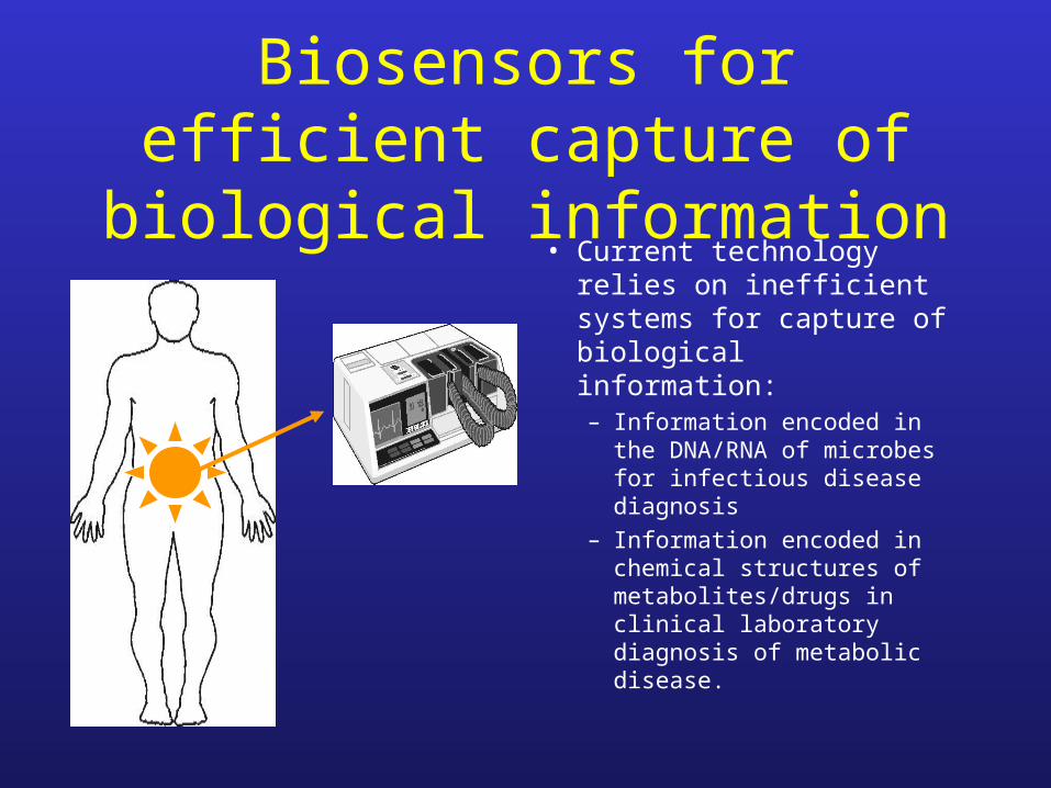
Biosensors for efficient capture of biological information
• Current technology relies on inefficient systems for capture of biological information:– Information encoded in the
DNA/RNA of microbes for infectious disease diagnosis
– Information encoded in chemical structures of metabolites/drugs in clinical laboratory diagnosis of metabolic disease.

DIG-ELISA PCR for diagnosis of meningococcal meningitis
substrate
product
SIGNAL
Newcombe, J., et al J.Clin.Microbiol. 34, 1637-1640.
Meningococcal DNA
enzyme
Patient sample
PCR amplification
DNA extraction

Nano-bioelectronic Devicesto efficiently capture biological signals
targetcapture
signaltransfer
signaltransduction
signalcapture

DNA chip biosensor (Cao et al, Science, 2002)DNA signal light signal
• Chip with bound capture probe – captures target DNA
• Target DNA (bound to chip) captures gold nanoparticles labelled with Ramen dye
• Laser bean Raman scattering signal
• Can detect 20 femtomoles – similar to PCR


Green Fluorescent protein to track cells and examine gene expression

Flow Cytometry
• SBLS has FACSCAN (Becton Dickenson) and FACSCalibur (BeckmanCoulter)
• Flow cytometry widely used to measure fluorescent intensity and proportion of target cells in cell population, particularly immune cells

Quantum Dots• fluoresce with a narrow and symmetric
emission spectrum that depends directly on the size of the crystal.
• can be fine-tuned to emit light at a variety of wavelengths simply by altering the size of the core
• constitute a set of multicoloured molecular beacons (up to 40,000 colours) for use in imaging
• Soluble quantum dots injected into frog embryos – only distributed to the offspring of the injected parent cell, and did not diffuse out of the cell used to track cell lineages.
• Can be tagged with antibody or DNA probes

Possible use of quantum dots?
• DNA fingerprinting with multiple allele-specific probes
• Widely used in forensic science, genetic typing and infectious disease diagnosis

2 Photon Microscopy• In confocal microscopy the
exciting laser must be very bright to allow an adequate signal-to-noise ratio.– photobleaching– Phototoxicity
• In 2 photon microscopy the signal is generated by two lower-energy (infrared) photons that are absorbed contemporaneously (within 1 femtosecond). – More focussed beam– Less toxicity

Biophotonics in SBLS• Dr George Kass (Toxicology)• Dr Nick Toms (Pharmacology)• Professor Ian Kitchen (Pharmacology)• Dr Lesley-Jane Reynolds (Microbiology)
1. Confocal Microscopy (Zeiss 510 META)Live cell (e.g. FRET) & fixed cell imaging (e.g. immunofluorescence)
2. Epifluorescence MicroscopySingle cell live dynamic fluorescence (e.g. intracellular Ca2+ imaging)
3. Flow CytometryCell population analysis of cellular fluorescence
4. Quantitative AutoradiographyRadioligand (e.g. [3H]drug) binding to tissue sections

Low Green Fluorescence High Red
Fluorescence
Excitation(488 nm) FRET
eYFPProtein
dsRedProtein
Fluorescence Resonance Energy Transfer (FRET)
Amino acid chain(Caspase-3 Target)
Healthy Neurones Dying Neurones
Excitation(488 nm)
FRETXNo Red
Fluorescence High Green Fluorescence
Death Enzyme“Caspase-3”

Green Fluorescence
Red Fluorescence
Excitation(488 nm)
FRETeYFP
ProteindsRedProtein
Healthy Neurones

A Dying Neurone
Excitation(488 nm)
FRETXNo Red
Fluorescence High Green Fluorescence
Death Enzyme“Caspase-3”

Confocal Analysis of Mitochondria

Immunofluorescence labelling of a Myelinating Brain Cell (Oligodendrocyte)
• Confocal Z-stack panoramic movie
(click on image to run)

Quantitative Autoradiography
fmol/mg
NSB
Binding of a [3H]drug to glutamate receptors in rat brain.