biorecognition by amino acid-based affinity chromatography...
TRANSCRIPT

UNIVERSIDADE DA BEIRA INTERIOR Ciências
Biorecognition by amino acid-based affinity chromatography for RNA purification
Ana Rita Nunes Martins
Tese para obtenção do Grau de Doutor em
Bioquímica (3º ciclo de estudos)
Orientadora: Prof. Doutora Fani Pereira de Sousa Co-orientador: Prof. Doutor João António de Sampaio Rodrigues Queiroz
Covilhã, Outubro de 2013

ii

iii
“Nothing is impossible; the word itself says
‘I'm possible’!”
Audrey Hepburn

iv

v
Dedication
Esta Tese de Doutoramento é dedicada de forma muito sentida ao meu Pai e à minha Mãe. Ao
meu Pai pelo exemplo que sempre foi para mim, pelo seu dinamismo, força de vontade e boa
disposição que sempre o acompanharam ao longo da vida, e por me ter ensinado que as
palavras “Não” e “Impossível” não existem. À minha Mãe, por me ter incutido a sua
generosidade, a sua paciência e a sua forma subtil e desembaraçada de enfrentar a vida.
Todos esses ensinamentos e atitudes definiram a minha personalidade e fizeram-me encarar a
vida com uma motivação adicional e com uma perspectiva lutadora, o que me ajudou a
alcançar sempre todos os objectivos a que me propus, como é o exemplo desta Tese de
Doutoramento.

vi

vii
This work was financed by the Portuguese Foundation for Science and Technology
(SFRH/BD/64100/2009) under the program QREN - POPH - Type 4.1 – Advanced Training, co-
founded by the European Social Fund and by national funds from the MCTES and also under a
research and development project (PTDC/EBB-BIO/114320/2009) and strategic programs
(PEst-C/SAU/UI0709/2011) co-founded by the operational program factors of competitiveness
– COMPETE and (EXPL/BBB-BIO/1056/2012) co-founded by FEDER funds (FCOMP-01-0124-
FEDER) of COMPETE.

viii

ix
Acknowledgments
I wish to thank all those who directly or indirectly contributed for the completion of this
doctoral thesis, including those who I will not specifically acknowledge here, but that were
also very important in realizing this goal.
First of all, I would like to express my gratitude to my supervisors. To my supervisor Prof. Fani
Sousa for her professionalism, dedication and all knowledge provided, for believing in me, for
all encouragement, support, and especially for her friendship, my very thank you! To my co-
supervisor Prof. João Queiroz, my sincerely appreciation for his geniality and inspiration, for
his friendship and availability, as well as the valuable scientific knowledge that he conveyed
me, all of which had a remarkable influence in the course of my academic career and in the
achievement of this thesis.
I would also like to thanks to Professor Cláudio Maia for his valuable contribution to this work
through his expertise in genetics and molecular biology. I am really thankful for the
availability and interest he demonstrated for my work through the great scientific discussions
and the knowledge transmitted.
To the Portuguese Foundation for Science and Technology (FCT) I want to acknowledge the
financial support through my doctoral fellowship (SFRH/BD/64100/2009) that allowed the
execution of all my research in Health Science Research Centre at University of Beira Interior
(CICS-UBI).
In addition, thanks to CICS-UBI for the host and to the technicians, Ana Sofia Duarte e
Margarida Carrilho, for their precious contribution in the organization of my laboratory tasks
and for the willingness that they responded to all my requests.
An appreciative note to Professor Luís Passarinha, for his friendship and support, and for
allowing me to extend my scientific knowledge and experience by participating in his
scientific projects and contributing with my slight experience for the formation of other
colleagues.
Moreover, an enormous thanks to my laboratory colleagues for the good working environment,
helpfulness and all the excellent moments of scientific discussions but also of fun. I wish
them very success in their research projects and their future lives. It was a pleasure working
with all of them. However, I could not let to name those who, in addition to co-workers, have
become great friends. Indistinctly, my deeply thankful to Ana Martinho, Ângela Sousa,
Catherine Nunes, Diogo Maio, Eduarda Coutinho, Fátima Santos, Filomena Silva, Marta Silva
and Patrícia Pereira.

x
To you, Miguel, I am deeply grateful for all the affection, caring, unconditional support and
patience throughout these months. You entered my life in the most difficult, demanding and
delicate phase of my existence, by coincidence or fate (I do not know!!), but you really
demonstrated to be a true companion for life.
Por último, gostaria de agradecer de uma forma muito especial e sentida à minha família pelo
amor, confiança, estímulo e apoio incondicional que sempre me deram. Aos meus Pais fica a
sentida dedicação desta Tese de Doutoramento. To my sister, I express my thanks for all her
caring and for being present at every stage of my life. The complicity that we still have, even
at distance, continues to keep us together and encouraged me to move forward, knowing that
she will always be there for me. E para o meu cunhado preferido, deixo o meu Obrigado pelos
momentos divertidos que em muito me ajudaram a manter o vigor mental.

xi

xii

xiii
Resumo
A sequenciação completa do genoma humano ofereceu novos horizontes relativamente à
prevenção, diagnóstico e tratamento de doenças humanas. Para além disso, com o avanço da
engenharia genética, têm surgido novas tecnologias terapêuticas, entre as quais se destacam
a terapia génica utilizando ácidos nucleicos. Apesar destas estratégias génicas se terem
iniciado com o DNA (ácido desoxirribonucleico), estudos recentes têm avaliado o potencial
interesse terapêutico do RNA (ácido ribonucleico).
O RNA foi recentemente reconhecido como uma molécula fundamental nos processos
celulares, com implicações fundamentais na evolução dos organismos, na hereditariedade e
na regulação de vários genes, o que destacou o seu vasto potencial terapêutico e conduziu ao
aparecimento de várias terapias baseadas em moléculas de RNA. Os resultados promissores
dessas novas abordagens terapêuticas têm vindo a reforçar a investigação relacionada com as
moléculas de RNA, a avaliar pelo número, cada vez mais elevado, de estudos estruturais,
biofísicos e biomédicos presentes na literatura. Além disso, a indústria biotecnológica e
farmacêutica começam a visar as moléculas de RNA como uma nova classe de produtos
bioterapêuticos.
Um requisito fundamental em todos esses estudos é a obtenção de grandes quantidades de
RNA isolado e puro e de integridade assegurada. Como por exemplo, em biologia molecular a
purificação de RNA é o primeiro passo chave para avaliar a expressão de um gene, uma vez
que a realização bem como a reprodutibilidade e relevância biológica dessa experiência está
dependente da quantidade e qualidade das preparações de RNA. Por outro lado, as terapias
promissoras e revolucionárias baseadas em RNA, como a vacinação ou utilização de
biofármacos recombinantes envolvem formulações de RNA que devem satisfazer critérios de
qualidade rigorosos recomendados por agências reguladoras internacionais. No entanto, o
processo de purificação das moléculas de RNA pode ser um passo bastante limitante para o
sucesso da sua aplicação terapêutica.
O RNA tem uma série de características químicas únicas que se reflectem na sua conformação
estrutural. Apesar do RNA ser uma molécula de cadeia simples na sua base, ele tem uma
elevada propensão para formar estruturas secundárias e terciárias bastante complexas. São
estas compactações próprias das moléculas de RNA que definem as suas importantes funções
biológicas. A elevada reactividade química é outra característica própria do RNA e com
grande relevância biológica a nível regulatório, por proporcionar mais instabilidade à
molécula aumentando a sua susceptibilidade à degradação. Num contexto laboratorial, estas
características moleculares do RNA são enormes desafios para a sua extracção e purificação,

xiv
pois a sua actividade biológica e integridade podem ser facilmente comprometidas durante os
procedimentos devido à presença ubíqua de enzimas que o degradam.
Várias técnicas têm sido desenvolvidas para superar os desafios inerentes ao isolamento e
purificação de moléculas de RNA, tais como a extracção com fenol e clorofórmio ou as
extracções em fase sólida empregando colunas ou esferas de sílica (SPE), bem como algumas
técnicas de purificação com cromatografia líquida de alta eficiência (HPLC) de fase reversa e
troca iónica. No entanto, estas ainda apresentam várias limitações nomeadamente em
relação ao elevado tempo despendido e à necessidade do uso de solventes tóxicos e condições
desnaturantes durante os procedimentos. Por todos estes motivos, torna-se evidente o
crescente interesse na avaliação e melhoria das metodologias actualmente utilizadas para o
isolamento e purificação de RNA de modo a satisfazer os requisitos necessários à sua
aplicação.
A cromatografia é um dos métodos mais diversos e potentes em biotecnologia, tanto a nível
analítico como preparativo devido à sua simplicidade, robustez, versatilidade e alta
reprodutibilidade. Por sua vez, a cromatografia de afinidade é reconhecida como uma técnica
poderosa apresentando grande aplicabilidade na purificação de muitas biomoléculas,
incluindo o DNA plasmídico (pDNA) e proteínas, porque explora o reconhecimento
biomolecular, ou seja, a capacidade de uma macromolécula biologicamente activa formar
complexos específicos e reversíveis com ligandos de afinidade.
Posto isto, o trabalho desenvolvido no âmbito desta tese incide na crescente necessidade de
desenvolver novas estratégias de isolamento e purificação para moléculas de RNA de modo a
ultrapassar as limitações ainda existentes nas metodologias actuais, contribuindo para a
evolução e sucesso da investigação e aplicações terapêuticas do RNA. Para isso, a
potencialidade da cromatografia de afinidade foi considerada nesta temática e foi explorada
a aplicação de aminoácidos como ligandos de afinidade. Este trabalho foi baseado em vários
estudos de reconhecimento molecular e atómico que descrevem a existência de diferentes
interacções entre proteínas e ácidos nucleicos nos sistemas biológicos, especialmente com
aminoácidos básicos como a histidina e a arginina, e também na hipótese de existirem
interacções preferenciais entre os aminoácidos e as bases nucleotídicas. Além disso, estudos
recentes de processos de purificação mostraram grande aplicabilidade destes aminoácidos em
isolar e purificar moléculas de pDNA biologicamente activas para aplicação em terapia génica
e vacinas.
Assim sendo, novas metodologias preparativas e analíticas foram alcançadas ao longo deste
trabalho, ou seja, foi possível obter preparações de RNA a partir de diferentes fontes
biológicas e de reacções sintéticas com elevados rendimentos e grau de pureza e de
integridade preservada, bem como também foi possível o desenvolvimento de um método
analítico para a sua quantificação e monitorização.

xv
A potencial aplicabilidade da cromatografia de afinidade, usando histidina como ligando de
afinidade, na purificação de moléculas de RNA foi demonstrada pela primeira vez com a
purificação do RNA 6S, um RNA regulatório não codificante presente nos procariotas,
nomeadamente em Escherichia coli (E. coli), que tem uma função reguladora importante no
processo de transcrição deste organismo. Nas estratégias de purificação com histidina foram
usados gradientes de sulfato de amónio devido à presença do anel de imidazol na cadeia
lateral aromática do aminoácido, o que permitiria explorar interacções maioritariamente
hidrofóbicas entre o RNA e a matriz, quer por interacção com o anel ou por pontes de
hidrogénio. Assim, foi utilizado um gradiente decrescente em concentração de sulfato de
amónio em três etapas que revelou um reconhecimento bioespecífico com RNA 6S, permitindo
a sua purificação de uma mistura complexa de outras moléculas de RNA de baixo peso
molecular (sRNA).
Uma segunda nova estratégia desenvolvida com a matriz de histidina permitiu obter
simultaneamente o isolamento e purificação das classes de sRNA e RNA ribossómico (rRNA) a
partir de lisados celulares de E. coli. Neste estudo, ambas as classes foram separadas das
impurezas do hospedeiro (DNA genómico e proteínas) com elevado rendimento e grau de
pureza quando comparadas com amostras preparadas com métodos de isolamento
convencionais baseados na extracção com fenol e clorofórmio. No entanto, a metodologia
baseada na cromatografia com histidina demonstrou a vantagem de evitar o uso de produtos
químicos tóxicos durante o processo.
A versatilidade da matriz de histidina na purificação de RNA, tanto de moléculas específicas
(RNA 6S) como das classes (sRNA e rRNA) sugeriu que o mecanismo de interacção envolveria
não só interacções hidrofóbicas, mas também um bioreconhecimento das bases do RNA pela
histidina. Apesar das preparações de RNA obtidas com estes métodos necessitarem de
caracterização funcional adicional para provar a sua aplicabilidade, o uso da cromatografia de
afinidade com histidina representa um grande avanço no isolamento de moléculas de RNA,
uma vez que as técnicas tradicionais não têm a capacidade de fraccionar RNA ao nível de um
tipo de molécula ou de isolar sRNA e rRNA num só processo. Todavia, a necessidade de aplicar
elevadas concentrações de sal nestas metodologias pode ser visto como uma desvantagem,
principalmente no que diz respeito à aplicação biotecnológica, pois acarreta maiores custos
de processo e tem maior impacto ambiental.
O uso de arginina como aminoácido imobilizado na cromatografia de afinidade foi utilizado na
perspectiva de melhorar as técnicas anteriores uma vez que este aminoácido se apresenta
sobretudo carregado positivamente, pelo que poderiam ser exploradas interacções
electrostáticas para a purificação de RNA utilizando condições de eluição moderadas. Além
disso, as interacções de arginina têm sido reconhecidas como as mais prevalentes nos
complexos RNA-proteína, aumentando a potencialidade de purificação por um maior
bioreconhecimento entre as moléculas de RNA e a matriz de arginina. Deste modo, um

xvi
gradiente com o aumento gradual da concentração de cloreto de sódio permitiu o isolamento
e purificação do RNA total de extractos celulares eucarióticas. O suporte de arginina
demonstrou uma aplicabilidade excepcional para interagir com todas as classes funcionais do
RNA apesar da sua diversidade estrutural e as suas diferentes conformações em condições
nativas. Essas interacções mais fortes e selectivas parecem advir da cadeia lateral do
aminoácido de arginina que apresenta uma multiplicidade para interacções, podendo
promover um contacto múltiplo com o RNA, através da sua estrutura açúcar-fosfato ou com as
suas bases, atendendo ao seu estado conformacional. Embora as interacções electrostáticas
entre os grupos fosfato do RNA e os ligandos de arginina possam desempenhar uma função
importante na retenção do RNA na coluna, as interacções com as bases também estão
envolvidas e modulam de alguma forma a interacção favorecida e a especificidade
encontradas na cromatografia com arginina. Assim, neste processo de purificação verificou-se
um elevado rendimento de recuperação do RNA total e pelas análises de controlo de
qualidade efectuadas mostrou-se que este apresentava uma elevada integridade bem como
uma boa pureza, demonstrada pela difícil detecção de proteínas nas amostras purificadas e
pela redução de DNA genómico para concentrações residuais. A eficiência desta técnica de
purificação e a aplicabilidade do RNA por ela obtido foi demonstrada num procedimento
habitualmente usado em biologia molecular para a análise de expressão génica. As amostras
de RNA total foram usadas com sucesso como moldes na reacção em cadeia da polimerase em
tempo real (qPCR) para a avaliação da expressão de dois genes controlo geralmente
empregues nestes procedimentos.
Reunindo os resultados anteriormente descritos, o isolamento e purificação de RNA de
amostras biológicas complexas usando as técnicas de cromatografia de afinidade com
aminoácidos imobilizados demostrou vários benefícios em relação a métodos de isolamento
actualmente empregues, como a extracção de fenol e clorofórmio ou de SPE, uma vez que
simplificam a integração do processo e minimizam o manuseamento das amostras, tornando a
cromatografia baseada em aminoácidos útil no desenvolvimento de metodologias em
condições não desnaturantes, livres de RNases e solventes orgânicos, particularmente
importantes em vários estudos estruturais e funcionais bem como de aplicabilidade
terapêutica.
Com os resultados positivos destas metodologias de afinidade na purificação de RNA foi
também desenvolvido e validado (de acordo com a legislação internacional e europeia para
métodos bioanalíticos) um método analítico para a quantificação e monotorização de RNA.
Com a crescente importância, a nível terapêutico, do desenvolvimento de novas ferramentas
analíticas para avaliar o RNA, uma vez que ainda existem várias lacunas nas técnicas actuais
de quantificação de RNA, tais como a falta de selectividade, tornou-se imperativo avaliar a
potencialidade dos suportes de afinidade neste campo. A versatilidade da metodologia foi
demonstrada pela sua aplicabilidade na quantificação de RNA de diferentes fontes

xvii
eucarióticas e também em amostras complexas de RNA quimicamente sintetizado, o que
demonstrou a usa utilidade em múltiplas áreas de investigação.
Desenvolveu-se ainda mais um estudo no isolamento e purificação de RNA com base nas
matrizes de aminoácidos, em particular com matriz de arginina, de modo a aproximar a
aplicabilidade desta técnica à prática terapêutica. Neste caso, o novo objectivo foi explorar o
seu potencial na purificação de moléculas de RNA mensageiro (mRNA) não a partir de células,
mas a partir de reacções sintéticas de transcrição in vitro, com o interesse de aplicar as
moléculas em terapias de vacinação com mRNA no cancro do colo do útero. As moléculas de
mRNA que codificavam para as proteínas E6 e E7 do vírus do papiloma humano (VPH) 16 foram
purificadas com sucesso de uma série de impurezas próprias das reacções de transcrição in
vitro, com são o pDNA molde, as enzimas, nucleótidos, sais e tampões, e a sua caracterização
em termos de rendimento, pureza e integridade foi avaliada. Neste trabalho, a cromatografia
de arginina voltou a demonstrar a sua capacidade singular em melhorar os processos de
purificação, pelas vantagens de eliminar passos adicionais e melhorar a economia global do
processo de produção.
Em suma, esta tese permitiu o desenvolvimento de novas metodologias para a purificação e
quantificação de RNA revelando várias características interessantes das moléculas, incluindo o
seu comportamento cromatográfico e interacções naturais que podem ocorrer com os
suportes de aminoácidos. Por conseguinte, estes métodos mostraram uma potencial
aplicabilidade polivalente, contribuindo para o progresso das investigações fundamentais e
terapêuticas baseadas no RNA, suportando a utilização da cromatografia de afinidade baseada
em aminoácidos no desenvolvimento futuro de novos processos de preparação de RNA.
Palavras-chave
Arginina, bioreconhecimento, cromatografia de afinidade, histidina, purificação, RNA.

xviii

xix
Abstract
Following the decoding of the human genome, a new era was opened for developing new gene
therapy strategies employing nucleic acids. Recently, RNA was renowned a central molecule
in cellular processes with implications in many diseases as well as in understanding of
evolution, becoming one of the most exciting research areas of molecular biology. From basic
to applied research, many procedures employ pure and intact RNA molecules. On one hand,
RNA purification is a first critical step of a number of molecular biology procedures and its
quality is crucial to ensure reproducibility and biological relevance of an experiment. On the
other hand, the promising and revolutionary RNA-based therapies of RNA vaccination, gene
therapy or recombinant biopharmaceuticals involves RNA formulations which should fulfill
rigorous quality criteria recommended by international regulatory agencies. However, the
isolation and purification of RNA are critical steps because of the easy degradability of RNA,
which can impair chemical stability and biological functionality essential for analysis. Many
techniques have been development to overcome the challenges of purifying RNA molecules;
nonetheless they still have several limitations in regard to time demanding and the
requirement of toxic solvents and denaturing conditions. Therefore, there is a growing
demand for the evaluation and improvement of the methodologies currently used for RNA
isolation and purification.
Chromatography is undoubtedly one of the most diverse and potent methods in
biotechnology, both at analytical, preparative and industrial level due to its simplicity,
robustness, versatility and high reproducibility. Affinity chromatography is recognized as a
powerful technique with great applicability in the purification of many biomolecules,
including plasmid DNA and proteins because it exploits the principle of biomolecular
recognition. The work that we have been developing considers new chromatographic
strategies for RNA purification, using amino acids as affinity ligands. These studies are based
on the fact that many different interactions exist between proteins and nucleic acids in
biological systems, involving in particular basic amino acids such as histidine or arginine. New
methodologies were accomplished that allowed obtaining RNA preparations from different
sources with high recovery yields, purity and integrity. A new analytical method for RNA
quantification was also developed in this work.
The applicability of histidine-based affinity chromatography in the purification of RNA
molecules was first demonstrated in the separation of 6S RNA, a regulatory non-coding RNA of
the prokaryotic Escherichia coli (E.coli). A specific recognition between the histidine support
and 6S RNA allowed its selective purification from a complex mixture of other small RNAs
(sRNA). In another strategy, the simultaneous isolation of sRNA and ribosomal RNA from E.coli
cell lysates, eliminating host DNA and proteins, was also attained by a histidine

xx
chromatographic-based method. Furthermore, arginine matrix was employed in RNA
purification from eukaryotic cells demonstrating an exceptional ability to interact with all
functional classes of RNA, despite their structural diversity and different folding states,
enabling their isolation from impurities of eukaryotic crude cell extracts. Moreover, an
analytical technique based on arginine affinity support for quantification and quality
assessment of total RNA from different eukaryotic cells and synthetic RNA samples was also
developed and validated, according to international and European legislation for bioanalytical
methods.
More efforts into RNA purification were developed with amino acid-based matrices, in
particular with arginine-agarose matrix, in order to approach this technique to therapeutic
application of RNA. The new goal was to exploit its applicability in purifying messenger RNA
(mRNA) molecules not from cells, but from synthetic crudes of in vitro transcription
reactions, pursuing mRNA vaccination for cervical cancer. In this work, arginine-based
chromatography also showed its singular capability to improve purification processes, showing
the advantages of eliminating additional steps and improving global economics of the
production process.
The development of these new methodologies revealed several interesting characteristics of
RNA molecules, including their chromatographic behavior and natural interactions that can
occur between amino acids-based supports and RNA molecules. Accordingly, these methods
demonstrated a potential multipurpose applicability by aiding in molecular biology RNA-based
analysis and RNA therapeutics, which support the interest in applying amino acid-based
affinity chromatography for the future development of new RNA isolation, purification and
quantification processes.
Keywords
Affinity chromatography, arginine, biorecognition, histidine, purification, RNA.

xxi

xxii

xxiii
Thesis Overview
This thesis is structured in four main chapters.
The first chapter includes an introduction that is divided in two sections. One section
explains the interest on RNA molecules to be potentially applied in novel therapies such as
gene therapy and RNA vaccination and the second section presents a brief explanation about
the relevance of purifying RNA molecules, as well as the main challenges and concerns
regarding RNA purification. In addition, a brief review of the literature related to the
techniques used in RNA purification regarding their advantages and disadvantages is discussed
together with the improvements that have been done lately. This second section is presented
as a publisher paper review form (Paper I).
The second chapter presents the main purpose and the specific goals that were established
for the development of this research work.
In the third chapter, the results obtained during this work are presented and discussed in the
form of original research papers organized as follows:
Paper II - A new affinity approach to isolate Escherichia coli 6S RNA with histidine-
chromatography
Paper III - Histidine affinity chromatography-based methodology for the simultaneous isolation
of Escherichia coli small and ribosomal RNA
Paper IV - A new strategy for RNA isolation from eukaryotic cells using arginine affinity
chromatography
Paper V - New approach in RNA quantification using arginine-affinity chromatography:
potential application in eukaryotic and chemically synthesized RNA
Paper VI - Arginine-affinity chromatography for mRNA vaccines purification
Finally, the fourth chapter outlines the concluding remarks about this work, regarding the
initial hypothesis of using affinity chromatography with immobilized amino acids in the
purification of RNA molecules. Furthermore, some future work are suggested to complement
the important findings achieve in this study.

xxiv

xxv
Index
List of Figures xxvii
List of Tables xxix
List of Scientific publications xxxi
List of Scientific communications xxxv
Chapter 1 1
1. Gene therapy and vaccination with RNA 3
1.1. Introduction 3
1.2. Overview of gene therapy 4
1.3. RNA-based therapies 11
1.3.1. Catalytic RNAs: Ribozymes 11
1.3.2. Antisense oligoribonucleotides 13
1.3.3. Agents of RNA interference 13
1.3.4. Aptamers 16
1.3.5. mRNA vaccination 17
References 19
2. Current issues in RNA preparation: approaching affinity chromatography
into RNA purification challenges (Paper I) 23
Chapter 2 67
Aims of the thesis 69
Chapter 3 73
Paper II. A new affinity approach to isolate Escherichia coli 6S RNA with
histidine-chromatography 75
Paper III. Histidine affinity chromatography-based methodology for the
simultaneous isolation of Escherichia coli small and ribosomal RNA 85
Paper IV. A new strategy for RNA isolation from eukaryotic cells using arginine
affinity chromatography 97
Paper V. New approach in RNA quantification using arginine-affinity
chromatography: potential application in eukaryotic and chemically
synthesized RNA 111
Paper VI. Arginine-affinity chromatography for mRNA vaccines purification 125
Chapter 4 151
Concluding remarks 153
Future perspectives 157

xxvi

xxvii
List of Figures
Figure 1 – Compiled data on gene therapy clinical trials provided by regulatory
agencies 5
Figure 2 – The in vivo and ex vivo paths used in gene therapy 6
Figure 3 – Vector systems commonly used in gene therapy clinical trials 7
Figure 4 – Ribozymes activities 12
Figure 5 – miRNAs, shRNAs and siRNAs pathways for RNAi in mammalian cells 15
Figure 6 - Treatment of cancer patients with tumour RNA-transfected DCs 18

xxviii

xxix
List of Tables
Table 1 - Viral vectors use for gene therapy 7
Table 2 – Non-viral delivery systems used in gene therapy approaches 9

xxx

xxxi
List of Scientific publications
Papers related to this Thesis
Current issues in RNA preparation: approaching affinity chromatography into RNA
purification challenges
R. Martins, J. A. Queiroz, F. Sousa
Submitted for publication (2013)
Arginine-affinity chromatography for mRNA vaccines purification
R. Martins, C. J. Maia, J. A. Queiroz, F. Sousa
To submit (2013)
New approach in RNA quantification using arginine-affinity chromatography:
potential application in eukaryotic and chemically synthesized RNA
R. Martins, J. A. Queiroz, F. Sousa
Analytical and Bioanalytical Chemistry. 2013. 405(27): 8849-8858
A new strategy for RNA isolation from eukaryotic cells using arginine affinity
chromatography
R. Martins, C. J. Maia, J. A. Queiroz, F. Sousa
Journal of Separation Science. 2012. 35(22): 3217-3226
Histidine affinity chromatography-based methodology for the simultaneous
isolation of Escherichia coli small and ribosomal RNA
R. Martins, J. A. Queiroz, F. Sousa
Biomedical chromatography. 2012. 26(7): 781-788
A new affinity approach to isolate Escherichia coli 6S RNA with histidine-
chromatography
R. Martins, J. A. Queiroz, F. Sousa
Journal of Molecular Recognition. 2010. 23(6): 519-524

xxxii
Papers not related to this Thesis
Performance of hydrophobic interaction ligands for human membrane-bound
catechol-O-methyltransferase purification
F. M. Santos, A. Q. Pedro, R. F. Soares, R. Martins, M. J. Bonifácio, J. A. Queiroz, L.
A. Passarinha
Journal Separation Sciences. 2013. DOI: 10.1002/jssc.201300010
Screening of gellan gum as an ionic and hydrophobic chromatographic matrix for
biomolecule purification
L. A. Rocha, A. Gonçalves, F. Silva, R. Martins, A. Sousa, L. A. Passarinha
Submitted for publication (2013)
Matriz cromatográfica baseada no polímero polissacárido gelana
L. A. Rocha, F. M. Santos, F. Silva, R. Martins, A. Sousa, L. A. Passarinha
Portuguese Patent 106446. Sep 7, 2012
Characterization of polyplexes involving small RNA
P. Pereira, A. F. Jorge, R. Martins, A. A. Pais, F. Sousa, A. Figueiras
Journal Colloid Interface Science. 2012. 387 (1):84-94

xxxiii

xxxiv

xxxv
List of Scientific communications
Oral communications related to this Thesis
Affinity-based method for RNA purification pursuing mRNA vaccination
R. Martins, C. J. Maia, J. A. Queiroz, F. Sousa
19th ISSS - International Symposium on Separation Sciences: New achievements in
Chromatography 2013. Poreč, Croatia
A new strategy for RNA isolation from eukaryotic cells using arginine affinity
chromatography.
R. Martins, C. J. Maia, J. A. Queiroz, F. Sousa
32th ISPPP - International Symposium on the Separation of Proteins, Peptides and
Polynucleotides 2012. Istanbul, Turkey
A new effective method for purifying Escherichia coli small and ribosomal RNA
using histidine affinity chromatography
R. Martins, J. A. Queiroz, F. Sousa
Affinity 2011. Tavira, Portugal
RNA purification by histidine affinity chromatography
R. Martins, J. A. Queiroz, F. Sousa
IV National Meeting of Biochemistry Students 2009. Covilhã, Portugal
Oral communications not related to this Thesis
Evaluation of human membrane-bound catechol-O-methyltransferase purification
by hydrophobic interaction chromatography
F. M. Santos, A. Q. Pedro, R. F. Soares, R. Martins, M. J. Bonifacio, J. A. Queiroz, L.
A. Passarinha
32th ISPPP - International Symposium on the Separation of Proteins, Peptides and
Polynucleotides 2012. Istanbul, Turkey

xxxvi
Poster communications related to this Thesis
Arginine based-chromatography as a new approach for prokaryotic and eukaryotic
RNA isolation
R. Martins, J. A. Queiroz, F. Sousa
7º National Meeting of Chromatography 2012. Porto, Portugal
Isolation of RNA from cell lysates using histidine affinity chromatography.
International
R. Martins, J. A. Queiroz, F. Sousa
30th ISPPP - Symposium on the Separation of Proteins, Peptides and Polynucleotides
2010. Bologna, Italy
Ribosomal RNA isolation from cell lysates by histidine affinity chromatography
R. Martins, J. A. Queiroz, F. Sousa
12th SBCN - International meeting and workshop of the Society for
Biochromatography and Nanoseparations 2010. Lyon, France
A new affinity approach to isolate RNA species with histidine-chromatography
R. Martins, J. A. Queiroz, F. Sousa
Affinity 2009. Reykjavik, Iceland
Poster communications not related to this Thesis
Structural and functional characterization of polyplexes for small RNA delivery
P. Pereira, A. F. Jorge, R. Martins, A. A. Pais, F. Sousa, A. Figueiras
8th World Meeting on Pharmaceutics, Biopharmaceutics and Pharmaceutical
Technology 2012. Istanbul, Turkey
Development of a Gellan Gum stationary phase as a new support for biomolecules
purification
L. A. Rocha, A. Gonçalves, F. Silva, R. Martins, A. Sousa, L. A. Passarinha
32th ISPPP - International Symposium on the Separation of Proteins, Peptides and
Polynucleotides 2012. Istanbul, Turkey

xxxvii

xxxviii

Chapter 1
1

2

1. Gene therapy and vaccination with RNA
1.1. Introduction
In recent decades, the advances in molecular biology combined with the culmination of the
decoding of the human genome have provided a genetic understanding of cellular processes
and disease pathogenesis. Numerous genes involved in disease have been identified as targets
for therapeutic approaches and a new era was opened for developing new gene therapy
strategies employing nucleic acids. Although the gene-based therapeutic strategies started to
be developed using DNA, a large number of studies are in progress in which the therapeutic
potential of RNA is evaluated.
The concept of using RNA molecules as therapeutic agents rose from a variety of newly
scientific discoveries that revealed RNA to be a versatile biological macromolecule
fundamental in mobilizing and interpreting genetic information and essential in cellular
processes of all living systems. The research in this area has been fuelled with the
exploitation of the inherent properties of RNAs with the purpose to interfere with or repair
dysfunctional nucleic acids or proteins and to stimulate the production of therapeutic gene
products in a variety of pathological situations. The simplicity of RNA engineering combined
with its versatility in structure and function has highlighted the use of RNA-based strategies
for therapy. The first generation of RNA therapeutics is now being evaluated in clinical trials,
raising significant interest in this emerging area of medical research.
3

1.2. Overview of gene therapy
Gene therapy is a highly promising therapeutic method to treat various diseases, including
both genetic and acquired disorders. In principle, gene therapy uses genetic information for
the treatment or prevention of a disease. It involves the transfer of a therapeutic genetic
material into specific cells of an individual in order to repair a defective gene or to introduce
a new gene whose function is to cure or to favourably modify the clinical course of a
condition (Verma and Weitzman, 2005).
Virtually all cells in the human body contain genes, making them potential targets for gene
therapy. Nevertheless, cells can be divided into two major categories: somatic cells (most
cells of the body) or cells of the germ-line (eggs or sperm). In theory it is possible to
transform either somatic cells or germ cells. However, somatic cells are non-reproductive and
therefore somatic cell therapy is viewed as a more conservative, safer approach, because it
affects only the targeted cells in the patient and is not passed on to future generations. All
gene therapy to date on humans has been directed at somatic cells, whereas germ-line
engineering in humans remains controversial and prohibited in for instance the European
Union (Wivel, 2002).
Historically, treating diseases by genetic engineering is an original conceptualization of the
investigators Avery, MacLeod and McCarthy that pioneered the notion and demonstrated that
genes could be transferred within nucleic acids in the early 1940s (Wolff and Lederberg,
1994). Soon after, viruses were envisioned as potential tools for human’s benefit, in
theoretical studies in somatic-cell genetics or possibly in gene therapy. Viral genomes were
then used for the development of the first relatively efficient methods for gene transfer into
mammalian cells in culture. In the late 1970s, the discovery of recombinant DNA technology
provided the tools to efficiently develop gene therapy. In the decades that followed,
tremendous advances in this technology enabled the manipulation of viral genomes, isolation
of genes, identification of mutations involved in human diseases, characterization and
regulation of gene expression, and engineer various delivery systems. In the early 1990s, the
first human gene therapy clinical trial was finally approved for treating a form of immune
deficiency called adenosine deaminase deficiency. Within a short time period, gene therapy
has moved from the conceptual stage to technology development and laboratory research to
clinical translational trials, which is clearly demonstrated by the increased number of gene
therapy clinical trials approved since 1989 as well as the widened range of diseases for which
gene therapy trials have been approved and the various gene types that have been used over
recent years (figure 1).
4
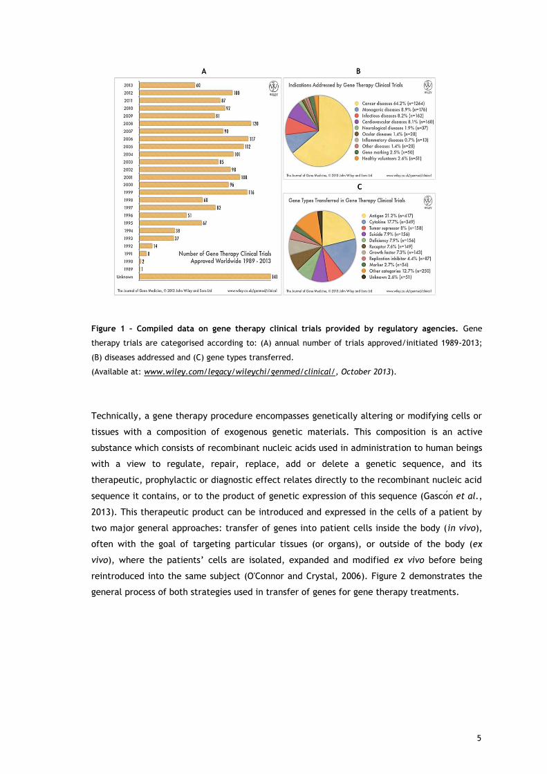
Figure 1 – Compiled data on gene therapy clinical trials provided by regulatory agencies. Gene
therapy trials are categorised according to: (A) annual number of trials approved/initiated 1989-2013;
(B) diseases addressed and (C) gene types transferred.
(Available at: www.wiley.com/legacy/wileychi/genmed/clinical/, October 2013).
Technically, a gene therapy procedure encompasses genetically altering or modifying cells or
tissues with a composition of exogenous genetic materials. This composition is an active
substance which consists of recombinant nucleic acids used in administration to human beings
with a view to regulate, repair, replace, add or delete a genetic sequence, and its
therapeutic, prophylactic or diagnostic effect relates directly to the recombinant nucleic acid
sequence it contains, or to the product of genetic expression of this sequence ( as o n et al.,
2013). This therapeutic product can be introduced and expressed in the cells of a patient by
two major general approaches: transfer of genes into patient cells inside the body (in vivo),
often with the goal of targeting particular tissues (or organs), or outside of the body (ex
vivo), where the patients’ ells are isolated, expanded and modified ex vivo before being
reintroduced into the same subject (O'Connor and Crystal, 2006). Figure 2 demonstrates the
general process of both strategies used in transfer of genes for gene therapy treatments.
5

Figure 2 – The in vivo and ex vivo paths used in gene therapy. The left side of the illustration shows
the in vivo approach to gene therapy where the therapeutic nucleic acid is directly delivers to the
patient. The gene can be delivered into the target cell by several delivery systems, commonly called as
gene therapy vectors. On the right side is the representation of the ex vivo approach that involves the
transfer of genes into cultured cells which were previously isolated from the patient or other donors.
These genetically altered cells are proliferated or cultured in vitro and subsequently implanted into the
patient. Gene transfer in vitro can be performed by the same delivery systems as those used in in vivo.
(Adapted from Kaji and Leiden (2001)).
In general, a therapeutic gene is delivered to the cell using a carrier, or vector, rather than
dire tly inserted into patient’s ells, due to the redu ed uptake into the ells of naked
therapeutic nucleic acids. A key factor in the success of gene therapy is the development of
delivery systems that are capable of efficient gene transfer in a variety of tissues, without
causing any associated pathogenic effects. A gene transfer system can be considered ideal if
the following aspects have been satisfied: specificity and efficiency of gene transfer;
magnitude and duration of expression; immunogenicity and manufacturing (Verma and
Weitzman, 2005). To make gene transfer more efficient, specific and safe, a variety of
different vectors and delivery techniques have been developed and studied, and applied in
gene therapy trials (Figure 3). Generally, these methods can be divided into two categories,
viral gene delivery and non-viral gene delivery, depending on the vectors involved.
6

Figure 3 – Vector systems commonly used in gene therapy clinical trials.
(Available at: www.wiley.com/legacy/wileychi/genmed/clinical/, October 2013).
Currently, the most common type of vectors are viruses that have been genetically altered to
carry normal human nucleic acids (Ginn et al., 2013). To date, five main classes of viral
vectors have been tested for clinical applications. These include adenoviruses, adeno-
associated viruses, retroviruses, lentiviruses and herpes simplex viruses (table 1). Viral
vectors are in fact the most effective because they offer higher transduction efficiency and
long-term gene expression, but their application can be limited by their immunogenicity,
oncogenicity and the small size of the nucleic acids they can transport (Walther and Stein,
2000).
Table 1 - Viral vectors use for gene therapy. (AAV, adeno-associated viruses; dsDNA, double-stranded
DNA; ssDNA/RNA, single-stranded DNA/RNA). (Adapted from Sheridan (2011)).
Adenovirus AAV Retrovirus/Lentivirus Herpesvirus
Family Adenoviridae Parvoviridae Retroviridae Herpesviridae
Genome dsDNA ssDNA ssRNA+ dsDNA
Infection/tropism
Dividing and
non-dividing
cells
Dividing and
non-dividing
cells
Dividing cells
Dividing and
non-dividing
cells
Host genome
interaction Non-integrating Non-integrating Integrating Non-integrating
Transgene
expression
Transient
Potential Long lasting Long lasting
Potential long
lasting
Packaging
capacity 7.5 kb 4.5 kb 8 kb >30 kb
7

Over the past decade, numerous non-viral methods for gene transfer have been proposed,
including physical methods and the employment of chemical vectors (table 2). These non-viral
vectors offer several advantages over viral vectors: simplicity of large scale production, low
immunogenicity, low toxicity and potential for more tissue specificity. The simplest non-viral
gene delivery system uses ‘naked’ nu lei a ids, su h as plasmid DNA, whi h an be delivered
into cells or tissues by physical methods such as electroporation, gene gun delivery,
sonoporation or hydrodynamic injection. Meanwhile, chemical vectors can be used to
encapsulate nucleic acids, exerting a protective effect. Currently in use, include inorganic
nanoparticles, as calcium phosphate, and synthetic or natural biodegradable particles such as
cationic lipids (forming lipoplexes upon mixing with nucleic acids) or cationic polymers
(forming polyplexes upon mixing with nucleic acids) ( as o n et al., 2013). However, despite
recent technological advances, the main limitation of non-viral systems is their low
transfection efficiency, although it has been improved by different strategies and the efforts
are still ongoing (Wang et al., 2013).
8
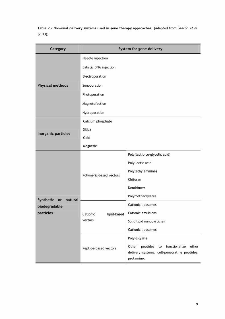
Table 2 – Non-viral delivery systems used in gene therapy approaches. (Adapted from as o n et al.
(2013)).
Category System for gene delivery
Physical methods
Needle injection
Balistic DNA injection
Electroporation
Sonoporation
Photoporation
Magnetofection
Hydroporation
Inorganic particles
Calcium phosphate
Silica
Gold
Magnetic
Synthetic or natural
biodegradable
particles
Polymeric-based vectors
Poly(lactic-co-glycolic acid)
Poly lactic acid
Poly(ethylenimine)
Chitosan
Dendrimers
Polymethacrylates
Cationic lipid-based
vectors
Cationic liposomes
Cationic emulsions
Solid lipid nanoparticles
Cationic liposomes
Peptide-based vectors
Poly-L-lysine
Other peptides to functionalize other
delivery systems: cell-penetrating peptides,
protamine.
9

Gene therapy is a relatively new paradigm in medicine with enormous therapeutic potential.
A major motivation for gene therapy has been the need to develop novel treatments for
diseases for which there is no effective conventional treatment. As previous stated, the
spectrum of gene therapy applications has now broadened considerably to every area of
molecular medicine offering new possibilities of mitigating, and even curing, numerous of
medical conditions ranging rare inherited monogenic disorders, metabolic diseases, infections
and even complex disorders such as cancer (Ginn et al., 2013). The traditional gene therapy
was focused on delivery of DNA encoding therapeutic proteins into cells. Depending on
purpose and delivery method, successful gene transfer could have several outcomes: to
modify defective host genes, to replace deficient host genes, to insert into the host genome
or to stay in the nucleus without integration into the host genome. The subsequent transgene
expression could restore normal cellular processes or induce new cellular responses. With the
research in recent years, current gene therapy is only restricted to deliver DNA. The delivery
of any other therapeutic nucleic acid materials, such as RNA who interferes with gene
expression by regulating post-transcription or translation, could also be included into the
concept of gene therapy (Strachan and Read, 1999 , Wang et al., 2013).
10

1.3. RNA-based therapies
RNA was once considered to be just an intermediate molecule in taking genetic information
from the genome to the ribosome, but that view has been changing rapidly by the recent
knowledge coming from basic RNA research. Recently, RNA was renowned as a central
molecule in cellular processes and gene regulation. This centrality of RNA reflects its
unprecedented biochemical properties. The linear sequence of RNA makes it a simple source
of genetic information. The property of RNA to form secondary structure, shielding some
sequences while exposing others for recognition, facilitates its interactions with other
molecules. In a more complex way, RNA can assume tertiary structures that present surfaces
for interactions and contain internal environments that create binding sites for metal ions
that can promote catalytic reactions (Sharp, 2009).
It is now clear that RNA is a versatile molecule that play key roles in many important
biological processes like splicing, editing, protein export and others, and it can also act
catalytically, like enzymes (Soll et al., 2001) which underscore the therapeutic potential of
RNA as a new gene therapy tool. RNA-based therapeutics make use of the mechanism of
activity of the various RNA molecules, which include catalytically active RNA molecules
(ribozymes), inhibitors of mRNA translation (antisense oligoribonucleotides), the agents of
RNA interference in gene expression (small RNAs), and RNAs that bind proteins and other
molecular ligands (aptamers) (Burnett and Rossi, 2012).
1.3.1. Catalytic RNAs: Ribozymes
The discovery that RNA can act as an enzyme changed the paradigm of the central dogma of
molecular biology (Lehman, 2010), and led to the development of a new class of therapeutics
based on RNAs enzymes. Ribozymes, or RNA enzymes, are RNA molecules that can mediate
their own cleavage or splicing or act as enzymes to promote reactions on substrate RNA
molecules (Khan, 2006) (figure 4).
Two types of RNA enzymes - hammerhead and hairpin ribozymes – have been the main focus
of efforts towards assessing the potential therapeutic utility of ribozymes. These ribozymes
were found to mediate inhibition of gene expression through the binding of messenger RNA
(mRNA) by complementarity and inducing its site-specific cleavage (figure 4a) with the
particularity that ribozymes recycle themselves ready to repeat this process multiple times.
Early preclinical works showed that such RNA enzymes could repeatedly cleave practically any
pathogenic transcript, which supported their use as therapeutic tools for manipulation of
gene expression (Hauswirth et al., 2001).
Several phase I and II clinical trials have been initiated using trans-cleaving ribozymes in a
small number of patients with infectious diseases or cancer. In these studies the ribozymes
11

have been delivered to the patients either by delivery systems or by direct injection of a
syntheti ribozyme that ontains hemi al modifi ations that greatly in rease the ribozyme’s
stability in biological fluids. However, the gene therapy-based trials have focused upon
developing ribozyme-based treatments for individuals infected with the human
immunodeficiency virus (HIV) (Burnett and Rossi, 2012).
Recently, the ribozymes with self-splicing ability were exploited to trans-splice RNA targets in
order to repair mutant mRNA molecules giving rise to genetic diseases (figure 4b). RNA repair
is an alternative way to control gene expression at the mRNA level, repairing mutant mRNAs
rather than destroying them. This method uses the splicing and editing processes that create
mRNAs from DNA to replace mutant regions in mRNA. The initial studies focused on RNA
repair used a trans-splicing version of a group I ribozyme to repair mutant lacZ transcripts in
bacteria and mammalian cells. These studies showed that the ribozyme was able to repair the
mutant RNA by recognizing the target transcript by base pairing with it, cleaving off mutant
sequences and linking a wild-type sequence onto the cleaved product (Phylactou et al.,
1998).
Figure 4 – Ribozymes activities. (a) Trans-cleaving ribozymes can bind pathogenic mRNAs through base-
pairing interactions and perform sequence-specific cleavage by phosphodiester isomerization, releasing
the reaction products. Ribozymes can repeat this process with multiple turnover. (b) Trans-splicing
ribozymes can repair a mutant RNA by recognizing the target transcript upstream of a mutation site
(Xm) by base pairing with it. The mutant sequence is cleaving off and an exon with a wild-type
sequence (Xwt) is ligated onto the cleavage product to generate a corrected transcript. (mRNA,
messenger RNA). (Adapted from Sullenger and Gilboa (2002)).
12

1.3.2. Antisense oligoribonucleotides
Antisense RNAs are small, diffusible, highly structured RNAs that act via sequence
complementarity on target RNAs called sense RNAs. In eukaryotes, some processes like
splicing or editing make use also of complementary small RNAs (Brantl, 2002). Antisense RNA
has long been thought of as a promising technique for disease therapy. The concept
underlying antisense technology is relatively straightforward: the use of a sequence,
complementary by virtue of Watson-Crick base pair hybridization, to a specific mRNA can
inhibit its expression and then induce a blockade in the transfer of genetic information from
DNA to protein. Conceptual simplicity, the possibility of rational design and relatively
inexpensive cost has led to the widespread use of these short fragments of RNA as therapeutic
agents (Dias and Stein, 2002). The success on clinical trials of this RNA-based therapy has led
to the first antisense RNA approved by the Food and Drug Administration (FDA) for
commercialization in 1998. Vitravene (Isis Pharmaceuticals/Novartis) is the brand name for
Fomivirsen, an antiviral drug used in the treatment of cytomegalovirus retinitis (CMV) in
immune compromised patients, including those with acquired immunodeficiency syndrome.
Fomivirsen is a synthetic 21-nucleotide sequence with phosphorothioate linkages (which are
resistant to degradation by nucleases), which blocks translation of viral mRNA by binding to
the complementary sequence of the mRNA transcribed from the coding segment of a key CMV
gene (Grillone and Lanz, 2001). Since then, the significant advances in RNA chemistry led to
the creation of second-generation antisense technology that was expected to overcome many
of the limitations of the original approaches and expand the use of this technology to other
diseases. In fact, this has been successful and in the beginning of this year FDA approved an
injectable antisense RNA, called KYNAMROTM (mipomersen sodium) from Genzyme Company,
which is an oligoribonucleotide inhibitor of apolipoprotein B-100 synthesis indicated as an
adjunct to lipid-lowering medications and diet to reduce low density lipoprotein-cholesterol,
apolipoprotein B, total cholesterol, and non-high density lipoprotein-cholesterol in patients
with homozygous familial hypercholesterolemia (Genzyme, 2013).
1.3.3. Agents of RNA interference
The Nobel-prize winning in 1998 for the discovery of the mechanism of activity of a class of
small RNA (sRNA) molecules produced by eukaryotes aroused a novel therapeutic approach to
treat human diseases, called RNA interference (RNAi) technology. The cellular process of RNAi
uses sRNAs to mediate resistance to both endogenous parasitic and exogenous pathogenic
nucleic acids, and regulates the expression of protein-coding genes through post-
transcriptional gene silencing (PTGS). PTGS is regulated by two distinct mechanisms:
sequence-specific cleavage of perfectly complementary mRNAs and translational repression
and degradation of mRNAs with imperfect complementarity. Small interfering RNA (siRNA)
and microRNA (miRNA) are the biological agents of RNAi, a family of regulatory non-coding
13

RNAs of 19-28 nucleotides in length (figure 5) (Burnett and Rossi, 2012, Kim, 2005). siRNAs are
short double-stranded RNA (dsRNA) with 2 nu leotides overhangs at the 3′- ends. In the
cytoplasm, siRNAs are loaded into a protein complex called the RNA-induced silencing
complex (RISC). The loaded RISC complex then scans all intracellular mRNA for a target mRNA
with a complementary sequence to the loaded siRNA. If a target mRNA is found by the loaded
RISC, the target mRNA is cleaved and degraded, successfully inhibiting the translation of the
target gene. siRNAs can be generated in several ways. In some cases, long dsRNA is
introduced in the cell, either by a virus or endogenous RNA expression (microRNA). The
enzyme Dicer cleaves the long duplex RNAs into siRNAs (Guo et al., 2010).
On the other hand, miRNAs are originated from endogenous genome DNA sequence and are
first transcribed in the nucleus as parts of long primary miRNA transcripts (referred to pri-
miRNA) with 5′- aps and 3′- polyA tails. miRNAs with hairpin structures are then processed
into pre-miRNAs by the ribonuclease Drosha. The pre-miRNAs are subsequently transported
out of the nucleus to cytoplasm by the dsRNA-binding protein Expotin-5, and processed to
mature miRNAs by the endoribonuclease Dicer. Similar to siRNA-mediated silencing pathway,
miRNA is then loaded into RISC. However, its mode of action is dependent on the extent of
sequence complementarity between the miRNA and the target mRNA. When a miRNA matches
the sequence of the mRNA completely, the miRNA/RISC complex mediates the cleavage of
the mRNA using the same mechanism as siRNA. For miRNAs that only partially match the
mRNA's sequence, the miRNA/RISC complex induces translational inhibition and subsequent
mRNA degradation. miRNAs silencing is arguably more complex than siRNA silencing, owing to
the fact that miRNAs only require partial sequence complementarity to silence genes (Guo et
al., 2010, Lin et al., 2006). The miRNA mechanism is not fully understood and some diseases
are suggested to be linked to aberrant miRNA expression and function (Soifer et al., 2007).
For therapeutic purposes, siRNA has been the focused molecule in the RNAi pathway. In fact,
siRNAs are even being used to interfere with aberrant miRNAs. PTGS may be efficiently
induced by delivering exogenously synthetic siRNA molecules to cells. Chemically synthesized
siRNA duplex or siRNA molecules prepared in vitro from dsRNAs by incubating with
recombinant Dicer protein, are commonly used in research for gene silencing. In this last
strategy, Dicer-processed siRNA products simply bypass the Dicer cleavage step. Another way
to introduce siRNA into cells is to express short hairpin RNAs (shRNA) genes in plasmid vectors
(Guo et al., 2010). shRNA is a sequence of RNA which is created in the cell from a DNA
construct encoding a sequence of single stranded RNA and its complement, separated by a
stuffer fragment, allowing the RNA molecule to fold back on itself, creating a dsRNA molecule
with a hairpin loop. In the cell, it is transcribed under the control of RNA Polymerase-II or
Polymerase-III promoters and folds into a structure resembling a siRNA duplex. shRNAs are
then processed by Dicer into siRNAs (figure 5) (Guo et al., 2010, Tokatlian and Segura, 2010,
Xiang et al., 2006).
14

Figure 5 – miRNAs, shRNAs, and siRNAs pathways for RNAi in mammalian cells. (A) miRNA genes are
transcribed by RNA polymerase II to generate the primary transcripts (pri-miRNAs) and processed into
stem-loop precursor miRNAs of 70 nucleotides by the Drosha-DGCR8 complex, which are then exported
by Exportin 5 to the cytoplasm. Upon export, Dicer participates in the second step of processing to
produce miRNA duplexes of 22 nucleotides. The imperfectly complementary miRNA duplexes are
associated to the AGO protein and are loaded into RISC, where the passenger strand is removed and the
guide strand remains to target mRNA for silencing. The final products (RISC complex) act as guide
molecules in translational control or cleavage of certain mRNAs. (B) Like miRNAs, engineered shRNAs are
transcribed from DNA and undergo similar processing. However, the perfect Watson-Crick base-pairing
between the guide strand and the target mRNA triggers AGO2-mediated cleavage of the mRNA target.
shRNA expression cassette can be delivered by viral vectors such as retroviral vector, lentiviral vector,
and adenoviral vector or it can be expressed in the nucleus from expression plasmids. (C) In contrast to
miRNA and shRNAs, siRNAs are processed in the cytoplasm. But, all steps of siRNA and shRNA are the
same after processing by Dicer/TRBP. siRNA can be artificially introduced into the cytoplasm in RNAi-
based therapeutics either as a chemically synthesized siRNA duplex or Dicer-processed siRNA molecules.
Viral and non-viral delivery systems are used for siRNA transfer into cells. (miRNAs, micro RNA; shRNAs,
small hairpin RNA; siRNAs, small interfering RNA; RNAi, RNA interference; RISC, RNA-induced silencing
complex; mRNA, messenger RNA). (Adapted from Burnett and Rossi (2012)).
15

The RNA interference technology is one of the most exciting biotechnology advances in the
last decade. It has revolutionized biology research, including drug target discovery and
revitalized interest in the clinical development of nucleic acid-based gene inhibition
approaches. Theoretically, RNAi can silence the expression of mRNA for any gene, including
growth factors, viral genes, or oncogenes and genes that were once considered
therapeutically unreached by small molecule inhibitors. The great effectiveness and the
simplicity of the design of a therapeutic siRNA, which only requires knowledge of the target
gene's sequence, have contributed for the increased development and success of this RNA-
based therapeutic approach. Moreover, the fact that siRNA-mediated RNAi mechanism takes
place in the cytoplasm is a potential advantage over other gene regulation mechanisms that
require penetrating the nucleus (Tokatlian and Segura, 2010). Therefore, several
pharmaceutical companies are focusing on the development of RNAi-based therapeutics for
the treatment of a wide range of diseases (Burnett and Rossi, 2012, Melnikova, 2007).
1.3.4. Aptamers
Many small RNAs can fold into three-dimensional structures that allow them to bind target
proteins with high affinity and specificity. This additional feature of RNAs makes them
tempting to consider as a therapeutic agent since they can bind to proteins and inhibit them
in an analogous manner to protein antagonists. This idea of ‘de oy’ RNAs has been shown in
preclinical work to slow down HIV replication, where trans-activation response region (TAR)
and rev response element (RRE), two decoy RNAs, could be used to competitively inhibit viral
protein function and replication. This suggested that other small-structured RNA molecules
might be able to bind pathogenic target proteins and inhibit their activity (Kohn et al., 1999).
Meanwhile, synthetic short oligoribonucleotides sequences that bind to a specific target
molecule with high affinity, called aptamers, emerged as potential molecules for both basic
research and clinical purposes as macromolecular drugs. Aptamers are highly specific,
relatively small in size, and non-immunogenic. Aptamers are essentially a chemical equivalent
of antibodies. However, aptamers can be chemically synthesized to produce large quantities
of these compounds for in vivo experimentation and clinical trials. Aptamers are usually
created by iterative in vitro selection methods that isolate high-affinity RNA ligands from
large pools of randomized RNA sequences (vast RNA shape libraries) that could bind to
proteins and small molecules. The selection process was named SELEX (systematic evolution
of ligands by exponential enrichment) (Ellington and Szostak, 1990), but natural aptamers
also exist as part of a nucleic acid-based genetic regulatory element named riboswitch
(Tucker and Breaker, 2005). Since the discovery of aptamers in the early 1990s, great efforts
have been made to make them clinically relevant for diseases like cancer, HIV, and macular
degeneration. For therapeutic purposes, aptamers can be used to bind and inhibit harmful
molecules or serve as targeting ligands for nanomedical constructs. RNA aptamers, simplify
the need for chemical conjugation or mixing with other moieties. Aptamers have been used as
16

ligands for specific delivery of siRNA to prostate cancer cells and lymphocytes (Ni et al.,
2011). The first aptamer based therapeutic was FDA approved in 2004 for the treatment of
age-related macular degeneration and several other aptamers are currently being evaluated
in clinical trials (Burnett and Rossi, 2012).
1.3.5. mRNA vaccination
Coding mRNA is emerging as a particularly attractive option in the development of new
approaches for the treatment of cancer or infection diseases focusing on immunotherapies
(Kreiter et al., 2011). The use of genetic vaccination, in which a genetically engineered
nucleic acid encoding an antigen is administered to an organism in order to stimulate an
immunological response against the antigen (Tang et al., 1992), is not a new therapeutic
strategy. Vaccines based on nucleic acids are already known to stimulate all effectors of the
adaptive immune response: B lymphocytes, cytotoxic T cells, and T helper cells have been
exploited for the creation of prophylactic vaccines for infectious diseases and for cancer
immunotherapy. However, vaccination approaches involving nucleic acids have focused
mostly on DNA-based strategies due to the instability and rapid degradation of RNA in the
human body, since RNA is prone to hydrolysis by ubiquitous ribonucleases. Nevertheless,
researchers have been overcoming these limitations by developing strategies for stabilization
and delivery of mRNA (Pascolo, 2008).
mRNA is a large family of RNA molecules that plays a fundamental and integral role in every
living cell. mRNAs are carriers of genetic information from DNA to the ribosome, where they
specify the amino acid sequence of the protein products of gene expression. Following
transcription of mRNA by RNA polymerase, the mRNA is translated into a polymer of amino
acids: a protein. Therefore, the concept of mRNA vaccination is to carry the information of an
antigenic protein to be translated in the cell cytoplasm and therefore generate an
immunological response. As an alternative to DNA-based vaccines, mRNA-based vaccines
present additional safety features including no persistence, no integration in the genome and
no autoimmune response. Moreover, mRNA which are generated by in vitro transcription, are
easy to produce in large amounts and with very high purity. This feature facilitates the good
manufacturing practices process and guaranties reproducibility (Pascolo, 2006).
Vaccination with mRNA can be achieved by several delivery methods, including direct
injection of naked mRNA, injection of mRNA encapsulated in liposomes, gene gun delivery of
mRNA loaded on gold beads, or in vitro transfection of the mRNA in cells followed by re-
injection of the cells into the patients (Pascolo, 2008). This last strategy is generating a lot of
interest in cancer immunotherapy. The approach uses tumour mRNA isolated from the patient
by biopsy, amplified and ombined with the immune ells from the patient’s bone marrow
called dendritic cells (DCs). These cells internalize the mRNA and present the proteins
encoded by the mRNA as an antigen on the cell surface, which ensures the stimulation of the
17
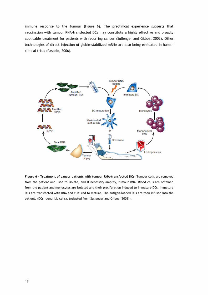
immune response to the tumour (figure 6). The preclinical experience suggests that
vaccination with tumour RNA-transfected DCs may constitute a highly effective and broadly
applicable treatment for patients with recurring cancer (Sullenger and Gilboa, 2002). Other
technologies of direct injection of globin-stabilized mRNA are also being evaluated in human
clinical trials (Pascolo, 2006).
Figure 6 - Treatment of cancer patients with tumour RNA-transfected DCs. Tumour cells are removed
from the patient and used to isolate, and if necessary amplify, tumour RNA. Blood cells are obtained
from the patient and monocytes are isolated and their proliferation induced to immature DCs. Immature
DCs are transfected with RNA and cultured to mature. The antigen-loaded DCs are then infused into the
patient. (DCs, dendritic cells). (Adapted from Sullenger and Gilboa (2002)).
18

References
Brantl S. Antisense-RNA regulation and RNA interference. Biochim Biophys Acta. 2002;
1575:15-25.
Burnett JC, Rossi JJ. RNA-based therapeutics: current progress and future prospects. Chem
Biol 2012; 19:60-71.
Dias N, Stein CA. Antisense oligonucleotides: basic concepts and mechanisms. Mol Cancer
Ther. 2002; 1:347-55.
Ellington AD, Szostak JW. In vitro selection of RNA molecules that bind specific ligands.
Nature. 1990; 346:818-22.
as o n AR, del ozo-Rodri guez A, olini s MA. Non-Viral Delivery Systems in Gene Therapy. In:
Martin F, editor. Gene Therapy - Tools and Potential Applications: InTech; 2013.
Genzyme C. FDA News Release: FDA approves new orphan drug Kynamro to treat inherited
cholesterol disorder. Food and Drug Administration; 2013.
Ginn SL, Alexander IE, Edelstein ML, Abedi MR, Wixon J. Gene therapy clinical trials
worldwide to 2012 – an update. J Gene Med. 2013; 15:65-77.
Grillone LR, Lanz R. Fomivirsen. Drugs Today (Barc). 2001; 37:245-55.
Guo P, Coban O, Snead NM, Trebley J, Hoeprich S, Guo S, et al. Engineering RNA for targeted
siRNA delivery and medical application. Adv Drug Deliv Rev. 2010; 62:650-66.
Hauswirth WW, Shaw LC, Whalen PO, Fritz JJ, White DA, Lewin AS. Inhibition of gene
expression by ribozymes. Methods Mol Med. 2001; 47:105-24.
Kaji EH, Leiden JM. Gene and stem cell therapies. JAMA 2001; 285:545-50.
Khan AU. Ribozyme: A clinical tool. Clin Chim Acta. 2006; 367:20-7.
Kim VN. Small RNAs: classification, biogenesis, and function. Mol Cells. 2005; 19:1-15.
Kohn DB, Bauer G, Rice CR, Rothschild JC, Carbonaro DA, Valdez P, et al. A clinical trial of
retroviral-mediated transfer of a rev-responsive element decoy gene into CD34(+) cells from
the bone marrow of human immunodeficiency virus-1-infected children. Blood. 1999; 94:368-
71.
19

Kreiter S, Diken M, Selmi A, Tureci O, Sahin U. Tumor vaccination using messenger RNA:
prospects of a future therapy. Curr Opin Immunol. 2011; 23:399-406.
Lehman N. RNA in evolution. Wiley Interdiscip Rev RNA. 2010; 1:202-13.
Lin SL, Miller JD, Ying SY. Intronic microRNA (miRNA). J Biomed Biotechnol. 2006;
2006:26818.
Melnikova I. RNA-based therapies. Nat Rev Drug Discov. 2007; 6:863-4.
Ni X, Castanares M, Mukherjee A, Lupold SE. Nucleic acid aptamers: clinical applications and
promising new horizons. Curr Med Chem. 2011; 18:4206-14.
O'Connor TP, Crystal RG. Genetic medicines: treatment strategies for hereditary disorders.
Nat Rev Genet. 2006; 7:261-76.
Pascolo S. Vaccination with messenger RNA. Methods Mol Med. 2006; 127:23-40.
Pascolo S. Vaccination with Messenger RNA (mRNA). In: Bauer S, Hartmann G, editors. Toll-
Like Receptors (TLRs) and Innate Immunity: Springer Berlin Heidelberg; 2008. p. 221-35.
Phylactou LA, Kilpatrick MW, Wood MJ. Ribozymes as therapeutic tools for genetic disease.
Hum Mol Genet. 1998; 7:1649-53.
Sharp PA. The Centrality of RNA. Cell. 2009; 136:577-80.
Sheridan C. Gene therapy finds its niche. Nat Biotechnol. 2011; 29:121-8.
Soifer HS, Rossi JJ, Saetrom P. MicroRNAs in disease and potential therapeutic applications.
Mol Ther. 2007; 15:2070-9.
Soll D, Nishimura S, Moore P. RNA. First ed. Oxford: Pergamon; 2001.
Strachan T, Read A. Gene therapy and other molecular genetic-based therapeutic
approaches. Human Molecular Genetics 2nd edition ed. New York: Wiley-Liss; 1999
Sullenger BA, Gilboa E. Emerging clinical applications of RNA. Nature. 2002; 418:252-8.
Tang DC, DeVit M, Johnston SA. Genetic immunization is a simple method for eliciting an
immune response. Nature. 1992; 356:152-4.
Tokatlian T, Segura T. siRNA applications in nanomedicine. Wiley Interdiscip Rev Nanomed
Nanobiotechnol. 2010; 2:305-15.
20

Tucker BJ, Breaker RR. Riboswitches as versatile gene control elements. Curr Opin Struct
Biol. 2005; 15:342-8.
Verma IM, Weitzman MD. Gene therapy: twenty-first century medicine. Annu Rev Biochem.
2005; 74:711-38.
Walther W, Stein U. Viral vectors for gene transfer: a review of their use in the treatment of
human diseases. Drugs. 2000; 60:249-71.
Wang W, Li W, Ma N, Steinhoff G. Non-viral gene delivery methods. Curr Pharm Biotechnol.
2013; 14:46-60.
Wivel NA. Gene Therapy, Ethics, Somatic Cell Gene Therapy. Encyclopedia of Ethical, Legal
and Policy Issues in Biotechnology John Wiley & Sons, Inc.; 2002.
Wolff JA, Lederberg J. An early history of gene transfer and therapy. Hum Gene Ther. 1994;
5:469-80.
Xiang S, Fruehauf J, Li CJ. Short hairpin RNA-expressing bacteria elicit RNA interference in
mammals. Nat Biotechnol. 2006; 24:697-702.
21

22
2. Current issues in RNA preparation:
approaching affinity chromatography into RNA
purification challenges (Paper I)
R. Martins, J. A. Queiroz, F. Sousa
(Submitted for publication)
23

24

Current issues in RNA preparation: approaching
affinity chromatography into RNA purification
challenges
R. Martins, J. A. Queiroz, F. Sousa
CICS-UBI – Health Sciences Research Centre, University of Beira Interior, Av.
Infante D. Henrique, 6200-506 Covilhã, Portugal.
25

26

Abstract
Research on RNA has led to many important biological discoveries and improvement of
therapeutic technologies. From basic to applied research, many procedures employ pure and
intact RNA molecules; however their isolation and purification are critical steps because of
the easy degradability of RNA, which can impair chemical stability and biological
functionality. The current techniques to isolate and purify RNA molecules still have several
limitations and the requirement for new methods able to improve RNA quality to meet
regulatory demands is growing. In fact, as basic research improves the understanding of
biological roles of RNAs, biopharmaceutical industry starts to focus on it as a biotherapeutic
tool.
Chromatographic bioseparation is the principal unit operation used for the purification of
biological compounds and its application in biopharmaceutical manufacturing is well
established. Thus, a number of chromatographic approaches have already been successfully
developed for RNA purification. In particular and in view of the unequalled specificity,
affinity chromatography has been recently applied, showing significant results and
improvements in RNA purification processes.
Therefore, this paper discusses the importance and the progress of RNA isolation and
purification procedures, considering the RNA applicability both in research and clinical field.
Accordingly, recent investigations using affinity approaches based on the biorecognition
between amino acids and RNA is focused, highlighting their potential contribution to
overcome the challenges of RNA purification.
Keywords
Affinity chromatography, amino acids, isolation methods, purification, RNA.
27

1. Introduction
Until recently, RNA was overlooked compared to DNA or proteins, consigned to a simple
intermediate role in the flow of information from genes to functioning molecules in living
cells. RNA is now known to play many more functional roles and to be responsible for a
multitude of essential biological processes (Sharp, 2009). In the last 20 years RNA was the
subject of four Nobel prizes winning discoveries - 1989 for catalytic RNA, 1993 for splicing,
2006 for RNA interference (RNAi), and 2009 for ribosomal structure (Lehman, 2010) and new
roles for RNA in biology continue to emerge at a glance. All of these discoveries have revealed
so far that RNA is truly a remarkable and multi-talented cellular component with fundamental
implication on biotic evolution and heredity. Furthermore, the widespread involvement of
RNA in the regulation of numerous genes has highlighted its vast therapeutic potential
(Burnett and Rossi, 2012). These and similar breakthroughs have led to the emergence of
numerous types of RNA-based therapeutics either using RNA as a therapeutic agent or a
therapeutic target. Table 1 shows potential therapeutic approaches for RNA, specifying the
involved RNA or RNA-based molecules and their activity mechanism of activity.
The successful results of these novel therapeutic approaches are reinforcing the focus on RNA
investigation and are rendering RNA molecules into new targets for pharmaceutical and
biotechnological industries (Melnikova, 2007). Due to the increasing number of structural,
biophysical and biomedical studies that require large quantities of homogeneous good-quality
RNA, a widespread need to improve production scale and RNA isolation and purification
schemes has been recognized.
As RNA emerges into the new class of biotherapeutic products, pharmaceutical-grade RNA
produced under the current good manufacturing practice (cGMP) is crucial. Thus, it will be
essential that the bioproduct fulfil the requirements of regulatory authorities such as Food
and Drug Administration (FDA), European Medicine Agency (EMA) and World Health
Organization (WHO). However, non-consensus still exists for regulation of mRNA vaccination
and RNA oligonucleotides-based therapies. In the European Union, mRNA-based therapies are
based on the regulation for advanced therapy medicinal products - EC No 1394/2007-
(Guideline on Human Cell-based Medicinal Products) which refers to directive 2001/83/EC
(Directive 2001/83/EC). Here, a ‘ ene therapy medi inal produ t is an a tive substan e
which contains or consists of a recombinant nucleic acid used in or administered to human
beings’. In the United tates of Ameri a, in ontrast, mRNA va ines are not ategorized as
gene therapy (Kreiter et al., 2011). On the other hand, RNA oligonucleotides products, such
as siRNAs or aptamers, are regulated as drugs under FDA’s Centre for Drug Evaluation and
Research and are not considered as an advanced therapy in the European Union (not classified
as gene therapy) (Srivatsa, 2012). Currently, none regulatory authority has formal guidelines
available for RNA oligonucleotide products or mRNA molecules. The guidelines established for
28

human cell-based medicinal products (Guideline on Human Cell-based Medicinal Products) and
DNA vaccines (Guidance for Industry: Consideration for plasmid DNA vaccines for infection
disease indications) are providing the guidance in the regulatory framework for RNA-based
therapies (Srivatsa, 2012, Weide et al., 2008). Nevertheless, those guidelines do not focus in
particular features of RNA, such that as shown in clinical trials, RNA-based therapies do not
confer the risks of integration into the genome. The scientific community, in the form of
volunteer members from the industry and regulatory agencies worldwide, are actively
engaged in addressing topics such as quality specifications and impurities in RNA bioproducts
(Srivatsa, 2011).
Consequently, economically feasible processes for RNA isolation and purification, as well as
the implementation of methodologies able to control RNA quality suitable for industrial
manufacturing, will be increasingly necessary, especially when the RNA products are finally
released to the market. However, the isolation and purification of RNA are critical steps
because of the easy degradability of RNA, consequence of the peculiar structural chemistry,
which can impair chemical stability and biological functionality, and can limit the success of
subsequent RNA investigations.
Therefore, this paper focuses the challenging task of isolating and purifying RNA molecules.
The current state of the isolation and purification methodologies used for RNA preparation
will be discussed regarding the growing demands in RNA applicability. Moreover, promising
affinity approaches based on chromatographic purification exploiting the biorecognition
between amino acids ligands and RNA molecules will be introduced. These new strategies
bring new insights into the way RNA can be accurately purified, contributing to the future
development of new and more robust bioseparation methods.
29

Table 1 - RNA molecules with therapeutic involvement. (Refs., References).
RNA type Cell function Therapeutic concept Refs.
Protein synthesis
Messenger RNA
(mRNA) Codes for protein Vaccination Kreiter et al. (2011)
Ribossomal RNA
(rRNA) Translation Antibiotic target
Tenson and Mankin
(2006)
Transfer RNA (tRNA) Translation
Apoptosis regulation
Understand many
human diseases
Belostotsky et al.
(2012), Raam and
Salvesen (2010)
Post-transcriptional modification or DNA replication
Small nuclear RNA
(snRNA)
Splicing and other
functions
Understand many
human diseases Matera et al. (2007)
Small nucleolar RNA
(snoRNA)
Nucleotide modification
of RNAs
Understand many
human diseases Kiss (2001)
Regulation
Antisense RNA
Transcriptional
attenuation
mRNA stabilization or
degradation
Translation block
Inhibitor of mRNA
translation
Brantl (2002), Dias and
Stein (2002)
MicroRNA (miRNA) mRNA cleavage and
Translation repression Gene silencing
Kusenda et al. (2006)
Lin et al. (2006)
Small interfering
RNA (siRNA)
mRNA cleavage and
Translation repression Gene silencing
Doench et al. (2003),
Ghildiyal and Zamore
(2009)
Ribozymes
RNA enzyme. Catalyze
RNA cleavage and
ligation reactions
mRNA reprogramming
and repair Phylactou et al. (1998)
Riboswitch
Regulate gene
expression by binding to
small metabolites
Regulate gene
expression
Antibacterial drug
target
Blount and Breaker
(2006), Tucker and
Breaker (2005),
Wittmann and Suess
(2012)
Aptamers
Oligoribonucleotide
part of a riboswitch
that binds to a specific
target molecule with
high affinity
Decoy mechanism that
inhibits various target
proteins
Mayer (2009), Ni et al.
(2011)
30

2. RNA chemistry
RNA has a number of unique chemical characteristics that have profound structural
consequences with remarkable implications in cell biology and are a real challenge in
research activities.
RNA is a polymer organized in a long chain of ribonucleotides monophosphates, but it
resembles DNA in many ways (Figure 1). RNA shares the same chemical units as DNA, except
that RNA has the nucleotide uracil (U) instead of thymine (T) and the sugar molecule is a
ribose rather than a desoxyribose. RNA has a hydroxyl group at the 2′ position of the ribose
while DNA lacks that group. This difference is manifested in the dissimilar shape that these
molecules adopt when they are base-paired into a double helix. RNA takes on the geometry
structure referred as an A - form helix while DNA takes on the B - form. But, unlike double-
strand DNA, RNA often comes as only a single-strand and is quite flexible (Murray et al.,
2000). RNA can twist itself into a variety of complex structures. Its propensity to form
secondary structures facilitates RNA interactions with other molecules by covering some
sequences and exposing others for recognition. Besides, RNA can assume tertiary structures
that present surfaces for interactions and contain internal environments that create binding
sites for metal ions, so that they can promote catalytic reactions (Soll et al., 2001).
The high chemical reactivity of RNA provides more instability to the molecule, increasing the
susceptibility to degradation. This instability is very important for cells, as they can change
their patterns of protein synthesis very quickly in response to biological needs (Arraiano et
al., 2010).
These properties consign RNA versatility in cellular processes, namely in gene regulation,
which open the possibility of exploring new therapeutic opportunities (Sullenger and Gilboa,
2002). On the other hand, the peculiar three-dimensional compaction and structural
instability of RNA are huge challenges in laboratory, as the biological activity and integrity
can be easily compromised during extraction and purification procedures. Thus, improved
methodologies for recovering RNA with high quality is a constant concern (Easton et al., 2010,
Fleige and Pfaffl, 2006, Jahn et al., 2008).
31

Figure 1- Structural characteristic of RNA. RNA differs from DNA in the nucleotide uracil, as a thymine exists
in DNA, as well as the constituent sugar molecule that is a ribose in RNA and a desoxyribose in DNA. In addition,
three-dimensional structures of RNA also greatly differ from that of DNA. While DNA molecules take a double helix
structure, which is formed through hydrogen bonding between the bases of two DNA strands, RNA molecules, which
are originally synthesized as single-strands, exhibit different conformational structures where single and double-
strands coexist in one RNA molecule because they often partially fold themselves to form double-strands. Compact
tertiary conformations can exhibit internal surfaces for interaction with other molecules.
32

3. RNA isolation and purification
The development of methodologies for the isolation and characterization of RNA has been
compared to the methods used for the analysis of its macromolecular counterparts, DNA and
proteins (Lehman, 2010). The extraction, isolation, and analysis of RNA are routinely more
difficult in comparison to that required for DNA. As raised before, RNA chemistry add
complexity to sample preparation because the ubiquitous presence of RNA-degrading enzymes
(RNases) both in biological samples and in laboratory environment easily degrade RNA,
compromising the integrity and biological activity of RNA molecules (Jahn et al., 2008).
Therefore, minimizing RNA degradation by protecting it against RNases requires that all
glassware, plastic ware, instrument tubing and reagents be RNase free. Additionally, among
the many challenges is the need for maximizing recovery yield, while removing unwanted
components, minimizing sample transfers, and avoiding non-specific binding to containers
(McGinnis et al., 2012). Therefore, the quality and quantity of RNA preparations are the main
concerns of isolation procedures, since the lack of integrity, the presence of contaminants or
the low RNA quantity may strongly compromise the success of several RNA based-procedures
in basic and clinical research (Bustin and Nolan, 2004). Additional challenges also emerge
with the advance of clinical trials using RNA intended to be administered in humans.
Presently, RNA molecules can be obtained by extraction from a biological matrix, such as
cells or tissues, or they can be produced by chemical or enzymatic (in vitro transcription)
synthesis. Chemical synthesis is normally used for the generation of short oligoribonucleotides
(<50 nucleotides) while in vitro transcription can produce longer RNAs. Synthetized RNAs are
being greatly employed in structural, biochemical and biophysical studies (Doudna, 1997,
Scott and Hennig, 2008) as well as in the development of new therapeutic approaches by RNA
interfering technology, RNA aptamers, ribozymes or mRNA vaccination (Lorenzi et al., 2010,
Wittmann and Suess, 2012, Zeng et al., 2002). In this case, the final RNA product needs to be
purified from impurities derived from the synthesis process. These impurities are, besides
enzymes, nucleotides, salts or buffer, aberrant oligonucleotides. Longer oligoribonucleotides
are more contaminated with aberrant species than short ones. The failure products are
prematurely halted as shorter oligonucleotides. Some are mismatch failure sequences where
there is missing nucleotides in the middle of sequence, rather than at the end. Other by-
products of synthesis may have greater molecular weight than the target oligoribonucleotide
(heterogeneous RNAs in length). This is a result of incomplete post-synthesis deprotection, or
due to the branching of an oligonucleotide backbone during the synthesis (Sherlin et al.,
2001).
On the other hand, biological RNAs are preferably used in basic research for the study of
cellular mechanisms, as they reflect intrinsic cell´s features (Dunin-Horkawicz et al., 2006),
in clinical investigations for pharmacokinetic and pharmacodynamics analysis (Yu et al., 2001)
33

and in some strategies of mRNA vaccination using bulk tumour mRNA (Kyte et al., 2006). In
RNA extraction from a biological matrix, the main principle is the disruption of cells and
subsequent elimination of host contaminants, such as genomic DNA (gDNA) and proteins, in
order to obtain intact and pure RNA molecules (Chomczynski and Sacchi, 2006).
Many methods have been developed in an attempt to circumvent the several challenges in
purifying RNA molecules and to achieve the goal of good-quality RNA (Farrell, 2012,
McGookin, 1988, Vomelova et al., 2009, Warren and Vella, 1995). Therefore, considering the
recent developments in RNA understanding and the growing demand on its purification, the
next discussion intends to briefly introduce the isolation and purification methods used in RNA
preparation. This description will draw particular attention to the main problems that can
compromise the success of RNA research or that can make the procedures not suitable to
obtain RNA to be further applied in clinical investigation, also focusing on the cost-
effectiveness for preparative-scale or large-scale industrial applications (Table 2 and 3).
3.1. RNA isolation methods
Traditionally two types of isolations are used in RNA preparations, (1) chemical extraction
using denaturing agents and organic solvent precipitation and (2) solid-phase extraction by
immobilizing RNA on a glass support. These methods generally include a chaotropic agent,
denaturant, or other chemical in the lysis step to inactivate RNases.
Chemical extraction that involves acid phenol/chloroform extraction is the most currently
employed method either performed with home-made solutions or commercial ready-to-use
reagents, because it leads to high recovery yields and purity of total RNA (RNAt)
(Chomczynski and Sacchi, 2006). However, these extractions are extremely toxic and
hazardous and highly operator dependent. Sometimes they can involve up to six or more steps
and three sample transfers leading to time consuming and laborious RNA preparations.
Although this method is almost always included in RNA purification schemes, the organic
solvents such as alcohols and phenol/chloroform may interfere in the majority of routine
molecular biology techniques because they can inhibit enzymes activity. Moreover, as those
compounds convey health risks, this procedure is not definitely tolerable for the welfare of
the researcher and should not be an integrant part of the process for a therapeutic
formulation. However, RNAs that are being employed in clinical trials are often extracted
using phenol/chloroform (Kyte et al., 2006). This should, certainly, be considered and advised
by regulatory authorities.
The other methods for RNA isolation are based in solid-phase purification using silica
membranes, as prefilled columns or as magnetic beads, in combination with
phenol/guanidine-based lysis of samples. This technology was developed to offer safer and
simpler operations, as they are amenable to automation using liquid handling robotics (Wen
34

et al., 2008). In fact, these techniques significantly reduce sample preparation time, but can
still involve multiple sample transfers and time-consuming evaporation steps. Low RNA yields
are often obtained due to the low binding capacity of the cartridges, which can be easily
overloaded (Chatterjee et al., 2010, Wen et al., 2008). Moreover, these processes lack
specificity as they are non-discriminatory for DNA or RNA, and in most cases the isolation of
pure RNA is achieved by a secondary enrichment, either through enzymatic removal of DNA
(DNase treatment) or by a second step using specific columns (Chatterjee et al., 2010). These
limitations can greatly diminish the success of several molecular biology investigations, in
particular gene expression analysis, because DNA contamination can interfere with real time-
PCR techniques compromising the interpretation of results (Laurell et al., 2012). In addition,
the treatment with enzymes, such as commercial DNases that are often animal-derived, is not
prudent in therapeutic applications (Guidance for Industry: Consideration for plasmid DNA
vaccines for infection disease indications).
Recent developments in high throughput technologies led to the design of large and complex
instrumentation using silica technology to simplify the isolation of nucleic acids. Automation
for nucleic acids extraction offers high-throughput sample processing, high quality and
effective purification since the human impact is constricted to a minimum (Chatterjee et al.,
2010). Therefore, less contaminants or inequalities concerning time, volumes of chemicals or
improper handling are avoided. Automation therefore increases the efficiency, accuracy and
velocity of RNA purification (Knepp et al., 2003). Nevertheless, cross contaminations can
occur by the sample collector arm. In these systems, the principles of purification are based
in solid-phase extraction, which presents some limitations, as previously mentioned. While
the high cost disadvantage of these systems could be overcome by the high-throughput of
automation, the lack of efficacy in the isolation method can commit the investment.
Actually, automated extraction systems may even be advantageous to some laboratories,
since consistent and reproducible purified samples in sufficient quality and purity are
achieved for subsequent application (Loens et al., 2007). However, in a biotechnological
process, these systems will not be cost-effective in attaining RNA-based therapeutic
formulations.
The use of preparative denaturing polyacrylamide gel electrophoresis (PAGE) has also been
extensively described to complement the schemes described above in the preparation of RNA,
especially synthetic RNA (Doudna, 1997, Hagen and Young, 1974, Price et al., 1995, Sherlin et
al., 2001). Preparative denaturing PAGE has been successful and continues to be the most
popular method to resolve RNA samples, but it presents several disadvantages. The protocol
is very lengthy as after denaturing PAGE, the desired RNA must be eluted from a gel matrix,
concentrated, equilibrated in the desired buffer, and refolded (Doudna, 1997). Moreover, this
introduces acrylamide contaminants, which are difficult to remove from RNA products and
can interfere with RNA analysis, namely by reducing the information content on nuclear
35

magnetic resonance (NMR). Although highly purified, the RNAs achieved by PAGE purification
show low yields since RNA can irreversibly aggregate due to many precipitation steps,
resulting in incomplete denaturation during electrophoresis and low accumulation in the
desired gel band (Kim et al., 2007, Lukavsky and Puglisi, 2004). Moreover, denaturation of
RNA molecules is another issue for RNA applicability. For many RNAs, significant time is spent
optimizing refolding conditions to minimize unproductive conformations. Some well-known
RNAs, such as Escherichia coli (E. coli) tRNAPhe, cannot be refolded into a conformational
homogeneous and active population (Kieft and Batey, 2004). Therefore, native purification
techniques should be favoured. From the therapeutic viewpoint, PAGE purification is rather
impracticable for many reasons. First, the prominent possibility of acrylamide contaminants
in RNA preparations is not adequate or safe for human therapy. Furthermore, structural
modifications due to denaturation can greatly compromise the effectiveness of RNA
therapeutic action.
Precipitation with high concentration of lithium chloride (LiCl) is another methodology often
used for synthetic RNA isolation (Baker et al., 1997, Nilsen, 2012, Pascolo, 2008). This
method can be used in the separation of small RNAs (<100 nucleotides long) (i.e., siRNA and
miRNAs) from other long RNAs or RNA-based impurities. Small RNAs are enriched in the
supernatant phase, while remaining impurities or all other long RNAs tend to precipitate
(Baker et al., 1997, Nilsen, 2012). However, the efficient fractionation typically requires
repetition of the LiCl precipitation procedure several times and the quality of resulting
preparation is in general dependent on the secondary or tertiary conformation of RNA
molecules (Romanovskaya et al., 2013). Other applications describe the use of LiCl
precipitation in in vitro transcripts reaction to eliminate most nucleotides, cap,
deoxynucleotides, oligodeoxynucleotides and proteins by selectively precipitating RNA of
more than approximately 100 bases, as mRNA transcripts (Pascolo, 2008). Nonetheless, most
of the time, this precipitation does not completely remove trace of contaminating plasmid
DNA (pDNA) or proteins and worse it introduces lithium metal into preparations. Thus, to
ensure long RNA purity, phenol/chloroform extraction often accompanies LiCl precipitation.
Although this method can be suitable for preparation of mRNA molecules for research, it is
not recommended to be used in clinical applications, as in vaccination approaches, since the
safeness of those preparations are not guaranteed to be applied in humans (Pascolo, 2008).
36
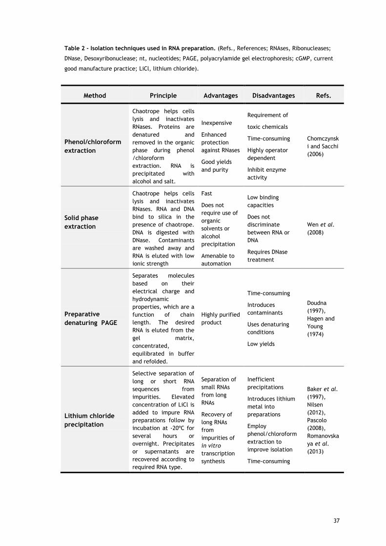
Table 2 - Isolation techniques used in RNA preparation. (Refs., References; RNAses, Ribonucleases;
DNase, Desoxyribonuclease; nt, nucleotides; PAGE, polyacrylamide gel electrophoresis; cGMP, current
good manufacture practice; LiCl, lithium chloride).
Method Principle Advantages Disadvantages Refs.
Phenol/chloroform
extraction
Chaotrope helps cells
lysis and inactivates
RNases. Proteins are
denatured and
removed in the organic
phase during phenol
/chloroform
extraction. RNA is
precipitated with
alcohol and salt.
Inexpensive
Enhanced
protection
against RNases
Good yields
and purity
Requirement of
toxic chemicals
Time-consuming
Highly operator
dependent
Inhibit enzyme
activity
Chomczynsk
i and Sacchi
(2006)
Solid phase
extraction
Chaotrope helps cells
lysis and inactivates
RNases. RNA and DNA
bind to silica in the
presence of chaotrope.
DNA is digested with
DNase. Contaminants
are washed away and
RNA is eluted with low
ionic strength
Fast
Does not
require use of
organic
solvents or
alcohol
precipitation
Amenable to
automation
Low binding
capacities
Does not
discriminate
between RNA or
DNA
Requires DNase
treatment
Wen et al.
(2008)
Preparative
denaturing PAGE
Separates molecules
based on their
electrical charge and
hydrodynamic
properties, which are a
function of chain
length. The desired
RNA is eluted from the
gel matrix,
concentrated,
equilibrated in buffer
and refolded.
Highly purified
product
Time-consuming
Introduces
contaminants
Uses denaturing
conditions
Low yields
Doudna
(1997),
Hagen and
Young
(1974)
Lithium chloride
precipitation
Selective separation of
long or short RNA
sequences from
impurities. Elevated
concentration of LiCl is
added to impure RNA
preparations follow by
incubation at -20ºC for
several hours or
overnight. Precipitates
or supernatants are
recovered according to
required RNA type.
Separation of
small RNAs
from long
RNAs
Recovery of
long RNAs
from
impurities of
in vitro
transcription
synthesis
Inefficient
precipitations
Introduces lithium
metal into
preparations
Employ
phenol/chloroform
extraction to
improve isolation
Time-consuming
Baker et al.
(1997),
Nilsen
(2012),
Pascolo
(2008),
Romanovska
ya et al.
(2013)
37

3.2. RNA purification methods
In recent years, several strategies employing chromatographic techniques have been explored
to overcome the increasing challenges in RNA purification. Advances in synthetic RNA
chemistry brought more efficient and affordable methods for large scale production of RNA.
Accordingly, synthetic techniques opened the possibility of inducing chemical modifications
on RNA molecules, reducing RNA instability and degradation (El-Sagheer and Brown, 2010),
benefiting structural, biochemical and biophysical studies, in techniques as crystallography,
spectroscopy, calorimetry or NMR, and many therapeutic investigations that employ
regulatory RNAs, as siRNA, miRNA, ribozymes and aptamers. However, not so many efforts
have been made for the improvement of biological RNAs purification.
Reversed-phase (RP) and ion-pairing (IP) RP (El-Sagheer and Brown, 2010, Maina et al., 2013,
Maroney et al., 2007), as well as anion-exchange chromatography (AEC) have been
extensively described for RNA purification (reviewed in (McGinnis et al., 2012)). In addition,
size exclusion chromatography (SEC) has also accompanied the development of schemes for
RNA preparation (McKenna et al., 2007).
RP chromatography relies on hydrophobicity as a mechanism of separation while IP
chromatography describes a RP-based improved technique in which an ion-pairing reagent,
such as a tri- or tetraalkylammonium salt, is capable of associating with the nonpolar
stationary phase through dynamic hydrophobic interactions, allowing enhanced resolution
(Cramer et al., 2011). In fact, a number of studies for the purification and analysis of RNA is
already available, demonstrating the versatility of IP RP HPLC in different applications with
synthetic oligoribonucleotides or biological RNA (reviewed in Dickman ( 2011b)). Although
these analytical studies include reliable information on RNA purification, very few of these
methods have been scaled up to a preparative context. The reason for this is that resins have
only moderate loading capacity and the cost of mobile-phase components and separation
matrices are considerably high. In addition, some organic solvents and ion-pairing agents are
difficult to remove from the purified oligonucleotides after chromatography and may even
require further chromatographic purification (Gilar, 2001). Furthermore, to avoid aggregation
of self-complementary or GC-rich oligonucleotides, RP HPLC columns are often placed in a
column oven at 60°C for temporarily destroying secondary structures to improve resolution
(Cramer et al., 2011), which can be disadvantageous to maintain the target RNA stability and
biological activity.
Therefore, recent investigations have explored the possibility to develop more rigorous and
complex IP RP HPLC methods, since great resolving properties of RP columns were evidenced
in analytical RNA separation. A recent approach describes the combination of SEC (porous
silica with a hydrophilic polymeric coating) and IP RP HPLC (polystyrene-divinyl benzene
beads) in a robust multidimensional platform to resolve, isolate and quantify non-coding RNA
38

species (ncRNA) in cells or tissues samples from several sources (Chionh et al., 2013). The
method takes advantage of the strengths of two types of HPLC techniques, thus increasing the
resolution of ncRNA across a wide size range. Many technical issues concerning ncRNA
preparations were improved, including the analysis speed, the application of non-denaturing
conditions, easy quantification, sensitivity, easy automation for fraction collection and use of
simple solvent mobile phases that do not contaminate the sample.
In addition, another recent strategy employed IP RP HPLC technique for the generation of a
therapeutic mRNA pursuing vaccination (Karikó et al., 2011), which purify long in vitro-
transcribed mRNA at a preparative scale. Despite the use of some organic solvents in the
procedure, the volatility of the ion-pairing agent was higher to be easily removed from
samples through evaporation and non-denaturing conditions were employed. The successful
applicability of the IP RP HPLC purification technique was characterized by the
immunogenicity of the samples, which demonstrated the effectiveness of the purified RNA at
an immunological level. This strategy can conveys important information about the
implementation of future purification strategies suitable for clinical applications (Kreiter et
al., 2011).
Regardless the success of these chromatographic techniques, the requirement of toxic
solvents continues to be their major weakness. The environmental impact and cost that the
use of organic compounds entails can be highly inconvenient for a biotechnological industry
besides, recovered RNA products might require further treatment to be adequate for
therapeutic purposes.
SEC has also accompanied the advances in RNA purification. This technique is able to
distinguish the components of a mixture of RNA on the basis of their molecular size, and is
the simplest form of chromatography for oligoribonucleotides purification. SEC has
contributed as a polishing step in removing salts from short oligoribonucleotides that have
been purified by HPLC. This extra form of desalting prevents cytotoxic effects from trace
synthesis by-products or trace solvents which may carry over from purification (Booy et al.,
2012). In addition, this technique has been explored as an alternative method to preparative
denaturing PAGE in purifying homogeneous-length RNA obtained by in vitro transcription (Kim
et al., 2007, Lukavsky and Puglisi, 2004, McKenna et al., 2007). Puglisi and workers developed
SEC-based purification schemes using fast performance liquid chromatography (FPLC) systems
that allowed the efficient elimination of unreacted nucleotides, enzymes, short abortive
transcripts, and the high molecular weight pDNA template from the desired RNA product. The
approaches were performed under non-denaturing conditions, which allows the exclusive
separation of monomeric RNA from oligomerized RNA, and avoided harsh precipitation steps
that may cause RNA aggregation and degradation (McKenna et al., 2007). However, SEC-based
methods still require several time-consuming preparatory steps, such as phenol/chloroform
39

extractions to remove proteins followed by desalting and sample concentration (Easton et al.,
2010).
Chromatographic studies on anion exchange matrices have been significantly explored
because of the polyanionic nature of RNA molecules. AEC mechanism relies primarily on
reversible electrostatic interactions that can be manipulated using an oppositely charged
buffer, typically simple salts and buffers are needed for separation. Furthermore, anion
exchange matrices are also described to have some inherent hydrophobicity which favours
oligonucleotides separation. AEC selectivity for oligonucleotides was found to be based on
molecular size and sequence and the success of separating double and single-stranded nucleic
acids relies on the distinct hydrophobic behaviours of the G-C, A-T and A-U base pairs (Gjerde
et al., 2009). Additionally, AEC demonstrates simplicity on the operations compared to RP,
since the separation is performed in aqueous conditions without the use of high cost eluents,
at low to moderate operating pressures and the secondary structures that interfere with
resolution are avoided using mild conditions to perform the purification. Hence,
oligonucleotides purification during mid- or large-scale manufacturing is typically performed
using AEC rather than RP chromatography (Shanagar, 2005).
Nonetheless, in some AEC strategies the necessity of denaturing conditions either by the use
of harsh alkaline conditions or elevated temperatures to accomplish full resolution of
oligoribonucleotides persists (Thayer et al., 2011a). As already mentioned, denaturation often
leads to misfolded or aggregated material or even leads to RNA degradation, which is
unsuitable for further usage. Therefore, the methods can reveal not cost-effective in the
preparation of RNA. Example of this is the preparation of double-strand RNAs (dsRNA), such as
siRNA or miRNA. The chromatographic purification is generally performed for the single-
strand intermediates rather than the final duplex, under denaturing conditions and therefore
further purification of the final siRNA duplex in native form is required and is determined by
SEC (Ge et al., 2003, Thayer et al., 2011a). Despite the successful application of this strategy
in the manufacture of virtually all therapeutic duplex oligoribonucleotides (Srivatsa, 2011),
the workload and process time to purify the separated single-strands, rather than the final
duplex is increased, which also increases process economics.
New developments in AEC purification strategies intended to address large scale duplex
preparation under native conditions are required. Good separations had already been
achieved in analytical non-denaturing AEC, and recently preparative methods were also
described (Noll et al., 2011). Therefore, the scale up production of dsRNA has been optimized
in two commercial AEC resins using the same quaternary amine as functional group, but
differing on matrix chemistry: one material consisted on a polystyrene/divinyl benzene beads
and the other on poly-(methyl-methacrylate). Both columns allowed the removal of most non-
hybridized strands as well as non-optimal duplexes from the hybridized duplex siRNA, with
higher yields than in parallel assays to purify single-strands intermediates before annealing.
40

Thus, subsequent duplex purification enabled the reduction of chromatographic steps, as the
final SEC is no longer required, and annealing was significantly simplified.
Furthermore, convective interaction media (CIM) monolithic columns immobilized with strong
anion exchange quaternary amines were also employed in the development of improved
dsRNA purification methods under non-denaturing conditions (Romanovskaya et al., 2013).
Differently from chemical synthesis, this work describes the purification of siRNAs from a
synthetic pool of functional siRNA produced by the enzymatic generation of long dsRNA
molecules followed by digestion with a recombinant Dicer enzyme in vitro. Multiple
contaminants, such as non-hybridized or unprocessed single-strand RNA, undigested or
abortive dsRNA molecules, nucleoside triphosphates, that are obtained in transcription
reaction were removed and siRNA was recovered at superior speed and scalability compared
to the conventional bead-based AEC columns and traditional stepwise LiCl precipitation.
Therefore, CIM monolithic columns were found to be suitable for industrial scale siRNA and
dsRNA purification, while retaining fast processing times and high resolution.
With the improvement of synthetic RNAs by chemical modification, other challenges in RNA
purification arose. Apart the regular impurities, many isomerizations, in luding 2′,5′-linkages,
phosphoramidate-linked RNA, phosphorothioate-linked, 2′-cyanoethoxymethyl-protected RNA
and oligonucleotides harboring phosphorothioate diastereoisomers, may also be formed during
chemical synthesis (Thayer et al., 2011b). Pellicular anion-exchangers (pAE) have been
reported in literature for its ability to analytically resolve those isomers. pAE phases were
designed to place the ion exchange sites only on the bead surface in order to overcome the
mass transfer limitation of porous beads. While they were capable of very high
chromatographic efficiency, they exhibit very low capacity, and they may harbor relatively
high nonspecific interactions (Stillian and Pohl, 1990). Recently, a surface-functionalized
monolith coated with pAE nanobeads (latexes) was developed, which intended to enhance
purifications of single and dsRNA, aptamers, and nucleic acids that may harbor isobaric
linkage isomers, such as phosphorothioate diastereoisomers and similar isobaric
oligonucleotide variants (Thayer et al., 2010). These new resins combine the selectivity and
mass transfer characteristics of pAE with the preparative capacity of monoliths in order to use
the successful analytical separations of pAE at a lab-scale. With this strategy it could be
possible to evaluate and characterize the oligonucleotides regarding its safety and
effectiveness as therapeutic and diagnostic products. However, further efforts are need to
scale up these methods so that the resultant RNA preparations can be readily used as
therapeutic products.
AEC has also been used as an alternative methodology to purify large scale in vitro
transcribed long RNAs in order to circumvent the laborious and hazardous denaturing gel
electrophoresis. Commonly weak and strong anion exchangers were reported for this purpose.
One method describes a series of three weak anion-exchange diethylaminoethyl (DEAE)-
41

Sepharose FPLC columns that can purify RNA from the direct loaded of crude transcription
reactions under non-denaturing conditions using mobile phases of sodium phosphate and
sodium chloride in concentration up to 1M (Easton et al., 2010). This technique enables to
remove free nucleotides, short abortive transcripts, linearized plasmids, and enzymes from
the desirable transcripts that can range in length from 30 to 500 nucleotides. Actually, this
method does not purify RNAs with homogeneous 3’ends often ru ial for X-ray
crystallographic studies, but it yields structurally homogeneous, natively folded RNA, which is
essential for many biochemical and biophysical applications.
Improving upon this strategy, a strong exchanger of quaternary amines was described as a
useful tool for transcripts purification (Koubek et al., 2013). The method was successfully
applied to a variety of RNAs with different chain lengths, including tRNACys, ribozyme
transcripts, and 4.5S RNA transcripts. All protocols were performed with only slight variations
without the need for special buffer systems, demonstrating the robustness of strong anion-
exchange support. When compared to DEAE Sepharose column, the resolving power of the
strong AEC was higher for the separation of RNA transcripts from the short oligonucleotide
templates. In addition, the purification by strong AEC led to the enrichment of the functional
tRNA from run-off transcripts. Nevertheless, the use of higher salt elution interfered with the
RNA folding.
Table 3 - Purification methods used in RNA preparation. (Refs., References; nt, nucleotides; ncRNA,
non-coding RNA; PAGE, polyacrylamide gel electrophoresis; SEC, size exclusion chromatography; dsRNA,
double-stranded RNA; CIM, convective interaction media; AE, anion-exchange; SPE, solid phase
extraction).
Chromatographic Method
Matrix Advantages Disadvantages Refs.
Reversed-phase
Modified silica with hydrocarbon chains (normally C8 or C18)
Based on differences in hydrophobicity
High resolution
Efficient purification
Limited to short RNA sequences (<50nt)
Requirement of toxic chemicals
Uses denaturing conditions
McGinnis et al. (2012)
42

Ion-pairing reversed-phase
Polystyrene-divinyl benzene beads
In combination with SEC provide more accurate purification of biological ncRNAs
Synthetic mRNA purification at preparative scale
Non-denaturing conditions
Use of simpler solvents
Difficult to scale up
Still use toxic solvents
Chionh et al. (2013), Dickman (2011a), Karikó et al. (2011)
Size exclusion
Cross-linked dextran Cross-linked agarose gel
Powerful as final polishing step in oligoribonucleotides purified by HPLC
Effective alternative to preparative PAGE purification of in vitro transcribed RNA
Non-denaturing conditions
Time-consuming
Highly operator dependent
Kim et al. (2007), Lukavsky and Puglisi (2004), McKenna et al. (2007)
Anion-exchange
Strong quaternary amine
Purification of synthetic dsRNA, rather than separated single-strands
Effective alternative to preparative PAGE purification of in vitro transcribed RNA
Non-denaturing conditions
Large scale preparation of dsRNA using CIM monoliths columns
RNA folding can be compromised by chromatographic conditions
Noll et al. (2011)
Koubek et al. (2013)
Romanovskaya et al. (2013)
Weak DEAE
Effective alternative to preparative PAGE purification of in vitro transcribed RNA
Natively folded RNA
Non-denaturing conditions
Scaled up methods
Three columns in series to achieve purification
Does not purify RNAs with homogeneous 3’ends
Easton et al. (2010)
Pelicular AE (latex)
Separates short synthetic RNAs from isomers impurities
Evaluation and characterization of therapeutic synthetic RNAs
High efficiencies and capacities
Lab-scale
Difficult to scale up
Thayer et al. (2010)
43

4. RNA affinity chromatography
The use of affinity chromatography (AC) has become invaluable as a platform in the
development of therapeutically useful products because of the unique property to simulate
and exploit natural biological processes such as molecular recognition for the selective
purification of the target molecule (Lowe et al., 2001). Actually, in general, affinity
techniques and concepts have become essential tools in bioscience with applications spanning
from protein purification, protein interaction mapping, development of biopharmaceuticals
and diagnostics, as well as new developments in genomics and proteomics (Uhlen, 2008).
The major advantage of AC relies in its distinctive capacity of establishing a combination of
different types of interactions, including electrostatic or hydrophobic interactions, van der
Waals forces, or hydrogen bonding, due to the specific biorecognition of the target
biomolecule by the specific ligand, which gives the technique an extremely high selectivity
and high resolution. Thus, the target biomolecule can easily be separated from a crude
sample in one step with thousands fold of increase in purity and high recovery (Roque and
Lowe, 2008). The choice of matrix and conditions for purification will depend on the
molecular properties of biomolecules and the physiochemical and thermodynamic nature of
their molecular interactions. Elution steps can be performed either specifically, using a
competitive ligand, or non-specifically, by changing the pH, ionic strength or polarity
depending on the matrix used and the chemical characteristics of the biomolecules
(Magdeldin and MoserTech, 2012). Overall, affinity methods have the advantages of
eliminating additional steps, increasing yields and improve process economics.
One trend that has always been present in AC has been the search for more selective, robust,
and/or reproducible ligands. Although the design of selective ligands for the purification of
biomolecules is complex, time consuming and expensive, their implementation into AC
processes would result in important economic advantages (Labrou, 2003), such as the
reduction of downstream steps and the improvement of the product quality, therefore
justifying the initial investments.
These are the main reasons justifying that AC processes have accompanied the growing
demands for highly selective, reliable and economical processes to conform to strict quality
assurance in production of therapeutic biomolecules (Ayyar et al., 2012, Sousa et al., 2008b).
Therefore, and in view of RNA peculiar chemical structure and versatility in biological
function that includes interaction with many molecules in cell, as well as, the single-stranded
nature of RNA, which is normally involved in RNA recognition, due to the high base exposure
and availability for interactions (Moore, 1999), AC could have a widespread application in RNA
preparation (table 4).
44

Currently, the most widely and successfully used affinity chromatography strategy is the
popular oligo(dT) for mRNA purification, based on the nucleotide base pairing specificity of A
with T, since a poly(A) tail is virtually found in all eukaryotic mRNA molecules. These
strategies allow mRNA purification directly from a biological sample or from previously
isolated total RNA. mRNA enrichment is typically achieved using home-made columns or kits
with oligo(dT)-cellulose (Aviv and Leder, 1972), but other oligo(dT)-derived media like
biotinylated oligo(dT), streptavidin-coated latex or silica are also used (Kyte et al., 2005,
Ruby et al., 1990). These methods are widely preferred among other isolation procedures
because of the simplicity and reliable effectiveness in purifying mRNA molecules with high
yields and purity suitable in cGMP production (Pascolo, 2008). However, because of particular
interactions explored in these affinity techniques, the purification of non-polyadenylated
mRNA, as exists in bacteria is not possible (Wendisch et al., 2001).
RNA affinity tags have also emerged as useful tools for the isolation of RNAs from cell extracts
in recent years (Ponchon and Dardel, 2011, Srisawat and Engelke, 2002). These tags are based
on RNA aptamers, which have been selected for affinity against a specific ligand by using the
systematic evolution of ligands by exponential enrichment (SELEX) method. A number of RNA
aptamers have been used for the purification of RNAs from cellular extracts, including
aptamers that bind to immobilized streptavidin or sephadex. These strategies use competitive
elution with biotin or dextran, respectively, in order to recover the RNA of interest (Srisawat
and Engelke, 2001, Srisawat et al., 2001). Although SELEX permit the selection of practically
all oligoribonucleotides sequences with the desired properties to bind to a specific molecule,
the cost and availability of appropriate affinity resins where only those aptamers can bind is a
huge limitation in the development of purification process based on affinity tags. In addition,
several considerations for tagging the RNA to be purified, such as folding, steric blockage, and
keeping the tag on the RNA prior to purification is required. Therefore, in some cases more
than one purification step is required or denaturant conditions are employed in order to
achieve higher RNA enrichments (Srisawat and Engelke, 2002, Srisawat et al., 2001, Walker et
al., 2008).
In line with this, later investigations developed more robust affinity systems when pursuing
the improvement of schemes to achieve rapid, large scale and native RNA purification of long
constructs produced by in vitro transcription methods. These strategies are based on protein–
RNA intera tions where the affinity tags were atta hed to the 3′ end of the target RNAs during
in vitro transcription, and specifically purified using an affinity column with a specific RNA-
binding protein immobilized (Batey and Kieft, 2007, Kieft and Batey, 2004). Kieft and Batey
described an affinity-based method where the RNA of interest was tagged by a signal
recognition particle (SRP) RNA with the ability to bind to the immobilized RNA-binding domain
of the SRP protein from Tetrahymena thermophile. Purification was achieved by cleavage
with a cis-acting mutated ribozyme from the hepatitis delta virus (Kieft and Batey, 2004). In
45

a second strategy, the MS2 coat protein binding stem–loops was used as affinity tags at the 3’
end of the RNA transcript, which binds to a Ni2+ affinity column via interaction with a
hexahistidine-tagged MBP-MS2 coat protein fusion. This RNA tag is preceded by the cis-acting
glmS ribozyme sequence which can be activated by glucosamine-6-phosphate to elute the
desired RNA product (Batey and Kieft, 2007). These robust affinity purification methodologies
allow the production of milligram quantities and the rapid purification of native RNAs with
homogeneous 3’ends, whi h an be essential for rystallographi appli ation. Although the
major advantage of these systems is the broadly applicability to any RNA of interest, these
methods are limited to the need of several design issues and binding the tags, which may lead
to longer optimization processes. In addition, the use of ribozyme cleavage is not always
suitable in purification procedures. The attached ribozyme may form alternative structures
with the desired RNA and thus disrupt the correct folding of the ribozyme, with its self-
cleaving power disabled (Sherlin et al., 2001).
Recently, the use of amino acids as immobilized ligands for AC was exploited and
implemented by our research group, as an effective methodology for the purification of RNA
molecules (Martins et al., 2010, Martins et al., 2012b). These studies were based on the fact
that many different specific interactions exist between proteins and nucleic acids in
biological systems, involving in particular basic amino acids such as histidine or arginine
(Sousa et al., 2010). Moreover, several atomic and molecular recognition studies performed
on RNA–protein interfaces have predicted preferential interactions occurring between
particular amino acids and nucleotide bases (Jeong et al., 2003, Treger and Westhof, 2001,
Yarus et al., 2009).
The use of less selective, but at the same time more robust, small molecular ligands (also
known as pseudo bioaffinity ligands) was first introduced by Vijayalakshmi and coworkers for
the purification of immunoglobulins and a wide variety of proteins, which has confirmed its
exceptional potential (Vijayalakshmi, 1996). The concept of using these ligands was then
applied by our research group for the first time to pDNA purification and their ability to
isolate supercoiled (sc) pDNA proved the presence of specific interactions occurring between
nucleic acids and the amino acid based matrices (Sousa et al., 2010, Sousa et al., 2008b).
Meanwhile, several studies are already available demonstrating the advantages of the amino
acid-based affinity chromatography in the purification process of sc pDNA preparations
intended for DNA vaccination or other gene therapy approaches (Sousa et al., 2006, Sousa et
al., 2008a, Sousa et al., 2009).
The potential of amino acids-based AC in obtaining RNA preparations under their native state
and with high integrity and purity was proved in various biological RNA molecules from
prokaryotic and eukaryotic cells rather than synthetic oligoribonucleotides. Although the use
of biological RNAs has been diminished in many applications because of the improvements in
46

oligoribonucleotides synthesis, most of the native RNAs have post-transcriptional
modifications (Dunin-Horkawicz et al., 2006) that may not be reproduce under chemical or
enzymatic synthesis and some of the modifications are quite important for their structure and
function. Therefore, efficient methods for extraction and purification of RNA molecules from
biological matrices are needed for the study of cellular mechanisms, as well as, in
pharmacokinetic and pharmacodynamics analysis and biomarker evaluations of regulatory
ncRNA (Wang et al., 2008).
The potential applicability of histidine-based AC in the purification of RNA molecules was first
demonstrated in the separation of 6S RNA, an ncRNA of the prokaryotic E. coli that has a
relevant regulatory function in the transcription process. Because of the aromatic imidazole
side chain of histidine amino acid the interactions between RNA and the matrix are suggested
to be mainly hydrophobic either by ring-staking or hydrogen bonds, thus ammonium sulphate
gradients were used in histidine strategies. Accordingly, a three stepwise decreasing gradient
of ammonium sulphate was employed to histidine matrix that revealed a specific recognition
for 6S RNA, allowing its accurate purification from a complex mixture of ncRNA molecules
(Martins et al., 2010). Subsequently, the simultaneous isolation of ncRNA and ribosomal RNA
(rRNA) from E.coli cell lysates was also attained by an histidine-based method but in a single
step gradient (Martins et al., 2012b). In this study, both RNA classes were separated from
host impurities (gDNA and proteins) showing a high purity degree and recovery yields
comparable to conventional phenol-based methods, but with the advantage of avoiding the
use of toxic chemicals during the isolation process. The versatility of histidine matrix in the
purification of both single 6S RNA molecule and ncRNA and rRNA classes suggested that the
underlying mechanism involves not only hydrophobic interaction, but also a bio-recognition of
RNA bases by histidine (Martins et al., 2010).
While the RNAs of these preparations need further functional characterization to prove their
applicability in RNA-based studies, histidine methodology represents a major advance in the
ability to accurately purify RNA molecules as the conventional procedures do not have the
capacity to fractionate RNA at a single molecule and the simultaneous isolation of ncRNA and
rRNA in a single procedure is not available yet. However, the requirement for high salt
concentration can be a disadvantage, especially with regard to biotechnological application,
because the use of salt is associated with higher costs and environmental impact.
The use of arginine as the immobilized ligand could improve the previous techniques. In fact,
arginine-base interactions have been recognized as the most prevalent interactions in several
protein-RNA complexes (Jeong et al., 2003). As arginine is a positively charged amino acid,
electrostatic interactions could be exploited for RNA purification using only mild elution
conditions. Thus, a stepwise increasing gradient of sodium chloride up to 1M permitted the
isolation of RNAt from impurities of eukaryotic cell extracts (Martins et al., 2012a). Arginine
support showed an exceptional ability to interact with all functional classes of RNA, despite
47

their structural diversity and different folding states as they are in their native state. These
strong and selective interactions with the affinity matrix may result from the multiplicity of
arginine side chain, which can promote multi contact with RNA backbone or RNA bases,
according to RNA folding. Although the electrostatic interactions between RNA phosphate
groups and the arginine ligands could be playing an important role on RNA retention, the
bases contacts are also involved and modulate some favored interaction and specificity found
in arginine–agarose chromatography. Hence, this process resulted in a high recovery yield of
RNA and quality control analysis showed a high integrity in RNA preparations as well as good
purity, demonstrated by the scarce detection of proteins and the reduction on genomic DNA
contamination to residual concentrations. The efficiency and applicability of this technique
was attested by gene expression analysis, where the RNAt samples were used as template in
real-time PCR and the expression of two common housekeeping genes was successfully
measured (Martins et al., 2012a). Therefore, this arginine-based procedure can be an
alternative method for the purification of total RNA pursuing gene expression analysis.
Taken together, the results on RNA isolation and purification from a complex biological
mixture using amino acids-based AC techniques showed several improvements over the
currently phenol/chloroform or SPE isolation methods, as it simplified the workflow
integration and miniaturizes sample handling process, making them useful for the
development of a RNase and organic solvents free methodology, particular important for
several structural and functional studies and clinical investigation.
48
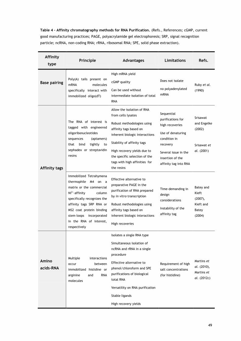
Table 4 - Affinity chromatography methods for RNA Purification. (Refs., References; cGMP, current
good manufacturing practices; PAGE, polyacrylamide gel electrophoresis; SRP, signal recognition
particle; ncRNA, non-coding RNA; rRNA, ribosomal RNA; SPE, solid phase extraction).
Affinity
type Principle Advantages Limitations Refs.
Base pairing
Poly(A) tails present on
mRNA molecules
specifically interact with
immobilized oligo(dT)
High mRNA yield
cGMP quality
Can be used without
intermediate isolation of total
RNA
Does not isolate
no polyadenylated
mRNA
Ruby et al.
(1990)
Affinity tags
The RNA of interest is
tagged with engineered
oligoribonucleotides
sequences (aptamers)
that bind tightly to
sephadex or streptavidin
resins
Allow the isolation of RNA
from cells lysates
Robust methodologies using
affinity tags based on
inherent biologic interactions
Stability of affinity tags
High recovery yields due to
the specific selection of the
tags with high affinities for
the resins
Sequential
purifications for
high recoveries
Use of denaturing
condition in
recovery
Several issue in the
insertion of the
affinity tag into RNA
Srisawat
and Engelke
(2002)
Srisawat et
al. (2001)
Immobilized Tetrahymena
thermophile M4 on a
matrix or the commercial
Ni2+-affinity column
specifically recognizes the
affinity tags SRP RNA or
MS2 coat protein binding
stem–loops incorporated
in the RNA of interest,
respectively
Effective alternative to
preparative PAGE in the
purification of RNA prepared
by in vitro transcription
Robust methodologies using
affinity tags based on
inherent biologic interactions
High recoveries
Time-demanding in
design
considerations
Instability of the
affinity tag
Batey and
Kieft
(2007),
Kieft and
Batey
(2004)
Amino
acids-RNA
Multiple interactions
occur between
immobilized histidine or
arginine and RNA
molecules
Isolates a single RNA type
Simultaneous isolation of
ncRNA and rRNA in a single
procedure
Effective alternative to
phenol/chloroform and SPE
purifications of biological
total RNA
Versatility on RNA purification
Stable ligands
High recovery yields
Requirement of high
salt concentrations
(for histidine)
Martins et
al. (2010),
Martins et
al. (2012c)
49

In fact, these purification schemes can a be valuable tool in recent emerged strategies to
produce stable structured RNA in vivo using recombinant approaches as an alternative to in
vitro transcription or chemical synthesis. Since synthetic methods can often be costly and
laborious and have their drawbacks with respect to sequence requirements, variations in
yield, non-templated nucleotide additions and/or the maximum length of the oligonucleotide,
tRNA-scaffolds have been described with some success as general method for producing a
large variety of recombinant RNA using E. coli as a host, providing an inexhaustible source of
RNA. (Ponchon and Dardel, 2011). At the end of the culture, cells are pelleted, lysed and
RNAt is isolated by direct phenol extraction and the recombinant RNA is purified by AEC or
alternatively, since the recombinant RNA molecules are equipped with a Sephadex aptamer,
by AC using Sephadex G-200, avoiding phenol-based purification. Finally, the product RNA is
separated from the tRNA-scaffold on preparative denaturing PAGE (Nelissen et al., 2012). The
use of amino-acid based methods to purify recombinant RNA would definitely improve the
present of purification strategies, since the phenol/chloroform extraction as well as the
preparative PAGE and all their disadvantages can be eliminated, maintaining the chemical
stability and biological competence of RNA molecules.
In biopharmaceutical industry, the use of an appropriate recombinant expression system
together with robust liquid chromatographic techniques is a well-established method for the
production of therapeutic bioproducts, such as recombinant proteins or pDNA (Cart and
Jungbauer, 2010, Ferreira et al., 1999). Accordingly, the recombinant production of RNA in E.
coli and the subsequent purification employing an amino acid-based AC method could be a
promising biotechnological process for the production of RNA bioproducts in large scale.
Figure 2 schematize a generic procedure for the preparation of recombinant RNA bioproduct
based on amino acid-based AC technique. This approach could provide a cost-effective
process, easily adopted by biopharmaceutical industries.
50

Figure 2 - Projected biotechnological process for RNA preparation in large scale. The downstream
processing is based on previously reported amino acid-based affinity chromatography works (Martins et
al., 2012a, Martins et al., 2012c). After amplification in E. coli, the recombinant RNA are lysed with a
guanidinium-based buffer, followed by concentration, wash and solubilization steps in order to change
to a simple buffer free of RNases, and a final step of amino acid-based affinity chromatography to
isolate and purify RNA from impurities. (E. coli, Escherichia coli; RNases, Ribonuclease; IsopOH pp,
isopropanol precipitation; EtOH pp, ethanol precipitation).
Whether the implementation of such method is feasible for the preparation of therapeutic
RNA molecules, new high-throughput analytical tools that can readily identify and quantify
RNA are required, since several flaws are still existing in the current RNA quantification
techniques, as the lack of selectivity for RNA (Martins et al., 2013). With the success of amino
acids as affinity ligands in the purification of RNA, an analytical technique based on arginine
AC for quantification and quality verification of RNAt was also developed and validated,
according to international and European legislation for bioanalytical methods. The versatility
of the methodology was demonstrated by its applicability in the quantification of RNA from
different eukaryotic cells and in crude samples of chemically synthetized RNA, which can
have a potential multipurpose applicability in molecular biology RNA-based analysis and RNA
therapeutics (Martins et al., 2013).
More efforts into RNA purification are being developed with amino acid based matrices, in
particular with arginine-based matrix. The new goal is to exploit their applicability in
purifying mRNA molecules not from cells, but from synthetic crudes of in vitro transcription,
pursuing mRNA vaccination for cervical cancer. So far, mRNA molecules encoding human
51

papillomavirus (HPV) 16 E6 and E7 proteins were successfully purified from the impurities of
pDNA template, enzymes, nucleotides, salts or buffer. In this work, arginine-based
chromatography is also demonstrating its singular ability in improving purification processes,
showing the advantages of eliminating additional steps and improving global economics of the
production process (unpublished results).
52

5. Conclusion and future trends
To attain large quantities of RNA with the demanded quality for structural and functional
studies as well as for application in medical therapies, more selective, reliable and efficient
isolation and purification processes are required. The difficulty of successfully handling RNA
in the laboratory, because of the structural susceptibility to RNases present in cell extracts
and in the environment, and the impurities present in cell samples or synthetic preparations,
result in numerous challenges for the selective purification of RNA molecules and in higher
costs for laboratories and biopharmaceutical industries.
The current methodologies used in RNA preparations circumvent some of those challenges,
however many difficulties still existing in order to obtain an innocuous, selective and
economically feasible procedure to purify RNA. As several clinical trials are ongoing provided
by many pharmaceutical and biotechnological companies, it seems that more accurate
methods exists but are not ready accessible. Despite that, the motivation of the scientific
community to disclose the knowledge on RNA purification processes is significantly increasing.
Recent investigations in chromatographic bioseparation are providing reliable data in process
development both for preparative-scale and for large-scale applications. In particular, AC
stands out because of combining the selectivity of a naturally occurring biological interaction
with the simplicity of a single small molecule. Moreover, the contribution of a novel affinity
approach based on amino acid ligands provided new insights in the way that the diverse RNA
molecules can be accurately purified. Moreover this methodology brings the prospect to be
combined in a recombinant approach rising the opportunity for the development of a robust
biotechnological process that might have a positive economic impact on the industrial
production of RNA.
Although AC represents a particular attractive option for RNA purification, there are
limitations regarding capacity of supports and the availability of suitable ligands that remain
to be solved. However, with the increasing understanding on RNA synthetic synthesis and
chemical modification, the design of highly selective and stable synthetic affinity ligands
might be more easily achieved. In addition, recent chromatographic technologies as
monolithic supports that combine the advantages of conventional chromatographic columns
packed with porous particles in terms of separation power, capacity and sample distribution
and those of membrane technology in terms of convective mass transport could be used with
immobilized affinity ligands, which should largely benefit the industrial production of RNA
biotherapeutics.
The combined efforts that are expected to occur in this field should help to accomplish
enhanced purification strategies with positive effects on RNA basic and applied research.
53

Acknowledgments
This work was supported by FCT, the Portuguese Foundation for Science and Technology
(PTDC/EBB-BIO/114320/2009, PEst-C/SAU/UI0709/2011 COMPETE and and EXPL/BBB-
BIO/1056/2012, COMPETE: FCOMP-01-0124-FEDER-027560). Rita Martins also acknowledges a
fellowship (SFRH/BD/ 64100/2009) from FCT.
The authors declared no conflict of interest.
54

References
Arraiano CM, Andrade JM, Domingues S, Guinote IB, Malecki M, Matos RG, et al. The critical
role of RNA processing and degradation in the control of gene expression. FEMS Microbiol Rev.
2010; 34:883-923.
Aviv H, Leder P. Purification of Biologically Active Globin Messenger RNA by Chromatography
on Oligothymidylic acid-Cellulose. Proc Natl Acad Sci U S A. 1972; 69:1408-12.
Ayyar BV, Arora S, Murphy C, O'Kennedy R. Affinity chromatography as a tool for antibody
purification. Methods. 2012; 56:116-29.
Baker BF, Lot SS, Condon TP, Cheng-Flournoy , Lesnik EA, asmor HM, et al. 2′-O-(2-
Methoxy)ethyl-modified Anti-intercellular Adhesion Molecule 1 (ICAM-1) Oligonucleotides
Selectively Increase the ICAM-1 mRNA Level and Inhibit Formation of the ICAM-1 Translation
Initiation Complex in Human Umbilical Vein Endothelial Cells. J Biol Chem. 1997; 272:11994-
2000.
Batey RT, Kieft JS. Improved native affinity purification of RNA. RNA. 2007; 13:1384-9.
Belostotsky R, Frishberg Y, Entelis N. Human mitochondrial tRNA quality control in health and
disease: a channelling mechanism? RNA Biol. 2012; 9:33-9.
Blount KF, Breaker RR. Riboswitches as antibacterial drug targets. Nat Biotechnol. 2006;
24:1558-64.
Booy EP, Meng H, McKenna SA. Native RNA Purification by Gel Filtration Chromatography In:
Conn GL, editor. Recombinant and In Vitro RNA Synthesis 2012. p. 69-81.
Brantl S. Antisense-RNA regulation and RNA interference. Biochim Biophys Acta. 2002;
1575:15-25.
Burnett JC, Rossi JJ. RNA-based therapeutics: current progress and future prospects. Chem
Biol. 2012; 19:60-71.
Bustin SA, Nolan T. Pitfalls of quantitative real-time reverse-transcription polymerase chain
reaction. J Biomol Tech. 2004; 15:155-66.
Cart G, Jungbauer A. Downstream Processing of Biotechnology Products. Protein
Chromatography Process Development and Scale-Up WILEY-VCH Verlag GmbH & Co.; 2010.
55

Chatterjee A, Mirer PL, Zaldivar Santamaria E, Klapperich C, Sharon A, Sauer-Budge AF. RNA
isolation from mammalian cells using porous polymer monoliths: an approach for high-
throughput automation. Anal Chem. 2010; 82:4344–56.
Chionh YH, Ho C-H, Pruksakorn D, Ramesh Babu I, Ng CS, Hia F, et al. A multidimensional
platform for the purification of non-coding RNA species. Nucleic Acids Res. 2013.
Chomczynski P, Sacchi N. The single-step method of RNA isolation by acid guanidinium
thiocyanate-phenol-chloroform extraction: twenty-something years on. Nat Protoc. 2006;
1:581-5.
Cramer H, Finn KJ, Herzberg E. Purity Analysis and Impurities Determination by Reversed-
Phase High-Performance Liquid Chromatography. In: Bonilla J.V., Srivatsa SG, editors.
Handbook of Analysis of Oligonucleotides and Related Products. Boca Raton, Florida: CRC
Press; 2011. p. 1-46.
Dias N, Stein CA. Antisense oligonucleotides: basic concepts and mechanisms. Mol Cancer
Ther. 2002; 1:347-55.
Dickman MJ. Ion Pair Reverse-Phase Chromatography: A Versatile Platform for the Analysis of
RNA. Chromatography Today 2011a. p. 22-6.
Dickman MJ. Ion Pair Reverse-Phase Chromatography: A Versatile Platform for the Analysis of
RNA. Chromatography Today http://www.chromatographytoday.com 2011b. p. 22-6.
Directive 2001/83/EC. European Parliament and of the Council of 6 November 2001 on the
Community code relating to medicinal products for human use: EC, Brussels, Belgium 2001
http://eceuropaeu/health/files/eudralex/vol-
1/dir_2001_83_cons/dir2001_83_cons_20081230_enpdf.
Doench JG, Petersen CP, Sharp PA. siRNAs can function as miRNAs. Genes Dev. 2003; 17:438-
42.
Doudna JA. Preparation of homogeneous ribozyme RNA for crystallization. Methods Mol Biol.
1997; 74:365-70.
Dunin-Horkawicz S, Czerwoniec A, Gajda MJ, Feder M, Grosjean H, Bujnicki JM. MODOMICS: a
database of RNA modification pathways. Nucleic Acids Res. 2006; 34:D145-9.
Easton LE, Shibata Y, Lukavsky PJ. Rapid, nondenaturing RNA purification using weak anion-
exchange fast performance liquid chromatography. RNA. 2010; 16:647-53.
56

El-Sagheer AH, Brown T. New strategy for the synthesis of chemically modified RNA constructs
exemplified by hairpin and hammerhead ribozymes. Proc Natl Acad Sci U S A. 2010;
107:15329-34.
Farrell RE. RNA Methodologies. In: Meyers RA, editor. Epigenetic Regulation and Epigenomics:
Advances in Molecular Biology and Medicine. 1st ed. Weinheim: Wiley-VCH; 2012. p. 1-37.
Ferreira GN, Cabral JM, Prazeres DM. Development of process flow sheets for the purification
of supercoiled plasmids for gene therapy applications. Biotechnol Prog. 1999; 15:725-31.
Fleige S, Pfaffl MW. RNA integrity and the effect on the real-time qRT-PCR performance. Mol
Aspects Med. 2006; 27:126-39.
Ge Q, McManus MT, Nguyen T, Shen CH, Sharp PA, Eisen HN, et al. RNA interference of
influenza virus production by directly targeting mRNA for degradation and indirectly inhibiting
all viral RNA transcription. Proc Natl Acad Sci U S A. 2003; 100:2718-23.
Ghildiyal M, Zamore PD. Small silencing RNAs: an expanding universe. Nat Rev Genet. 2009;
10:94-108.
Gilar M. Analysis and purification of synthetic oligonucleotides by reversed-phase high-
performance liquid chromatography with photodiode array and mass spectrometry detection.
Anal Biochem. 2001; 298:196-206.
Gjerde DT, Hoang L, Hornby D. RNA Purification and Analysis: Sample Preparation, Extraction,
Chromatography: Wiley-VCH Verlag GmbH & Co; 2009.
Guidance for Industry: Consideration for plasmid DNA vaccines for infection disease
indications. FDA, US 2007
http://wwwfdagov/downloads/BiologicsBloodVaccines/GuidanceComplianceRegulatoryInform
ation/Guidances/Vaccines/ucm091968pdf
Guideline on Human Cell-based Medicinal Products. EMA, L, Great Britain 2007
http://wwwemaeuropaeu/docs/en_GB/document_library/Scientific_guideline/2009/09/WC5
00003898pdf.
Hagen FS, Young ET. Preparative polyacrylamide gel electrophoresis of ribonucleic acid.
Identification of multiple molecular species of bacteriophage T7 lysozyme messenger
ribonucleic acid. Biochemistry. 1974; 13:3394-400.
Jahn CE, Charkowski AO, Willis DK. Evaluation of isolation methods and RNA integrity for
bacterial RNA quantitation. J Microbiol Methods. 2008; 75:318-24.
57
Jeong E, Kim H, Lee SW, Han K. Discovering the interaction propensities of amino acids and
nucleotides from protein-RNA complexes. Mol Cells. 2003; 16:161-7.
Karikó K, Muramatsu H, Ludwig J, Weissman D. Generating the optimal mRNA for therapy:
HPLC purification eliminates immune activation and improves translation of nucleoside-
modified, protein-encoding mRNA. Nucleic Acids Res. 2011; 39:e142.
Kieft JS, Batey RT. A general method for rapid and nondenaturing purification of RNAs. RNA.
2004; 10:988-95.
Kim I, McKenna SA, Viani Puglisi E, Puglisi JD. Rapid purification of RNAs using fast
performance liquid chromatography (FPLC). RNA. 2007; 13:289-94.
Kiss T. Small nucleolar RNA-guided post-transcriptional modification of cellular RNAs. EMBO J.
2001; 20:3617-22.
Knepp JH, Geahr MA, Forman MS, Valsamakis A. Comparison of automated and manual nucleic
acid extraction methods for detection of enterovirus RNA. J Clin Microbiol. 2003; 41:3532-6.
Koubek J, Lin KF, Chen YR, Cheng RP, Huang JJT. Strong anion-exchange fast performance
liquid chromatography as a versatile tool for preparation and purification of RNA produced by
in vitro transcription. RNA. 2013.
Kreiter S, Diken M, Selmi A, Tureci O, Sahin U. Tumor vaccination using messenger RNA:
prospects of a future therapy. Curr Opin Immunol. 2011; 23:399-406.
Kusenda B, Mraz M, Mayer J, Pospisilova S. MicroRNA biogenesis, functionality and cancer
relevance. Biomed Pap Med Fac Univ Palacky Olomouc Czech Repub. 2006; 150:205-15.
Kyte JA, Kvalheim G, Aamdal S, Saeboe-Larssen S, Gaudernack G. Preclinical full-scale
evaluation of dendritic cells transfected with autologous tumor-mRNA for melanoma
vaccination. Cancer Gene Ther. 2005; 12:579-91.
Kyte JA, Mu L, Aamdal S, Kvalheim G, Dueland S, Hauser M, et al. Phase I/II trial of melanoma
therapy with dendritic cells transfected with autologous tumor-mRNA. Cancer Gene Ther.
2006; 13:905-18.
Labrou NE. Design and selection of ligands for affinity chromatography. J Chromatogr B. 2003;
790:67-78.
Laurell H, Iacovoni JS, Abot A, Svec D, Maoret J-J, Arnal J-F, et al. Correction of RT–qPCR
data for genomic DNA-derived signals with ValidPrime. Nucleic Acids Res. 2012.
58

Lehman N. RNA in evolution. Wiley Interdiscip Rev RNA. 2010; 1:202-13.
Lin SL, Miller JD, Ying SY. Intronic microRNA (miRNA). J Biomed Biotechnol. 2006;
2006:26818.
Loens K, Bergs K, Ursi D, Goossens H, Ieven M. Evaluation of NucliSens easyMAG for
automated nucleic acid extraction from various clinical specimens. J Clin Microbiol. 2007;
45:421-5.
Lorenzi J, Trombone A, Rocha C, Almeida L, Lousada R, Malardo T, et al. Intranasal
vaccination with messenger RNA as a new approach in gene therapy: Use against tuberculosis.
BMC Biotechnol. 2010; 10:77.
Lowe CR, Lowe AR, Gupta G. New developments in affinity chromatography with potential
application in the production of biopharmaceuticals. J Biochem Biophys Methods. 2001;
49:561-74.
Lukavsky PJ, Puglisi JD. Large-scale preparation and purification of polyacrylamide-free RNA
oligonucleotides. RNA. 2004; 10:889-93.
Magdeldin S, MoserTech A. Affinity Chromatography: Principles and Applications. In:
Magdeldin S, editor. Affinity Chromatography: InTech; 2012.
Maina A, Blackman BA, Parronchi CJ, Morozko E, Bender ME, Blake AD, et al. Solid-phase
synthesis, characterization and RNAi activity of branch and hyperbranch siRNAs. Bioorg Med
Chem Lett. 2013; 23:5270-4.
Maroney PA, Chamnongpol S, Souret F, Nilsen TW. A rapid, quantitative assay for direct
detection of microRNAs and other small RNAs using splinted ligation. RNA. 2007; 13:930-6.
Martins R, Maia CJ, Queiroz JA, Sousa F. A new strategy for RNA isolation from eukaryotic
cells using arginine affinity chromatography. J Sep Sci. 2012a; 35:3217-26.
Martins R, Queiroz JA, Sousa F. A new affinity approach to isolate Escherichia coli 6S RNA
with histidine-chromatography. J Mol Recognit. 2010; 23:519-24.
Martins R, Queiroz JA, Sousa F. Histidine affinity chromatography-based methodology for the
simultaneous isolation of Escherichia coli small and ribosomal RNA. Biomed Chromatogr.
2012b; 26:781-8.
59

Martins R, Queiroz JA, Sousa F. New approach in RNA quantification using arginine-affinity
chromatography: potential application in eukaryotic and chemically synthesized RNA. Anal
Bioanal Chem. 2013: 405:8849-58.
Matera AG, Terns RM, Terns MP. Non-coding RNAs: lessons from the small nuclear and small
nucleolar RNAs. Nat Rev Mol Cell Biol. 2007; 8:209-20.
Mayer G. The chemical biology of aptamers. Angew Chem Int Ed Engl. 2009; 48:2672-89.
McGinnis AC, Chen B, Bartlett MG. Chromatographic methods for the determination of
therapeutic oligonucleotides. J Chromatogr B Analyt Technol Biomed Life Sci. 2012; 883-
884:76-94.
McGookin R. Purification of Synthetic Oligonucleotides by Preparative Gel Electrophoresis.
1988. p. 215-20.
McKenna SA, Kim I, Puglisi EV, Lindhout DA, Aitken CE, Marshall RA, et al. Purification and
characterization of transcribed RNAs using gel filtration chromatography. Nat Protoc. 2007;
2:3270-7.
Melnikova I. RNA-based therapies. Nat Rev Drug Discov. 2007; 6:863-4.
Moore PB. Structural motifs in RNA. Annu Rev Biochem. 1999; 68:287-300.
Murray RK, Granner DK, Mayes PA, Rodwell VW. Harper´s Biochemistry. 25 ed. USA: McGraw-
Hill; 2000.
Nelissen FH, Leunissen EH, van de Laar L, Tessari M, Heus HA, Wijmenga SS. Fast production
of homogeneous recombinant RNA--towards large-scale production of RNA. Nucleic Acids Res.
2012; 40:e102.
Ni X, Castanares M, Mukherjee A, Lupold SE. Nucleic acid aptamers: clinical applications and
promising new horizons. Curr Med Chem. 2011; 18:4206-14.
Nilsen TW. Selective Precipitation of Large RNAs. Cold Spring Harb Protoc. 2012;
2012:pdb.prot072322.
Noll B, Seiffert S, Hertel F, Debelak H, Hadwiger P, Vornlocher HP, et al. Purification of small
interfering RNA using nondenaturing anion-exchange chromatography. Nucleic Acid Ther.
2011; 21:383-93.
Pascolo S. Vaccination with Messenger RNA (mRNA). In: Bauer S, Hartmann G, editors. Toll-
Like Receptors (TLRs) and Innate Immunity: Springer Berlin Heidelberg; 2008. p. 221-35.
60
Phylactou LA, Kilpatrick MW, Wood MJ. Ribozymes as therapeutic tools for genetic disease.
Hum Mol Genet. 1998; 7:1649-53.
Ponchon L, Dardel F. Large scale expression and purification of recombinant RNA in
Escherichia coli. Methods. 2011; 54:267-73.
Price SR, Ito N, Oubridge C, Avis JM, Nagai K. Crystallization of RNA-protein complexes. I.
Methods for the large-scale preparation of RNA suitable for crystallographic studies. J Mol
Biol. 1995; 249:398-408.
Raam BJv, Salvesen GS. Transferring Death: A Role for tRNA in Apoptosis Regulation. Mol Cell.
2010; 37:591-2.
Romanovskaya A, Sarin LP, Bamford DH, Poranen MM. High-throughput purification of double-
stranded RNA molecules using convective interaction media monolithic anion exchange
columns. J Chromatogr A. 2013; 1278:54-60.
Roque AC, Lowe CR. Affinity chromatography: history, perspectives, limitations and
prospects. Methods Mol Biol. 2008; 421:1-21.
Ruby SW, Goelz SE, Hostomsky Z, Abelson JN. Affinity chromatography with biotinylated
RNAs. Methods Enzymol. 1990; 181:97-121.
Scott LG, Hennig M. RNA structure determination by NMR. Methods Mol Biol. 2008; 452:29-61.
Shanagar J. Purification of a synthetic oligonucleotide by anion exchange chromatography:
Method optimisation and scale-up. J Biochem Biophys Methods. 2005; 64:216-25.
Sharp PA. The Centrality of RNA. Cell. 2009; 136:577-80.
Sherlin LD, Bullock TL, Nissan TA, Perona JJ, Lariviere FJ, Uhlenbeck OC, et al. Chemical and
enzymatic synthesis of tRNAs for high-throughput crystallization. RNA. 2001; 7:1671-8.
Soll D, Nishimura S, Moore P. RNA. First ed. Oxford: Pergamon; 2001.
Sousa F, Cruz C, Queiroz JA. Amino acids-nucleotides biomolecular recognition: from
biological occurrence to affinity chromatography. J Mol Recognit. 2010; 23:505-18.
Sousa F, Freitas S, Azzoni AR, Prazeres DM, Queiroz JA. Selective purification of supercoiled
plasmid DNA from clarified cell lysates with a single histidine-agarose chromatography step.
Biotechnol Appl Biochem. 2006; 45:131-40.
61

Sousa F, Matos T, Prazeres DM, Queiroz JA. Specific recognition of supercoiled plasmid DNA in
arginine affinity chromatography. Anal Biochem. 2008a; 374:432-4.
Sousa F, Prazeres DM, Queiroz JA. Affinity chromatography approaches to overcome the
challenges of purifying plasmid DNA. Trends Biotechnol. 2008b; 26:518-25.
Sousa F, Prazeres DMF, Queiroz JA. Improvement of transfection efficiency by using
supercoiled plasmid DNA purified with arginine affinity chromatography. J Gene Med. 2009;
11:79-88.
Srisawat C, Engelke DR. Streptavidin aptamers: affinity tags for the study of RNAs and
ribonucleoproteins. RNA. 2001; 7:632-41.
Srisawat C, Engelke DR. RNA affinity tags for purification of RNAs and ribonucleoprotein
complexes. Methods. 2002; 26:156-61.
Srisawat C, Goldstein IJ, Engelke DR. Sephadex-binding RNA ligands: rapid affinity purification
of RNA from complex RNA mixtures. Nucleic Acids Res. 2001; 29:e4.
Srivatsa GS. Regulatory Considerations for the Development of Oligonucleotide Therapeutics.
In: Bonilla JV, Srivatsa SG, editors. Handbook of Analysis of Oligonucleotides and Related
Products. Boca Raton, Florida: CRC Press 2011. p. 465–86.
Srivatsa S. Regulatory Challenges. European Biopharmaceutical Review. 2012; Sping 2012.
Stillian JR, Pohl CA. New latex-bonded pellicular anion exchangers with multi-phase
selectivity for high-performance chromatographic separations. J Chromatogr A. 1990;
499:249-66.
Sullenger BA, Gilboa E. Emerging clinical applications of RNA. Nature. 2002; 418:252-8.
Tenson T, Mankin A. Antibiotics and the ribosome. Mol Microbiol. 2006; 59:1664-77.
Thayer J, Murugaiah V., WU Y. Purity Analysis and Impurities Determination by AEX-HPLC. In:
Bonilla J.V., Srivatsa SG, editors. Handbook of Analysis of Oligonucleotides and Related
Products. Boca Raton, Florida: CRC Press; 2011a. p. 47–104.
Thayer JR, Flook KJ, Woodruff A, Rao S, Pohl CA. New monolith technology for automated
anion-exchange purification of nucleic acids. J Chromatogr B. 2010; 878:933-41.
Thayer JR, Rao S, Pohl C, Wu Y, Angelino M. Pellicular anion-exchange chromatography
applied to RNAi assays for monitoring strand stoichiometry and RNA stability.
Chromatography Today http://www.chromatographytoday.com 2011b. p. 28-32.
62

Treger M, Westhof E. Statistical analysis of atomic contacts at RNA-protein interfaces. J Mol
Recognit. 2001; 14:199-214.
Tucker BJ, Breaker RR. Riboswitches as versatile gene control elements. Curr Opin Struct
Biol. 2005; 15:342-8.
Uhlen M. Affinity as a tool in life science. Biotechniques. 2008; 44:649-54.
Vijayalakshmi MA. Histidine Ligand Affinity Chromatography. Mol Biotechnol. 1996; 6:347-57.
Vomelova I, Vanickova Z, Sedo A. Methods of RNA purification. All ways (should) lead to
Rome. Folia Biol (Praha). 2009; 55:243-51.
Walker SC, Scott FH, Srisawat C, Engelke DR. RNA affinity tags for the rapid purification and
investigation of RNAs and RNA-protein complexes. Methods Mol Biol. 2008; 488:23-40.
Wang WX, Wilfred BR, Baldwin DA, Isett RB, Ren N, Stromberg A, et al. Focus on RNA
isolation: obtaining RNA for microRNA (miRNA) expression profiling analyses of neural tissue.
Biochim Biophys Acta. 2008; 1779:749-57.
Warren W, Vella G. Principles and methods for the analysis and purification of synthetic
deoxyribonucleotides by high-performance liquid chromatography. Molecular Biotechnology.
1995; 4:179-99.
Weide B, Garbe C, Rammensee HG, Pascolo S. Plasmid DNA- and messenger RNA-based anti-
cancer vaccination. Immunol Lett. 2008; 115:33-42.
Wen J, Legendre LA, Bienvenue JM, Landers JP. Purification of Nucleic Acids in Microfluidic
Devices. Anal Chem. 2008; 80:6472-9.
Wendisch VF, Zimmer DP, Khodursky A, Peter B, Cozzarelli N, Kustu S. Isolation of Escherichia
coli mRNA and comparison of expression using mRNA and total RNA on DNA microarrays. Anal
Biochem. 2001; 290:205-13.
Wittmann A, Suess B. Engineered riboswitches: Expanding researchers' toolbox with synthetic
RNA regulators. FEBS Lett. 2012; 586:2076-83.
Yarus M, Widmann JJ, Knight R. RNA-amino acid binding: a stereochemical era for the genetic
code. J Mol Evol. 2009; 69:406-29.
Yu RZ, Zhang H, Geary RS, Graham M, Masarjian L, Lemonidis K, et al. Pharmacokinetics and
Pharmacodynamics of an Antisense Phosphorothioate Oligonucleotide Targeting Fas mRNA in
Mice. J Pharmacol Exp Ther. 2001; 296:388-95.
63

Zeng Y, Wagner EJ, Cullen BR. Both Natural and Designed Micro RNAs Can Inhibit the
Expression of Cognate mRNAs When Expressed in Human Cells. Mol Cell. 2002; 9:1327-33.
64

65

66

Chapter 2
67

68

Aims of the thesis
The main goal of this thesis is to explore the possibility of using affinity chromatography with
amino acids as specific ligands to develop new methodologies to efficiently isolate, purify and
analyze RNA molecules. Because of the particularities of RNA and the challenges implicit to
its purification, the interactions between amino acids and RNA will be exploited so that RNA
molecules can be recovered under their native state and with high integrity and purity, in
order to follow the growing demands of the therapeutic application of RNA. Since various
limitations still exist in the current techniques to isolate and purify RNA, namely the use of
chemical solvents and denaturing conditions, the use of amino acid supports in affinity
chromatography is intended to aid in the development of new approaches to recover a
product to be biologically applied and that lead to an efficient, tolerable and scalable
process, improving workload, process time and economics. To achieve this principal purpose,
the work will be developed concerning the following specific aims:
1. Optimization of the lysis and separation processes to recover and isolate RNA
molecules or total nucleic acids from prokaryotic or eukaryotic cells, for application
in chromatographic studies;
2. Study of the binding/elution conditions of RNA or total nucleic acids extracts in
affinity supports with immobilized histidine or arginine by testing the influence of
elution conditions such as the temperature, flow rate and buffer composition (pH,
ionic strength and type of salt), in order to evaluate the ability of the supports to
specifically recognize the various RNA molecules.
3. Monitoring the influence of the chromatographic conditions in the structure and
stability of RNA and control its quality throughout the purification process;
4. Development of purification strategies to selectively purify RNA molecules from
complex and impure cell extracts in order to eliminate the use of organic solvents and
denaturing conditions from RNA isolations;
5. Evaluation of the biological activity of the purified RNAs by integrating the
chromatographic process with the more reliable and promising support in the global
process of RNA preparation to be applied in RNA research or RNA therapeutics.
69

70

71

72

Chapter 3
73

74

Paper II
A new affinity approach to isolate Escherichia coli 6S
RNA with histidine-chromatography
R. Martins, J. A. Queiroz, F. Sousa
Journal of Molecular Recognition. 2010. 23(6): 519-524
75

76

Research Article
Received: 13 November 2009, Revised: 19 July 2010, Accepted: 20 July 2010, Published online in Wiley Online Library: 2010
(wileyonlinelibrary.com) DOI:10.1002/jmr.1078
A new affinity approach to isolate Escherichiacoli 6S RNA with histidine-chromatographyy
R. Martinsa, J. A. Queiroza and F. Sousaa*
6S RNA is an abundant non-coding RNA in Escherichia c
J. Mol. Rec
oli (E. coli), but its function has not been discovered until recently.The first advance on 6S RNA function was the demonstration of its ability to bind the s70-holoenzyme form of RNApolymerase, inhibiting its activity and consequently the transcription process. The growing interest in the investigationof non-coding small RNAs (sRNA) calls for the development of new methods for isolation and purification of RNA. Thiswork presents an optimized RNA extraction procedure and describes a new affinity chromatography method using ahistidine support to specifically purify 6S RNA from other E. coli sRNA species. The RNA extraction procedure wasoptimized, and a high yield was obtained in the separation of sRNA and ribosomal RNA (rRNA) from total RNA (RNAt).This improved method takes advantage of its simplicity and significant cost reduction, since some complex operationshave been eliminated. A purification strategy was also developed to separate 6S RNA from an sRNA mixture. Pure RNAcan be advantageously obtained using the histidine-affinity chromatography method, aiming at its application tostructural or functional studies. Copyright � 2010 John Wiley & Sons, Ltd.
Keywords: affinity chromatography; histidine; RNA; RNA extraction; small RNA; 6S RNA
* Correspondence to: F. Sousa, CICS – Centro de Investigacao em Ciencias daSaude, Universidade da Beira Interior, 6201-001 Covilha, Portugal.E-mail: [email protected]
a R. Martins, J. A. Queiroz, F. Sousa
CICS – Centro de Investigacao em Ciencias da Saude, Universidade da Beira
Interior, 6201-001 Covilha, Portugal
y This article is published in Journal of Molecular Recognition as a special issueon Affinity 2009, edited by Gideon Fleminger, Tel-Aviv University, Tel-Aviv, Israeland George Ehrlich, Hoffmann-La Roche, Nutley, NJ, USA. 5
INTRODUCTION
Small RNAs (sRNA) are a family of regulatory non-coding RNAs,which are targeted in several research studies on all organisms.Their activities have been associated to RNA processing, mRNAstability, translation, protein stability and secretion in bacteria,acting mostly by base pairing and, to a lesser extent, by proteinbinding (Wassarman et al., 1999). Escherichia coli (E. coli) 6S RNAwas one of the first sRNA to be discovered in the late 1960s, itssequence and secondary structure having been promptlyproposed (Willkomm and Hartmann, 2005). Although 6S RNAwas soon recognized, its function remained unknown for severalyears. This RNA species has always been found at high abundancein total cellular RNA (RNAt). However, studies of 6S RNA depletionand over-expression have not demonstrated any evident changein cell metabolism behaviour (Willkomm and Hartmann, 2005).In 2000, the first breakthrough on 6S RNA function was thedemonstration of its ability to bind the s70-holoenzyme form ofRNA polymerase, inhibiting its activity in the stationary phase ofcell growth. The high structural similarity observed between 6SRNA and a DNA open promoter in RNA transcription process, aswell as the identification of 6S RNA in a specific complex linked toRNA polymerase, has suggested that this RNA type functions bymimicking the transcription promoter. Hence, 6S RNA seems todirectly compete with DNA promoters for the s70-RNA poly-merase active site to inhibit the transcription process (Wassarmanand Storz, 2000; Wassarman, 2007).Over the last years, new insights triggered a better under-
standing of the molecular mechanism of 6S RNA (Gildehaus et al.,2007; Karen, 2007), while the techniques employed in the isola-tion of this RNA species were still plasmid design and enzymaticpurification. These techniques involve time-consuming andexpensive procedures. Therefore, the development of new toolsfor 6S RNA purification would be of great importance to simplifythe current genetic-based approaches.
ognit. 2010; 23: 519–524 Copyright � 20
The use of amino acid-based affinity chromatography has beendescribed as a promising approach for nucleic acids purification.This is due to the biological selectivity, which occurs between theamino acid ligand and the nucleic acid molecule under study. Ingeneral, the selectivity found in affinity chromatography can beexplained by some biological recognition or individual chemicalstructure, which favours the interaction (Sousa et al., 2008). In thepresent study, the isolated E. coli sRNA population was tested inan affinity chromatographic support with immobilized histidine,combining the reliable and economical chromatographic opera-tion with the high selectivity of histidine, which has been shownto have great applicability in the purification of nucleic acids.Histidine–agarose chromatographic support has demonstratedto efficiently separate supercoiled (SC) and open circular (OC)plasmid DNA (pDNA) isoforms, via a bio-recognition phenom-enon with the nucleic acid bases, involving also hydrophobicinteractions between the support and the pDNA molecules(Sousa et al., 2005). Furthermore, in these previous studies, theRNA showed to be strongly retained on the column, because ofthe higher base exposure, and a decrease in salt concentrationwas deemed necessary for its elution (Sousa et al., 2006). Buildingon the interesting results obtained for RNA separation, this reportexplores the possibility of using histidine–agarose chromatog-raphy to purify 6S RNA from other sRNA.
10 John Wiley & Sons, Ltd.
1977

R. MARTINS ET AL.
520
MATERIALS AND METHODS
Materials
L-histidine–agarose gel, oligo(dT) cellulose and all the organiccompounds used in the RNAt extractions were obtained fromSigma (St. Louis, MO, USA). Glycogen was from Roche (Mannheim,Germany) and the RNA/DNA midi kit was purchased from Qiagen(Hilden, Germany). The PD-10 desalting columns were from GEHealthcare (Uppsala, Sweden) and the RNA molecular weightmarker was obtained from Invitrogen (Carlsbad, CA, USA).Specific primers for E. coli 6S RNA cDNA were purchased fromStab Vida (Lisbon, Portugal). All salts used were of analyticalgrade.
Bacterial growth conditions
The RNA used in this study was obtained from a cell culture of E.coli DH5a. Growth was carried out in shake flasks at 378C and250 rpm with 250mL of Terrific Broth medium (12 g/L tryptone,24 g/L yeast extract, 4mL/L glycerol, 0.017M KH2PO4, 0.072MK2HPO4). It was suspended in the late log phase (OD600� 9). Cellswere recovered by centrifugation and were stored at �208C.
Lysis and RNAt isolation
Cells were lysed and RNAt was extracted using the acidguanidinium thiocyanate–phenol–chloroform method describedby Chomczynski and Sacchi (2006) with some modifications. Thebacterial pellets were resuspended in 5mL of Solution D (4Mguanidinium thiocyanate; 25mM sodium citrate, pH 4.0; 0.5% (w/m)N-laurosylsarcosine (sarcosyl) and 0.1M b-mercaptoethanol) toperform lysis. After incubating on ice for 10min, cellular debris,genomic DNA and proteins were precipitated by gently addingand mixing 10mL of water-saturated phenol and 1mL of 2Msodium acetate (pH 4.0). The RNAt isolation was achieved byadding 2mL of chloroform/isoamyl alcohol (49:1), and by mixingvigorously until two immiscible phases were obtained. The upperaqueous phase, which contained mostly RNA, was recovered andconcentrated by the addition of 11mL of 20mL/mL glycogen and10mL of ice-cold isopropanol. After incubating for 10min at�208C, RNAt was recovered by centrifugation at 15 000 g for20min at 48C. The RNAt pellet was resuspended in 3mL ofSolution D. It was concentrated again with glycogen and 3mL ofice-cold isopropanol. After centrifuging for 10min at 15 000 g(48C), the RNAt pellet was washed with 7.5mL of 75% ethanol andincubated at room temperature for 10min, followed by a 5mincentrifugation at 15 000 g (48C). The air-dried RNAt pellet wassolubilized in 400mL of 0.05% DEPC-treated water, and its opticaldensity was determined in order to assess RNAt quantity andpurity.
Isolation of sRNA population from RNAt extract
The isolation of the sRNA population was first performed usingthemethod previously described by Stellrecht and Gandhi (2002).However, the complexity of this protocol and its frequent use ineucaryotic cells led us to develop a new methodology for sRNAextraction from procaryotic cells.In the optimized protocol, RNAt extract was precipitated with
2M (NH4)2SO4 and incubated at 48C for several hours. The sRNArecovery was accomplished by centrifugation at 15 000 g for20min at 48C, as the sRNA population was in the supernatant,
View this article online at wileyonlinelibrary.com Copyright � 2
78
while the ribosomal RNA (rRNA) was in the pellet. Next, thesupernatant was applied to a PD-10 desalting column (GEHealthcare, Uppsala, Sweden), following the manufacturer’sinstructions. The sRNA concentration was achieved with 2volumes of ice-cold isopropanol and 11mL of 20mL/mL glycogen,followed by centrifugation at 15 000 g for 20min at 48C. Finally,the sRNA pellet was solubilized with 200mL of 0.05%DEPC-treated water. The quantification and purity of sRNA wasassessed as previously described for RNAt.
Affinity chromatography
Chromatography was performed in an AKTA purifier system withUNICORN software (GE Healthcare, Sweden). A 10mmdiameter�20mm long (�2mL) column was packed with the commercialL-histidine–agarose gel. The manufacturer characterizes thissupport as a cross-linked 4% beaded agarose matrix with a12-atom spacer and an extent of labelling between 1 and 2mmol/mL. The column was equilibrated with 2.2M (NH4)2SO4 in 10mMTris–HCl (pH 7.0) buffer at a flow rate of 1mL/min. The isolatedsRNA samples were injected onto the column using a 100mL loopat the same flow rate. The absorbance of the eluent was conti-nuously monitored at 260 nm. After elution of unbound specieswith 2.2M (NH4)2SO4 in 10mM Tris–HCl (pH 7.0) buffer, the ionicstrength of the buffer was decreased stepwise to 1.6M (NH4)2SO4
in 10mM Tris–HCl (pH 7.0) buffer. Finally, tightly bound RNAspecies were removed by changing to ammonium sulphate-free10mM Tris–HCl (pH 7.0) buffer. Fractions were pooled accordingto the chromatograms obtained. Following concentration anddesalting with Vivaspin concentrators (Vivascience), the poolswere kept for further analysis as described below.
Polyacrylamide electrophoresis
Pooled fractions were analysed by vertical electrophoresis usingAmersham Biosciences system (GE Healthcare, Sweden) with7.5% polyacrylamide gel and were then stained with ethidiumbromide (0.5mg/mL). Electrophoresis was carried out at 120 V for90min with TBE buffer (0.84M Tris base, 0.89M boric acid and0.01M EDTA, pH 8.3). The pooled fractions were previouslydenatured with 97.5% formamide and the denatured conditionswere kept in the gel due to the presence of 8M urea. sRNA in thegel was visualized using a Vilber Lourmt system (ILC Lda).
Reverse-transcription PCR analysis
6S RNA identification was assessed using reverse-transcriptasepolymerase chain reaction (PCR) in a MyCyclerTMThermal Cycle(Biorad). Samples collected after the chromatographic purifi-cation process with histidine–agarose column were pre-treatedwith DNase I (Sigma Chemical Co., St. Louis, MO) according to themanufacturer’s instructions. cDNA was synthesized from 150 or500 ng of 6S RNA which was denatured for 5min at 658C with500mM of deoxynucleotide triphosphates (Amersham, Uppsala,Sweden) and 250 ng of random primers (Invitrogen, KarlsruheLMA, Germany). Reverse transcription was carried out at 378C for60min in a 20mL reaction containing reverse transcriptase buffer(50mM Tris–HCl, 75mM KCl and 3mM MgCl2), 0.1M DTT, 60 U ofRNaseOUT (Invitrogen) and 200U of M-MLV RT (Invitrogen). Thereaction was stopped at 758C for 15min. PCR reactions werecarried out using 1mL of synthesized cDNAs in a 25mL reactioncontaining 1� Taq DNA polymerase buffer (20mM Tris–HCl and50mM KCl), 500mM deoxynucleotide triphosphates (Amersham),
010 John Wiley & Sons, Ltd. J. Mol. Recognit. 2010; 23: 519–524

NEW AFFINITY APPROACH TO ISOLATE E. coli 6S RNA
3mM of MgCl2 (Promega, Madison), 300 nM of each primer and0.125U of Taq DNA polymerase (Promega). Specific primers for E.coli 6S RNA cDNA (sense: 50-GCT CCG CGG TTG GTG AGC AT-30;antisense: 50-GAT GCC GCC GCA GGC TGT AA-30) whose designwas achieved on RNA database were used to amplify a fragmentof 95 bp. After an initial denaturation at 958C for 5min, the cyclingconditions were used as follows: 25 cycles consisting ofdenaturation at 958C for 30 s, annealing at 608C for 30 s andextension at 728C for 10 s. PCR products were analysed using1.5% agarose-gel electrophoresis.
RESULTS AND DISCUSSION
The available protocols to obtain sRNA molecules are complex,time-consuming and require the use of highly toxic compounds.Therefore, they may not be adequate to recover a product forbiological application, which justifies the need for the develop-ment of an efficient, tolerable and scalable process to purifynon-coding RNA. This study was developed to evaluate theapplicability of amino acid-based affinity chromatography,namely the potential of histidine ligand, in the purification ofthe different sRNA molecules.
Optimization of the RNA extraction methods
The methods commonly used to isolate the different types ofRNA require a variety of organic compounds and saline solutions,as well as the application of commercial kits. In fact, this may beconsidered a drawback to their broader application. However, thedeveloped protocol allowed the efficient isolation of RNA whilesignificantly reducing the number of operation steps.
Figure 1. RNAt extraction and isolation of rRNA and sRNA (NaAc, sodium
isopropanol; etOH, ethanol; DEPC, diethylpyrocarbonate; (NH4)2SO4, ammon
J. Mol. Recognit. 2010; 23: 519–524 Copyright � 2010 John Wiley & So
Figure 1 shows the global extraction procedure used in ourexperiments to obtain sRNA molecules while Figure 2 highlightsthe differences between the described multi-step method(Stellrecht and Gandhi, 2002) and the optimized protocol thatwe have developed to isolate the sRNA population from RNAt.The herein proposed protocol was based on the stringent
properties of RNA, consisting of the manipulation of severalvariables, such as the ionic strength, which influence the nucleicacid structure (Farrell, 2005). The effect of high salt concentrationson stringency allows single-stranded nucleic acid molecules toform stable hydrogen bonds between their complementarybases. The monovalent cations present in the salts minimize thetendency for natural electrostatic repulsion between twonegatively charged phosphodiester backbones and, as a result,the nucleic acid structure is stabilized and compacted. Inparticular, salts with NHþ
4 cations stabilize the tertiary structure ofrRNA due to the formation of specific bonds involving hydrogenbonds with a tetrahedral rearrangement of the rRNA carbonylbases (Wang et al., 1993). Thus, the separation of sRNA moleculeswas achieved via the precipitation of rRNA in the RNAt extractwith 2M (NH4)2SO4 at 48C for several hours. This procedurerevealed to be practical, economical and effective in the isolationof the sRNA population for chromatographic application.
Chromatographic separation of sRNA 6S
The ability of the histidine–agarose support to separate the SCand OC pDNA isoforms present in a ‘native’ (SCþOC) pDNAsample prepared with a commercial purification kit or from a E.coli lysate has been recently described (Sousa et al., 2005; Sousaet al., 2006). The previous works demonstrated that RNA interacts
acetate; G, glacial; CI, chloroform/isoamyl alcohol; Gg, glycogen; IsopOH,
ium sulphate; pp, precipitation).
ns, Ltd. View this article online at wileyonlinelibrary.com
52179

Figure 2. (a) Separation of mRNA, rRNA and sRNA using themethod previously described by Stellrecht and Gandhi (b) rRNA and sRNA separation using
the optimized protocol (NaAc, sodium acetate; G, glacial; CI, chloroform/isoamyl alcohol; Gg, glycogen; IsopOH, isopropanol; etOH, ethanol; DEPC,
diethylpyrocarbonate; (NH4)2SO4, ammonium sulphate; pp, precipitation).
R. MARTINS ET AL.
522
with the histidine–agarose matrix in the presence of higher saltconcentration and elutes with the decrease of (NH4)2SO4
concentration in the elution buffer to 1.5M. Following theseresults, the general aim of the present study was to explore theretention pattern of the different sRNA in the histidine–agarosesupport in order to investigate the interactions that might arisefrom RNA retention.The 6S RNA purification process started with the extraction
procedure for RNAt followed by the isolation of the sRNA species,which were required to perform the chromatographic studies inthe histidine–agarose matrix.The chromatographic assays performed initially were intended
to test the ionic strength effects on the sRNA retention (data notshown). In the first set of experiments, a two-step elution with2.2M (NH4)2SO4 in 10mM Tris–HCl (pH 7.0) buffer andammonium sulphate-free Tris buffer was carried out. The highersalt concentration promoted total sRNA retention. Its elution wasachieved with ammonium sulphate-free Tris buffer. In another setof experiments, we used 1.6M (NH4)2SO4 in 10mM Tris–HCl (pH7.0) buffer in the first step and the Tris buffer in the last step. It wasobserved that sRNA molecules eluted immediately with 1.6M(NH4)2SO4. These results were important to study the behaviourof the sRNA mixture in different salt concentrations and to
View this article online at wileyonlinelibrary.com Copyright � 2
80
develop the best purification strategy for 6S RNA. In fact, high saltconcentration plays a key role on the 6S RNA binding to histidineligand.Figure 3a shows the chromatographic profile obtained after
injection of the sRNA sample (�100mg). The presence of differentpeaks in the chromatogram indicates that the RNAs present inthe sRNA population interact differently with the histidi-ne–agarose support. A denaturing polyacrylamide electropho-resis was used to detect and identify the different species elutingin each peak (Figure 3b). The sRNA sample injected on thehistidine–agarose matrix (Figure 3b, lane S) was also run on thegel for comparative purposes. This sample was qualitativelycharacterized by three distinct bands, which corresponded to 6SRNA, 5S RNA and other sRNA, including tRNAs. The electropho-resis analysis indicates that the first peak corresponds to theelution of a small part of 5S RNAs and other sRNA (peak 1 and lane1 in Figure 3). However, the complete elution of these RNAs wasachieved in the second peak, following the reduction of the ionicstrength of the elution buffer (peak 2 and lane 2). Finally, with theTris buffer, 6S RNAwas eluted, as it is observed in the third peak ofthe chromatogram (peak 3 and lane 3).The main explanation for the specific interactions occurring
between the sRNA population and the histidine–agarosematrix is
010 John Wiley & Sons, Ltd. J. Mol. Recognit. 2010; 23: 519–524

Figure 3. (a) Chromatogram showing the purification of 6S RNA from sRNA population by histidine–agarose chromatography. Step elution wasperformed at 1mL/min by stepwise decreasing (NH4)2SO4 concentration in the eluent from 2.2 to 1.6M, finishing with ammonium sulphate-free 10 mM
Tris-HCl (pH 7.0) buffer (b) Polyacrylamide gel electrophoresis analysis of the samples collected at the column outlet. Fractions corresponding to peaks (1),
(2) and (3) are shown in lanes 1–3, respectively. Lane M, molecular weight marker; lane S, sRNA sample injected onto the column.
Figure 4. Polyacrylamide electrophoresis analysis of sRNA samplesrecovered from E. coli cell growth was suspended at 3 and 24 h. Cell
cultures of E. coli DH5a were fermented and their growth was suspended
at 3 h and at 24 h of cell growth. Cells were recovered by centrifugationand followed by sRNA extraction procedures. Note the 6S RNA accumu-
lation at 24 h of growth. Lane M, molecular weight marker; Lane S3 and
lane S24, sRNA population obtained from 3 and 24h of cell growth,
respectively.
NEW AFFINITY APPROACH TO ISOLATE E. coli 6S RNA
5
the single-stranded nature of RNA, which is normally involved inRNA recognition, due to the high base exposure and availabilityfor interactions. Furthermore, atomic studies performed onprotein-RNA complex structures have shown that histidine has astrong tendency to interact with nucleotides (Jeong et al., 2003).As for the exact type of interactions, these may include (i)hydrogen-bonding between H-donor (NtH) and H-acceptor (Np)atoms in the non-protonated histidine with base edges; (ii) ringstacking/hydrophobic interactions and (iii) water-mediatedhydrogen bonds (Hoffman et al., 2004; Morozova et al., 2006).Thus, and considering the fact that at the working pH (7.0),histidine (pKa¼ 6.5) is not significantly protonated (Ozkara et al.,2002; Pitiot and Vijayalakshmi, 2002) the elution of sRNA whensalt concentration is decreased suggests that ring stacking/hydrophobic interactions and histidine–RNA direct hydrogen-bonding are the dominant effects.Additionally, 6S RNA structural features seem to be relevant on
its distinct behaviour with the histidine–agarose matrix. 6S RNApresents a DNA promoter-like secondary structure consisting oftwo long irregular double-stranded stem regions, which areinterrupted by small bulge loops and a largely single-strandedinternal loop in the central region. Along the central bulge andthrough the continuous stem sequences there are mostlyadenines (A) and guanines (G) (Barrick et al., 2005), which weredescribed to interact preferably with histidine (Hoffman et al.,2004). Interestingly, in newly described aspects on the function of6S RNA, A and G were also identified as specific nucleotidesinvolved in close contact with RNA polymerase (Gildehaus et al.,2007).
Identification of the purified sRNA species
A more accurate identification of 6S RNA species purified by thehistidine–agarose matrix was performed. Hence, 6S RNA levelswere determined during different stages of E. coli growth, takinginto account the fact that this RNA is highly expressed in thestationary phase which is already known (Wassarman and Storz,
J. Mol. Recognit. 2010; 23: 519–524 Copyright � 2010 John Wiley & So
2000). After the purification process, the fractions of 6S RNA werefurther identified by reverse-transcription PCR.The 6S RNA accumulation in the late growing phase of cells is
clearly attested in the electrophoretic analysis of the sRNA
ns, Ltd. View this article online at wileyonlinelibrary.com
2381

Figure 5. 6S RNA identification by reverse-transcription PCR. The agar-ose-gel-electrophoretic analysis of PCR products shows amplification of
6S RNA cDNA fragments from E. coli cells grown in 3 and 24 h. The controls
had no band intensification. Lane M, DNAmolecular weight marker; lanes1 and 2, 6S RNA cDNA from cells cultured for 3 and 24 h, respectively; lane
3, negative control; lanes 4 and 5, 6S RNA samples from cells cultured for 3
and 24 h, respectively, treated with DNase I.
R. MARTINS ET AL.
524
samples (Figure 4) because there was a significant increase in 6SRNA concentration from 3 to 24 h of cell growth (Figure 4, lanesS3 and S24). The 6S RNAs from the sRNA populations of thedifferent cell extracts were successfully purified using theabove-described chromatographic technique with a histidi-ne–agarose support. For the PCR analysis, the purified 6S RNApools were treated with DNase I because contaminated DNAcould be amplified giving rise to false-positive results. As it can beobserved from the electrophoresis analysis of the PCR products(Figure 5), by using specific primers for 6S RNA cDNA, the PCRreaction allowed the amplification of 6S RNA cDNA fragmentswithin 3 h (lane 1) and 24 h (lane 2) of cell growth. No furtheramplification was seen in the controls (lanes 3–5). The negativecontrol was made of PCR reaction solutions without cDNA, whilethe positive controls consisted of 6S RNA samples treated withthe DNase I, which was used to initially synthesize cDNAs.Therefore, we verified the identity of the sRNA isolated from theoriginal population as the 6S RNA.The isolation of biologically competent and chemically stable
RNA continues to be a central procedure in molecular biology,which has even greater relevance today due to the progress ofnew disciplines, such as functional genomics and proteomics. Thedevelopment of new RNA isolation methodologies, such as thepurification strategy for 6S RNA isolation that is presented here, isan asset to the research now regularly exploring the transcrip-tome, the proteome, the metabolome and the genome.
CONCLUSION
In conclusion, this study provided an optimization of the protocolfor the separation of sRNA and rRNA from RNAt by reducing thecomplexity of the general procedure. Moreover, we have
View this article online at wileyonlinelibrary.com Copyright � 2
82
developed a purification strategy to separate 6S RNA from E.coli sRNA mixture by affinity chromatography with immobilizedamino acids, using histidine as a specific ligand. The underlyingmechanism involves not only hydrophobic interaction, but also abio-recognition of nucleic acid bases with histidine. Thesuccessful results obtained with this support reveal an efficienttechnique to obtain a reproducible and appropriate RNA qualitywith potential applicability for RNA structural and functionalstudies and gene therapy.
REFERENCES
Barrick JE, Sudarsan N, Weinberg Z, Ruzzo WL, Breaker RR. 2005. 6S RNA isa widespread regulator of eubacterial RNA polymerase thatresembles an open promoter. RNA 11(5): 774–784.
Chomczynski P, Sacchi N. 2006. The single-step method of RNA isolationby acid guanidinium thiocyanate-phenol-chloroform extraction:twenty-something years on. Nat. Protoc. 1(2): 581–585.
Farrell RE. 2005. RNA Methodologies. Academic Press: New York, USA; 688.Gildehaus N, Neusser T, Wurm R, Wagner R. 2007. Studies on the function
of the riboregulator 6S RNA from E. coli: RNA polymerase binding,inhibition of in vitro transcription and synthesis of RNA-directed denovo transcripts. Nucleic Acids Res. 35(6): 1885–1896.
HoffmanMM, KhrapovMA, Cox JC, Yao J, Tong L, Ellington AD. 2004. AANT:the amino acid-nucleotide interaction database. Nucleic Acids Res. 32(Database issue): D174–D181.
Jeong E, Kim H, Lee SW, Han K. 2003. Discovering the interactionpropensities of amino acids and nucleotides from protein-RNAcomplexes. Mol. Cells 16(2): 161–167.
Karen MW. 2007. 6S RNA: a regulator of transcription.Mol. Microbiol. 65(6):1425–1431.
Morozova N, Allers J, Myers J, Shamoo Y. 2006. Protein-RNA interactions:exploring binding patterns with a three-dimensional superpositionanalysis of high resolution structures. Bioinformatics 22(22): 2746–2752.
Ozkara S, Yavuz H, Patir S, Arica MY, Denizli A. 2002. Separation ofhuman-immunoglobulin-G from human plasma with L-histidineimmobilized pseudo-specific bioaffinity adsorbents. Sep. Sci. Technol.37(3): 717–731.
Pitiot O, Vijayalakshmi MA. 2002. Immobilized histidine ligand affinitychromatography. In Biochromatography: Theory and Practice,Vijayalakshmi MA (ed). 1st ed. Taylor & Francis Inc: New York, USA;301–325.
Sousa F, Tomaz CT, Prazeres DM, Queiroz JA. 2005. Separation of super-coiled and open circular plasmid DNA isoforms by chromatographywith a histidine-agarose support. Anal. Biochem. 343(1): 183–185.
Sousa F, Freitas S, Azzoni AR, Prazeres DM, Queiroz J. 2006. Selectivepurification of supercoiled plasmid DNA from clarified cell lysateswith a single histidine-agarose chromatography step. Biotechnol.Appl. Biochem. 45(Pt 3): 131–140.
Sousa F, Prazeres DM, Queiroz JA. 2008. Affinity chromatographyapproaches to overcome the challenges of purifying plasmid DNA.Trends Biotechnol. 26(9): 518–525.
Stellrecht CM, Gandhi V. 2002. Concurrent isolation of ribosomal, messenger,and low molecular weight RNA. Biotechniques 33(5): 1122–1124.
Wang YX, Lu M, Draper DE. 1993. Specific ammonium ion requirement forfunctional ribosomal RNA tertiary structure. Biochemistry 32(46):12279–12282.
Wassarman KM. 2007. 6S RNA: a small RNA regulator of transcription. Curr.Opin. Microbiol. 10(2): 164–168.
Wassarman KM, Storz G. 2000. 6S RNA regulates E. coli RNA polymeraseactivity. Cell 101(6): 613–623.
Wassarman KM, Zhang A, Storz G. 1999. Small RNAs in Escherichia coli.Trends Microbiol. 7(1): 37–45.
WillkommDK, Hartmann RK. 2005. 6S RNA – an ancient regulator of bacterialRNA polymerase rediscovered. Biol. Chem. 386(12): 1273–1277.
010 John Wiley & Sons, Ltd. J. Mol. Recognit. 2010; 23: 519–524

83

84

Paper III
Histidine affinity chromatography-based methodology
for the simultaneous isolation of Escherichia coli small
and ribosomal RNA
R. Martins, J. A. Queiroz, F. Sousa
Biomedical Chromatography. 2012. 26(7): 781-788
85

86

Research article
Received: 19 July 2011, Accepted: 11 September 2011 Published online in Wiley Online Library
(wileyonlinelibrary.com) DOI 10.1002/bmc.1729
Histidine affinity chromatography-basedmethodology for the simultaneous isolation ofEscherichia coli small and ribosomal RNARita Martins, João António Queiroz and Fani Sousa*
ABSTRACT: Research on RNA has led to many important biological discoveries and the improvement of therapeutic technol-ogies. In particular, there is a great focus on small RNA and ribosomal RNA owing to their key functions in the cell, whichmake them excellent therapeutic targets. Although the study of these RNA classes is progressing, some limitations have beenfound regarding the use of suitable techniques that are able to produce and isolate biologically competent and chemicallystable RNA. To address this, we have developed a novel histidine affinity chromatography-based isolation methodologyfor small and ribosomal RNA molecules. The new procedure involves three main steps: (1) cell lysis with guanidinium buffer,(2) RNA primary isolation with ammonium sulfate precipitation and (3) histidine affinity chromatography to specifically purifysmall RNA and ribosomal RNA from other Escherichia coli impurities (genomic DNA and proteins). The RNA quality assessmentrevealed that both RNA species were obtained with a high recovery, integrity and purity. The potential of this method toachieve a reproducible RNA isolation with appropriate quality has been demonstrated and it should have broad applicationin the structural, biophysical and biomedical investigation of systems involving RNA components. Copyright © 2011 JohnWiley & Sons, Ltd.
Keywords: affinity chromatography; histidine; ribosomal RNA; RNA extraction; small RNA
* Correspondence to: F. Sousa, CICS-UBI – Health Sciences Research Centre,University of Beira Interior, Av. Infante D. Henrique, 6200-506 Covilhã,Portugal. E-mail: [email protected]
CICS-UBI, Health Sciences Research Centre, University of Beira Interior, Av.Infante D. Henrique 6200-506 Covilhã, Portugal
Abbreviations used: SPE, solid-phase extraction.
IntroductionThe increasing awareness of the role played by RNA in manybiological processes and the progression of diseases makes thediscovery of new RNA targets an emerging field for therapeutictreatment. In disciplines such as biotechnology and pharmacol-ogy, ribosomal RNA (rRNA) and small RNA (sRNA) are new tar-gets for therapeutic product design and for the developmentof novel gene-based therapies. RNA is a key drug target for anti-bacterial treatment, since ribosomal RNA has long been knownto be the receptor for antibiotics in clinical practice (Tensonand Mankin, 2006). However, the development of new ribosomalRNA-binding drugs is severely limited by the lack of chemistrystudies dedicated to RNA and by the poor understanding ofrRNA structure and recognition principles (David-Eden et al.,2010; Foloppe et al., 2006). On the other hand, the noncodingsRNA molecules have been recently recognized as crucial regula-tory molecules in all organisms and are revolutionizing medicineowing to their specific functions in the cell, such as genesilencing (Fischer, 2010). The research focusing on these promisingmolecules is making rapid progress, and sRNA species shouldcontinue tobe investigated. The possibility of existing undiscov-ered types of sRNAs in higher organisms (Kawaji et al., 2008;Lagos-Quintana et al., 2001), and the fact that there are still sRNAswhose exact function is unknown, make these molecules astimulating research area (Gottesman, 2004; Kim, 2005; Wassarmanet al., 1999). Therefore, to exploit the potential of rRNA and sRNA,it is necessary to develop new tools to aid in the isolation andpurification of large quantities of RNA, as this remains a signifi-cant limitation in many structural and biophysical studies(Doudna, 2000).
Biomed. Chromatogr. 2011 Copyright © 2011 John
RNA isolation remains a sensitive and critical process owing tothe ubiquitous presence of RNA-degrading enzymes (RNases) inaqueous buffers, on labware, and associated with humanhandling. The remarkable stability of some forms of RNases contri-butes to the difficulty of successful handling of RNA in the labora-tory. To overcome the problems associated with RNA isolation,several kits are commercially available for safe and easy RNA isola-tion from a diverse variety of samples. Some of the largest suppli-ers of such kits are Qiagen (Dusseldorf, Germany), Ambion (Austin,TX, USA) and Promega (Madison, WI, USA) (Chatterjee et al., 2010).The different kits can be used to isolate different types of RNAmolecules from different biological sources and provide very sim-ilar RNA yield in terms of quality and quantity (Guarino et al., 1997).Most of the existing kits use either silica-based solid-phase extrac-tion (SPE) technology or a magnetic-bead based extraction tech-nology (Boom et al., 1990). Such processes are nondiscriminatoryfor DNA or RNA, and in most cases the isolation of pure RNA isachieved by a secondary enrichment, either through enzymaticremoval of DNA (DNase treatment) or by a second step usingspecific columns. Also still available are the phenol-based RNA iso-lation reagents such as TRI reagent (Ambion) or TRIZOL (Invitrogen),which have higher yields than the SPE kits (Chatterjee et al., 2010;
Wiley & Sons, Ltd.
87
R. Martins et al.
Vomelova et al., 2009). Although phenol-based products areextremely hazardous and the procedure is laborious, this remainsthe most widely used method in molecular biology to obtainRNA, and is further applied in a large number of analytical tech-niques (Cristóvão et al., 2009; Elkon et al., 2005; Jin et al., 2009; Maiaet al., 2008; Martinho et al., 2010). Nevertheless, none of the existingkits allow the simultaneous isolation of rRNA and sRNA in a singleprocedure. In addition, very few reports describe methodologiesfor the concurrent isolation of these RNA classes (Chen et al.,2010; Stellrecht and Gandhi, 2002).
The main goal of this work was to develop a new techniquefor RNA isolation that enables the isolation and purification ofrRNA and sRNA with high integrity and purity, suitable for fur-ther applications. To accomplish this purpose, we explored thepossibility of using histidine affinity chromatography to selec-tively isolate the different RNA populations from Escherichia coli(E. coli) cell lysate. The potential of histidine has already beendemonstrated in plasmid DNA purification (Sousa et al., 2006,2008, 2009b) and recently in RNA purification (Martins et al.,2010). In this last work, we optimized an RNA extraction proce-dure and developed a new affinity chromatography methodusing histidine–agarose support to specifically purify 6S RNAfrom other E. coli small RNA species. The study revealed severalinteresting characteristics of RNA molecules, including theirchromatographic behavior and natural interactions that canoccur between histidine support and RNA. These results supportthe interest in applying amino acid-based affinity chromatogra-phy to develop new RNA isolation and purification processes.
Materials and method
Material
L-Histidine–agarose gel, the guanidinium salt and all the chemi-cals used in the lysis buffer were obtained from Sigma-Aldrich(St Louis, MO, USA). Other compounds used in the elution bufferwere ammonium sulfate purchased from Panreac (Barcelona,Spain) and Tris from Merck (Darmstadt, Germany). All solutionswere freshly prepared using 0.05% diethyl pyrocarbonate (DEPC;Sigma-Aldrich, St Louis, MO, USA)-treated water and the elutionbuffers were filtered through a 0.20 mm pore size membrane(Schleicher Schuell, Dassel, Germany) and degassed ultrasoni-cally. The DNA molecular weight marker was obtained fromVivantis Technologies (Selangor DE, Malaysia) and RNA ladderwas from Invitrogen (Carlsbad, CA, USA). All the materials usedin the experiments were RNase-free.
Bacterial growth conditions
The RNA used in this study was obtained from a cell culture ofE. coli DH5a. Growth was carried out in shake flasks at 37 �Cand 250 rpmwith 250mL of Terrific Brothmedium (12g/L tryptone,24 g/L yeast extract, 4mL/L glycerol, 0.017 M KH2PO4 and 0.072 M
K2HPO4). Cell growth was suspended in the early exponentialphase (OD600 = ~2.6). Cells were recovered by centrifugationand stored at �20 �C.
Lysis and RNA primary isolation
Cells were lysed based on a lysis solution reported by Chomczynskiand Sacchi (2006). Briefly, 50mL of bacterial pellets were resus-pended by successive pipeting in 5mL of guanidinium salt
Copyright © 2011 Johnwileyonlinelibrary.com/journal/bmc
88
buffer [4 M guanidinium thiocyanate; 25mM sodium citrate, pH4.0; 0.5% (m/v) N-laurosylsarcosine (Sarcosyl); and 0.1 M
b-mercaptoethanol) and were then incubated on ice for 10min.Insoluble material was removed by centrifugation at 19,000g for30min at 4 �C and the soluble nucleic acids present in thesupernatant were concentrated by adding 5mL of ice-cold iso-propanol. The precipitate was recovered by centrifugation at16,000g for 20min at 4 �C. After centrifuging, the pellet waswashed with 2.5mL of 75% ethanol and incubated at room tem-perature for 10min, followed by a 5min centrifugation at16,000g (4 �C). The air-dried pellet was dissolved in 2mL of0.05% DEPC-treated water and incubated for 10min at 60 �C toensure complete solubilization. The optical density of thesample was determined at 260/280 nm using a Nanodrop spec-trophotometer to assess its quantity and purity. Next, ammo-nium sulfate was dissolved in the lysate extract up to a finalconcentration of 1.6 M, followed by 15min incubation on ice.The precipitated fraction [genomic DNA (gDNA) and rRNA]was separated by centrifugation at 16,000 g for 30min at 4 �C,washed with 75% ethanol and solubilized in 0.05% DEPC-treatedwater. The supernatant (gDNA and sRNA) was desalted and con-centrated by Vivaspin concentrators (Vivascience). The concen-tration and purity of the resulting samples were also determinedas previously described. Finally, both samples were loaded directlyonto the histidine chromatographic column.
To set a point of reference for the new RNA isolation proce-dure described, total RNA was also isolated from E. coli cellsusing the commercial solution TRI reagent (Ambion), accordingto the manufacturer’s instructions.
Affinity chromatography
The chromatographic technique was performed in an ÄKTApuri-fier system with UNICORN software (GE Healthcare, Sweden). A10mm diameter� 20mm long (~2mL) column was packed withthe commercial L-histidine–agarose gel. This support is character-ized by the manufacturer as a cross-linked 4% beaded agarosematrix with a 12-atom spacer and an extent of labeling between1 and 2 mmoL/mL. The experiments performed with the partiallypurified sRNA molecules were initiated with the equilibration ofthe column with 2.0 M of (NH4)2SO4 in 10mM Tris–HCl (pH 8.0)buffer at a flow-rate of 1mL/min while the experiments for rRNApurification were performed equilibrating the column with 1.5 M
of (NH4)2SO4 in 10mM Tris–HCl (pH 8.0). In the two set of experi-ments, both samples were injected onto the column using a100 mL loop at the same flow-rate. The absorbance of the eluateswas continuously monitored at 260 nm. After the elution ofunbound species with 2.0 M (NH4)2SO4 in 10mM Tris–HCl (pH 8.0)buffer or 1.5 M (NH4)2SO4 in 10mM Tris–HCl (pH 8.0), the ionicstrength of the buffer was decreased to ammonium sulfate-free10mM Tris–HCl (pH 8.0) buffer. Chromatographic runs wereperformed at room temperature (~25 �C). The fractions werepooled according to the chromatograms obtained, and followingconcentration and desalting with Vivaspin concentrators, thepools were kept for quantification and further analysis.
Agarose gel electrophoresis
Pooled fractions were analyzed by horizontal electrophoresisusing 15 cm 1.2% agarose gels (Hoefer, San Francisco, CA, USA),stained with ethidium bromide (0.5mg/mL). Electrophoresis wascarried out at 120 V with TAE buffer in DEPC-treated water
Biomed. Chromatogr. 2011Wiley & Sons, Ltd.

sRNA and rRNA isolation by His-chromatography
(40mM Tris base, 20mM acetic acid and 1mM EDTA, pH 8.0). Thenucleic acids were visualized using a Vilber Lourmat system (ILCLda, Lisbon, Portugal).
Polyacrylamide electrophoresis
The sRNA supernatant and the fractions recovered from the sRNAchromatographic experiment were also analyzed by vertical elec-trophoresis using an Amersham Biosciences system (GE Health-care, Sweden) with 10% polyacrylamide gel. Electrophoresis wascarried out at 120V for 90min with TBE buffer (0.84 M Tris base,0.89 M boric acid and 0.01 M EDTA, pH 8.3). sRNA samples werepreviously denatured with 97.5% formamide and denatured con-ditions were kept in the gel owing to the presence of 8 M urea.Thus, sRNA molecules in the gel were visualized using the VilberLourmat system after staining with ethidium bromide (0.5mg/mL).
Protein analysis
The protein concentration was measured by the micro-BCA(bicinchoninic acid) assay (Thermo Fisher Scientific Inc.). A frac-tion of each sample (50 mL) was added to 200 mL of BCA reagentin a microplate and incubated for 30min at 60 �C. Absorbancewas measured at 570 nm in a microplate reader. The calibrationcurve was prepared using bovine serum albumin standards(0.01–0.1mg/mL).
Results and discussionThe importance of RNA analysis has increased owing to the prog-ress of basic and applied science considering this macromolecule,as well as its application in clinical fields. Although rRNA and sRNAare two important classes of RNA that can trigger great therapeuticevolution, the lack of structural and chemical information on thesemolecules delays the enhancement of medical strategies forsevere diseases. Therefore, the development of new tools for theisolation of RNA species provides greater availability of pure mole-cules that certainly will aid studies.
This work describes a new method in which rRNA andsRNA can be isolated from E. coli cell lysate, demonstrating the
Figure 1. Overall procedure of the new methodology developed for isolationsulfate precipitation step to get an initial separation between the different RNA pchromatography (C). (DEPC-treated H2O, diethyl pyrocarbonate-treated water;
Biomed. Chromatogr. 2011 Copyright © 2011 John
successful application of amino acid-based affinity chromatogra-phy using histidine as a biospecific ligand in the purification ofRNA species from gDNA and proteins.Figure 1 schematically shows the new chromatographic-
based method for the isolation and purification of both RNAclasses. Each step of the method is detailed and discussed inthe following sections. RNA was also isolated from E. coli cellswith TRI reagent from Ambion, a commonly used commercialreagent based on phenol–chloroform methodology, to comparethe efficacy and applicability.
E. coli cell lysis
The first steps of the new RNA isolation procedure include E. colicell lysis with guanidinium-based lysis buffer and the removal ofcell debris to obtain a clarified cell lysate (Fig. 1 A). Guanidiniumthiocyanate is a stronger protein denaturant that appears re-peatedly in the literature and in various commercial formula-tions for the preparation of RNA from sources enriched in RNaseactivity (Farrell, 2005). In general, the inclusion of guanidiniumbuffers in the majority of the procedures relies on their: (1) effi-ciency in protein denaturation, inhibiting RNases activity; (2)recovery of high-quality RNA samples; and (3) chaotropicity.Figure 2, lane L, shows the nucleic acid profile of the clarifiedlysate, containing different RNA species and gDNA obtained aftercell lysis. The total RNA sample obtained with TRI reagent (Fig. 2,lane T) was also loaded on the gel for comparative purposes. It isimportant to note that, unlike the samples resulting from TRI ex-traction that only contain RNA species, the presence of gDNA inthe lysate was expected, since, at this stage of the new method,no further purification has been performed. Nevertheless, theelectrophoresis indicates an accurate banding profile for bothsamples compared with the manufacturing information of thecommercial reagent. As observed in the electrophoresis of thelysate (Fig. 2, lane L), the use of guanidinium-based lysis buffersmandates a procedure for the partitioning of both RNA classesand DNA. Therefore, we have developed a new strategy toaccomplish the successful isolation and purification of sRNAand rRNA, combining salt precipitation (Fig. 1 B) and affinity-chromatography (Fig. 1 C).
of RNA species. The method includes E. coli cell lysis (A) and an ammoniumopulations (B). Finally, RNA purification is accomplished by histidine affinityetOH, ethanol; His, histidine; IsopOH, isopropanol; pp, precipitation).
Wiley & Sons, Ltd. wileyonlinelibrary.com/journal/bmc
89

Figure 2. Agarose gel electrophoresis analysis of E. coli cell lysate recov-ered by guanidinium-based lysis and total RNA isolated with TRI reagent.Lane M, DNA molecular weight marker; lane T, total RNA sample isolatedwith TRI reagent; lane L, cell lysate obtained by primary isolation.
R. Martins et al.
sRNA and rRNA primary isolation
In order to accomplish pure RNA populations from the celllysate, sRNA and rRNA were first separated from each other bya precipitation step (Fig. 1 B). In a previous work (Martins et al.,2010), we have successfully exploited RNA stringency properties,which enables rRNA separation from sRNA population throughan ammonium sulfate precipitation. Building on these results,the separation between sRNA and rRNA in the crude lysatewas accomplished by adjusting the ionic strength of the lysatesolution with 1.6M of (NH4)2SO4, as schematically shown in Fig. 1(B).The nucleic acid content of the supernatant and the pelletsamples was analyzed by agarose gel electrophoresis as shownin Fig. 3. The sRNA and gDNA were recovered in the supernatant(Fig. 3, lane S) while the pellet contained rRNA and a few gDNAmolecules (Fig. 3, lane P). Thus, the ammonium sulfate precipita-tion was revealed to be an interesting approach for the prelim-inary separation of RNA molecules from cell lysates.
Figure 3. Agarose gel electrophoresis analysis of the E. coli cell lysateafter ammonium sulfate precipitation. Lane M, DNA molecular weightmarker; lane L, cell lysate; lane S, supernatant of salt precipitation; LaneP, pellet formed in the precipitation step.
sRNA and rRNA chromatographic purification
Affinity chromatography is a unique separation method, as it isthe only technique that permits the purification of biomoleculeson the basis of biological functions rather than individual physi-cal or chemical properties. Therefore, the affinity chromatographyhas been widely used in downstream processes, owing to thehighly specific interaction between the ligand and target biomole-cules. Our research group has been developing a systematic andcontinuous work in plasmid DNA purification highlighting thepotential of amino acid-based affinity chromatography for thispurpose (Sousa et al., 2005, 2006, 2008, 2009a, 2009c, 2010; Sousaand Queiroz, 2011). These studies revealed the possibility todevelop new purification approaches by exploiting favored inter-actions of amino acids and other nucleic acids. In fact, RNA purifi-cation using amino-acid based affinity chromatography wasrecently initiated with an isolation strategy for E. coli 6S RNA using
Copyright © 2011 Johnwileyonlinelibrary.com/journal/bmc
90
histidine affinity chromatography (Martins et al., 2010). In thatwork, the histidine support was able to isolate 6S RNA from a mix-ture of sRNAs owing to a biorecognition phenomena occurringbetween the ligands and the RNA bases.
The present work evaluates the possibility of exploiting favoredinteractions between histidine and the different nucleic acids pres-ent in an E. coli lysate in order to develop an affinity chromato-graphic operation to specifically purify each RNA population fromDNA and proteins (Fig. 1 C), from a complex cell lysate.
Different chromatographic conditions were required to selec-tively purify sRNA and rRNA molecules. In order to evaluate andadjust the chromatographic conditions for purification purposes,a set of chromatographic assays were first performed to studythe ionic strength effects on RNA retention (data not shown).Briefly, (NH4)2SO4 concentrations of 2 and 1.6 M were tested forimpure sRNA samples. These preliminary studies showed thatthe higher 2 M salt concentration promotes total sRNA retentionbut gDNA is readily eluted, while the application of 1.6 M (NH4)2SO4 to the histidine column causes the immediate elution ofboth nucleic acids. The behavior of sRNA molecules in theseexperiments is in accordance with the results previouslydescribed (Martins et al., 2010), where the retention pattern ofsRNA population was evaluated to accomplish 6S RNA isolation.In this previous work, the sRNA species were totally retainedwith 2.2 M (NH4)2SO4, and its elution was achieved with thereduction of the ionic strength of the elution buffer to 1.6 M
(NH4)2SO4. In the present work, the main goal is the isolationof the whole sRNA population from a mixture containing gDNAmolecules, thus the specificity of histidine–agarose matrix,which selectively retains sRNA molecules even in more complexfeed samples, is noteworthy.
Because ionic strength influences RNA conformational chemis-try (Farrell, 2005), particularly in rRNA species (Martins et al.,2010), the concentrations of (NH4)2SO4 tested for rRNA sampleswere up to 1.5 M, which also prevents precipitation. The resultsdemonstrated that below 1.5 M (NH4)2SO4, gDNA molecules areco-eluted with rRNA, which precludes accurate rRNA purification.Thus, the best condition to accomplish rRNA isolation from gDNAis the application of 1.5 M (NH4)2SO4.
Based on the above preliminary studies, it was possible to de-velop a new purification strategy using the histidine–agarosesupport, to successfully attain isolated and pure sRNA and rRNAmolecules. Figure 4 shows the chromatograms with the
Biomed. Chromatogr. 2011Wiley & Sons, Ltd.

sRNA and rRNA isolation by His-chromatography
optimized ionic strength conditions applied to histidine–agarosechromatography for the purification of each RNA population,sRNA and rRNA (Fig. 4A and B, respectively) and the electropho-resis analysis of the pure factions (Fig. 4C). The presence of twomain peaks in both chromatograms indicates that the nucleicacids present in the samples interact differently with the histi-dine support. The agarose gel electrophoresis was used to de-tect and identify the nucleic acids eluting in each peak (Fig. 4C).The impure sRNA and rRNA samples injected (Fig. 4, lanes S andP, respectively) were also run on the gel for comparative pur-poses. The electrophoretic results indicate that the first peakobtained in both chromatograms (Fig. 4A, peak 1 and B, peak3) corresponds to gDNA (Fig. 4C, lanes 1 and 3). This nucleic acidwas not retained in the histidine–agarose support at either 2.0 or1.5 M (NH4)2SO4 salt concentration. Curiously, in the rRNA purifi-cation chromatogram were observed two first peaks (Fig. 4B,peaks 3) with similar retention, both identified as gDNA by elec-trophoresis analysis (Fig. 4C, lane 3). In addition, gDNA was alsoverified both in supernatant and pellet samples after the ammo-nium sulfate precipitation step (Fig. 3). These two facts suggestthe existence of a heterogeneous mixture of gDNA molecules,probably owing to the lysis process. The possibility of the exis-tence of single- and double-stranded gDNA molecules, whichprovide size heterogeneity in the lysate solution, explains theambiguous behavior of gDNA during the RNA isolation method.
To recover the bound sRNA and rRNA from the histidine–agarose matrix, the elution buffer was changed to ammoniumsulfate-free 10mM Tris–HCl (pH 8) buffer, as can be observedfrom the electrophoresis analysis of the peaks 2 and 4 (Fig. 4C,lanes 2 and 4, respectively), where both sRNA and rRNA wererecovered with high purity.
Figure 4. Affinity chromatographic profile of nucleic acid retention on hi(B) rRNA purification chromatogram. (C) Agarose gel electrophoresis analysisto peaks 1–4 are shown in lanes 1–4, respectively. Lane M, DNA molecular wcipitation injected onto the column; lanes 1 and 3, gDNA; lane 2, sRNA molecstep injected onto the column; lane 4, rRNA molecules 16S and 23S.
Biomed. Chromatogr. 2011 Copyright © 2011 John
The different interaction behavior of double- and single-stranded nucleic acids during the purification process has beendescribed (Sousa et al., 2006, 2009b), where plasmid and gDNApresented lower retention when compared with RNA and single-stranded oligonucleotides. In line with these results, gDNA wasobserved to be less retained, revealing a weak affinity for thehistidine–agarose support. This could be due to the doublestranded structure of this biomolecule, which causes the cover-age of the principal contact surfaces, the nucleotide bases(Sousa et al., 2006). On the other hand, the stronger and selec-tive interaction of both isolated RNA species with the histi-dine–agarose matrix can be explained by the single-strandedstructure of RNA, which is normally involved in RNA recognition,owing to the high nucleotide base exposure and availability forinteractions. Furthermore, atomic studies performed on protein–RNA complex structures have shown that histidine has a strongtendency to interact with nucleotides (Jeong et al., 2003). As forthe exact type of interactions, these may include (i) hydrogen-bonding between H-donor (NtH) and H-acceptor (Np) atoms inthe nonprotonated histidine with base edges; (ii) ring stacking/hydrophobic interactions; and (iii) water-mediated hydrogenbonds (Hoffman et al., 2004; Morozova et al., 2006). Moreover,analysis of nucleotides binding to histidine chromatographysupports were recently provided by STD-NMR spectroscopy(Cruz et al., 2011). This technique enabled the binding character-ization and understanding of the interactions involved in therecognition of 5’-mononucleotides by the histidine support.Thus, and considering the fact that, at the working pH (8.0), his-tidine (pKa = 6.5) is not significantly protonated (Özkara et al.,2002; Pitiot and Vijayalakshmi, 2002) the elution of RNA whensalt concentration is decreased suggests that ring stacking/
stidine–agarose support, at 25 �C. (A) sRNA purification chromatogram.of the samples collected at the column outlet. Fractions correspondingeight marker; lane S, sample obtained from the supernatant of salt pre-ules; lane P, sample obtained from the pellet formed in the precipitation
Wiley & Sons, Ltd. wileyonlinelibrary.com/journal/bmc
91

Table 1. Quantitative analysis of the cell lysate throughout the primary isolation process. Data are presented as means with SD(n= 3)
Sample Volume(mL)
Nucleic acids Ratio(260/280nm)
Proteins(mg/mL) (mg) (mg/mL) (mg)
Cell lysate 2.00 1132� 12.77 2264.00� 25.53 1.97� 0.02 206.15� 5.32 412.30� 10.65(NH4)2SO4 precipitationSupernatant 2.00 890.00� 16.93 1780� 33.86 1.96� 0.01 53.08� 4.88 106.15� 9.75Pellet 0.50 478.00� 6.35 239� 3.18 2.04� 0.01 140.28� 7.03 70.14� 3.52
R. Martins et al.
hydrophobic interactions and histidine–RNA direct hydrogen-bonding are the dominant effects.
The present data shows the development of an affinity chro-matography approach based on natural occurrence interactions,between histidine and nucleic acids, aiming the purification ofRNA molecules. The exploitation of these affinity interactionscan trigger new insights not only in isolation strategies but alsoin many other RNA research fields owing to its implication inmolecular recognition phenomena.
Assessment of sRNA and rRNA yield and quality
To ensure the success of the purification methodology here de-scribed, it is essential to control the quality of RNA preparations.In order to check RNA quality, two strategies are commonly fol-lowed: spectrophotometry analysis and agarose gel electropho-resis (Farrell, 2005; Madabusi et al., 2006). The RNA recovery yieldand purity were determined by measuring absorbance at 260and 280 nm. The spectrophotometry analysis estimated thenucleic acid concentration using the absorbance values at260 nm, while the purity of each sample was determined by cal-culating the 260/280 ratio. Electrophoresis analysis is an impor-tant complementary technique that enables assessing RNAintegrity and purity, as well.
The quantitative analysis of purity and recovery yield of RNAthroughout the isolation procedure is summarized in Tables 1and 2. In sRNA and rRNA primary separation (Table 1), the spec-trophotometric data show that the ammonium sulfate precipita-tion step has a good recovery yield, because 1780� 33.86 mg ofnucleic acids was recovered in the supernatant and 239� 3.18mgin the pellet, considering the initial mass of nucleic acids in thecell lysate (2264� 25.53 mg). In addition, the ratio between the
Table 2. Quantitative analysis of purity and recovery yield of the Rare presented as means with SD (n= 3)
Sample Nucleic acids (mg) Recoveryyield (%)gDNA RNA
Supernatant 89.00� 1.71Peak 1 23.00� 1.20 ND 90.00Peak 2 ND 56.00� 0.26Pellet 47.80� 0.64Peak 3 11.00� 0.30 NDPeak 4 ND 13.60� 0.69 51.50TRI reagenta ND 66.00� 0.24 —
ND, not detected by agarose gel electrophoresis.aControl sample.
Copyright © 2011 Johnwileyonlinelibrary.com/journal/bmc
92
absorbance at 260 nm and 280 nm for lysate samples was belowthe reference ratio of 2.00� 0.1 for a pure RNA sample (Farrell,2005). The low ratio usually indicates protein contaminationowing to protein absortion at 280 nm. Hence, total protein con-tent was evaluated by micro-BCA assay. The results revealed thatprotein concentration is practically maintained during the pre-cipitation process. However, proteins tend to precipitate alongwith rRNA. The rRNA pellet presented a concentration of140.28� 7.03 mg/mL of protein, which is almost 3-fold the pro-tein measured in the supernatant (53.08� 4.88 mg/mL).
Table 2 shows the results for purity and recovery yield of RNAspecies purified by histidine affinity chromatography. The chro-matographic process demonstrates an excellent performance inthe isolation of each RNA species, attaining a recovery yield of90.0% for sRNA and 51.5% for rRNAmolecules. To establish a pointof reference for this new RNAmethod, total RNA was isolated withTRI reagent. TRI samples were obtained with 66.00� 0.24mg oftotal RNA while the total mass of RNA species recovered by thenew RNA purification strategy was 69.6� 0.67mg (56.00� 0.24mgof sRNA and 13.60� 0.69mg of rRNA; Table 2). Therefore, the over-all yield of the proposed RNA isolation technique is comparable tothat of phenol-based methodologies, which have been describedas the most effective methods in RNA isolation in terms of yieldand quality (Sambrook et al., 2001).
The purity of the samples was also evaluated by measuringthe 260/280 nm ratio. Similar to total RNA sample isolated withTRI reagent, sRNA and rRNA species have shown 260/280 nmratios of 2.00� 0.01, which is often characteristic of a pure RNApreparation (Farrell, 2005). The protein content is a commonimpurity in RNA preparations that can be omitted in spectropho-tometry analysis. In fact, a relatively large amount of protein con-tamination is necessary to significantly affect the 260/280 ratio
NA species isolated by histidine–agarose chromatography. Data
Ratio(260/280nm)
Proteins(mg/mL) (mg)
1.96� 0.02 53.08� 4.88 5.31� 0.492.00� 0.02 22.82� 7.10 2.28� 0.712.00� 0.00 30.67� 8.11 3.07� 0.812.04� 0.01 140.28� 7.03 70.14� 3.521.86� 0.02 15.08� 1.52 1.51� 0.152.15� 0.01 22.49� 7.04 2.25� 0.702.00� 0.01 41.35� 0.16 4.14� 0.02
Biomed. Chromatogr. 2011Wiley & Sons, Ltd.

Figure 5. Polyacrylamide gel electrophoresis analysis of sRNA molecules.Lane M, RNA molecular weight marker; lane T, sRNA population of totalRNA isolated with TRI reagent; lane S, sample of the supernatant of saltprecipitation injected onto the column; lane 2, sRNA molecules purifiedby histidine–agarose support.
sRNA and rRNA isolation by His-chromatography
in an RNA solution (Sambrook et al., 2001). In addition, the pro-tein analysis in the sample of total RNA isolated with TRI reagentindicates the presence of proteins in a concentration of41.35� 0.16 mg/mL, which suggests that RNA samples are notentirely pure, even presenting a ratio of 2.00. Thus, protein con-tent was also measured in the RNA fractions collected from thechromatographic peaks. It was found that the chromatographicgradient used to purify sRNA eliminates almost half of the pro-teins (22.82� 7.10mg/mL) contained in the supernatant samplethroughout the simultaneous elution of proteins with gDNA athigh salt concentration [2 M (NH4)2SO4]. As a result, the proteincontamination level was significantly reduced in sRNA samples(30.67� 8.11mg/mL). Curiously, in the rRNA gradient the reduc-tion of proteins is more pronounced as only 22.49� 7.04mg/mLis present in the final rRNA sample. The analysis of proteincontent in the fractions collected along the rRNA purificationgradient revealed that proteins tend to elute steadily through-out the gradient (data not show). Therefore, the chromato-graphic strategy used in this new RNA isolation method alsoprovides a reduction of protein contamination in both sRNAand rRNA samples, achieving a similar protein concentration asin total RNA samples isolated with TRI reagent.
The previous analyses allowed the assessment of RNA quality,indicating that the developed RNA isolation methodology canpurify the different RNA species with high recovery yield andpurity. However, electrophoresis analysis allows the visualizationof the nucleic acid components of a sample, which is crucial toguarantee RNA quality. The integrity of the rRNA subunits (23S,16S and 5S), the presence of lowmolecular weight RNA-degradationproducts and the presence of gDNA contamination are commonlyassessed using agarose gel electrophoresis. Ideally, all expectedrRNA subunits should be observed, with no signs of RNA degrada-tion products or presence of gDNA (Farrell, 2005). Figure 4(C)shows a native agarose gel electrophoresis analysis performedfor both purified RNA populations. Similarly to what was achievedfor the commercial RNA preparation (Fig. 2), RNA bands are sharpand clear with a characteristic banding profile. These results are
Biomed. Chromatogr. 2011 Copyright © 2011 John
good indicators of chemically intact and biologically competentsRNA and rRNA (Fig. 4C, lanes 2 and 4). In addition, the polyacryl-amide gel electrophoresis analysis in Fig. 5 confirmed thatthere was no degradation of sRNA population and the integ-rity of 5S RNA was maintained. These conclusions can beassumed owing to the similar electrophoretic profile forsRNA isolated with TRI reagent (lane T) and with the newRNA isolation strategy (lanes S and 2).The successful isolation of sRNA and rRNA species with high
yield in terms of quality and quantity is a step forward in RNA ex-traction methodologies and can widely contribute to studies inmany RNA research fields. Affinity chromatography is a uniquetechnique that simulates and exploits biological processes suchas molecular recognition for the selective purification of thetarget molecule, while eliminating additional steps, increasingyields and improving process economics. In a single step, histi-dine affinity purification offered RNA isolation from a complexbiological mixture, eliminating the use of other chemicals duringthe isolation process. The chromatographic technique simplifiesworkflow integration and miniaturizes sample handling process,making it useful for the development of an RNase and organicsolvents free methodology. Moreover, an RNA isolation proce-dure based on affinity columns can be automated by the devel-opment of specialized equipment. Over recent years, demandfor automated systems designed for medium to large laborato-ries has grown. Automating nucleic acid extraction processes ispotentially beneficial for a number of reasons, including reduc-ing working time, decreasing labor costs and increasing workersafety. At the same time they provide opportunities to increasereproducibility and quality of results (Chatterjee et al., 2010;Tan and Yiap, 2009).
ConclusionThe present work demonstrates the development of a novel pro-tocol for isolation and purification of different RNA classes. Thepeculiar chemical properties of RNA allowed the separation oftwo main RNA populations, sRNA and rRNA, from E. coli celllysates, whereas histidine affinity chromatography enables theirselective purification, eliminating gDNA and proteins. The chro-matographic system with histidine–agarose is demonstrated tobe a multifunctional technique, which attests to its successfulperformance in RNA purification, supported by accurate recov-ery yields and purity degree achieved with this new method.To conclude, the new isolation methodology for sRNA and rRNArepresents a major advance in the ability to accurately purifyRNA molecules that can also be applicable to a broad range ofbiochemical applications.
AcknowledgmentsThis work was supported by FCT, the Portuguese Foundation forScience and Technology (PTDC/EBB-BIO/114320/2009). RitaMartins also acknowledges a fellowship (SFRH/BD/ 64100/2009)from FCT.
ReferencesBoom R, Sol CJ, Salimans MM, Jansen CL, Wertheim-van Dillen PM and
van der Noordaa J. Rapid and simple method for purification ofnucleic acids. Journal of Clinical Microbiology 1990; 28(3): 495–503.
Chatterjee A, Mirer PL, Zaldivar Santamaria E, Klapperich C, Sharon A andSauer-Budge AF. RNA isolation from mammalian cells using porous
Wiley & Sons, Ltd. wileyonlinelibrary.com/journal/bmc
93

R. Martins et al.
polymer monoliths: an approach for high-throughput automation.Analytical Chemistry 2010; 82(11): 4344–4356.
ChenP, JagerGandZhengB. Transfer RNAmodifications andgenes formod-ifying enzymes inArabidopsis thaliana.BMCPlant Biology2010;10(1): 201.
Chomczynski P and Sacchi N. The single-step method of RNA isolationby acid guanidinium thiocyanate–phenol–chloroform extraction:twenty-something years on. Nature Protocols 2006; 1: 581–585.
Cristóvão AC, Choi D-H, Baltazar G, Beal MF and Kim Y-S. The role ofNADPH oxidase 1-derived reactive oxygen species in paraquat-mediated dopaminergic cell death. Antioxidants & Redox Signaling2009; 11(9): 2105–2118.
Cruz C, Cabrita EJ and Queiroz JA. Analysis of nucleotides binding to chro-matography supports provided by nuclear magnetic resonance spec-troscopy. Journal of Chromatography. A 2011; 1218(22): 3559–3564.
David-Eden H, Mankin AS and Mandel-Gutfreund Y. Structural signaturesof antibiotic binding sites on the ribosome. Nucleic Acids Research2010; 38(18): 5982–5994.
Doudna JA. Structural genomics of RNA. Nature Structural Biology 2000;7(suppl.): 954–956.
Elkon R, Rashi-Elkeles S, Lerenthal Y, Linhart C, Tenne T, Amariglio N,Rechavi G, Shamir R and Shiloh Y. Dissection of a DNA-damage-inducedtranscriptional network using a combination of microarrays, RNA inter-ference and computational promoter analysis. Genome Biology 2005;6(5): R43.
Farrell RE. RNA Methodologies, 3rd edn. Academic Press: New York, 2005.Fischer SE. Small RNA-mediated gene silencing pathways in C. elegans.
The International Journal of Biochemistry & Cell Biology 2010; 42(8):1306–1315.
Foloppe N, Matassova N and Aboul-ela F. Towards the discovery of drug-like RNA ligands? Drug Discovery Today 2006; 11(21–22): 1019–1027.
Gottesman S. The small RNA regulators of Escherichia coli: roles andmechanisms. Annual Review of Microbiology 2004; 58: 303–328.
Guarino H, Goyal SM andMurtaughMP. Comparison of four RNA extractionmethods for the detection of porcine reproductive and respiratory syn-drome virus by RT-PCR. The New Microbiologica 1997; 20: 319–324.
Hoffman MM, Khrapov MA, Cox JC, Yao J, Tong L and Ellington AD. AANT:the Amino Acid-Nucleotide Interaction Database. Nucleic AcidsResearch 2004; 32: 174–181.
Jeong E, Kim H, Lee SW and Han K. Discovering the interaction propen-sities of amino acids and nucleotides from protein–RNA complexes.Molecules and Cells 2003; 16: 161–167.
Jin ZB, Hirokawa G, Gui L, Takahashi R, Osakada F, Hiura Y, Takahashi M,Yasuhara O and Iwai N. Targeted deletion of miR-182, an abundantretinal microRNA. Molecular Vision 2009; 15: 523–533.
Kawaji H, Nakamura M, Takahashi Y, Sandelin A, Katayama S, Fukuda S,Daub C, Kai C, Kawai J, Yasuda J, Carninci P and Hayashizaki Y. Hiddenlayers of human small RNAs. BMC Genomics 2008; 9(1): 157.
Kim VN. Small RNAs: classification, biogenesis, and function. Moleculesand Cells 2005; 19(1): 1–15.
Lagos-Quintana M, Rauhut R, Lendeckel W and Tuschl T. Identificationof novel genes coding for small expressed RNAs. Science 2001;294(5543): 853–858.
Madabusi LV, Latham GJ and Andruss BF. RNA extraction for arrays. InMethods in Enzymology, Alan K and Brian O (eds). Academic Press:San Diego, CA, 2006; 1–14.
Maia C, Socorro S, Schmitt F and Santos C. STEAP1 is over-expressed inbreast cancer and down-regulated by 17b-estradiol in MCF-7 cellsand in the rat mammary gland. Endocrine 2008; 34(1): 108–116.
Copyright © 2011 Johnwileyonlinelibrary.com/journal/bmc
94
Martinho A, Gonçalves I, Cardoso I, Almeida MR, Quintela T, Saraiva MJand Santos CRA. Human metallothioneins 2 and 3 differentially affectamyloid-beta binding by transthyretin. FEBS Journal 2010; 277(16):3427–3436.
Martins R, Queiroz JA and Sousa F. A new affinity approach to isolateEscherichia coli 6S RNA with histidine-chromatography. Journal ofMolecular Recognition 2010; 23(6): 519–524.
Morozova N, Allers J, Myers J and Shamoo Y. Protein–RNA interactions:exploring binding patterns with a three-dimensional superposition anal-ysis of high resolution structures. Bioinformatics 2006; 22: 2746–2752.
Özkara S, Yavuz H, Patr S, Arca MY and Denizli A. Separation of human-immunoglobulin-G from human plasma with L-histidine immobilizedpseudo-specific bioaffinity adsorbents. Separation Science and Tech-nolology 2002; 37: 717–731.
Pitiot O and Vijayalakshmi MA, Immobilized histidine ligand affinity chro-matography. In Biochromatography: Theory and Practice, VijayalakshmiMA (ed.). Taylor & Francis: New York, 2002; 301–324.
Sambrook J, Fritsch EF and Maniatis T. Molecular Cloning: a LaboratoryManual, 3rd edn. Cold Spring Harbor Laboratory Press: New York,2001.
Sousa F and Queiroz JA. Supercoiled plasmid quality assessment by ana-lytical arginine-affinity chromatography. Journal of Chromatography.A 2011; 1218(1): 124–129.
Sousa F, Tomaz CT, Prazeres DM and Queiroz JA. Separation of super-coiled and open circular plasmid DNA isoforms by chromatographywith a histidine–agarose support. Analytical Biochemistry 2005; 343:183–185.
Sousa F, Freitas S, Azzoni AR, Prazeres DM and Queiroz JA. Selectivepurification of supercoiled plasmid DNA from clarified cell lysateswith a single histidine–agarose chromatography step. Biotechnologyand Applied Biochemistry 2006; 45: 131–140.
Sousa F, Prazeres DM and Queiroz JA. Affinity chromatographyapproaches to overcome the challenges of purifying plasmid DNA.Trends in Biotechnology 2008; 26(9): 518–525.
Sousa A, Sousa F and Queiroz JA. Biorecognition of supercoiled plasmidDNA isoform in lysine-affinity chromatography. Journal of Chroma-tography B 2009a; 877(27): 3257–3260.
Sousa A, Sousa F, Prazeres DM and Queiroz JA. Histidine affinity chroma-tography of homo-oligonucleotides. Role of multiple interactions onretention. Biomedical Chromatography 2009b; 23: 745–753.
Sousa A, Sousa F and Queiroz JA. Differential interactions of plasmidDNA, RNA and genomic DNA with amino acid-based affinity matrices.Journal of Separation Science 2010; 33(17–18): 2610–2618.
Sousa F, Prazeres DMF and Queiroz JA. Improvement of transfectionefficiency by using supercoiled plasmid DNA purified with arginineaffinity chromatography. The Journal of Gene Medicine 2009c; 11(1):79–88.
Stellrecht CM and Gandhi V. Concurrent isolation of ribosomal, messenger,and low molecular weight RNA. Biotechniques 2002; 33(5): 1122–1124.
Tan SC and Yiap BC. DNA, RNA, and protein extraction: the past and thepresent. Journal of Biomedicine and Biotechnology 2009; 2009: 1–10.
Tenson T and Mankin A. Antibiotics and the ribosome. Molecular Microbi-ology 2006; 59(6): 1664–1677.
Vomelova I, Vanickova Z and Sedo A. Methods of RNA purification. Allways (should) lead to Rome. Folia Biologica (Praha) 2009; 55(6):243–251.
Wassarman KM, Zhang A and Storz G. Small RNAs in Escherichia coli.Trends in Microbiology 1999; 7(1): 37–45.
Biomed. Chromatogr. 2011Wiley & Sons, Ltd.

95

96

Paper IV
A new strategy for RNA isolation from eukaryotic cells
using arginine affinity chromatography
R. Martins, C. J. Maia, J. A. Queiroz, F. Sousa
Journal of Separation Science. 2012. 35(22): 3217-3226
97

98

J. Sep. Sci. 2012, 35, 3217–3226 3217
Rita MartinsClaudio J. MaiaJoao A. QueirozFani Sousa
CICS-UBI—Health SciencesResearch Centre, University ofBeira Interior, Covilha, Portugal
Received March 30, 2012Revised May 28, 2012Accepted June 02, 2012
Research Article
A new strategy for RNA isolation fromeukaryotic cells using arginine affinitychromatography
The relevance of RNA in many biological functions has been recognized, broadening thescope of RNA research activities, from basic to applied sciences, also aiming the transla-tion to clinical fields. The preparation and purification of RNA is a critical step for furtherapplication, since the quality of the template is crucial to ensure reproducibility and bi-ological relevance. Therefore, the establishment of new tools that allows the isolation ofpure RNA with high quality is of particular importance. New chromatographic strategiesfor RNA purification were considered, exploiting affinity interactions between amino acidsand nucleic acids. In the present study, a single arginine-affinity chromatography step wasemployed for the purification of RNA from a total eukaryotic nucleic acid extract, thuseliminating several steps compared with current RNA isolation procedures. The applicationof this process resulted in a high RNA recovery yield of 96 ± 17% and the quality controlanalysis revealed a high integrity (28S:18S ratio = 1.96) in RNA preparations as well as agood purity, demonstrated by the scarce detection of proteins and the reduction on genomicDNA contamination to residual concentrations. Furthermore, the performance of the newRNA isolation method was tested regarding the applicability of the isolated RNA in modernmolecular biology techniques. Hence, this new affinity approach will simplify the isolationand purification of RNA, which can bring great improvements in biomedical investigation.
Keywords: Affinity chromatography / Arginine / Gene expression / Real-time PCR /RNA isolationDOI 10.1002/jssc.201200338
1 Introduction
The increasing awareness that RNA is one of the centralmolecules in cellular processes is broadening the RNA-basedresearch [1]. Understanding the role of the various types ofRNA present in cellular events is critical to develop new meth-ods of diagnosis and treating diseases [2]. On the other hand,RNA is also the starting material in numerous molecular biol-ogy procedures involving the characterization of known mes-senger RNA (mRNA), the identification of unknown genes,and the assignment of function to several proteins. Moreover,
Correspondence: Dr. Sousa, CICS-UBI—Health Sciences ResearchCentre, University of Beira Interior, Avenida Infante D. Henrique,6200-506 Covilha, PortugalE-mail: [email protected]: +351 275 329 099
Abbreviations: AC, affinity chromatography; BCA, bicin-choninic acid; cDNA, complementary DNA; Ct, thresholdcycle; DEPC, diethylpyrocarbonate; gDNA, genomic DNA;hGAPDH, human glyceraldehyde 3-phosphate dehydroge-nase; ncRNA, noncoding RNA; PC-3 cells, human prostatecancer cell line; pDNA, plasmid DNA; qPCR, real-time PCR;RT, reverse transcription; rRNA, ribosomal RNA; RNases,RNA-degrading enzymes; STD-NMR, saturation transferdifference-nuclear magnetic resonance; SPR, surface plas-mon resonance; sc, supercoiled; tRNA, transfer RNA
in applications such as complementary DNA (cDNA) libraryconstruction, Northern blot analysis, reverse transcription(RT), and in situ hybridization analysis, a major factor de-termining the rate of success is the quality of initial RNA. Al-though RNA purification is a first critical step of a number ofpreparative and analytical methods, the commonly used isola-tion techniques present several limitations and have changedlittle in the past years [3]. To overcome this issue, the devel-opment of new tools that allows the isolation of biologicaland chemically stable RNA is of particular importance. Themainly used procedure for total RNA isolation employs a com-bination of denaturing agents, acid phenol chloroform extrac-tion followed by precipitation of the nucleic acids [3, 4]. Thisprocedure has the disadvantages of using hazardous prod-ucts and of being very time consuming and highly operatordependent.
Liquid chromatography, especially HPLC, has beenlargely applied in attempting to overcome the limitationson RNA purification [5–9]. In these cases, the RNA is pre-pared through linear plasmid DNA (pDNA) templates forlarge-scale in vitro transcription and the HPLC methods,mainly reversed-phase, size-exclusion, or anion-exchangechromatography, are used to separate the desired RNAoligonucleotide from the transcription mixture. More re-cently, a robust affinity-purification protocol via ribozyme-cleavable RNA affinity purification tags was also proposed
C© 2012 WILEY-VCH Verlag GmbH & Co. KGaA, Weinheim www.jss-journal.com
99

3218 R. Martins et al. J. Sep. Sci. 2012, 35, 3217–3226
[10]. In those applications, in vitro RNA transcripts are usedas substitutes for native RNAs. However, most of the nativeRNAs have posttranscriptional modifications [11] and someof the modifications are quite important for their structureand function. Thus, isolation of intact RNAs from cells isessential for their study and application.
In this work, affinity chromatography (AC) with immo-bilized arginine is considered a potential technique for RNAisolation from a biological source, because of its unique char-acteristic of using a biospecific ligand to purify biomolecules,on the basis of their biological function or individual chemi-cal structure. In fact, the use of amino acids as immobilizedligands for AC has been recently exploited and implemented,by our research group, as an effective methodology for nu-cleic acids purification [12–14]. Histidine [15,16] and arginine[17–19] have been used as amino acid ligands, and their abil-ity to isolate supercoiled (sc) pDNA proved the presence ofspecific interactions occurring between pDNA and the aminoacid based matrices. Furthermore, histidine AC was also ap-plied in the purification of RNA [20,21]. As a result, histidinematrix showed a specific recognition for 6S RNA, allowingits purification from a complex mixture of Escherichia coliregulatory noncoding RNA (ncRNA) molecules [20]. Subse-quently, the simultaneous isolation of ncRNA and ribosomalRNA (rRNA) was also accomplished using histidine AC. Inthis strategy, both RNA classes were accurately purified fromE. coli impurities (genomic DNA (gDNA) and proteins) [21].
On the other hand, the application of arginine matrixshowed some improvements in pDNA purification over otherchromatographic techniques [13]. The arginine-based sup-port allowed the efficient separation of plasmid isoforms,revealing the presence of a specific recognition for sc isoform[22]. Moreover, the different interactions of arginine ligandswith pDNA, RNA, and gDNA suggested its potential applica-tion for the selective recovery of any nucleic acid. Additionally,it was found that the simplified purification process achievedwith this support had a significant impact on sc pDNA stabil-ity, enhancing its biological function [18].
Building on the interesting results obtained, we will ex-plore the possibility of using arginine AC to selectively iso-late total RNA with high quality, in view of the applicationin molecular biology procedures, namely for gene expres-sion analysis. This work intends to aid in the developmentof new procedures for RNA isolation and purification, whichare generally recognized to be crucial for the overall successof RNA-based analyses [23, 24]. Therefore, it is expected thatthis new affinity protocol for RNA isolation can offer advan-tages over other less-selective and time-consuming multistepprocedures and can improve process economics.
2 Materials and methods
2.1 Materials
Human caucasian prostate adenocarcinoma cell line (PC-3,ECACC 90112714) was purchased from the European Collec-
tion of Cell Cultures (ECACC, Salisbury, UK). Cell culturereagents, namely RPMI 1640, and trypsin/EDTA were pur-chase from Sigma (St. Louis, MO, USA). Fetal bovine serumwas obtained in Biochrom (Berlin, Germany) while peni-cillin/streptomycin solution was obtained from Invitrogen(Carlsbad, CA, USA). All the chemicals used in the cell lysisbuffer were obtained from Sigma. Arginine–Sepharose 4B gelwas obtained from GE Healthcare Biosciences (Uppsala, Swe-den). The compounds used for chromatographic experimentswere sodium chloride purchased from Panreac (Barcelona,Spain) and Tris base from Fisher Scientific (Leicestershine,UK). Glycogen was obtained from USB (Cleveland, OH,USA). All solutions were freshly prepared using 0.05% di-ethylpyrocarbonate (DEPC)-treated water from Fluka (Sigma)and the elution buffers were filtered through a 0.20-�mpore size membrane (Schleicher Schuell, Dassel, Germany)and degassed ultrasonically. The DNA molecular weightmarker, HyperLadder I, was obtained from Bioline (London,UK). All the experiments were performed with RNase-freedisposables.
2.2 Methods
2.2.1 Cell culture and lysis
PC-3 cells were initially cultured in 25-cm2 flasks in RPMI1640 at 37�C in a humidified incubator in 95% air/ 5% CO2.The culture medium was supplemented with 10% (v/v) fe-tal bovine serum and 1% (v/v) penicillin/streptomycin. ThePC-3 cells were routinely passaged at 90–95% confluence to75-cm2 flasks. In order to obtain total nucleic acid extracts,PC-3 cells from passages number 19, 21, and 22 were col-lected by washing and detaching with 0.25% trypsin/EDTA.The trypsin solution was neutralized with RPMI 1640 sup-plemented culture medium and the cells were spun downwith a centrifuge (Sigma 3K18C, Bioblock Scientific; SigmaLaboratory Centrifuges) at 1500 rpm for 5 min at room tem-perature, and resuspended in culture medium. The cells spinand resuspension was repeated to ensure complete removalof trypsin. The recovered cells were resuspended in 1 mLof PBS and the total cells were counted using a Neubauerchamber. Next, approximately 10 × 106 cells were spun downand the pellets were resuspended in 2 mL denaturing cell ly-sis solution (4 M guanidinium thiocyanate; 25 mM sodiumcitrate, pH 4.0; 0.5% (m/v) N-laurosylsarcosine; and 0.1 M�-mercaptoethanol). After 5-min incubation at room temper-ature, 2 mL of ice-cold isopropanol was added. The lysatesolution was homogenized by inversion and incubated onice for 5 min. The precipitated molecules were recovered bycentrifugation at 16 000 × g for 20 min at 4�C. The pelletwas washed with 1 mL of 75% ethanol in DEPC-treated wa-ter and incubated at room temperature for 10 min, followedby a 5-min centrifugation at 16 000 × g (4�C). The air-driedpellet was dissolved in 240 �L of 0.05% DEPC-treated wa-ter and incubated for 5 min at room temperature to ensure
C© 2012 WILEY-VCH Verlag GmbH & Co. KGaA, Weinheim www.jss-journal.com
100

J. Sep. Sci. 2012, 35, 3217–3226 Liquid Chromatography 3219
complete solubilization. The concentration of total nucleicacid preparation was estimated using NanoPhotometer (IM-PLEN, Munich, Germany).
Besides the preparation of total nucleic acid extracts, totalRNA was also isolated by using a conventional procedure.Thus, approximately 107 PC-3 cells from the same cell pas-sages mentioned above were treated with commercial TRIreagent (Ambion, Carlsbad, CA, USA), according to manu-facturer’s instructions.
2.2.2 Preparative arginine–agarose chromatography
Chromatographic experiments were carried out using anAKTA Avant system with UNICORN 6 software (GE Health-care). It was used a commercial arginine–Sepharose 4Bgel characterized by the manufacturer as a cross-linked 4%beaded agarose matrix with a 12-atom spacer and an extent oflabeling between 14 and 20 �moL/mL. The stationary phasewas packed in a 10 mm diameter × 20 mm long (∼2 mL)column. Temperature was maintained at 7�C during the ex-periments by a circulating water bath.
The stationary phase was equilibrated with 190 mM NaClin 10 mM Tris-HCl (pH 8.0) buffer at a flow rate of 1 mL/min.Conductivity was controlled at 19 mS/cm. With relation tothe sample application, 200 ng/�L of total nucleic acid prepa-ration, from passages 19, 21, and 22, was injected onto thecolumn using a 100-�L loop at the same flow rate. After wash-ing out the unbound material with 190 mM NaCl in 10 mMTris-HCl (pH 8.0) buffer, the ionic strength of the buffer wasincreased to 1 M NaCl in 10 mM Tris-HCl (pH 8.0) buffer. Thedifferent peaks were monitored with a UV detector at 260 nm.Each peak was automatically collected in a climate-controlledfraction collector chamber and was concentrated by additionof 20 �g glycogen and four volumes of 100% ethanol. After a2-h incubating period at –80�C, the fractions were recoveredby centrifugation at 16 000 × g for 20 min at 4�C. Pelletswere air-dried for 15 min and reconstituted in DEPC-treatedwater. After chromatographic runs, the column was cleanedwith three column volumes of 0.2 M NaOH.
For the identification of eluting species and evaluation ofRNA integrity, the samples were resolved on a 1.2% nativeagarose gel in TAE buffer (40 mM Tris base, 20 mM aceticacid, and 1 mM EDTA, pH 8.0) prepared in DEPC-treatedwater. The gels were stained with ethidium bromide (0.5�g/mL), and photographed. The assessment of RNA qual-ity and downstream analysis were further performed as de-scribed below.
2.2.3 RNA quantification
AKTA Avant system (GE Healthcare) with arginine–agarosecolumn was also used to quantify the RNA present in eachsample recovered from the chromatographic purification. Acalibration curve was prepared with RNA standards (10–80 �g/mL) purified with TRI reagent (Ambion). The stan-dard experiments were undertaken in triplicate. The experi-ments were performed by injecting 100 �L of RNA standards
onto arginine–agarose matrix after an equilibration step with190 mM NaCl in 10 mM Tris-HCl (pH 8.0) buffer, using aflow rate of 1 mL/min. The elution buffer of 1 M NaCl in 10mM Tris-HCl (pH 8.0) buffer was immediately applied to thecolumn in order to favor entire RNA elution. The areas of thepeaks obtained in the AKTA histogram were calculated usingUNICORN 6 software. A standard curve was obtained by alinear fit between the sample concentration and peak inte-gration area, with a correlation coefficient of 0.998. The RNAquantification in other samples was accomplished by com-paring their peak areas with the respective standard curve.
2.2.4 Protein analysis
Protein residual contamination in RNA samples, either col-lected from the purification with arginine–agarose supportor isolated by TRI reagent, was assessed by using the micro-BCA (bicinchoninic acid) assay (Thermo Fisher ScientificInc., Rockford, IL, USA), according to manufacturer’s in-structions. Briefly, the calibration curve was prepared usingBSA standards (0.01–0.1 mg/mL). A total of 50 �L of eachstandard or RNA samples was added to 200 �L of BCA reagentin a microplate and incubated for 30 min at 60�C. Absorbancewas measured at 570 nm in a microplate reader.
2.2.5 RT-PCR and real-time PCR (qPCR)
All RNA samples in study were amplified by RT-PCR in athermo cycler (Biometra, Goettingen, Germany). cDNAs syn-thesis was performed using RevertAid First Strand cDNASynthesis Kit (Fermentas, Thermo Fisher Scientific Inc.), ac-cording to manufacturer’s instructions. A total of 1 �g oftotal RNA was used to initiate cDNA synthesis. PCR reac-tions were carried out using 1 �L of the synthesized cDNAsin a 25 �L reaction containing 1 × Taq DNA polymerasebuffer (Xnzytech, Lisboa, Portugal), 500 �M deoxynucleotidetriphosphates (Amersham Biosciences, Uppsala, Sweden),3 mM of magnesium chloride (Fermentas, Thermo FisherInc.), 300 nM of each primer, and 0.125 U of Taq DNApolymerase (Xnzytech). The used human glyceraldehyde 3-phosphate dehydrogenase (hGAPDH) primers were providedby RevertAid First Strand cDNA Synthesis Kit, which amplifyproducts of 496 bp. The cycling conditions were used in ac-cordance to the instructions of cDNA synthesis kit in ControlPCR amplification section. To confirm the presence and pu-rity of amplicons, RT-PCR products were analyzed by 1%agarose gel electrophoresis.
gDNA contamination and gene expression was evalu-ated using total RNA purified with the arginine matrix orTRI reagent in an iQ5 Multicolor Real-Time PCR DetectionSystem (Bio-Rad Laboratories, Hercules, CA, USA). Specificprimers to hGAPDH and 18S rRNA were used in gDNA andcDNAs amplification according to Table 1. The qPCR effi-ciency was determined for all primer sets using serial dilu-tions of cDNA samples (1:1, 1:10, 1:100, and 1:1000). A stan-dard curve was generated by serial dilution of PC-3 gDNA(purified with the Wizard gDNA purification kit; Promega,
C© 2012 WILEY-VCH Verlag GmbH & Co. KGaA, Weinheim www.jss-journal.com
101

3220 R. Martins et al. J. Sep. Sci. 2012, 35, 3217–3226
Table 1. Technical features of the specific primers used in qPCR analysis
Primer Sequence Amplified fragment (bp) Annealing temperature (�C)
Sense Antisense
hGAPDH_introna) 5′-CCCACACACATGCACTTACC-3′ 5′-CCCACCCCTTCTCTAAGTCC-3′ 176hGAPDH_74 60
5′-CGCCCGCAGCCGACACATC-3′ 5′-CGCCCAATACGACCAAATCCG-3′ 75hGAPDH_14918S _980
5′-AAGACGGACCAGAGCGAAAG-3′ 5′-GGCGGGTCATGGGAATA-3′ 14818S _1128
a) The hGAPDH primers inside the first gene intron were used for gDNA amplification.
Madison, WI, USA) in the range of 0.8–800 ng/�L. qPCRreactions were carried out using 1 �L of gDNA standardsor synthesized cDNA in a 20 �L reaction containing 10 �LMaxima SYBR Green/ROX qPCR Master mix (Fermentas,Thermo Fisher Scientific Inc.) and 300 nM hGAPDH or 18Sprimers. All reactions were performed in 96-well plates forPCR heat-sealed with heat sealing film (Bio-Rad Laborato-ries). After an initial denaturation at 95�C for 5 min, cyclingconditions were as follows: 35 cycles consisting of denatura-tion at 95�C for 10 s, annealing at 60�C for 30 s, and extensionat 72�C for 10 s. The amplified PCR fragments were checkedby melting curves: reactions were heated from 55 to 95�C with10 s holds at each temperature (0.05�C/s). Samples were runin triplicate for each assay. Results were analyzed using iQ5optical system software version 2.0 after manual adjustmentof the baseline and fluorescence threshold. Fold differenceswere calculated following the mathematical model proposedby Pfaffl using the formula 2−�Ct [25].
3 Results and discussion
3.1 RNA purification from PC-3 total nucleic acid
extracts
In this work, the applicability of a method based on arginineAC for the isolation of RNA from PC-3 cells, a well-establishedprostate cancer cell line [26], was evaluated.
Total nucleic acid preparations were obtained by chemi-cal lysis with guanidinium buffer, which due to its chaotropiceffect disrupts the plasma membrane and induces organellelysis. This process liberates heterogeneous nuclear RNA andgDNA from nucleus and mitochondria, all of which are re-covered with the cytoplasmic RNA. In addition, guanidiniumbuffer causes efficient unfolding of proteins, by which RNA-degrading enzymes (RNases) tertiary structure is distorted,inhibiting their activity [27]. Therefore, it is not necessary toadd additional RNase inhibitors.
Figure 1 shows the electrophoretic profile of the nucleicacids present in a PC-3 total nucleic acid preparation afterchemical lysis (Fig. 1, lane L). This sample contains four dis-tinct nucleic acid species, which correspond to gDNA, 28SrRNA, 18S rRNA, and low molecular weight RNA species,
Figure 1. Agarose gel electrophoresis of the nucleic acids in PC-3total nucleic acid extract recovered by guanidinium-based lysis(lane L) and in samples isolated with TRI reagent (lane T). Lane M,DNA molecular weight marker. Data are representative of threeindependent experiments with three cell passages.
from the slower to faster migrating biomolecules. Total RNAisolated by TRI reagent was also loaded onto the gel as a con-trol (Fig. 1, lane T). The electrophoretic analysis indicates anaccurate banding profile for RNA molecules, with a clear defi-nition of 28S and 18S rRNA species. The greater fluorescenceof the 28S rRNA demonstrates the integrity of the samples.All transfer RNA (tRNA) and the low molecular weight 5Sand 5.8S rRNA species comigrate and appear at the bottomof the gel. Because electrophoretic analysis was performedunder native conditions, a resultant smear can be occasion-ally visualized. The smear is not due to sample degradationbut instead it reflects persistent RNA secondary structure [27].Therefore, this qualitative analysis suggests that RNA in totalnucleic acid extracts is chemically intact.
Total RNA isolation with arginine-chromatography wasachieved after several optimizing experiments using differ-ent salt concentrations, buffer types, and temperatures inorder to select the best conditions for RNA binding and elu-tion (results not shown). Therefore, mild chromatographicconditions using low NaCl concentrations in Tris buffer pH8.0 were used while maintaining the temperature at 7�C toprevent RNA degradation. Total RNA retention was achievedwith NaCl concentrations between 150 and 200 mM and itselution occurred when using 1 M NaCl. Curiously, when nosalt was present in the Tris buffer, gDNA and rRNA were
C© 2012 WILEY-VCH Verlag GmbH & Co. KGaA, Weinheim www.jss-journal.com
102

J. Sep. Sci. 2012, 35, 3217–3226 Liquid Chromatography 3221
Figure 2. (A) Chromatographic profile of the purification of total RNA from PC-3 total nucleic acid extract by arginine–agarose chromatog-raphy. Elution was performed at 1.0 mL/min by stepwise increasing NaCl concentration in the eluent from 190 mM to 1 M, as representedby the arrows. The conductivity was followed along the chromatographic purification as indicated by the dashed line. (B) Agarose gelelectrophoresis of the samples collected at the column outlet. Fractions corresponding to peaks (1) and (2) are shown in lanes 1 and 2,respectively. Lane L, total nucleic acid extract injected onto the column. Data are representative of three independent experiments withthree cell passages.
observed to promptly elute, while sRNAs were retained. Dur-ing these experiments, the need for strict control of chro-matographic conditions was verified in order to maintain thereproducibility since a slight variation in conductivity (saltconcentration and/or temperature) affected total RNA reten-tion.
Figure 2A shows the chromatographic profile obtainedafter the injection of total nucleic acid preparation on thearginine support. The chromatographic run was initiated atlow ionic strength with 190 mM NaCl in 10 mM Tris buffer(pH 8.0). Under these conditions, it was observed a flow-through peak containing unbound species. The elution ofhighly bound species was then achieved by increasing theionic strength of the buffer to 1 M NaCl. The presence of twodifferent peaks in the chromatogram is a consequence of thedifferent interaction that nucleic acids exhibit with arginine–agarose matrix. The agarose gel electrophoresis was used toidentify the different nucleic acid species from each peak(Fig. 2B). The electrophoretic profile presented in lanes 1and 2 corresponds to the samples pooled from the respectivepeaks in the chromatogram. The total nucleic acid preparationinjected on arginine matrix (Fig. 2B, lane L) was also run in thegel for comparative purposes. Hence, electrophoretic analysisshowed that the first peak of unbound species correspondsto gDNA (lane 1), while the second peak mainly refers toRNA species (lane 2). These results suggest that the differentfunctional classes of RNA present a stronger interaction withthe arginine matrix than gDNA.
Although the second peak mainly includes RNA species, aslight band of gDNA is still visible in the electrophoretic anal-ysis (lane 2). Thus, it is important to determine the extent ofgDNA contamination on RNA samples. Further experimentson the quantification of gDNA by qPCR were performed and
they are discussed in the next section of RNA quality charac-terization.
Additionally, it was attempted to improve the quality ofRNA preparations by performing some changes in the elutioncondition on arginine AC.
In AC, the elution of a target solute that is bound to theaffinity ligands can be achieved through addition of a com-peting agent in the elution buffer rather than changing theionic strength, pH, or polarity of elution buffer. In this study,the competitive studies were performed by adding 250 mM ofarginine to the elution buffer in linear or stepwise gradients(data not shown). Arginine was used as competing agent toexploit specific elution of RNA from the column and thereforeto evaluate the possibility to reach higher purification factors.However, the experiments did not result in an improvementin RNA purification, because higher quantity of gDNA wasrecovered in RNA fractions. These results suggest that thepresence of arginine in the elution buffer has also an effectin gDNA elution (data not shown).
In AC, the interactions occurring between a targetbiomolecule and its specific ligand are responsible for thehigh selectivity achieved in this technique. Those interac-tions are similar to the contacts described in many biologicalsystems [28]. Thus, the binding mechanism is suggested to in-volve phenomenological interactions, such as biorecognition,between the amino acid and RNA, including, electrostatic,hydrophobic interactions, multiple hydrogen bonds, dipole–dipole forces, or cation–� interactions [29]. However, depend-ing on the environmental conditions established, some inter-actions can be more favored than others, becoming more evi-dent under those conditions. Since RNA is negatively chargeddue to the phosphate groups in its backbone, it is reasonableto predict a favored electrostatic interaction between RNA
C© 2012 WILEY-VCH Verlag GmbH & Co. KGaA, Weinheim www.jss-journal.com
103

3222 R. Martins et al. J. Sep. Sci. 2012, 35, 3217–3226
phosphate groups and arginine ligands. In fact, in somemolecular recognition studies, arginine is reported as a pref-erential amino acid to contact with RNA when the overall neg-ative charge of RNA is considered [29,30]. Additionally, satu-ration transfer difference-nuclear magnetic resonance (STD-NMR) spectroscopy and surface plasmon resonance (SPR)biosensor techniques recently reported that adenine, cyto-sine, and guanine polynucleotides bind to arginine–agarosesupport mainly through the sugar-phosphate backbone [31].
On the other hand, gDNA was observed to be the less-retained nucleic acid, revealing a not so strong affinity for thearginine–agarose support as in case of RNA species. Thesefindings are in agreement with some previous studies focusedon the purification of pDNA by arginine chromatography[18, 32]. According to the authors, the negative charge of thebiomolecules is important for their interaction with arginine,nonetheless the nucleotide bases exposure is also suggestedto have a crucial role in nucleic acid retention [18, 32]. Thus,despite the negative charge of gDNA, its double-strandedstructure causes the coverage of the nucleotides bases, dis-abling gDNA interactions. In line with this, the bases expo-sure on RNA species should play an important role in thefavorable interaction found with arginine–agarose.
Furthermore, structural diversity of RNAs was recentlydescribed to be of significant importance in protein–RNAinteractions because RNA can exhibit different moieties ac-cording to its folding state [1, 33]. Bioinformatics predictionsshowed that amino acids complexes with mRNA and tRNA,exhibiting less-compact secondary structures, have a greaternumber of base-specific contacts and fewer backbone con-tacts, while the amino acids complexes with rRNA (morecompact secondary structures) have less base-specific con-tacts [33].
In this study, the purification approach described enabledthe separation of gDNA and RNA. Moreover, the argininesupport has shown ability to interact with all RNA classes evenwith different conformational rearrangements. The multi-position interaction of arginine with RNA sites [34, 35] canexplain this result. The multiplicity of interaction sites canoccur because arginine has two different polar centers withwhich RNA can strongly associate: at �-carbon group and theside chain guanidinium [29]. Thus, it is reasonable to sup-pose that the retention of all functional classes of RNA inarginine–agarose matrix is due to arginine side chain, whichcan promote multicontact with RNA backbone or RNA bases,according to RNA folding. Overall, it is suggested that al-though electrostatic interactions could play an important roleon RNA retention, the bases contacts are also involved andmodulate some favored interaction and specificity found inarginine–agarose chromatography.
3.2 RNA quality characterization
Fundamental criteria for extraction and purification proce-dures of total RNA concerning molecular biology application,in particular qPCR, have been reported [36, 37]. Accordingly,
Table 2. Total RNA quantification in different PC-3 total nucleicacid preparations injected onto arginine column
Total nucleic acid Integrated Total RNApreparation peak area (�g)injected (�g)
30 247.91 23.3535 257.49 24.3540 273.15 25.9750 355.02 34.49
155 108.20
the RNA preparation should be free of proteins and gDNA,especially if the target is an intronless gene, should be unde-graded (28S:18S ratio should be roughly between 1.8 and 2.0),free of enzymatic inhibitors for RT and PCR reactions, freeof any substance that complexes essential reaction cofactors,such as Mg2+ or Mn2+, and free of nucleases [36, 37].
To validate these criteria, RNA samples obtained by argi-nine affinity purification were quantified, the RNA integritywas evaluated by ribosomal band intensity, and the presenceof gDNA and proteins was assessed. Total RNA control sam-ples obtained by using the commercial TRI reagent were alsoincluded in the measurements.
The results of RNA quantification are shown in Table 2that presents the concentration of total RNA in differentamounts of PC-3 nucleic acid extracts, which were appliedto arginine chromatography. These results indicated thatarginine–agarose matrix allowed the quantitation of differentRNA contents in the complex mixture. In addition, the massreduction relative to total nucleic acid extract corresponds togDNA that is separated from total RNA by arginine–agarosematrix. Total RNA quantification by peak integration pro-vided a reliable and accurate method because spectrometricmethods often fail in sensitivity and are highly variable lead-ing to over- or underestimation of the real RNA concentration[37,38]. Regarding the recovery yields obtained, it was verifiedthat from the chemical lysis of around 107 PC-3 cells, it waspossible to obtain about 282 �g of nucleic acids. In Table 2, itcan also be seen that from 114 ± 19 �g of total RNA presentin the nucleic acids extract, 108 ± 11 �g of pure RNA wasobtained after arginine AC purification, achieving a recoveryyield of 96 ± 17% (Table 3). This RNA recovery is similar tothe one obtained with TRI reagent, which enables the recov-ery of an RNA averaging amount of 157 ± 17 �g from thesame starting number of cells.
The RNA integrity was assessed by agarose gel elec-trophoresis using UVIband–1D gel analysis software (Uvitec,Cambridge, UK) to determine the intensity of ribosomalbands. The proportion of the ribosomal bands (28S:18S) iscrucial to guarantee RNA integrity, followed by the absenceof low molecular weight RNA-degradation products. Figure2B lane 2 shows the electrophoretic analysis of the RNA sam-ple obtained after arginine AC isolation and Fig. 3 shows thesemiquantitative analysis of the intensity of ribosomal bands.
C© 2012 WILEY-VCH Verlag GmbH & Co. KGaA, Weinheim www.jss-journal.com
104

J. Sep. Sci. 2012, 35, 3217–3226 Liquid Chromatography 3223
Table 3. Quantitative analysis of purity and recovery yield of RNA isolated by arginine–agarose chromatography. The correlation coefficientsof gDNA and protein calibration curves were 0.995 and 0.993, respectively. Data are presented as means with SD (n = 3). TNA,total nucleic acid extract; Arg, arginine
Method Sample Volume (�L) Nucleic acids (�g) Recovery yield (%) Proteins (ng/�L)
gDNA RNA 200 TNA 400 TNA
Arg–AC TNA extract 120 155 ± 21 18 ± 6 42 ± 441 ± 29 114 ± 19
Peak 2 (RNA) 1 ± 0.27 108 ± 11 96 ± 17 NDb) 5 ± 2
TRIa) RNA 120 0.39 ± 0.44 157 ± 17 ____ NDb) 4 ± 2
a) Control sample.b) ND, not detected by micro-BCA assay.
The ratio of 1.96 indicates that the intensity of 28S rRNA bandis two times higher than the intensity of 18S rRNA, demon-strating a good RNA integrity. In fact, the integrity of theRNA molecules is a key factor for the overall success of fur-ther application. Furthermore, RNA is one of the most diffi-cult materials to separate under chromatographic conditions.One reason for this is that RNA is degraded very quickly innature and its stabilization is very difficult. Hence, the finalRNA integrity will depend on maintaining the stability of thesample before separation, throughout the purification pro-cess, and also during the recovery of RNA fractions when theseparation has been completed. Our results demonstrate thatthe isolation steps involved in the affinity procedure allowedmaintaining the RNA stability. As previously discussed, thechaotropicity of guanidinium buffers inhibits RNase action,preventing RNA degradation. Additionally, the application ofarginine AC can also be strongly associated with the preservedintegrity observed in RNA samples since arginine, owing toits multiplicity for interactions, has been largely associatedwith stabilizing effects on RNA conformations [35, 39, 40].
With relation to the residual contamination of gDNA, itis an inherent problem during RNA purification due to thesimilar physicochemical properties of RNA and DNA. Quan-tification by qPCR indicated that the residual concentration
Figure 3. Semiquantitative analysis of the intensity of rRNAbands using UVIband–1D gel analysis software. 28S:18S ratiowas calculated using the peak volumes. Data are representativeof three independent experiments.
of gDNA was of 8 ± 0.27 and 3 ± 0.44 ng/�L in RNA sam-ples after arginine AC and in total RNA samples isolated byTRI reagent, respectively (Table 3). Nevertheless, arginine ACallowed a significant decrease in gDNA levels in total RNApreparations. As mentioned above, the double-stranded struc-ture of gDNA does not favor the interaction with the argininematrix, so reduced gDNA content on total RNA fraction wouldbe expected.
Some of the concerns associated with the presence ofgDNA in RNA preparations are related to the interfering ef-fect that it can promote on several molecular biology analyses,particularly in qPCR analysis. However, the impact of gDNAcontamination on qPCR signals is often dependent on primerdesign strategy [41].
The protein analysis (Table 3) performed by micro-BCAmethod showed that when 200 ng/�L of total nucleic acidpreparation was injected on arginine–agarose column, noprotein was detected in total RNA or gDNA pools, indicatingthat protein content is inferior to the detection limit of themethod (<5 ng/�L). However, a residual protein concentra-tion of 5 ± 2 ng/�L was determined in total RNA samples,when the injection of total nucleic acid preparation was in-creased to 400 ng/�L. These results may indicate that proteinshave a propensity to elute steadily throughout the gradient,which is probably due to the heterogeneous proteins con-tent, promoting different interactions with arginine–agarosematrix. In line with these results, total RNA samples iso-lated with TRI reagent demonstrated undetectable proteinsin concentrations of RNA up to 400 ng/�L, while above thisconcentration the residual protein level was of 4 ± 2 ng/�L.
Overall, regarding the described criteria for an accurateRNA extraction and purification method [36, 37], the resultsobtained so far demonstrate that the arginine affinity basedprotocol described here is a potential methodology for RNAisolation. The chemical lysis with guanidinium buffers usedin this method is extensively described in literature for RNAextraction, including in many commercial kit and reagents,with no significant implications in downstream applications[27, 42]. Moreover, total nucleic acid extract is purified byAC controlled by an automatic system that improves repro-ducibility. In addition, arginine chromatography makes use
C© 2012 WILEY-VCH Verlag GmbH & Co. KGaA, Weinheim www.jss-journal.com
105

3224 R. Martins et al. J. Sep. Sci. 2012, 35, 3217–3226
Figure 4. qPCR output showing amplification plots of hGAPDH expression (A) and PCR efficiencies (B). The squares and triangles linesrepresent hGAPDH expression profile from total RNA samples purified by arginine AC or TRI reagent, respectively. Data in (A) and (B) arerepresentative of three separated experiments with total RNA samples isolated from three cell passages using both methods and wereconfirmed in other three experiments with 18S gene.
of low NaCl concentrations, rather than organic or toxic com-pounds, and the use of enzymes is not necessary. Finally,RNA concentration step employs ethanol precipitation in thepresence of glycogen, which is used as a carrier agent to in-crease the recovery of RNA and to help in the visualizationof the pellet. Ethanol is easily removed by centrifugation andevaporation and the use of 20 �g of glycogen is described notaffecting downstream analysis [43]. Furthermore, total RNAsamples were found with good integrity, low gDNA content,and the protein content was negligible.
Although chromatography is used more often in RNAisolation methodologies [6, 7, 10, 44], this is the first attemptto purify total RNA from eukaryotic cells using AC in anagarose support. Thus, it is considered that the implemen-tation and optimization of this methodology can bring newinsights to RNA purification.
3.3 Evaluation of total RNA isolated by arginine AC
With the aim to characterize arginine affinity based methodfor RNA isolation, total RNA samples obtained were usedas template in qPCR, which is one of the most widely usedtechniques in modern molecular biology. Since TRI reagent isextensively used to isolate RNA for gene expression analysis,total RNA samples extracted by TRI reagent were used as areference group.
First, the cDNA synthesis was successfully carried outusing total RNA isolated by arginine column or TRI reagent.The quality of cDNAs was evaluated by conventional PCR,which enabled the amplification of hGAPDH fragments with496 bp in both RNA samples (data not shown).
In order to evaluate if there is significant differences indetection of gene expression levels between RNA samples,the mRNA expression of hGAPDH and 18S genes was an-alyzed because they are two common housekeeping genesoften used as endogenous references in qPCR [45, 46].
Figure 4 shows the qPCR plots (A) and PCR efficiencyslops (B) for hGAPDH expression profiling from cDNAs syn-thesized from total RNA samples isolated by arginine affinitybased method or TRI reagent. The amplification plots allowedobtaining the threshold cycle values (Ct), while PCR standardcurves demonstrate the primers efficiencies. The spaced am-plification curves from each sample produced a linear stan-dard curve with reaction efficiencies between 90 and 110%,which is indicative of a good efficiency [47].
The Pfaffl method (2−�Ct) is one of the most recurrentapproaches for calculating relative gene expression and itgives the possibility to present the expression data as “foldvariation” [48, 49].
Table 4 indicates the mean of Ct values obtained for eachsample and the fold variation between the two methods used
Table 4. Ct values obtained from the qPCR plots of each house-keeping gene and calculations of fold variation in ex-pression between the two methods used for total RNAisolation. Data are presented as means with SD (n = 3)
Gene Method Ct 2–�Ct
GAPDH Arg–AC 21 ± 3 16TRI 17 ± 2
18S Arg–AC 12 ± 4 4TRI 10 ± 3
C© 2012 WILEY-VCH Verlag GmbH & Co. KGaA, Weinheim www.jss-journal.com
106

J. Sep. Sci. 2012, 35, 3217–3226 Liquid Chromatography 3225
Figure 5. Electrophoretic analysis of mRNA samples isolated byOligotex mRNA Mini Kit from total RNA purified by arginine ACand isolated by TRI reagent. Lane mT, mRNA from total RNAsamples isolated by TRI; lane mA, mRNA from total RNA samplesisolated by arginine–agarose column. Data were confirmed inthree independent experiments with total RNA samples isolatedfrom three cell passages using both methods.
to extract total RNA. The hGAPDH or 18S gene expressionwas found to be diminished in samples where total RNA wasisolated by arginine affinity method, showing different foldvariations between genes. The number of hGAPDH mRNAmolecules in total RNA samples isolated by arginine AC isdecreased by 16-fold relatively to hGAPDH mRNA moleculesisolated by TRI reagent, while the expression of 18S rRNA isdecreased by fourfold. These expression differences might bedue to the fact that the methods may isolate different propor-tion of RNA molecules, leading to a higher concentration ofrRNA comparatively to mRNA in total RNA samples.
Thus, it was supposed that Ct values obtained by qPCRwere probably affected by the quantities of the starting mate-rial, since the same amounts of RNA should be used whencomparing different samples using qPCR [37]. Therefore,mRNA was isolated from total RNA samples (Oligotex mRNAMini Kit, Qiagen Hilden, Germany) obtained either by argi-nine column or TRI reagent, in order to guarantee that thesame initial concentration of molecules was present whenperforming qPCR. The mRNA concentration and qualitywere measured using NanoPhotometer and through agarosegel electrophoresis (Fig. 5). Curiously, no smear was visual-ized in mRNA sample obtained from arginine affinity basedmethod (Fig. 5, lane mA), contrarily to what was observedin the other sample (lane mT). Next, hGAPDH amplifica-tion was quantified by qPCR in both mRNA samples, whichrevealed a similar fold variation comparatively to the exper-iments using total RNA samples isolated by arginine AC orTRI reagent (data not shown). These findings sustain thehypothesis that the expression differences can be due to dif-ferent amounts of starting molecules. This is supported bythe fact that mRNA molecules from arginine AC purificationwere not visualized in agarose gel, suggesting that the realRNA concentration was not the one measured in the spec-trophotometer. Considering the 16-fold variation between thetwo methods for hGAPDH expression, the mRNA injected in
the agarose gel was indeed less than the expected and it wasbelow the detection limit of electrophoresis technique. It ispossible that the mRNA quantified spectrophotometricallyhas been overestimated due to the presence of contaminantsin the sample. Regarding the gDNA contamination in totalRNA samples, a higher gDNA level in total RNA obtained byarginine affinity method than in TRI samples was found (Ta-ble 3). The primers used to detect hGAPDH did not amplifygDNA because the primers are located in different exons.On the other hand, 18S primers can amplify contaminatinggDNA. Our results suggest that the residual gDNA in totalRNA samples did not affect the gene amplification becausethe Ct values obtained from arginine affinity protocol arehigher than the Ct values determined in total RNA extractedby TRI reagent. This means that more PCR amplificationcycles in samples from arginine AC method were necessarythan in TRI samples, in order to detect the presence of RNAmolecules. If qPCR was affected by gDNA contamination, Ctvalues in the samples from arginine AC method would belower than in TRI samples.
Nevertheless, the overall fold variation (<16) for thehousekeeping genes used are considered minor differencesin RNA concentrations [24], which encourage the use of argi-nine affinity based method to prepare RNA samples to be ap-plied in downstream analysis aiming gene expression stud-ies. Yet, the use of total RNA isolated by the arginine ACshould be taken cautiously in qPCR analysis of genes withlow-expression levels. Moreover, the accuracy of the ampli-fication plots and PCR efficiencies obtained in qPCR quan-tification attest the performance of arginine affinity basedmethod to isolate total RNA with high quality, since degradedor impure RNA can limit the efficiency of the RT reaction andreduce yield [23].
4 Concluding remarks
In the present study, a new affinity approach for total RNA iso-lation from PC-3 total nucleic acid extracts using arginine ACis introduced. Amino acid based AC has shown a great poten-tial for the purification of nucleic acids, because it combinesthe selectivity of a naturally occurring biological interactionwith the simplicity of a single small molecule used as a lig-and. The exploitation of specific affinity interactions betweenRNA and arginine allowed obtaining total RNA preparationswith high integrity and purity, which were attested by qPCRanalysis.
The results of control analysis and performance in-dicated that the chromatographic separation is a promis-ing strategy for total RNA isolation. Nevertheless, someexperimental setting should be reevaluated in order to renderarginine–agarose chromatography viable for the purificationof total RNA pursuing gene expression analysis.
This work was supported by FCT, the Portuguese Foundationfor Science and Technology (PTDC/EBB-BIO/114320/2009).
C© 2012 WILEY-VCH Verlag GmbH & Co. KGaA, Weinheim www.jss-journal.com
107

3226 R. Martins et al. J. Sep. Sci. 2012, 35, 3217–3226
Rita Martins also acknowledges a fellowship (SFRH/BD/64100/2009) from FCT.
The authors have declared no conflict of interest.
5 References
[1] Doudna, J. A., Nat. Struct. Biol. 2000, 7 (Suppl), 954–956.
[2] Sharp, P. A., Cell 2009, 136, 577–580.
[3] Tan, S. C., Yiap, B. C., J. Biomed. Biotechnol. 2009, 2009,1–10.
[4] Vomelova, I., Vanickova, Z., Sedo, A., Folia Biol. 2009, 55,243–251.
[5] Azarani, A., Hecker, K. H., Nucleic Acids Res. 2001, 29,E7.
[6] Dickman, M. J., Hornby, D. P., RNA 2006, 12, 691–696.
[7] Kim, I., McKenna, S. A., Viani Puglisi, E., Puglisi, J. D.,RNA 2007, 13, 289–294.
[8] McCarthy, S. M., Gilar, M., Gebler, J., Anal. Biochem.2009, 390, 181–188.
[9] Easton, L. E., Shibata, Y., Lukavsky, P. J., RNA 2010, 16,647–653.
[10] Batey, R. T., Kieft, J. S., RNA 2007, 13, 1384–1389.
[11] Dunin-Horkawicz, S., Czerwoniec, A., Gajda, M. J., Feder,M., Grosjean, H., Bujnicki, J. M., Nucleic Acids Res. 2006,34, D145–149.
[12] Sousa, F., Prazeres, D. M., Queiroz, J. A., Trends Biotech-nol. 2008, 26, 518–525.
[13] Sousa, A., Sousa, F., Queiroz, J. A., J. Sep. Sci. 2010, 33,2610–2618.
[14] Sousa, A., Sousa, F., Queiroz, J. A., J. Chromatogr. B2009, 877, 3257–3260.
[15] Sousa, F., Freitas, S., Azzoni, A. R., Prazeres, D. M.,Queiroz, J. A., Biotechnol. Appl. Biochem. 2006, 45, 131–140.
[16] Sousa, A., Sousa, F., Prazeres, D. M., Queiroz, J. A.,Biomed. Chromatogr. 2009, 23, 745–753.
[17] Sousa, F., Queiroz, J. A., J. Chromatogr. A 2011, 1218,124–129.
[18] Sousa, F., Prazeres, D. M. F., Queiroz, J. A., J. Gene Med.2009, 11, 79–88.
[19] Sousa, A., Sousa, F., Queiroz, J. A., J. Sep. Sci. 2009, 32,1665–1672.
[20] Martins, R., Queiroz, J. A., Sousa, F., J. Mol. Recognit.2010, 23, 519–524.
[21] Martins, R., Queiroz, J. A., Sousa, F., Biomed. Chro-matogr. 2012 26, 781–788.
[22] Sousa, F., Prazeres, D. M., Queiroz, J. A., Biomed. Chro-matogr. 2009, 23, 160–165.
[23] Fleige, S., Pfaffl, M. W., Mol. Aspects Med. 2006, 27, 126–139.
[24] Bustin, S. A., Benes, V., Garson, J. A., Hellemans, J.,Huggett, J., Kubista, M., Mueller,R., Nolan, T., Pfaffl,
M. W., Shipley, G. L., Vandesompele, J., Wittwer, C. T.,Clin. Chem. 2009, 55, 611–622.
[25] Pfaffl, M. W., Nucleic Acids Res. 2001, 29, e45.
[26] Tai, S., Sun, Y., Squires, J. M., Zhang, H., Oh, W. K., Liang,C.-Z., Huang, J., Prostate 2011, 71, 1668–1679.
[27] Farrell, R. E., RNA Methodologies, Academic Press, NewYork 2005.
[28] Hage, D. S., Clin. Chem. 1999, 45, 593–615.
[29] Yarus, M., Widmann, J. J., Knight, R., J. Mol. Evol. 2009,69, 406–429.
[30] Treger, M., Westhof, E., J. Mol. Recognit. 2001, 14, 199–214.
[31] Cruz, C., Cabrita, E. J., Queiroz, J. A., Anal. Bioanal.Chem. 2011, 401, 983–993.
[32] Sousa, F., Matos, T., Prazeres, D. M., Queiroz, J. A., Anal.Biochem. 2008, 374, 432–434.
[33] Ellis, J. J., Broom, M., Jones, S., Proteins 2007, 66, 903–911.
[34] Janas, T., Widmann, J. J., Knight, R., Yarus, M., RNA2010, 16, 805–816.
[35] Calnan, B. J., Tidor, B., Biancalana, S., Hudson, D.,Frankel, A. D., Science 1991, 252, 1167–1171.
[36] Pfaffl, M. W., in: Worsfold, P. J., Gallagher, P. K. (Eds.),Nucleic Acids, Encyclopedia of Analytical Science, Aca-demic Press, Elsevier Ltd., Philadelphia, USA 2005, pp.417–426.
[37] Bustin, S. A., Nolan, T., J. Biomol. Tech. 2004, 15, 155–166.
[38] Imbeaud, S., Graudens, E., Boulanger, V., Barlet, X.,Zaborski, P., Eveno, E., Mueller, O., Schroeder, A., Auf-fray, C., Nucleic Acids Res. 2005, 33, e56.
[39] Jiang, F., Gorin, A., Hu, W., Majumdar, A., Baskerville,S., Xu, W., Ellington, A., Patel, D. J., Structure 1999, 7,1461–S1412.
[40] Puglisi, J. D., Wyatt, J. R., Tinoco Jr, I., J. Mol. Biol. 1990,214, 437–453.
[41] Laurell, H., Iacovoni, J. S., Abot, A., Svec, D., Maoret,J. J., Arnal, J. F., Kubista, M., Nucleic Acids Res. 2012, 40(7), e51.
[42] Chomczynski, P., Sacchi, N., Nature Protocols 2006, 1,581–585.
[43] Hengen, P. N., Trends Biochem. Sci. 1996, 21, 224–225.
[44] Perica, M. C., Sola, I., Urbas, L., Smrekar, F., Krajacic, M.,J. Chromatogr. A 2009, 1216, 2712–2716.
[45] Eisenberg, E., Levanon, E. Y., Trends Genet. 2003, 19,362–365.
[46] Thellin, O., Zorzi, W., Lakaye, B., De Borman, B.,Coumans, B., Hennen, G., Grisar, T., Igout, A., Heinen,E., J. Biotechnol. 1999, 75, 291–295.
[47] Taylor, S., Wakem, M., Dijkman, G., Alsarraj, M., Nguyen,M., Methods 2010, 50, S1–S5.
[48] VanGuilder, H. D., Vrana, K. E., Freeman, W. M., Biotech-niques 2008, 44, 619–626.
[49] Schmittgen, T. D., Livak, K. J., Nat. Protocols 2008, 3,1101–1108.
C© 2012 WILEY-VCH Verlag GmbH & Co. KGaA, Weinheim www.jss-journal.com
108

109

110

Paper V
New approach in RNA quantification using arginine-
affinity chromatography: potential application in
eukaryotic and chemically synthesized RNA
R. Martins, J. A. Queiroz, F. Sousa
Analytical and Bioanalytical Chemistry. 2013. 405(27): 8849-8858.
111

112

RESEARCH PAPER
New approach in RNA quantification using arginine-affinitychromatography: potential application in eukaryoticand chemically synthesized RNA
R. Martins & J. A. Queiroz & F. Sousa
Received: 11 June 2013 /Revised: 20 August 2013 /Accepted: 30 August 2013 /Published online: 14 September 2013# Springer-Verlag Berlin Heidelberg 2013
Abstract The knowledge of RNA’s role in biological systemsand the recent recognition of its potential use as a reliablebiotherapeutic tool increase the demand for development andvalidation of analytical methods for accurate analysis of RNA.Affinity chromatography is a unique technique because of theversatility of applications reliant on the affinity ligand used.Recently, an arginine-based matrix has been effectively ap-plied in the purification of RNA because of the specificrecognition mechanism for RNA molecules. This interactionis suggested to be due to the length of arginine side chain andits ability to produce good hydrogen bonding geometries,which promote multi-contact with RNA backbone or RNAbases, based on RNA folding. Thus, this work presents thedevelopment and validation of an analytical method withultraviolet detection for the quantification of RNA using affin-ity chromatography with arginine amino acid as immobilizedligand. The method was validated according to Internationaland European legislation for bioanalytical methods. The resultsrevealed that the proposed method is suitable for the reliabledetection, separation, and quantification of RNA, showing thatthe method is precise and accurate for concentrations up to200 ng/μL of RNA. Furthermore, the versatility of the meth-odology was demonstrated by its applicability in the quantifi-cation of RNA from different eukaryotic cells and in crudesamples of chemically synthesized RNA. Therefore, the pro-posed method demonstrates a potential multipurpose applica-bility in molecular biology RNA-based analysis and RNAtherapeutics.
Keywords Affinity . Arginine . Chromatography . RNA .
Transcription
Introduction
RNAwas recently recognized as a central molecule in cellularprocesses with implications in many diseases as well as inunderstanding of evolution, becoming one of the most excit-ing research areas of molecular biology [1]. From basic toapplied research, many procedures employ pure and intactRNA molecules [2]. Working with low-quality and quantityRNA may strongly compromise the success of several proce-dures that are often labour-intensive, time-consuming, andhighly expensive [3, 4]. Thus, the improvement of RNAisolation and purification methods, as well as the implemen-tation of high-throughput analytical tools that can readilyseparate, quantify, and analyze RNA samples assumes anincreasing significance, especially if diagnostic, therapeutic,or prognostic conclusions will be drawn.
Current methods for RNA quality assessment include spec-trophotometric, gel electrophoresis, and other fluorescence-based quantifications [5, 6]. Ultraviolet (UV) absorption usinga spectrophotometer is one of themost widely usedmethods fornucleic acid quantification because of its wide detection range,small sample volume, fast analysis, and low cost [7]. However,this method lacks specificity and sensitivity for RNA mole-cules. All nucleic acids (dsDNA, RNA, and ssDNA) absorb at260 nm, and the method is not capable of distinguishingbetween the various forms of nucleic acid. Moreover, contam-inants that absorb around 260 nm will also contribute to thefinal quantification, resulting in an overestimation of nucleicacid concentration. Agarose or acrylamide gel electrophoresisis another popular method for RNA analysis [8]. RNA concen-tration can be qualitatively measured by comparing the relativefluorescence intensity of unknown RNA bands to that ofknown RNA standards or quantitatively by using sophisticatedequipment that use software to analyze a gel image, known asgel densitometry. Additionally, the contamination by othernucleic acids, such as genomic DNA (gDNA), can be assessed
R. Martins : J. A. Queiroz : F. Sousa (*)CICS-UBI – Health Sciences Research Centre, University of BeiraInterior, Av. Infante D. Henrique, 6200-506 Covilhã, Portugale-mail: [email protected]
Anal Bioanal Chem (2013) 405:8849–8858DOI 10.1007/s00216-013-7334-y
113

as they can be visualized in distinct bands in the gel because ofthe different molecular weights [8]. Although the cost of usinggel electrophoresis is relatively low, analysis requires signifi-cant amount of handling and additional precautions consideringthat fluorescent dyes are potentially hazardous. Thus, a properhandling and disposal of the reagents are required, especiallywith ethidium bromide, as it is a potential carcinogen [9].Improving upon this approach, other intercalating fluorescentdye based systems, such as QuantiFluor™ RNA Dye Systemand Quant-iT™ RiboGreen® RNA Reagent, have been devel-oped becoming a standard practice because of their highersensitivity and more specific detection [10]. Nevertheless, in-accurate quantifications may occur as fluorescence dyes are notalways specific for RNA and will bind to DNA as well.Recently, new instruments conjugating fluorescence and re-verse transcriptase polymerase chain reaction (RT-qPCR) [11]and micro-fluidic capillary electrophoresis (2100 Bioanalyzer(Agilent Technologies), Experion (BioRad)) [12] were devel-oped to minimize the limitations still existing in quality assess-ment of RNA samples. While these methods have unique andpowerful features, the main disadvantage is cost, because spe-cific instrumentation, reagents, and chips are required, whichcan make them inaccessible for most laboratories.
Chromatography is undoubtedly one of the most di-verse and potent methods in biotechnology, both atanalytical and preparative level. Ion pair reverse-phasechromatography has provided great advances in RNAcharacterization and analysis [13–17], nonetheless theenvironmental impact and cost that the use of organiccompounds entails can be highly inconvenient for abiotechnological industry. Although chromatography isa reliable method with inherent simplicity and ease ofoperation, no other chromatographic-based techniqueshave been employed in RNA assessment so far.
Affinity chromatography is recognized as a powerful tech-nique with great applicability in the purification of manybiomolecules [18] because it exploits the principle of biomo-lecular recognition [19]. Recently, affinity chromatographywas introduced in the development of new RNA purificationprocesses [20, 21]. Newly, we have reported successful RNApurification approaches using amino acids as affinity ligands[22–24]. These studies were based on the fact that manydifferent interactions exist between proteins and nucleic acidsin biological systems, involving in particular basic aminoacids such as histidine or arginine [25]. Moreover, severalatomic and molecular recognition studies performed onRNA–protein interfaces have predicted preferential interac-tions between amino acids and nucleotide bases [26–28]. Infact, we have described the use of arginine amino acid as anaffinity ligand for the development of a RNA purificationprocedure that enabled the isolation of pure and intact RNAwith appropriate quality, proven by its use in gene expressionanalysis [24].
Given the simplicity, robustness, versatility, and high re-producibility of arginine affinity chromatography, the valida-tion of an analytical method based on this technique, wasconsidered imperative.
In light of the above, we developed and validated a simple,fast, and reliable analytical affinity chromatography method forthe separation, identification, and quantification of total RNA.Furthermore, the potential applicability of the proposed approachwas evaluated in cell lysates or total RNA samples obtained fromdifferent eukaryotic cell types and in crude samples of chemicallysynthesized RNA by in vitro transcription. The versatile applica-tion of the arginine affinity method can be significant in severalstandard molecular biology methods and in monitoring RNAbioproduct in therapeutic approaches.
Experimental section
Materials
Human Caucasian prostate adenocarcinoma cell line (PC-3,European Collection of Cell Cultures (ECACC) 90112714)was purchased from the ECACC (Salisbury, UK), and humannegroid cervix epitheloid carcinoma (HeLa) cells were purchasedfrom ATCC (Middlesex, UK). Cell culture reagents, namelyRoswell Park Memorial Institute (RPMI) 1640 medium,Dulbecco’s modified Eagle’s medium (DMEM), trypsin/EDTA, and antibiotic–antimicotic solutions were obtained fromSigma (St Louis,MO,USA)while fetal bovine serum (FBS)wasobtained in Biochrom (Berlin, Germany). In sample processing,TRI Reagent and the MEGAscript® Kit were acquired inAmbion (Carlsbad, CA, USA), and all chemicals used in the celllysis buffer were from Sigma (St Louis, MO, USA). GreenSafeDirect Load stain for nucleic acid gel electrophoresis was pur-chased in NZYTech (Lisbon, Portugal). Arginine Sepharose 4Bgel was obtained in Amersham Biosciences (Uppsala, Sweden).The compounds used in chromatographic experiments weresodium chloride purchased from Panreac (Barcelona, Spain)and Tris base from Fluka (Sigma). All solutions were freshlyprepared using 0.01 % diethylpyrocarbonate (DEPC)-treatedwater from Fluka (Sigma), and the elution buffers were filteredthrough a 0.20-μm pore size membrane (Schleicher Schuell,Dassel, Germany) and degassed ultrasonically. The DNA mo-lecular weight marker, HyperLadder I, was obtained in Bioline(London,UK). All experiments were performedwith RNase-freedisposables.
Cell culture
PC-3 or HeLa cells were initially cultured in 25 cm2 flasks inRPMI 1640 or DMEM media, respectively at 37 °C in ahumidified incubator in 95 % air⁄5 % CO2. The culture mediawere supplemented with 10 % (v /v ) FBS and 1 % (v /v )
8850 R. Martins et al.
114

antibiotic–antimicotic solution. The PC-3 or HeLa cells wereroutinely passaged at 90–95 % confluence to 75 cm2 flasks.
Total RNA samples, cell lysates, and chemically synthesizedRNA
To obtain total RNA samples and cell lysates, PC-3 or HeLa cellswere collected after washing and detaching with 0.25 % trypsin/EDTA. The trypsin solution was neutralized with RPMI 1640 orDMEM supplemented culture medium and the cells were spundown in a centrifuge (Sigma 3K18C, Bioblock scientific) at1,500 rpm for 5 min at room temperature, and resuspended inculture medium. The cells spin and resuspension was repeated toensure complete removal of trypsin. The recovered cells wereresuspended in 1 mL of PBS and counted using a Neubauerchamber. Subsequently, approximately 10×106cells were spundown and pellets were lysed according to the sample required.Thus, total RNA samples from PC-3 or HeLa cells wereextracted and purified by TRI Reagent according to manufac-turer’s instructions while cell lysates were obtained as we havepreviously described [24]. Briefly, the pellets were resuspendedin 2 mL denaturing cell lysis solution (4 M guanidinium thiocy-anate; 25 mM sodium citrate, pH 4.0; 0.5 % (m /v ) N -laurosylsarcosine; and 0.1 M β-mercaptoethanol) and after5 min incubation at room temperature, 2 mL of ice-coldisopropanol were added. The lysate solution was homoge-nized by inversion and incubated on ice for 5 min. Theprecipitated molecules were recovered by centrifugation at16,000×g for 20 min at 4 °C. The pellet was washed with1 mL of 75 % ethanol in DEPC-treated water and incubatedat room temperature for 10 min, followed by a 5-mincentrifugation at 16,000×g (4 °C). The air-dried pellet wasdissolved in 200 μL of 0.01 % DEPC-treated water andincubated 5 min at room temperature to ensure completesolubilization. Crude samples of chemically synthesizedRNA were obtained by in vitro transcription using pTRI-Xef control template provided by the kit and following themanufacturer’s instructions, but no further purification ofRNA transcript was performed.
Total RNA samples from PC-3 cells were used as referencefor the analytical method validation.
Agarose gel electrophoresis
All RNA-based samples were qualitatively assessed for purityand integrity by horizontal electrophoresis using 15-cm 1.0 %agarose gels (Hoefer, San Francisco, CA, USA), which werestained with GreenSafe Direct Load (0.25 μg/mL). Gel elec-trophoresis was carried out at 110 V with TAE buffer inDEPC-treated water (40 mM Tris base, 20 mM acetic acid,and 1 mM EDTA; pH 8.0). The nucleic acids were visualizedunder UV light in an UVIdoc system (Uvitec, Cambridge,UK). Next, the samples were applied in analytical arginine
affinity chromatography to either perform method validationor test its applicability.
Chromatographic conditions
An ÄKTA Avant system controlled by UNICORN 6.1software, both from GE Healthcare (Uppsala, Sweden),was used in the analytical chromatographic study. Theseparation of analytes was performed at room temperatureon a Tricorn High-Performance Column (5/20) packedwith 2 mL of the commercial Arginine–sepharose 4Bgel, characterized by the manufacturer as a cross-linked4 % beaded agarose matrix with a 12-atom spacer and anextent of labeling between 14 and 20 μmoL/mL. Thesystem was initially prepared with ice-cold solutions of10 mM Tris–HCl buffer, pH 8.0 in pump A and 1 MNaCl in 10 mM Tris–HCl buffer, pH 8.0 in pump B. Thecolumn was equilibrated with a mobile phase consisting of21 % of buffer B at a flow rate of 0.5 mL/min. Conduc-tivity was rigorously controlled at 19 mS/cm. RNA stan-dards or other RNA-based samples were injected using a50-μL loop using the same conditions (flow rate, bufferpercentage, and conductivity). After a 6-min run, the ionicstrength of the mobile phase was increased to 100 %buffer B and the flow rate decreased to 0.3 mL/min.These conditions were kept for 15 min. The chromato-graphic runs were monitored by an UV detector at260 nm. To identify the eluting species, peak fractionswere pooled according to the chromatograms obtainedand comprising the full amplitude of the peak. After eachrun, the column was washed with DEPC-treated water andre-equilibrated with equilibration buffer for 10 min.
Results and Discussion
Recent findings concerning RNA isolation demonstrated theapplicability of arginine affinity chromatography in eukaryot-ic RNApurification, as confirmed by gene expression analysis[24]. Therefore, in the present work the analytical method wasdeveloped based on the binding and elution conditionspreviously described for preparative arginine-based chro-matography. Briefly, a NaCl concentration of approxi-mately 210 mM can be used to retain RNA moleculesand eliminate impurities and by increasing the ionicstrength to 1 M NaCl, bound RNA is recovered [24].Selectivity, linearity, accuracy, lower quantification anddetection limits, reproducibility, and repeatability wereevaluated as these are the essential characteristics of abioanalytical method to ensure the reliability of an an-alytical result, according to Food and Drug Administra-tion [29] and European Medicines Agency (EMA) [30]guidelines.
RNA quantification using arginine-affinity chromatography 8851
115

Selectivity
Selectivity is an extremely relevant parameter as it is requiredfor the implementation of a new analytical method able torecognize and quantify RNA molecules. Actually, specificRNA recognition was one of the main capacities found inarginine-based affinity chromatography in our previous work[24]. Overall, it was suggested that electrostatic interactionswere playing an important role on RNA retention due to thepredominant negative charge of the phosphate groups and thepositive charge of arginine amino acid, at the pH in study.Additionally, the nucleotide-based contacts with arginine sidechain were also involved and modulate the interaction andspecificity found in arginine–agarose chromatography.Figure 1 illustrates the suggested interactions.
Therefore, the ability of the proposed bioanalytical methodtomeasure and differentiate RNA in the presence of impuritieswas confirmed by analyzing the elution profile and retentiontimes of PC-3 cell lysate samples containing gDNA, totalRNA and proteins, against PC-3 total RNA samples used asreference. Nonetheless, purity and integrity of the sampleswere qualitatively evaluated by electrophoresis before thechromatographic runs. Figure 2 (lane R) shows the electro-phoretic profile of a total RNA sample, including 28S rRNA,18S rRNA, mRNA, and small RNAs, as 5S RNA and regu-latory non-coding RNAs, such as siRNA and miRNA; in laneL, it is presented the cell lysate profile which besides RNAspecies also shows a high molecular weight band correspond-ing to gDNA.
Figure 3a, b shows the chromatograms obtained after in-jection of each sample onto the analytical arginine column.The sample containing total RNA (Fig. 3a) displays a very
small peak in the first gradient step and a distinguished secondpeak at 1 M NaCl in Tris buffer step, with a retention time of15.46 min. The RNA peak is often broad and tailing becauseof the different RNA species eluting at this experimentalcondition. For RNA standards, this last peak was the onlyexpected, however, chromatographic analysis showed thepresence of another peak, which was found to be related to apersistent gDNA contamination that was not detected underquality control electrophoresis analysis (Fig. 2). In fact, themanufacturer’s instructions of TRI Reagent forewarn for aresidual presence of DNA and recommends treating RNAsamples with DNase for subsequent applications. In this study,the analytical arginine affinity chromatography was able todetect gDNA contamination and remove it completely fromtotal RNA samples. An evidence of this ability is shown inFig. 3b which refers to the injection of a complex lysate
Fig. 1 Proposed interactionsbetween arginine–agarose matrixand RNA molecules. Given themultiplicity of arginine side-chaininteractions and depending uponRNA folding state, arginine willpreferably bind to phosphategroups of RNA backbone orRNA bases
Fig. 2 Agarose gel electrophoresis of reference RNA (lane R) and celllysates (lane L) from PC-3 cells. Lane M , DNAmolecular weight marker
8852 R. Martins et al.
116
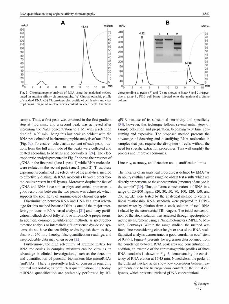
sample. Thus, a first peak was obtained in the first gradientstep at 4.32 min., and a second peak was achieved afterincreasing the NaCl concentration to 1 M, with a retentiontime of 14.99 min., being this last peak coincident with theRNA peak obtained in chromatographic analysis of total RNA(Fig. 3a). To ensure nucleic acids content of each peak, frac-tions from the full amplitude of the peaks were collected andtreated according to Martins and co-workers [24]. The elec-trophoretic analysis presented in Fig. 3b shows the presence ofgDNA in the first peak (lane 1; peak 1) while RNA moleculeswere isolated in the second peak (lane 2; peak 2). Thus, theseexperiments confirmed the selectivity of the analytical methodto effectively distinguish RNA molecules between other bio-molecules present in cell lysates. Moreover, despite the fact ofgDNA and RNA have similar physicochemical properties; agood resolution between the two peaks was achieved, whichsupports the specificity of arginine-based chromatography.
Discrimination between RNA and DNA is a great advan-tage for this method because DNA is one of the major inter-fering products in RNA-based analysis [31] and many purifi-cation methods do not fully remove it fromRNA preparations.In addition, common quantification methods, as spectropho-tometric analysis or intercalating fluorescence dye-based sys-tems, do not have the sensibility to distinguish them as theyabsorb at 260 nm, thereby, false quantification readings, andirreproducible data may often occur [32].
Furthermore, the high selectivity of arginine matrix forRNA molecules in complex mixtures can be view as anadvantage in clinical investigations, such as the detectionand quantification of potential biomarkers like microRNAs(miRNAs). There is presently a lack of consensus regardingoptimal methodologies for miRNA quantification [33]. Today,miRNAs quantification are preferably performed by RT-
qPCR because of its substantial sensitivity and specificity[34]; however, this technique follows several initial steps ofsample collection and preparation, becoming very time con-suming and expensive. The proposed method presents theadvantage of detecting and quantifying RNA molecules insamples that just require the disruption of cells without theneed for specific extraction procedures. This will simplify theprocess and improve economics.
Linearity, accuracy, and detection and quantification limits
The linearity of an analytical procedure is defined by EMA “asits ability (within a given range) to obtain test results which aredirectly proportional to the concentration (amount) of analyte inthe sample” [30]. Thus, different concentrations of RNA in arange of 20–200 ng/μL (20, 30, 50, 70, 100, 120, 150, and200 ng/μL) were tested by the analytical method to verify alinear relationship. RNA standards were prepared in DEPC-treated water by dilution from a stock solution of total RNAisolated by the commercial TRI reagent. The initial concentra-tion of the stock solution was assessed through spectrophoto-metric measurement using a NanoPhotometer (IMPLEN, Mu-nich, Germany). Within the range studied, the method wasfound linear considering either height or area of the RNA peak.Statistical analysis demonstrated a good correlation coefficientof 0.9991. Figure 4 presents the regression data obtained fromthe correlation between RNA peak area and concentration. Inaddition, an example of the chromatographic profiles of threeRNA standards is shown in Fig. 5, demonstrating the consis-tency of RNA elution at 15.45 min. Nonetheless, the peaks ofthe different nucleic acids show low correlation between ex-periments due to the heterogeneous content of the initial celllysates, which presents unrelated gDNA concentrations.
Fig. 3 Chromatographic analysis of RNA using the analytical methodbased on arginine affinity chromatography. (A) Chromatographic profileof standard RNA. (B) Chromatographic profile of cell lysates and elec-trophoresis image of nucleic acids content in each peak. Fractions
corresponding to peaks (1) and (2) are shown in lanes 1 and 2 , respec-tively. Lane L , PC-3 cell lysate injected onto the analytical argininecolumn
RNA quantification using arginine-affinity chromatography 8853
117

Table 1 shows the results of accuracy assessed by back-calculated concentration for all standards and for an interme-diate RNA concentration of 80 ng/μL. Thus, the closenessagreement between the values of the nominal concentrationsand RNA concentrations calculated using the regression equa-tion was evaluated by determining the coefficient of variation(CV) and the mean relative error. Both parameters were withinthe range of ±15 % of the nominal concentration for all RNAconcentrations studied, including the lowest concentrationstandard of 20 ng/μL which shows a relative error of 5.24 %(Table 1), which is far below the value of 20% accepted by theguidelines [30]. In fact, the lowest RNA standard was
established as the limit of quantification, that was defined asthe minimal RNA amount for which the arginine-based chro-matography method has a suitable level of precision (a CVlower than ±20%) and accuracy (within ±20% of the nominalconcentration). Additionally, the 20 ng/μL was also consid-ered the limit of detection taking into account the baselinenoise. This parameter was determined by comparing chro-matographic signals of blank samples containing 1 M NaClin Tris buffer pH 8 and the lowest RNA concentration. Theassays showed a signal-to-noise ratio of 2:1 which is generallyacceptable for estimating the detection limit [30]. Althoughcommon methods for RNA quantification show lowerlimits of quantification than the proposed method, theirrange of quantification is also low, normally between3 μg/μL and 2 ng/μL for nano-spectrophometers or500–0.05 ng/μL, for bioanalyzers [35]. This can limitthe quantification of RNA produced in a large scale, asin the case of RNA vaccines [36]. Therefore, the pro-posed method presents an advantage over other quanti-fication methods since it offers the possibility to in-crease the upper limit of the quantification range vali-dated here. Based on our previous work, arginine affin-ity chromatography allows the quantification of at least35 μg of RNA [24] nevertheless the determination ofdynamic binding capacity of this matrix is required toestablish the highest quantification limit.
Fig. 4 Linear regression data obtained from the plot of peak areas asfunction of RNA concentration. R2 coefficient of determination of thelinear regression
Fig. 5 Representativechromatograms of three RNAstandards with concentrations of20, 50, and 100 ng/μL used in thevalidation of the analyticalmethod
8854 R. Martins et al.
118

Precision
The investigation of precision reflects the closeness agreementof individual measurements of RNAwhen the analytical pro-cedure is applied repeatedly to multiple sampling of homoge-neous RNA reference material, and it can be studied at twolevels: repeatability and reproducibility, also termed as intra-and inter-day precision, respectively. The values for intra-dayprecision were obtained from consecutive injections of stan-dard RNA of a minimum of three concentration levels (low,medium, and high) over a minimal of nine determinations.The same samples were also analyzed over a period of threeconsecutive days to determine the inter-day precision. Theseries of measurements were analyzed by determining statis-tical parameters related to precision which are presented inTables 2 and 3. The calculated mean, standard deviation, CV,and mean relative error for three RNA concentrations of 30,80, and 150 ng/μL concerning repeatability (Table 2) andreproducibility (Table 3) showed an adequate precision forthis new method. The CV values were lower than 2 or 3 % inintra- and inter-day precision, respectively. Moreover, thedeviation from the nominal concentration (mean relative er-ror) was between −1.17 and 3.59 % for intra-day experimentsand inter-day values did not exceed 5.33 %, which alsoconfirms the accuracy found for the proposed analyticalmethod.
Building on the successful results present so far, the meth-od is concluded to be simple, selective, linear, precise, and
accurate in a concentration range adequate to measure RNAcontent and to assess its purity. Moreover, several flaws incurrent RNA quantification techniques may be overcomeusing the described analytical arginine affinity method.
Method application
The applicability and robustness of the analytical arginine-affinity chromatography method for RNA quantification wasfurther explored by using RNA samples from different sourcesand preparation methods. The intention of these assays was toevaluate the broad use of the method regardless the origin ofthe RNA or its purpose. Therefore, total RNA obtained fromHeLa cells and crude samples of RNA chemically synthesizedby in vitro transcription were tested according to the procedurepreviously described for RNA-based samples from PC-3 cells.Figure 6 shows the chromatographic analysis of total RNAsamples from HeLa cells with an electrophoretic image of theinjected sample (lane H). The displayed peaks show similarretention times when compared with the chromatographicprofiles of total RNA from PC-3 cells (Fig. 3). In case of peakheight, RNA from HeLa cells revealed lower peaks butbroader than PC-3 samples. The differences in chromato-graphic signals were found to be due to the predominance ofdifferent species of RNA in the samples. Accordingly, elec-trophoretic analysis of RNAmolecules isolated from each cellline also allowed the virtual perception of lower molecular
Table 1 Evaluation of accuracyof the proposed analytical method(n =3)
SD standard deviation, CV coef-ficient of variation
[Nominal] (ng/μL) Mean (RNA)±SD (ng/μL) CV (%) Mean relative error (%)
20 20.63±0.39 5.24 0.03
30 32.98±0.75 5.55 0.10
50 50.02±0.71 3.22 0.00
70 68.28±0.28 0.92 −0.02100 97.89±0.47 1.03 −0.02120 121.96±0.29 0.51 0.02
150 148.58±0.08 0.11 −0.10200 204.81±0.35 0.36 0.02
80 81.48±0.57 1.52 0.02
Table 2 Determination of intra-day variability of the analytical methodfor RNA assessment (n=3)
Intra-day precision
[Nominal](ng/μL)
Mean (RNA)±SD(ng/μL)
CV (%) Mean relativeerror (%)
30 31.10±0.30 0.98 3.59
80 81.59±1.18 1.45 1.99
150 148.24±0.86 0.58 −1.17
SD standard deviation, CV coefficient of variation
Table 3 Determination of inter-day variability of the analytical methodfor RNA assessment (n=3)
Inter-day precision
[Nominal](ng/μL)
Mean (RNA)±SD(ng/μL)
CV (%) Mean relativeerror (%)
30 31.60±0.94 2.97 5.33
80 81.73±0.39 0.48 2.16
150 148.00±0.41 0.27 −1.33
SD standard deviation, CV coefficient of variation
RNA quantification using arginine-affinity chromatography 8855
119

weight RNA species in HeLa cells (Fig. 6) than in PC-3 cells(Fig. 2). The isolation of dissimilar RNA species from eachcell line could be expected because of their unparalleled lifecycles, which are differently favoring the production of onetype of RNA over another to accomplish physiological cellfunction [37]. Nevertheless, peak areas of the injected con-centrations of RNA from HeLa cells are consistent with RNAstandards from PC-3 cells, giving concordant results betweenthe injected concentration and the concentration achieved byextrapolation of the peak area on the regression equation.
With regard to the applicability of the proposed analyticalmethod to monitor and quantify chemically synthesized RNAfrom a crude sample obtained by in vitro transcription, argi-nine column also demonstrated an appropriate performancefor this purpose. In these assays, the applicability of the
method was attested either by the evaluation of the chromato-graphic behavior or by the electrophoretic analysis of theeluting peaks. Figure 7 shows the chromatographic profile ofa crude sample of chemically synthesized RNA together withthe electrophoretic image of the injected sample and theresulting peaks. Again, the second peak corresponds to RNAelution, confirmed by the similar retention times and electro-phoretic analysis (peak 2; lane 2). However, the first peakpresented a retention time of 4.94 min., which is not coinci-dent with the retention time of gDNA from PC-3 cell lysates(4.32 min). Based on the electrophoretic result, it was hypoth-esized that the peak did not refer to the elution of a nucleicacid. Thus, as no previous purification was performed in thetranscription-based RNA samples, the existence of a first peakcan be due to impurities, such as enzymes, nucleotides, salts,
Fig. 6 Chromatographic analysisof RNA isolated from HeLa cellsand electrophoretic profile of theRNA sample (lane H) injectedonto the analytical argininecolumn
Fig. 7 Chromatographic profileof crude sample of chemicallysynthesized RNA andelectrophoretic analysis of theinjected sample and resultingpeaks. Fractions corresponding topeaks (1) and (2) are shown inlanes 1 and 2 , respectively. LaneC , crude sample of chemicallysynthesized RNA by in vitrotranscription injected onto theanalytical arginine column
8856 R. Martins et al.
120

buffer or other components that derive from the in vitro tran-scription process and may absorb at 260 nm. Therefore, theaccurate quantification of chemically synthesized RNA canalso be achieved by arginine affinity chromatography method,nonetheless further evaluation of the presence of impurities inRNA peaks should be assessed by complementary techniques.
Conclusions
The requirement of new analytical techniques for rigorousanalysis and quantification of RNA samples meeting regula-tory demands is becoming evident as basic research improvesthe understanding of biological roles of RNA and pharmaceu-tical industry starts to focus on it as a biotherapeutic tool.
In this work, an analytical technique based on arginine-affinity chromatography for quantification and quality verifi-cation of total RNAwas developed and validated. The methodwas found linear in the range of 20–200 ng/μL, precise andaccurate, according to international guidelines. The specificitywas determined with cell lysates, showing that gDNA is wellseparated from RNA molecules, which confirms the greatselectivity of arginine column and highlights the great advan-tage of this method over other quantification techniques.Moreover, the current methods used for RNA detection andmonitorization were discussed and compared with the pro-posed analytical technique, underlining the several advantagesof the new methodology.
Furthermore, the proposed method was successfullyemployed in the quantification and monitorization of differentRNA types, namely RNA isolated from HeLa cells and chem-ically synthesized by in vitro transcription, which demon-strates its potential applicability in basic research and RNA-based therapies.
Acknowledgments This work was supported by FCT, the PortugueseFoundation for Science and Technology (PTDC/EBB-BIO/114320/2009 andPEst-C/SAU/UI0709/2011 COMPETE). Rita Martins also acknowledges afellowship (SFRH/BD/ 64100/2009) from FCT.
Conflict of interest The authors declare no conflict of interest.
References
1. Sharp PA (2009) The centrality of RNA. Cell 136(4):577–5802. Kreiter S, Diken M, Selmi A, Tureci O, Sahin U (2011) Tumor
vaccination using messenger RNA: prospects of a future therapy.Curr Opin Immunol 23(3):399–406
3. Vermeulen J, De Preter K, Lefever S, Nuytens J, De Vloed F,Derveaux S, Hellemans J, Speleman F, Vandesompele J (2011)Measurable impact of RNA quality on gene expression results fromquantitative PCR. Nucleic Acids Res 39(9):e63
4. Gjerde DT, Hoang L, Hornby D (2009) RNA purification and anal-ysis: sample preparation, extraction, chromatography. Wiley,Weinheim
5. Farrell RE (2012) RNA methodologies. In: Meyers RA (ed) Epige-netic regulation and epigenomics, vol 2, 1st. Wiley, Weinheim, pp 1–37
6. Wieczorek D, Delauriere L, T. S (October 2012) Methods of RNAquality assessment. Promega Corporation Website. http://worldwide.promega.com/resources/articles/pubhub/methods-of-rna-quality-assessment. Accessed 20 Feb 2013
7. Heptinstall J, Rapley R (2000) Spectrophotometric analysis ofnucleic acids. In: Rapley R (ed) The nucleic acid protocols handbook.Humana Press, Totowa, pp 57–60. doi:10.1385/1-59259-038-1:57
8. Barril P, Nates S (2012) Introduction to agarose and polyacrylamidegel electrophoresis matrices with respect to their detection sensitivi-ties. In: Magdeldin DS (ed) Gel electrophoresis—principles andbasics. InTech, Rijeka. doi:10.5772/38573
9. Ohta T, Tokishita S, Yamagata H (2001) Ethidium bromide andSYBRGreen I enhance the genotoxicity of UV-irradiation and chem-ical mutagens in E . coli . Mutat Res 492(1–2):91–97
10. Jones LJ, Yue ST, Cheung C-Y, Singer VL (1998) RNA quantitationby fluorescence-based solution assay: RiboGreen reagent character-ization. Anal Biochem 265(2):368–374
11. Schmittgen TD, Lee EJ, Jiang J, Sarkar A, Yang L, Elton TS, Chen C(2008) Real-time PCR quantification of precursor and maturemicroRNA. Methods 44(1):31–38
12. Masotti A, Preckel T (2006) Analysis of small RNAswith the Agilent2100 Bioanalyzer. Nat Methods. doi:10.1038/NMETH908
13. Dickman MJ (2011) Ion pair reverse-phase chromatography: a ver-satile platform for the analysis of RNA. Chromatography Today 4:22
14. McCarthy SM, Gilar M, Gebler J (2009) Reversed-phase ion-pairliquid chromatography analysis and purification of small interferingRNA. Anal Biochem 390(2):181–188
15. Azarani A, Hecker KH (2001) RNA analysis by ion-pair reversed-phase high performance liquid chromatography. Nucleic Acids Res29(2):E7
16. Dickman MJ, Hornby DP (2006) Enrichment and analysis of RNAcentered on ion pair reverse phase methodology. RNA 12(4):691–696
17. Waghmare SP, Pousinis P, Hornby DP, DickmanMJ (2009) Studyingthe mechanism of RNA separations using RNA chromatography andits application in the analysis of ribosomal RNA and RNA:RNAinteractions. J Chromatogr A 1216(9):1377–1382
18. Clonis YD (2006) Affinity chromatography matures as bioinformaticand combinatorial tools develop. J Chromatogr A 1101(1–2):1–24
19. Magdeldin S, Moser A (2012) Affinity chromatography. In:Magdeldin DS (ed) Affinity chromatography: principles and appli-cations. InTech, Rijeka. doi:10.5772/39087
20. Srisawat C, Goldstein IJ, Engelke DR (2001) Sephadex-bindingRNA ligands: rapid affinity purification of RNA from complexRNA mixtures. Nucleic Acids Res 29(2):E4
21. Batey RT, Kieft JS (2007) Improved native affinity purification ofRNA. RNA 13(8):1384–1389
22. Martins R, Queiroz JA, Sousa F (2010) A new affinity approach toisolate Escherichia coli 6S RNA with histidine-chromatography. JMol Recognit 23(6):519–524
23. Martins R, Queiroz JA, Sousa F (2012) Histidine affinitychromatography-based methodology for the simultaneous isolationof Escherichia coli small and ribosomal RNA. Biomed Chromatogr26(7):781–788
24. Martins R, Maia CJ, Queiroz JA, Sousa F (2012) A new strategy forRNA isolation from eukaryotic cells using arginine affinity chroma-tography. J Sep Sci 35(22):3217–3226
25. Sousa F, Cruz C, Queiroz JA (2010) Amino acids–nucleotides bio-molecular recognition: from biological occurrence to affinity chro-matography. J Mol Recognit 23(6):505–518
RNA quantification using arginine-affinity chromatography 8857
121

26. Treger M, Westhof E (2001) Statistical analysis of atomic contacts atRNA–protein interfaces. J Mol Recognit 14(4):199–214
27. Jeong E, Kim H, Lee SW, Han K (2003) Discovering the interactionpropensities of amino acids and nucleotides from protein–RNA com-plexes. Mol Cells 16:161–167
28. Yarus M,Widmann JJ, Knight R (2009) RNA–amino acid binding: astereochemical era for the genetic code. J Mol Evol 69(5):406–429
29. U.S. Department of Health and Human Services (2001) Guidance forindustry, bioanalytical method validation. UD Food and Drug Ad-ministration, Rockville
30. Kaighn ME, Narayan KS, Ohnuki Y, Lechner JF, Jones LW (1979)Establishment and characterization of a human prostatic carcinomacell line (PC-3). Investig Urol 17(1):16–23
31. Laurell H, Iacovoni JS, Abot A, Svec D,Maoret JJ, Arnal JF, KubistaM (2012) Correction of RT-qPCR data for genomic DNA-derivedsignals with ValidPrime. Nucleic Acids Res 40(7):e51
32. Fleige S, Pfaffl MW (2006) RNA integrity and the effect onthe real-time qRT-PCR performance. Mol Aspects Med 27(2–3):126–139
33. Mo MH, Chen L, Yebo F, Wang W, Fu SW (2012) Cell-free circu-lating miRNA biomarkers in cancer. J Cancer 3:432–448
34. Kang K, Peng X, Luo J, Gou D (2012) Identification of circulatingmiRNA biomarkers based on global quantitative real-time PCRprofiling. J Anim Sci Biotechnol 3(1):4
35. Aranda R, Dineen SM, Craig RL, Guerrieri RA, Robertson JM(2009) Comparison and evaluation of RNA quantification methodsusing viral, prokaryotic, and eukaryotic RNA over a 10(4) concen-tration range. Anal Biochem 387(1):122–127
36. Pascolo S (2006) Vaccination with messenger RNA. Methods MolMed 127:23–40
37. Salem C, El-AlfyM, Leblond CP (1998) Changes in the rate of RNAsynthesis during the cell cycle. Anat Rec 250(1):6–12
8858 R. Martins et al.
122

123

124

Paper VI
Arginine-affinity chromatography for mRNA vaccines
purification
R. Martins, C. J. Maia, J. A. Queiroz, F. Sousa
(to submit)
125

126

Arginine-affinity chromatography for mRNA
vaccines purification
R. Martins, C. J. Maia, J. A. Queiroz, F. Sousa*
CICS-UBI – Health Sciences Research Centre, University of Beira Interior, Av.
Infante D. Henrique, 6200-506 Covilhã, Portugal.
*corresponding author:
E-mail: [email protected]
Telephone: +351 275 329 002
Fax: +351 275 329 099
127

128

Abstract
Coding messenger RNA (mRNA) is emerging as a particularly attractive option in the
development of new approaches for the treatment of cancer or infection diseases focusing on
immunotherapies. Cervical cancer is the second most common cancer in women worldwide.
Human papillomavirus (HPV) is the primary etiologic agent of cervical cancer and the two
HPV16 proteins, E6 and E7, are prospective therapeutic vaccines as they are consistently
expressed in tumour cells.
Because of the increasing success of mRNA-based immunotherapies, the development of new
tools to improve RNA purification is of great significance since rigorous quality criteria
recommended by international regulatory authorities should be fulfilled. Therefore, a
recently described chromatographic strategy for RNA purification was considered, exploiting
affinity interactions between immobilized amino acids and nucleic acids.
In the present study, a single arginine affinity chromatography step was employed for the
purification of mRNA, produced by enzymatic synthesis, encoding HPV16 E6 and E7 proteins.
The chromatographic experiments were developed by optimizing a sodium chloride elution
gradient to purify the mRNA transcripts from impurities resultant from the production
process. The evaluation of the purification process revealed that both E6 and E7 mRNAs are
recovered with high yield, 97.5% and 98%, respectively. Furthermore, the quality control
showed that mRNA preparations presented high integrity as well as an improved purity. The
results obtained so far indicated that the chromatographic separation is a promising strategy
for mRNA purification pursuing therapeutic applications.
Keywords
Affinity chromatography, arginine, messenger RNA, in vitro transcription.
129

1. Introduction
Recently, messenger RNA (mRNA) is being an important target of several research studies,
due to its potential applicability in the treatment of cancer or infection diseases focusing on
immunotherapies (Kreiter et al., 2011). The physiological role of mRNA is to transfer genetic
information from the nucleus to the cytoplasm where this information is translated into the
corresponding protein. The concept of mRNA vaccination is to carry the information of an
antigenic protein to be translated in the cell cytoplasm and therefore generate an
immunological response (Bringmann et al., 2010). Recent studies describe that mRNAs
encoding antigens are able to stimulate all effectors of the adaptive immune response: B
lymphocytes, cytotoxic T cells, and T helper cells (Ponsaerts et al., 2003). Besides, it has
demonstrated several advantages to be applied as a vaccine including feasibility,
applicability, safeness, and effectiveness when it comes to the generation of immune
responses (Pascolo, 2008, Van Lint et al., 2013). In particular, the safety of mRNA-based
treatments supports the use of mRNA-vaccination for therapeutic or prophylactic approaches
as it addresses some shortcomings of recombinant virus or DNA-based vaccination therapies
(Vergati et al., 2010).
Cervical cancer is the second most common cancer in women worldwide. Human
papillomavirus (HPV) is the primary etiologic agent of cervical cancer and HPV16 is by far the
most common carcinogenic type (Huh, 2001). Two HPV16 proteins, E6 and E7, are consistently
expressed in tumour cells and are critical to the induction and maintenance of cellular
transformation. Thus, E6 and E7 proteins represent good targets for developing therapeutic
vaccines for cervical cancer (Bharti et al., 2009). Currently, a prophylactic vaccine of HPV16
and HPV18 has been registered with successful results in cervical cancer prevention, but it
has no benefit in women who were already infected with those HPV types (Franco et al.,
2009). Thus, the necessity to develop effective therapeutic HPV vaccines continues and the
use of mRNA-based vaccination has been described to be a promising approach (Thornburg et
al., 2000).
Due to the increasing success of clinical application of mRNA-based immunotherapies, the
development of new tools to improve RNA manufacture process is of great significance. Of
particular importance is the purification process, since the RNA bioproduct should fulfill
rigorous quality criteria recommended by international Regulatory Authorities, such as the
World Health Organization (WHO), Food and Drug Administration (FDA) and European
Medicines Agency (EMA) (Pascolo, 2008).
Therapeutic mRNA is currently produced by cloning the gene encoding a desired protein in a
plasmid containing regulatory sequences (poly(A) tail, untranslated regions (UTRs)), followed
by amplification in Escherichia coli (E. coli). Next, the plasmid is recovered, linearized,
130

purified and in vitro transcribed using a RNA polymerase that recognizes the bacteriophage
promoter (Weide et al., 2008). However, final RNA products need to be isolated from
impurities derived from the synthesis process. In general, these impurities are the plasmid
DNA (pDNA) template, proteins, nucleotides and aberrant oligonucleotides, salts and buffers.
The current purification strategies for synthetic mRNA include the precipitation with high
concentration of lithium chloride (LiCl) (Pascolo, 2006). However, most of the time this
precipitation does not completely remove traces of contaminating pDNA or proteins and
worse it introduces lithium metal into preparations. Thus, to improve mRNA purity,
phenol/chloroform extraction often accompanies LiCl precipitation (Pascolo, 2006). Although,
this methodology achieves highly pure mRNA preparations, it employs extremely hazardous
compounds, which are not recommended to be used in clinical applications. Otherwise, mRNA
can be purified using affinity chromatography and, in fact, this is strongly recommended for
therapeutic applications of mRNA, as in vaccination approaches (Pascolo, 2008). However, the
description of these affinity strategies is not totally available in the literature.
Therefore, new chromatographic strategies for mRNA purification were considered,
exploiting affinity interactions between amino acids and nucleic acids. The potential of using
affinity chromatography with histidine or arginine as ligands has been recently demonstrated
by our research group to selectively isolate and monitor different RNA species of various cell
sources (Martins et al., 2012a, Martins et al., 2010, Martins et al., 2012b, Martins et al.,
2013). Histidine matrix demonstrated to mediate a specific recognition for a regulatory non-
coding RNA (ncRNA), 6S RNA, allowing its purification from a complex mixture of other E.coli
ncRNAs (Martins et al., 2010). Furthermore, the simultaneous isolation of ncRNA and
ribosomal RNA from E.coli cell lysates, eliminating host DNA and proteins, was also attained
by using an histidine chromatography-based method (Martins et al., 2012b). Moreover,
arginine-agarose support enabled the isolation of total RNA from impurities of an eukaryotic
cell extract (Martins et al., 2012a) and was also used in the development of an analytical
methodology for quantification and monitorization of RNA molecules (Martins et al., 2013).
Hence, this work intends to explore the possibility of using arginine affinity chromatography
to improve the purification of mRNA transcripts encoding the oncogenic proteins of HPV16
pursuing the development of a new and enhanced methodology for mRNA based-vaccination.
131

2. Experimental section
2.1. Materials
For plasmid design, 1321 HPV16 E6/E7 and T7TS plasmids were obtained in Addgene, a
plasmid repository (Cambridge, USA). Synthetic oligonucleotides used as primers were
purchased in StabVida (Lisboa, Portugal). Restriction enzymes BglII and EcoRV were acquired
in Fermentas (Thermo fisher scientific Inc) and T4 DNA ligase was from Promega (Madison,
USA). For production of mRNA molecules, MEGAscript® Kit was acquired in Ambion (Carlsbad,
CA, USA) and Sma1 restriction enzyme was from Takara Bio Inc. (Shiga, Japan).
For the purification process, Arginine Sepharose 4B gel was obtained in GE Healthcare
(Uppsala, Sweden). The compounds used in chromatographic experiments were sodium
chloride (NaCl) purchased from Panreac (Barcelona, Spain) and Tris base from Fluka (Sigma,
St Louis, MO, USA). All solutions were freshly prepared using 0.01% diethylpyrocarbonate
(DEPC)-treated water from Fluka (Sigma) and buffers were filtered through a 0.20 µm pore
size membrane (Schleicher Schuell, Dassel, Germany) and degassed ultrasonically. For the
nucleic acid gel electrophoresis it was used GreenSafe Direct Load stain from NZYTech
(Lisbon, Portugal) and DNA molecular weight marker, HyperLadder I, was obtained in Bioline
(London, UK). All experiments were performed with RNase-free disposables.
2.2. Plasmid construct
HPV16 E6 and E7 genes were isolated by polymerase chain reaction (PCR) from the 1321 HPV-
16 E6/E7 plasmid (Addgene plasmid 8641 (Munger et al., 1989)), which contains the E6 and E7
genes. A set of primers corresponding to the 5’ and 3’ regions of E6 or E7 genes were used
(Table 1). PCR was performed under standard conditions (95ºC for 30 sec, 60ºC for 30 sec and
72ºC for 15 sec, for 35 cycles) and the products were separated on a 1% agarose gel
electrophoresis. The band corresponding to the expected PCR product size was cut out and
the DNA was purified by NucleoSpin® Gel and PCR clean-up (Macherey-Nagel GmbH & Co,
Düren, Germany). Purified DNA was cut with BglII and EcoRV and ligated using T4 DNA ligase
into a similarly cut prokaryotic expression vector T7TS plasmid (Addgene 17091) (figure 1).
The ligation mixture was used to transform DH5α E. coli competent cells by heat shock and
selected for ampicillin resistance. Isolated colonies were collected to check for the presence
of plasmid. The cloned HPV16 E6 or E7 genes in pT7TS were verified by PCR after performing
the two restriction enzymes digestion (figure 2). E6 and E7 coding regions were verified by
sequencing (StabVida, Lisboa, Portugal). Finally, the pT7TS E6 and pT7TS E7 (figure 1) were
amplified by fermentation of E. coli DH5 in shake flasks at 37ºC and 250 rpm with 250 mL of
Terrific Broth medium (12 g/L tryptone, 24 g/L yeast extract, 4 mL/L glycerol, 0.017 M
KH2PO4, 0.072 M K2HPO4). Cell growth was suspended in the late log phase (OD600 ≈ 12). Cells
132

were recovered by centrifugation and the plasmids were extracted by NZYMaxiprep
(NZYTech, Lisboa, Portugal) and stored at -20ºC or readily used in the production of mRNA
molecules.
Figure 1 - Graphic map of the pT7TS plasmid backbone with the inserted fragments HPV16 E6 or
HPV16 E7 genes. The restriction sites used for cloning E6 or E7 genes (BglII and EcoRV) and the
restriction site used for linerarization (Sma1) are shown. The hatched box represent the β-globin 5' UTR
(approx. 60 bp); the gray box represents the place where HPV16 E6 or HPV16 E7 coding genes (E6,
approx 476bp and E7, approx 296bp) are inserted; the dotted box represents the β-globin 3' UTR (approx
200 bp) and the poly-A tail (30 A); the dotted ellipse represents the origin of replication and the white
box represents the ampicilin-resistance gene. (HPV16, human papillomavirus 16; UTR, untranslated
region: bp, base pairs, A, adenine).
133

Figure 2 - Construction of the prokaryotic recombinant expression vectors of pT7TS E6 and pT7TS
E7. (A) P1, pT7TS E6; P2, pT7TS E7; (B) 1, E6 PCR product; 2, enzyme digestion (BglII/EcoRV) of pT7TS
E6; 3, E7 PCR product; 4, enzyme digestion (BglII/EcoRV) of pT7TS E7. (M, DNA molecular weight
marker; bp, base pair).
2.3. Preparation of mRNA-based samples
mRNA encoding E6 or E7 proteins were enzymatically synthetized using MEGAscript® Kit,
according to the manufacturer’s recommendations. Briefly, the constructed plasmids
containing E6 or E7 genes were linearized with Sma1 enzyme and concentrated to a final
concentration of 1 μg/μL. Then, the linearized plasmids were in vitro transcribed with the
reagents provided in the kit for 6h at 37ºC in order to produce the mRNA molecules. Next, the
kit procedure was interrupted because it was intended to purify mRNAs by the proposed
arginine affinity chromatography method. Thus, differently from the kit´s instructions, the
reactions were terminated by adding 1/20th volume 0.5 M EDTA, 1/10th volume of 3M sodium
acetate and two volumes of ethanol, followed by incubation at –20°C for at least 15 min.
After centrifugation at 4°C for 15 min at maximum speed, the supernatant was discarded and
the tubes were re-spin for a few seconds to eliminate residual fluid. Then, the pellets were
solubilized in DEPC-treated water. Final mRNA-based preparations were qualitatively
evaluated for purity and integrity by non-denaturing agarose gel electrophoresis and their
concentration was estimated by spectrophotometric analysis. The samples were stored frozen
at –80°C until be used in purification studies by arginine affinity chromatography. As a
reference point, for the new purification procedure, other crude transcription reactions were
concluded and purified using the conventional procedure of precipitation with LiCl. In this
case, the procedure was performed according to the manufacturer’s instruction of
MEGAscript® Kit (Ambion). Briefly, transcription reaction was stopped and mRNAs were
precipitated by adding 1/2 of transcription reaction volume of nuclease-free water and 1/2
volume of LiCl precipitation solution and mixed thoroughly. After ≥30 min incubation at –
20°C, the samples were centrifuged at 4°C for 15 min at maximum speed to pellet the
134

mRNAs. The pellets were washed once with ~1 mL 75% ethanol and re-centrifuged. Finally,
mRNA preparations were resuspended in DEPC-treated water and stored frozen at –80°C, to
be used as comparison in the quality verification and concentration assessment.
2.4. Purification of mRNAs encoding E6 and E7 proteins
Chromatographic experiments were carried out using an ÄKTA Avant system with UNICORN 6
software (GE Healthcare, Uppsala, Sweden). It was used a commercial arginine–sepharose 4B
gel characterized by the manufacturer as a cross-linked 4% beaded agarose matrix with a 12-
atom spacer and an extent of labelling between 14 and 20 μmoL/mL. The stationary phase
was packed in a 10 mm diameter×20 mm long (~2 mL) column. Temperature was maintained
at 15ºC during the experiments by a circulating water bath. The system was initially
prepared with ice-cold solutions of 10 mM Tris-HCl buffer, pH 8.0 in pump A and 1 M NaCl in
10 mM Tris-HCl buffer, pH 8.0 in pump B. The column was equilibrated with a mobile phase
consisting of 34% of buffer B at a flow rate of 1.0 mL/min. Conductivity was rigorously
controlled at 31 mS/cm. Crude transcription samples containing mRNA encoding E6 or E7
proteins were injected using a 50 µL loop at the same conditions (flow rate, mobile phase and
conductivity). After 5 min run, the ionic strength of the mobile phase was increased to 100%
buffer B at the same flow rate of 1 mL/min. These conditions were kept for 7 min. The
chromatographic runs were monitored by an UV detector at 260 nm. To identify the eluting
species, peak fractions were pooled according to the chromatograms obtained and comprising
the full amplitude of the peak. After each run, the column was washed with DEPC-treated
water and re-equilibrated with equilibration buffer for 20 min. For the identification of the
nucleic acid species eluting in each peak, the samples were resolved on a 1.2% native agarose
gel in TAE buffer (40 mM Tris base, 20 mM acetic acid and 1 mM EDTA, pH 8.0) prepared in
DEPC-treated water. The gels were stained with GreenSafe (0.5 μg/mL), and photographed.
The optical density of the crude transcription samples and pooled fractions was determined
at 260 nm using a Nanodrop spectrophotometer to assess their quantity. The quality
characterization of mRNA molecules was further performed as described below.
2.5. Protein contamination
Protein contamination in mRNA samples resulting either from purification with arginine-
agarose support or purified by LiCl, was assessed using the micro-BCA (bicinchoninic acid)
assay (Thermo fisher scientific Inc.), according to manufacturer’s instructions. Briefly, the
calibration curve was prepared using BSA standards (0.01–0.1 mg/mL). 25 μL of each standard
or RNA sample were added to 200 μL of BCA reagent in a microplate and incubated for 30 min
at 60 ºC. Absorbance was measured at 570 nm in a microplate reader.
135

2.6. Plasmid DNA template contamination
pT7TS E6 or pT7TS E7 contamination was evaluated in mRNA samples purified by arginine
matrix or LiCl precipitation in an iQ5 Multicolor Real-Time PCR Detection System (Bio-Rad
Laboratories, Hercules, CA, US). Specific primers to HPV16 E6 or HPV16 E7 (Table 1) were
used for the template amplification. The real-time PCR efficiency was determined for both
primers using serial dilutions of mRNA samples (1:1, 1:10, 1:100 and 1:1000). Real-time PCR
reactions were carried out using 1 µL of all mRNA preparations in a 20 µL reaction mix
containing 10 µl Maxima SYBR Green/ROX qPCR Master mix (Fermentas, Thermo fisher
scientific Inc.) and 300 nM HPV16 E6 or HPV16 E7 primers. All reactions were performed in
96-well plates for PCR heat-sealed with heat sealing film (Bio-Rad Laboratories, Hercules, CA,
US). After an initial denaturation at 95ºC for 5 min, cycling conditions were used as follows:
40 cycles consisting of denaturation at 95ºC for 30 s, annealing at 60ºC for 30 s and extension
at 72ºC for 1 min. Final elongation was at 72ºC for 5min. The amplified PCR fragments were
checked by melting curves: reactions were heated from 55 to 95ºC with 10 s holds at each
temperature (0.05ºC/s). Samples were run in triplicate for each assay. Results were analysed
using iQ5 optical system software version 2.0 after manual adjustment of the baseline and
fluorescence threshold.
Table 1 Primer sequences of E6 and E7 genes used for amplification. BglII and EcoRV restriction sites
are shown in bold and the start and stop codons are underlined.
Primer Sequence Restriction site
HPV16 E6 Sense: 5’-CCAGATCTATGCACCAAAAGAGAACTGC-3’
Antisense: 5’-CCGATATCTTACAGCTGGGTTTCTCTAC-3’
BglII
EcoRV
HPV16 E7 Sense: 5’-CCAGATCTATGCATGGAGATACACCTAC-3’
Antisense: 5’-CCGATATCTTATGGTTTCTGAGAACAGA-3’
BglII
EcoRV
136

3. Results and Discussion
The newly explored approaches of mRNA-based gene therapy are rapidly gaining consistency
in cancer immunotherapy and in treating infection diseases. Furthermore, therapeutic mRNA
can be easily produced and up scaled to gram quantities for clinical trials within 3 months
(Pascolo, 2006), which is an advantage considering the number of patients that can be
treated following this approach. While the production process of mRNA vaccines under
current good manufacture practices (cGMP) is advantageously described for simplicity, the
purification strategies encompass various intermediate steps of precipitation with LiCl and
phenol/chloroform extractions. In addition, a final polishing step with affinity
chromatography is usually used, however, the procedure is not clearly described (Pascolo,
2006). Therefore in this work, we describe a new affinity method in which mRNA molecules
can be directly purified from crude transcription reactions using amino acid-based affinity
chromatography with arginine as a biospecific ligand, eliminating additional steps and
improving economics. Figure 3 schematically shows an overview of all the procedures
employed in this study in order to demonstrate the applicability of the proposed purification
method with arginine affinity chromatography.
Figure 3 - General procedure of the new methodology developed for purification of E6 and E7 mRNA
from crude transcription reactions. The process includes the design of a vector encoding for HPV16 E6
and E7 proteins and amplification in E. coli. The plasmid is extracted from the host and linearized to
facilitate in vitro transcription of the target mRNA. Next, crude transcription samples are purified by
affinity chromatography with arginine as immobilized ligand. After purification, the characterization of
mRNA molecules is performed in regard to recovery yield and purity. Finally, the performance of the
arginine affinity chromatography-based method should be evaluated by cell transfection experiments.
(pDNA, plasmid DNA; HPV16, human papillomavirus 16; E. coli, Escherichia coli; mRNA, messenger RNA).
137

3.1. Construction of the prokaryotic expression vectors
To synthetically produce mRNA encoding for E6 or E7 proteins, recombinant procedures were
used. Figure 1 represents the pDNA backbone, pT7TS, used for cloning and recombinant
production of E6 or E7 genes. This plasmid is already described in the literature for the
production of mRNA for vaccination because it contains regulatory sequences, such as UTR’s,
and a poly(A) tail. These confer structural modifications to mRNA molecules, which improve
the transcripts' stability and enhance their translation in cells. HPV16 E6 or E7 genes were
obtained from the eukaryotic expression vector p1321 HPV16 E6/E7 by PCR and were then
cloned into pT7TS through the two restriction sites of BglII and EcoRV to complete the design.
pT7TS also presents a linearization site that is useful for in vitro transcription as it improves
the process. The final preparation of the recombinant pT7TS E6 and pT7TS E7 were confirmed
by PCR and restriction enzyme digestion as it is shown in figure 2.
3.2. In vitro transcription of mRNA molecules
After amplification of the cloned vectors in E. coli DH5α, the plasmids were extracted from
the cells and linearized using SmaI restriction site originally present in pT7TS backbone. This
step is of particular importance for in vitro transcription of mRNA molecules since the use of
circular plasmid templates will generate extremely long, heterogeneous RNA transcripts
because RNA polymerases are very processive.
Transcription is a biological process of gene expression, in which a particular segment of DNA
is copied into RNA by the enzyme, RNA polymerase. Both RNA and DNA are nucleic acids,
which use base pairs of nucleotides as a complementary language that can be converted back
and forth from DNA to RNA by the action of the correct enzymes. During transcription, a DNA
sequence is read by an RNA polymerase, which produces a complementary, antiparallel RNA
strand. When the gene transcribed encodes a protein, the result of transcription is mRNA,
which will then be used to create that protein via the process of translation. In vitro
transcription techniques rely on the same principle but require a purified linear engineered
DNA template containing a bacteriophage promoter sequence, ribonucleotide triphosphates, a
buffer system that includes dithiothreitol and magnesium ions, and an appropriate phage RNA
polymerase (Beckert and Masquida, 2011).
The mRNA molecules produced by this process always need further purification since the
reagents used and other products of reaction, like aberrant oligonucleotides, can still be
present in the final preparations. Figure 4A shows the electrophoretic profile of the crude
transcription samples obtained in this study, which include the produced E6 and E7 mRNA
molecules and also residual pT7TS template and other long molecular weight impurities
resultant from the in vitro transcription reaction. Nonetheless, these samples did not present
abortive low molecular weight oligonucleotides that typically appear in the bottom of the gel.
138

After purification through the currently used LiCl precipitation procedure, those impurities
were no longer detected by electrophoretic analysis (figure 4B). The electrophoresis was
performed under non-denaturing conditions which do not reflect the correct molecular
weights of E6 and E7 mRNA molecules because of the presence of secondary structures. In
fact, in long transcripts as E6 mRNA, double strands structures by self-complementation can
also cause artefacts in mRNAs bands, displaying two bands instead of one as it visualized in
figure 4A and B.
Figure 4 - Non-denaturing agarose gel electrophoresis of mRNA-based samples. (A) Samples
recovered from crude transcription reactions with E6 and E7 mRNA molecules together with pDNA and
other nucleotide-based impurities. (B) E6 and E7 mRNA molecules purified by LiCl precipitation
compared with crude transcription samples without purification. Data are representative of several
independent experiments of in vitro transcription reaction. (Lane M, DNA molecular weight marker; bp,
base pair; pDNA, plasmid DNA template; MW, molecular weight).
3.3. Purification of mRNA molecules by arginine affinity
chromatography
The crude transcription samples previously analysed were purified by using affinity
chromatography with an arginine-agarose support in order to eliminate the impurities derived
from the production process. Chromatographic conditions were optimized concerning mRNA
retention in the column and the molecular stability. The temperature in the column was set
at 15ºC and ice cold solutions of 10mM Tris-HCl, pH 8 in pump A and 1M NaCl in 10mM Tris-
HCl, pH 8 in pump B were used. Different salt concentrations were tested to achieve mRNA
retention, in order to exploit favoured interactions between mRNA molecules and arginine-
agarose matrix that allow their purification. During these experiments it was verified the
139

need for strict control of chromatographic conditions in order to maintain the reproducibility,
since a slight variation in conductivity (salt concentration and/or temperature) affected
mRNAs retention (results not shown).
Figure 5 shows the chromatograms with the optimized elution gradient applied to arginine–
agarose chromatography for the purification of E6 and E7 mRNAs and the electrophoretic
analysis of the pure factions (figure 5A and B, respectively). The agarose gel electrophoresis
was used to detect and identify the nucleic acids eluting in each peak. Impure E6 and E7
samples (figure 5A and 5B, lanes C) were also run on the gel for comparative purposes.
mRNAs purification by arginine-agarose support was performed in two steps. After column
equilibration with 34% Pump B, crude transcription samples were injected onto the column
and in the first gradient step with the same ionic strength, pT7TS template was eluted in the
first peak, as it can be seen in the lane 1 of electrophoretic analyses (figure 5A and 5B). In a
second step, the ionic strength was increased to 100% Pump B allowing pure mRNAs to elute,
as it is observed in lane 2 (figure 5A and 5B). Purification was achieved in a single step in
approximately 12 min, which greatly improves the time expended in mRNA purification when
employing the multi-step procedure of LiCl.
Interestingly, the first peak, where the elution of pDNA was hardly detected by
electrophoresis, due to low initial concentration in the injected transcription reactions,
showed a higher peak area than the peak where mRNA molecules were eluting with much
higher concentration. This difference can be explained by the fact that the electrophoresis
only detects eluting nucleic acids, but other impurities from transcription process that are
not detected by this electrophoresis can also absorb at 260nm. In fact, while assessing
proteins contamination in mRNA preparations purified by arginine affinity chromatography,
we verified that the first recovered peak contained a higher concentration of proteins than
the second peak (data not shown).
The presence of two main peaks in both chromatograms indicates that nucleic acids present
in the samples interact differently with arginine support. Curiously, E6 and E7 mRNAs were
purified from impurities of the synthesis by using the same chromatographic conditions, which
suggests that the major interactions occurring between mRNAs and the support are the same.
In affinity chromatography, the interactions occurring between a target biomolecule and its
specific ligand are responsible for the high selectivity achieved in this technique. Those
interactions are similar to the contacts described in many biological systems (Roque and
Lowe, 2008). Thus, the binding mechanism is suggested to involve a combination of
phenomenological interactions, such as biorecognition, between the amino acids and RNA,
including electrostatic or hydrophobic interactions, van der Waals forces, or hydrogen
bonding (Yarus et al., 2009). However, depending on the environmental conditions
140

established, some interactions can be more favored than others. Since RNA is negatively
charged due to the phosphate groups in its backbone, it is reasonable to predict a favored
electrostatic interaction between mRNAs phosphate groups and arginine ligands that are
positively charged at the pH in study. In fact, some molecular recognition studies reported
arginine as the preferential amino acid to contact with RNA when the overall negative charge
of RNA is considered (Treger and Westhof, 2001, Yarus et al., 2009). Additionally, saturation
transfer difference-nuclear magnetic resonance (STD-NMR) spectroscopy and surface plasmon
resonance (SPR) biosensor techniques recently reported that adenine, cytosine, and guanine
polynucleotides bind to arginine–agarose support mainly through the sugar-phosphate
backbone (Cruz et al., 2011).
On the other hand, pT7TS template is also a negative charged nucleic acid and it was
observed to be less retained, revealing a not so strong affinity for the arginine-agarose
support as in case of RNA transcripts. Therefore, nucleotide bases exposure is suggested as
having a crucial role in nucleic acid retention. The double-stranded structure of pDNA causes
the coverage of the nucleotides bases disabling the interactions with arginine-agarose matrix,
while single-stranded RNA can expose them, strengthening interactions with the support. In
fact, previous studies in regard to the purification of pDNA by arginine chromatography also
reported similar findings (Sousa et al., 2008, Sousa et al., 2009).
141

A
B
Figure 5 Arginine affinity chromatography purification of E6 (A) and E7 (B) mRNA molecules from
crude transcription samples with the corresponding non-denaturing agarose gel electrophoresis of
the samples collected at the column outlet. Chromatographic conditions for the mRNA purification
included arginine column at 15ºC and ice cold solutions. Solution in pump A was 10mM Tris-HCl, pH 8
and in pump B 1M NaCl in 10mM Tris-HCl, pH 8. Elution was performed at 1.0 mL/min by stepwise
increasing NaCl concentration in the eluent from 34% pump B to 100% pump B, as represented in the
arrows. The conductivity was followed along the chromatographic purification as indicated by the light
grey line. Fractions corresponding to peaks (1) and (2) are shown in lanes 1 and 2, respectively. Lane C,
crude transcription samples injected onto the column. Data are representative of several independent
experiments from different in vitro transcription reaction. (Abs, absorbance; nm, nanometers, pDNA,
plasmid DNA template; mRNA, messenger RNA).
142

3.4. mRNA quality characterization
To certify the efficacy and applicability of arginine affinity method in the purification of
mRNA molecules, the recovery yield and the quality of the preparations in regard to purity,
were evaluated. Recovery yields were semi-quantitatively assessed by spectrophotometric
analysis, measuring absorbance at 260 nm of the resulting peaks. Moreover, the quality of
mRNA preparations was evaluated concerning total protein content, determined by micro-BCA
assay, pDNA template contamination identified by real-time PCR and aberrant
oligonucleotides detected by electrophoretic analysis. Crude transcription reactions purified
by LiCl precipitation were used as a reference point for the new purification procedure in
order to control and compare the quality of mRNA prepared by both methods.
Table 2 summarizes the recovery yields and total protein content. Spectrophotometric
quantification demonstrated a high recovery yield for both E6 and E7 mRNAs purified by the
arginine-based process, achieving 97.5% and 98% of recovery, respectively. Protein
contamination was also determined and compared with samples precipitated with LiCl. The
results indicated that arginine affinity chromatography efficiently purified mRNA with low
protein content. However, while E6 mRNA purified by arginine-agarose matrix presented
lower protein levels than precipitated with LiCl, E7 mRNA had similar protein concentration in
both arginine-based or control preparations.
pT7TS template contamination was also assessed in mRNA samples purified by arginine-based
method or precipitated by LiCl. Figure 6 shows the good efficiencies (around 100%) of the
primers used to amplify pDNA. The results of real-time PCR were based on the evaluation of
threshold cycles (Ct), which is defined as number of amplifications cycles necessary to detect
molecules on a sample (Schmittgen and Livak, 2008). Thus, the results showed no detection
of pDNA in samples purified by arginine affinity chromatography however, pDNA was detected
in samples precipitated with LiCl after 22 cycles of amplification.
143

Figure 6 - Real time PCR output showing PCR efficiencies of the primers used to detect pT7TS E6
and E7 templates in mRNA preparations. (mRNA, messenger RNA).
During all the methodology employed in this study, electrophoretic analyses were used as
complementary technique in order to detect and identify the nucleic acids in each sample
(figures 4 and 5), but it also enabled the verification of aberrant oligonucleotides, since they
can be detected by the presence of bands with higher or lower molecular weight than E6 or
E7 mRNAs (Beckert and Masquida, 2011). While electrophoresis performed in crude
transcription reactions demonstrated the presence of high molecular weight oligonucleotides
impurities (figure 4A and B, lane Crude), the purification by arginine affinity chromatography
or LiCl eliminated the presence of aberrant oligonucleotides (figure 4B, lane LiCl and 5A and
B, lane 2). Additionally, this technique can also provide important qualitative information on
RNA integrity. Typically, electrophoretic profile of degraded RNA presents a smear-like band
instead of a compact band. Our results demonstrate an accurate banding profile for mRNA
molecules, suggesting that mRNAs are chemically intact. Although a resultant smear can be
occasionally visualized together with mRNA bands (figure 5, lane 2), that effect is not due to
sample degradation but instead it reflects persistent mRNA secondary structure, since
electrophoretic analyses are performed under native conditions (Farrell, 2005).
In order to render arginine affinity chromatography as an effective methodology for mRNA
purification pursuing vaccine-based therapies, endotoxins content together with cell
transfection experiments should also be considered essential parameters.
Primers pT7TS E6 Primers pT7TS E7
144

Table 2 - Characterization of mRNA preparations in relation to recovery yield and protein
contamination. Protein concentration is compared in samples purified by the new arginine affinity
chromatography method or the conventional LiCl purification. The correlation coefficient of protein
calibration curve was 0.998. Recovery yield data is presented as means with SD (n=3). (mRNA,
messenger RNA; Arg-AC, arginine affinity chromatography; LiCl pp, lithium chloride precipitation; ND,
not detected by gel electrophoresis analysis).
Method Sample Volume
(µL) Impurities (µg) mRNA (µg)
Recovery
yield (%)
Proteins
(µg/µL)
Arg-AC
E6
Crude
samples 1000 200.0±21
97.5±17
26.2
Peak 1 500 72.4±29 ND 3.1
Peak 2 500 ND 122.6±11 1.6
E7
Crude
samples 1000 200±14
98±14
27.6
Peak 1 500 62.0±19 ND 11.6
Peak 2 500 ND 134.0±17 7.6
LiCl pp
E6 200 30.5±25 ____ 13.2
E7 200 22.7±18 ____ 8.0
4. Conclusion
The present study introduces a new affinity chromatography method using arginine as a
biospecific ligand for mRNA purification, which reveals several advantages over the existing
procedures, namely LiCl precipitation. The exploitation of specific affinity interactions
between RNA and arginine allowed obtaining mRNA preparations with high recovery yield and
good purity, eliminating additional steps and improving global economics of the production
process. The results obtained so far indicated that the chromatographic separation is a
promising strategy for mRNA purification pursuing therapeutic applications.
145

Acknowledgments
The authors acknowledge to Prof. Peter Howley from Harvard Medical School, Boston, USA
and to Prof. Paul Krieg from University of Arizona College of Medicine, USA for kindly provide
their plasmids p1321 HPV-16 E6/E7 and pT7TS, respectively. This work was supported by FCT,
the Portuguese Foundation for Science and Technology (PTDC/EBB-BIO/114320/2009; PEst-
C/SAU/UI0709/2011 COMPETE and EXPL/BBB-BIO/1056/2012, COMPETE: FCOMP-01-0124-
FEDER-027560). Rita Martins also acknowledges a fellowship (SFRH/BD/ 64100/2009) from
FCT.
The authors declared no conflict of interest.
146

References
Beckert B, Masquida B. Synthesis of RNA by in vitro transcription. Methods Mol Biol. 2011;
703:29-41.
Bharti AC, Shukla S, Mahata S, Hedau S, Das BC. Anti-human papillomavirus therapeutics:
facts & future. Indian J Med Res. 2009; 130:296-310.
Bringmann A, Held SA, Heine A, Brossart P. RNA vaccines in cancer treatment. J Biomed
Biotechnol. 2010; 2010:623687.
Cruz C, Cabrita EJ, Queiroz JA. Screening nucleotide binding to amino acid-coated supports
by surface plasmon resonance and nuclear magnetic resonance. Anal Bioanal Chem. 2011;
401:983-93.
Farrell RE. RNA Methodologies: A Laboratory Guide for Isolation and Characterization. 3rd ed.
New York: Elsevier Academic Press; 2005.
Franco EL, Coutlee F, Ferenczy A. Integrating human papillomavirus vaccination in cervical
cancer control programmes. Public Health Genomics. 2009; 12:352-61.
Huh WK, Alvarez, R. D., Curiel, D. T. Gene therapy and cervical cancer: an overview. CME
Journal of gynecologic oncology. 2001; 6:377-82.
Kreiter S, Diken M, Selmi A, Tureci O, Sahin U. Tumor vaccination using messenger RNA:
prospects of a future therapy. Curr Opin Immunol. 2011; 23:399-406.
Martins R, Maia CJ, Queiroz JA, Sousa F. A new strategy for RNA isolation from eukaryotic
cells using arginine affinity chromatography. J Sep Sci. 2012a; 35:3217-26.
Martins R, Queiroz JA, Sousa F. A new affinity approach to isolate Escherichia coli 6S RNA
with histidine-chromatography. J Mol Recognit. 2010; 23:519-24.
Martins R, Queiroz JA, Sousa F. Histidine affinity chromatography-based methodology for the
simultaneous isolation of Escherichia coli small and ribosomal RNA. Biomed Chromatogr.
2012b; 26:781-8.
Martins R, Queiroz JA, Sousa F. New approach in RNA quantification using arginine-affinity
chromatography: potential application in eukaryotic and chemically synthesized RNA. Anal
Bioanal Chem. 2013; 405:8849-58.
147

Munger K, Phelps WC, Bubb V, Howley PM, Schlegel R. The E6 and E7 genes of the human
papillomavirus type 16 together are necessary and sufficient for transformation of primary
human keratinocytes. J Virol. 1989; 63:4417-21.
Pascolo S. Vaccination with messenger RNA. Methods Mol Med. 2006; 127:23-40.
Pascolo S. Vaccination with Messenger RNA (mRNA). In: Bauer S, Hartmann G, editors. Toll-
Like Receptors (TLRs) and Innate Immunity: Springer Berlin Heidelberg; 2008. p. 221-35.
Ponsaerts P, Van Tendeloo VF, Berneman ZN. Cancer immunotherapy using RNA-loaded
dendritic cells. Clin Exp Immunol. 2003; 134:378-84.
Roque AC, Lowe CR. Affinity chromatography: history, perspectives, limitations and
prospects. Methods Mol Biol. 2008; 421:1-21.
Schmittgen TD, Livak KJ. Analyzing real-time PCR data by the comparative CT method. Nat
Protoc. 2008; 3:1101-8.
Sousa F, Matos T, Prazeres DM, Queiroz JA. Specific recognition of supercoiled plasmid DNA in
arginine affinity chromatography. Anal Biochem. 2008; 374:432-4.
Sousa F, Prazeres DMF, Queiroz JA. Improvement of transfection efficiency by using
supercoiled plasmid DNA purified with arginine affinity chromatography. J Gene Med. 2009;
11:79-88.
Thornburg C, Boczkowski D, Gilboa E, Nair SK. Induction of cytotoxic T lymphocytes with
dendritic cells transfected with human papillomavirus E6 and E7 RNA: implications for
cervical cancer immunotherapy. J Immunother. 2000; 23:412-8.
Treger M, Westhof E. Statistical analysis of atomic contacts at RNA-protein interfaces. J Mol
Recognit. 2001; 14:199-214.
Van Lint S, Heirman C, Thielemans K, Breckpot K. mRNA: From a chemical blueprint for
protein production to an off-the-shelf therapeutic. Hum Vaccin Immunother. 2013; 9:265-74.
Vergati M, Intrivici C, Huen N-Y, Schlom J, Tsang KY. Strategies for Cancer Vaccine
Development. J Biomed Biotechnol. 2010; 2010.
Weide B, Garbe C, Rammensee HG, Pascolo S. Plasmid DNA- and messenger RNA-based anti-
cancer vaccination. Immunol Lett. 2008; 115:33-42.
Yarus M, Widmann JJ, Knight R. RNA-amino acid binding: a stereochemical era for the genetic
code. J Mol Evol. 2009; 69:406-29.
148

149

150

Chapter 4
151

152

Concluding remarks
Although over the recent years therapeutic potential of RNA has been evidenced, intensifying
the studies on RNA in many research areas, various limitations have been found regarding the
use of suitable techniques to isolate and purify RNA molecules.
Due to the particular chemical structure of RNA, its formulations are prepared with methods
that employ chemical solvents and denaturing conditions to ensure chemical stability and
enhanced purification. However, these procedures turned out to be inadequate to recover a
product to be biologically applied and do not conduct to an efficient, tolerable and scalable
process.
This thesis established a general hypothesis of using amino acid-based affinity
chromatography to specifically and accurately purify RNA molecules by exploiting the
interaction between the histidine or arginine amino acids and RNA, in order to overcome the
challenges in RNA purification and to improve the purification process. The progress of this
work disclosed several interesting characteristics of RNA molecules, including
chromatographic behavior and natural interactions that can occur between amino acids-based
supports and RNA molecules, which evidenced the remarkable versatility of amino acids-
based affinity chromatography in the purification and analysis of RNA.
Histidine-agarose was the first support to be tested in RNA purification by exploiting the
interaction that might accrue with RNA molecules from E. coli. Because of the presence of
the aromatic imidazole ring in histidine structure the interactions between RNA and the
matrix were suggested to be mainly hydrophobic either by ring-staking or hydrogen bonds,
thus ammonium sulphate gradients were used in histidine strategies.
As described in paper I, the first studies employed the sRNA class because of their increasing
importance as regulatory molecules in gene expression in all organisms and thus, the
availability of techniques to accurately purify these molecules would be of great benefit for
their research. Because of the several limitations in the extraction procedures to obtain the
sRNA samples to be used in the chromatographic studies, namely the deficiency of convenient
techniques to fractionate RNA classes or a single RNA type, a first scheme to obtain an
isolated pool of sRNA had to be developed based on a precipitation with ammonium sulphate.
Consequently, the application of histidine affinity chromatography showed a specific
recognition between the histidine support and 6S RNA, which allowed its selective isolation
from the complex mixture of other sRNAs, demonstrating the potential of this technique in
RNA purification purposes.
153

Driven from the results and conclusions obtained in the first study, we found necessary to
explore the use of histidine affinity chromatography in a method allowing the isolation and
purification of the main RNA classes from the impurities of host cells without the use of
hazardous extractions (paper II). A lysis step was successfully optimized to extract the nucleic
acids from the cells, avoiding the use of phenol/chloroform, and a preliminary separation
between the sRNA and rRNA classes in the cell lysates was positively accomplished by the
precipitation with ammonium sulphate (developed in the previous work). But, the
performance of histidine matrix was highlighted, since it had the ability to specifically
interact with sRNA or rRNA, eliminating genomic DNA and proteins and preserving the
integrity of RNA molecules. Therefore, this method allowed the preparation of both RNA
classes with high purity degree and recovery yields.
The versatility of histidine matrix in both strategies suggested that the underlying mechanism
in the purification of single 6S RNA molecule or both RNA classes involves not only
hydrophobic interactions, but also a biorecognition of RNA bases by histidine. Therefore,
histidine affinity chromatography represents a major advance in the ability to accurately
purify RNA molecules, since the conventional procedures do not have the capacity to
fractionate RNA at a single molecule and the simultaneous isolation of sRNA and rRNA in a
single procedure is not available yet.
Despite the effectiveness of histidine-based methodologies, the requirement for high salt
concentration can be a disadvantage, especially with regard to biotechnological application,
because the use of salt is associated with higher costs and environmental impact. The use of
affinity supports with arginine as the immobilized ligand could overcome this limitation and
improve RNA purification. Since arginine is a positively charged amino acid, electrostatic
interactions could be exploited using only mild condition with biological salts as sodium
chloride. Moreover, arginine-base interactions have been recognized as the most prevalent
interactions in several protein-RNA complexes.
In paper III, arginine affinity chromatography was used to develop a strategy involving RNAt
purification from impurities of eukaryotic cell extracts to aid in molecular biology analysis,
such as gene expression, since these experiments employ high amounts of RNAt and are
critically dependent on the quality of RNA preparations. In this study, arginine support
demonstrated an exceptional ability to interact with all functional classes of RNA (RNAt),
despite their structural diversity and different folding states as they are in their native state.
These strong and selective interactions with the affinity matrix were suggested to be due to
the multiplicity of interactions that arginine side chain is able to establish, promoting, in this
case, a multi contact with RNA backbone or RNA bases, according to RNA folding. Those
properties enabled a high recovery yield, a high integrity and good purity of RNA
preparations. The successful applicability of arginine-based technique in the purification of
154

RNAt attested by gene expression analysis, proved the reliability and potential of arginine
support in the purification of RNA.
Taken together the results of paper I, II and III, the RNA isolation and purification from a
complex biological mixture using the amino acids-based affinity techniques showed several
improvements over the currently phenol/chloroform or SPE isolation methods. Moreover, this
method simplified the workflow integration and miniaturizes sample handling process, making
them useful for the development of a RNase and organic solvents free methodology. The
developed approaches can be very supportive in structural and functional studies and clinical
investigations.
Furthermore, due to the multiple interactions that arginine matrix evidenced in the previous
work enabling the accurate purification of RNAt, an analytical technique for quantification
and quality assessment of RNAt was also developed and validated, according to international
and European legislation for bioanalytical methods (paper IV). The development of this
approach with arginine support was imperative, since several flaws still exist in the current
RNA quantification techniques, as the lack of selectivity. The versatility of arginine support
was demonstrated by its applicability in the quantification of RNA from different eukaryotic
cells and in crude samples of synthetized RNA, which highlighted the potential multipurpose
applicability in molecular biology RNA-based analysis and RNA therapeutics.
The reliability and potential of arginine-based chromatography in RNA purification triggered
the development of a new approach in order to render this technique useful in therapeutic
application of RNA (paper V). This work showed the singular ability of arginine-based
chromatography in purifying mRNA molecules from synthetic crudes of in vitro transcription
reactions, pursuing mRNA vaccination for cervical cancer. Moreover, it advantageously
eliminated additional steps and improved the global economics of the production process,
proving to be an interesting option for the downstream processing of RNA therapeutic
products.
Overall, this thesis shows that the hypothesis of using affinity chromatography based on
natural occurring biological interactions between amino acids and RNA, to develop new
improved approaches for RNA isolation and purification was successfully accomplished. This
work provided new insights in the way RNA can be accurately purified, expectantly
contributing for the future development of new and more robust bioseparation methods.
155

156

Future perspectives
Despite all the efforts performed during this thesis, further investigations is still need to
increase the significance of these important findings and to realize the development of new
and more robust purification methods for RNA.
As future perspectives and following the work already performed, we propose more studies
concerning the improvement on affinity ligands and the capacity of supports. The progression
on synthetic synthesis and chemical modification would promisingly aid in the development of
highly selective and stable synthetic affinity ligands. Furthermore, ready available
technologies as monoliths could be experienced as affinity chromatographic supports based
on the immobilization of amino acids, which would potentially improve the speed, resolution
and capacity of the chromatographic operation while combining the distinctive property to
simulate and exploit natural biological interactions to selectively purify RNA. This should
largely benefit the industrial production of RNA biotherapeutics.
Moreover, complementary studies on the understanding of the biorecognition phenomenon
between the amino acids and RNA molecules would be of great value, since the knowledge of
the involved interactions in the specific recognition of RNA and the understanding of the
chromatographic conditions responsible by favoring or not the underling interactions could
more easily permit the control and manipulation of the selective retention and/or elution of
the different RNA molecules and impurities. Accordingly, the molecular identification of
those interactions could be improved through the determination of affinity constants, and
characterization of the nucleotide regions involved in these interactions by SPR-biosensor and
NMR spectroscopy.
Finally, the applicability of this work would greatly increase if RNA biotherapeutic product
was tested in in vivo systems, becoming interesting to further evaluate the therapeutic
function.
157