biopsy studiescorrelateddm5migu4zj3pb.cloudfront.net/manuscripts/101000/101407/jci43101407.pdf ·...
TRANSCRIPT
RENALBIOPSY STUDIES CORRELATEDWITH RENALCLEARANCEOBSERVATIONSIN HYPERTENSIVEPATIENTS TREATED
BY RADICAL SYMPATHECTOMY1
By JOHN H. TALBOTT, BENJAMIN CASTLEMAN, REGINALD H. SMITHWICK,ROBERTS. MELVILLE, AND L. J. PECORA
(From the Departments of Medicine, Pathology, and Surgery, Massachusetts General Hospital,and the Fatigue Laboratory, Harvard University, Boston)
(Received for publication September 15, 1942)
The morphologic changes in the kidneys of pa-tients who have died from essential hypertensionand its complications are well known. The struc-tural pattern at death is informative, but it offerslittle clue as to the intervening degenerative proc-esses responsible for the terminal picture. A note-worthy effort has been made in recent years to fillthe void. Various investigators (1, 2, 3) have ap-plied the ingenious renal clearance procedures de-vised by Smith (4) to hypertensive patients andhave obtained consistent and definite data. Ana-tomical studies also have been pursued in livingpatients, and recently one of us (B. C.) reporteda small series of renal biopsies, taken during lifefrom patients with various degrees of essentialhypertension (5). The specimens were obtainedduring operation for sympathectomy, and allshowed diffuse vascular disease. Since this pre-liminary report the number of biopsies has beenincreased to more than 100.2 No attempt wasmade at that time to consider anatomic findings inrelation to renal clearance data or to the clinicalstatus. In the first portion of the present com-munication, such an attempt has been made, andthe microscopic appearance of renal tissue from20 living patients has been correlated with thefunction of the kidneys. The quantitative clear-ance procedures include measurement of rate offormation of glomerular filtrate, renal plasma flow,and maximal capacity of the tubules to excrete dio-drast.3 The second portion of the paper deals
1 This investigation was aided in part by a grant fromthe Corn Industries Research Foundation.
2 A report on the microscopic findings in the first 100cases will be made separately by two of us (B. C.;R. H. S.) J. A. M. A. (In press.)
8 The clearance studies were performed by two of us(J. H. T. and R. S. M.) according to methods describedby Smith and associates (4, 6). The patients were pre-pared for the test by allowing them to consume one literof tap water, 12 and 2 hours, respectively, before each
with renal clearance observations as affected bysympathectomy.
CORRELATIONOF RENAL BIOPSIES AND
RENAL CLEARANCEDATA
The clinical status of each of the 20 patientsconformed to the type usually associated with ar-terial hypertension. Headache, dyspnea, vertigo,blurring of vision, nausea, vomiting, frequency ofurination, nocturia, precordial pain, palpitation,paresthesia, faintness, weakness, and nervousnesswere frequent complaints. The systolic bloodpressure usually was over 200 and the diastolicover 100 mm. Hg. The majority had evidence ofhypertensive heart disease as suggested by theelectrocardiogram and the measurement of heartsize by x-ray. The patients were equally dividedin regard to sex. Five of the females gave a his-tory of having had a kidney disturbance or eleva-tion of blood pressure during one or more preg-nancies. Two males gave a history of having hadtest. Most urine specimens were collected by a soft rub-ber ureteral catheter. Levels of mannitol and inulin inthe plasma were maintained at approximately 125 mgm.per 100 cc. The plasma level of diodrast at low levelsvaried between 1 and 2 mgm. per 100 cc. At high plasmalevels for determination of TmD, it ranged from 40 mgm.to 25 mgm. per cc. The duration of the average collectionperiod was 10 minutes. Blood samples for determinationof concentration of clearance constituents in the serumwere taken at the half-way time in many periods. Whenthis was not deemed necessary, the concentrations wereinterpolated from data collected during periods imme-diately before and after. Venous bloods were taken atall times. A stop-watch was used for timing. The ob-servations on glomerular filtration, renal plasma flow, andthe effective renal blood flow are the mean for 4 or more10-minute collection periods. The observations for TmDare the mean of 3 10-minute periods. All the data arecorrected for a body surface area of 1.73 sq.M. The de-terminations were carried out with the patients in thehorizontal position in a quiet room where the temperaturewas 70° F.
387
J. H. TALBOTT, B. CASTLEMAN, R. H. SMITHWICK, R. S. MELVILLE, L. J. PECORA
cystitis. Three patients had had a cerebral hemor-rhage before admission. The duration of symp-toms attributed to hypertension varied from 6months to 14 years.
Particular attention was given to the examina-tion of the ocular fundi. The eyeground findingswere graded 1 through 4; the higher the grade, themore extensive the vascular damage observed.Fifteen showed grade 2, 3, or 4.
Clinical tests of renal function included abilityto concentrate solids following abstinence fromfluid for 12 hours, urinary excretion of phenol-sulfonephthalein dye 15 minutes after 1.0 cc. hadbeen given intravenously, determination of con-centration of non-protein nitrogen in the serum,and intravenous pyelography. These proceduresgave results which were interpreted usually asconsistent with unimpaired renal function. Nonehad an elevation of the non-protein nitrogen inthe serum, nor was the specific gravity of the urinefixed at a low level. All except 2 were able toconcentrate to 1.020. Five were unable to excrete25 per cent or more of phenolsulfonephthalein dyein the first 15 minutes after the intravenous injec-tion. The examination of the urinary sediment,however, showed casts or blood cells in a pre-ponderant number, while nearly half of the pa-tients showed albuminuria.
An extensive removal of the sympathetic chainsby one of us (R. H. S.) was performed at opera-tion (7). In most patients, the lower 4 dorsalganglia and the upper 1 or 2 lumbar ganglia wereexcised as well as a splanchnic denervation, re-moving the great splanchnic nerve from the semi-lunar ganglion to the mid-thoracic level. Theextensive retroperitoneal exposure permitted ac-cess to the kidney and the opportunity to excise aportion for study. The specimens taken were 6 to7 mm. wide and 5 mm. deep. Specimens weretaken from both kidneys in a few patients. Thegross appearance of the kidneys, when fully ex-posed at operation, was not remarkable. Usu-ally, the size was normal and only rarely was thecapsule firmly adherent to the renal cortex. Afew minute scars were visible in approximatelyone-third of the cases, while the remainder of theparenchyma was smooth and appeared normal.
The 20 biopsies were graded 0, I, II, III, IVby one of us (B. C.), according to the severity ofthe vascular lesions. The criteria for these grades
are described in more detail in a study of the largeseries of 100 renal biopsies of hypertensive pa-tients.4 A valid objection might be raised that abiopsy is not an adequate sample of the wholekidney. However, great care was taken in eachcase to select a representative piece for biopsy,and in 25 patients out of the larger series of 100,specimens removed from both kidneys were of thesame grade. It is noteworthy that not all speci-mens graded I and II appeared abnormal at casualinspection. Careful search, however, showedvascular changes in all 20 patients except Nos. 19and 20, which were graded 0. The vessels weredivided into three groups: (1) small arteriole, theexternal diameter of which measured up to 25microns; (2) large arteriole, from 25 to 50 mi-crons; and (3) small artery, greater than 50microns. The last group rarely exceeded 100 mi-crons, since the specimens were from the periph-eral portions of the cortex. The vascular lesionscould be classified readily under the three termsemployed by Moritz and Oldt (8): intimal hya-linization, medial hypertrophy and degeneration,and endotheli'al hyperplasia. No necrotizing ar-teriolitis was observed, confirming the theory thatthis lesion is a terminal one. Most of the speci-mens showed combinations of these types, but ina few, one type was predominant. No specific typeof process was limited to any one of the differentsized vessels, although, by and large, the arteriesshowed endothelial hyperplasia with reduplicationof the internal elastic lamella or fibrous intimalthickening, and the arterioles showed either me-dial hypertrophy or intimal hyalinization or veryfrequently both processes. Although the biopsygrades were based solely on the severity of thearterial and arteriolar disease, it is interesting tonote that a large proportion of the glomeruli ap-peared normal in most of the cases. Glomerularchanges were entirely absent throughout the bi-opsied material of kidneys classed under grade I.Some slight thickening of the capillary walls andan occasional sclerosed glomerulus were seen inthe kidneys of grade II; there was otherwise littlechange in the glomerular tufts. In the more ad-vanced cases, grades III and IV, often associatedwith visible scarring, the glomeruli adjacent to orwithin the scarred area were partially or com-
4 See footnote 2.
388
RENAL BIOPSY AND CLEARANCESTUDIES IN HYPERTENSION
pletely hyalinized. Careful examination was madeof the juxta-glomerular group of cells (9) forevidence of hyperplasia, but no abnormality was
observed. There were 3 patients classified ingrade IV, 8 patients in grade III, 3 patients ingrade II, 4 patients in grade I, and 2 patients ingrade 0. Renal vascular lesions have been so
well illustrated in Moritz and Oldt's paper (8),that it was felt unnecessary to include photomicro-graphs in this report.
Grade IV renal vascular disease. Advancedvascular disease in both arteries and arterioles was
present in the specimens removed from 3 patients.The average glomerular filtration rate and renalplasma flow were 64 cc. and 283 cc. per minute,respectively (Table I). This represents a 50 per
cent depression below the normal mean filtrationrate, and a 60 per cent depression in renal plasmaflow. Patient No. 2 died of a cerebral hemorrhageand uremia before discharge from the hospital.
TABLE I
Correlation of renal cearance observations ith renal biopsis *
PSP Glomer- EffectivePatient Blood Retinal exetion ular Plama whole Diodrast Filtrationnumber pressure chge in first filtration flow blood iodine Tm fraction
15 minutes rate flow t
POdc cc. of pama deared per misuSk mm. per per cmisuSe
GRADEIV RENAL VASCULARDISEASE
1 46 208/138 3 10 56 220 370 19 252 36 210/142 3 10 67 250 420 273 50 230/110 2 35 69 380 680 18
________________ |iAverage 64 283 457 23.3
GRADEIII RENAL VASCULARDISEASE
4 38 190/140 4 25 86 400 660 215 34 175/135 1 40 90 480 840 196 27 225/145 1 25 70 420 780 177 34 205/120 3 30 86 430 770 208 34 220/140 3 40 71 320 570 229 45 205/112 2 20 105 500 850 21
10 30 215/145 1 45 86 450 760 46 1911 35 230/120 1 35 121 510 720 41 24
Average 89.4 438.8 743.8 43.5 20.4
GRADEII RENAL VASCULARDISEASE
12 48 204/116 2 25 100 450 750 37 2213 38 180/100 2 35 90 490 880 42 1814 40 180/110 1 20 83 470 800 43 18
Average 91 470 810 40.7 19.3
GRADEI RENAL VASCULA DISEASE
15 23 163/114 1 45 127 700 67 1916 37 148/98 2 38 11417 32 1 40 10018 56 200/126 1 20 76 410 710 45 19
Average 104.2 552 56 19
GRADE0 RENAL VASCULARDISEASE
19 39 144/122 1 40 92 520 990 1820 18 140/100 3 40 96 730 1210 42 13
1 1I I Average 94 625 1100 15.5
The 20 cases are divided into 5 groups according to the grade of renal vascular disease found in the kidney biopsiestaken at operation. The renal clearance data are given for the patients in each group.
t Calculated from renal plasma flow by taking hematocrit into account.
389
390~J.H. TALBOTT, B. CASTLEMAN, R. H. SMITHWICK, R. S. MELVILLE, L. J. PECORA
Grade
43-
3a,a
0
0
C0
15 16 17 18 19 20 21 22 23 24
Filtration Fraction- (per cent)
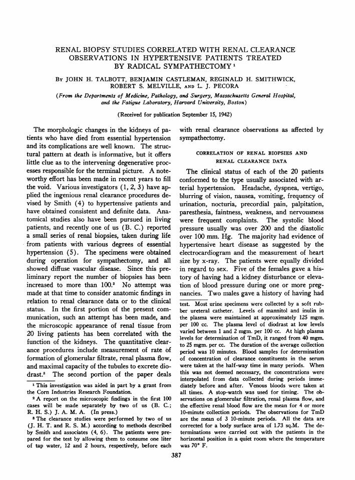
FIG.
The average of the filtration f ractions f or the cases in
each biopsy group is plotted against the grade of renal
vascular disease. The curve indicates that the filtration
f raction increases as does the evidence of renal vascular
disease, but was not above the normal of 20 per cent in
the lower biopsy groups. The filtration f ractions for each
case are given in Table L.
This is the only patient reported in this com-
munication who has died.
Grade III renal vascular disease. There were
8 patients in this group. The intimal hyaliniza-
tion of the large and small arterioles in this group
was similar to that in grade IV, while the arterial
changes and parenchymal scarring was slightlyless severe. Approximately 10 per cent of the
glomeruli were sclerosed, although in some speci-mens no hyalinized glomeruli were seen after a
careful search. The glomerular filtration rate
averaged 89 cc. per minute, and the renal plasma
flow, 439 cc. per mninute (Table I). Both of these
values represent about a 30 per cent depressionbelow the normal.
Grade II renal vascular disease. The 3 cases
in this group showed moderate vascular changesof all types, but especially intimal hyalinization.The average glomerular filtration rate, 91 cc. per
mninute, was only slightly below the average range
for normals, while plasma flow, 470 cc. per minute,
was depressed significantly (Table I).
Grade I renal vascular disease. There were 4
patients in this group. Slight but definite vascu-
lar disease was noted. Glomerular filtration rate
in all except patient No. 18 was normal, although
plasma flow was depressed slightly (Table I).Patient No. 15 showed high normal values forboth functions, the highest observed in any patientreported in this communication. These figureswere checked at subsequent examinations and thegrade I anatomnical changes were confirmed. Theaverage glomerular filtration rate and plasma flowwere 104 and 552 cc. per minute, respectively.
Grade 0 normal renal vascular findings. The2 patients in this group, Nos. 19 and 20 (Table I),had renal clearance data that were at, or slightlybelow, the lower limit of normal. Nothing wasnoted preoperatively to suggest an adrenal tumoras being responsible for the hypertension. Parox-ysmal episodes were absent in both patients. Nev-ertheless, patient No. 19 had an adrenal corticaltumor and patient No. 20 had an adrenal medul-lary tumor. The tumor was on the right side inboth patients. That these 2 instances are notunique may be assumed from the observation of5 additional cases in the larger series of 100 bi-opsied cases, 4 exhibiting a cortical tumor andthe other a medullary tumor.5 Equally interestingis the fact that the renal vessels were normal inboth patients, the only patients who showed norenal anatomical changes in this small series.6 Agood clinical result followed unilateral sympa-thectomy and excision of the tumor in patientNo. 20. Patient No. 19 experienced partial r&.lief from symptoms. He returned 4 months afterthe first operation for a sympathectomy on theintact side. Renal clearance observations at thistime checked remarkably well with those obtainedat the first admission.
Filtration fraction. Goldring and associatesfound that patients with hypertension have agreater percentile depression in renal plasma flowthan in glomerular filtration rate, so that theratio, (glomerular filtration rate) deintdfl
(renal plasma flow) deintdfltration fraction by Smith (6), is increased abovethe normal of 20 per cent. An increase of thisfraction has been interpreted as an indication ofconstriction of efferent renal arterioles. The av-
5 These 7 cases will form the basis of a more detailedcommunication by Drs. Smithwick and Castleman.
65 In the larger series of renal biopsies, there was an-other patient with cortical adrenal tumor and 4 patientswithout adrenal tumors who showed no renal vasculardisease.
390

RENAL BIOPSY AND CLEARANCESTUDIES IN HYPERTENSION
Renal Vascular DiseaseGrode 0 o
I .o a
E 700 - 4 X °
* 600 _
500390 400 /Ia:o 300 /
CL 200 L
° 100
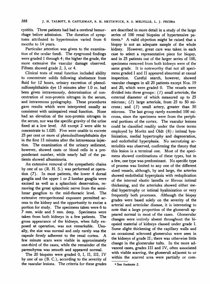
5 10 15 20 25 3 5 40 45 50 55 60Diodrast Tm Mgm per minute
FIG. 2In this figure, diodrast Tm is plotted against renal plasma flow.
These data are available in 9 of the 20 cases. The grade of the renalbiopsy is indicated in each instance.
erage filtration fraction in our series of patientsshowed a slight increase, 21.6 per cent, and therewas a definite trend for the percentage to decreasewith the less severe renal vascular disease (Fig-ure 1). In biopsy groups 0, I, and II, the filtra-tion fraction was normal in 7 of 8 cases (Table I),while in groups III and IV, the filtration fractionwas above normal in 6 of 11 cases.
Diodrast Tm (maximal capacity for excretingdiodrast iodine) was determined in approximatelyhalf of the patients (Table I). If 45 mgm. perminute is assumed to be the lower level of the nor-mal, only 3 patients had a normal value and thepercentile depression followed closely the depres-sion in glomerular filtration rate. In Figure 2,renal plasma flow is plotted against TmD, accord-ing to Goldring. All of the data except one are be-low line M, which is drawn so as to represent a re-duction in renal plasma flow and tubular excretorymass of proportionate amounts. That is, if someprocess were operating to reduce plasma flow andtubular excretory mass proportionately, the re-duction would follow line M. In a heterogeneousgroup of renal disorders, there should be a scatterof data with points above line Mas well as below.Inspection of Figure 2 shows that some factor isoperating in hypertensive subjects to produce a
relative ischemia in the residual functioning tubu-lar tissue.
RENAL CLEARANCEOBSERVATIONSFOLLOWING
SYMPATHECTOMY
Renal clearance observations following sympa-thectomy were obtained in 3 types of patients.(1) In 9 patients, the observations were obtainedwithin 2 weeks after the second stage sympa-thectomy for comparison with the studies beforeoperation. (2) In 9 patients, the studies wereobtained from 4 to 13 months after the secondoperation for comparison with studies both be-fore and within 2 weeks after operation. (3) In6 patients, no preoperative or immediately post-operative studies were obtained, but they weremade from 18 months to 4 years following opera-tion.
Inspection of Table II shows that the glomeru-lar filtration rate decreased about 20 per cent dur-ing the immediate postoperative period, but withina year had returned to about its preoperative level.Although there were no preoperative studies onthe 6 patients that were followed from 1% to 4:years, their postoperative filtration rates did notdiffer materially from the preoperative values ofthe other cases. It seems reasonable to conclude,
391
J. H. TALBOTT, B. CASTLEMAN,R. H. SMITHWICK, R. S. MELVILLE, L. J. PECORA
TABLE II
Effe-t of sympathectomy on renal dcarance
Glomerular filtmation rate Plaa flow Effective whole blood flow Diodrast iodine Tm FSltration fraction
Pa- Renal Postopemrtive Pertive Postoperative Postoptive Postoprativetient biopsy~ pre-. Pre- re- Pre- Pr,
ber (Grade) op- op op.. op- op-era- With- 4 to 1} era- With- 4 to 1j era- W-ith- 4 to 1} ora- With- 4 to Ij era- With- 4 to 1}tive in 2 13 to 4 tivo in 2 13 to 4 tive in 2 13 to 4 tivo in 2 13 to 4 tive in 2 13 to 4
weeks months years week months yer weeks months yea weeks months yas weeks months Yer2 IV 67 26 250 110 420 170 27 248 III 71 73 320 320 670 620 22 23
10 III 86 45 450 270 760 430 46 20 19 1713 II 90 83 490 540 880 860 42 45 18 154 III 86 58 104 400 340 370 660 80 630 21 17 285 III 90 91 94 480 630 290 840 1020 480 19 14 326 III 70 92 92 420 500 480 780 740 800 39 17 18 197 III 86 71 65 430 440 300 770 660 540 20 16 189 III 105 84 90 600 420 270 850660 440 38 21 20 331 IV 56 56 220 240 370 385 19 21 25 26
12 II 100 118 450 400 750 670 37 42 22 2716 I 127 700 760 67 1921 91 83 350 170 620 300 49 43 26 4922 II 57 69 290 250 470 410 28 20 2423 III 93 64 300 230 530 410 43 30 31 2824 98 4325 74 370 600 41 2026 96 400 680 2427 76 320 540 39 24
* The renal clearance data are arranged under S headings: glomerular filtration rate, plasma flow, effective wholeblood flow, diodrast Tm, and filtration fraction. The relation to the time of operation is indicated in each instance.The number of the patient and the biopsy grades are given., (Refer to Table I.)
therefore, that glomerular filtration rate is un-altered by sympathectomy. In only 1 patient, No.4, was a below normal rate before operation re-stored to normal by operation.
The renal plasma flow studies did not show anysignificant change in the immediate postoperativeperiod, but did decrease about 17 per cent withina year and remained approximately the same forthe next few years.
The filtration fraction improved somewhat dur-ing the 2-week period after operation, but grad-ually rose during the first year and showed notendency to return to normal later. This findingsuggests that efferent constriction had not beenlessened by sympathectomy.
The diodrast Tmobservations are too few andscattered to warrant any conclusions.
DISCUSSION
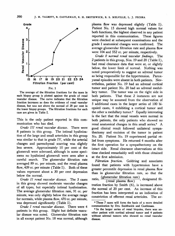
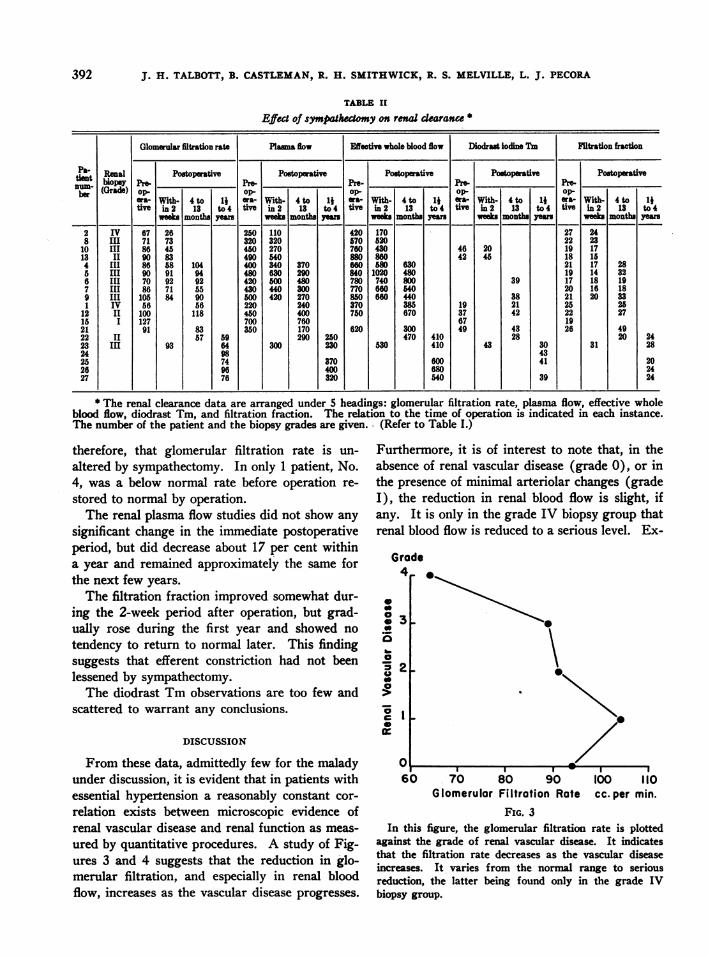
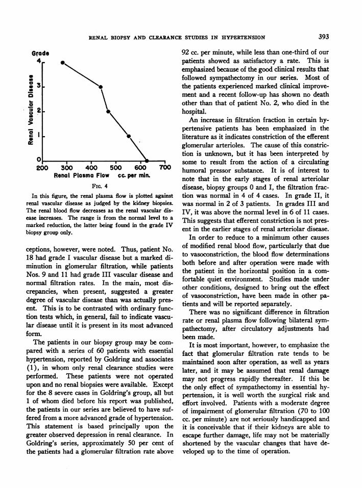
From these data, admittedly few for the maladyunder discussion, it is evident that in patients withessential hypertension a reasonably constant cor-relation exists between microscopic evidence ofrenal vascular disease and renal function as meas-ured by quantitative procedures. A study of Fig-ures 3 and 4 suggests that the reduction in glo-merular filtration, and especially in renal bloodflow, increases as the vascular disease progresses.
Furthermore, it is of interest to note that, in theabsence of renal vascular disease (grade 0), or inthe presence of minimal arteriolar changes (gradeI), the reduction in renal blood flow is slight, ifany. It is only in the grade IV biopsy group thatrenal blood flow is reduced to a serious level. Ex-
Grode4,
0
o.3
0aa
0a
' I
0
IL
L-
0
N
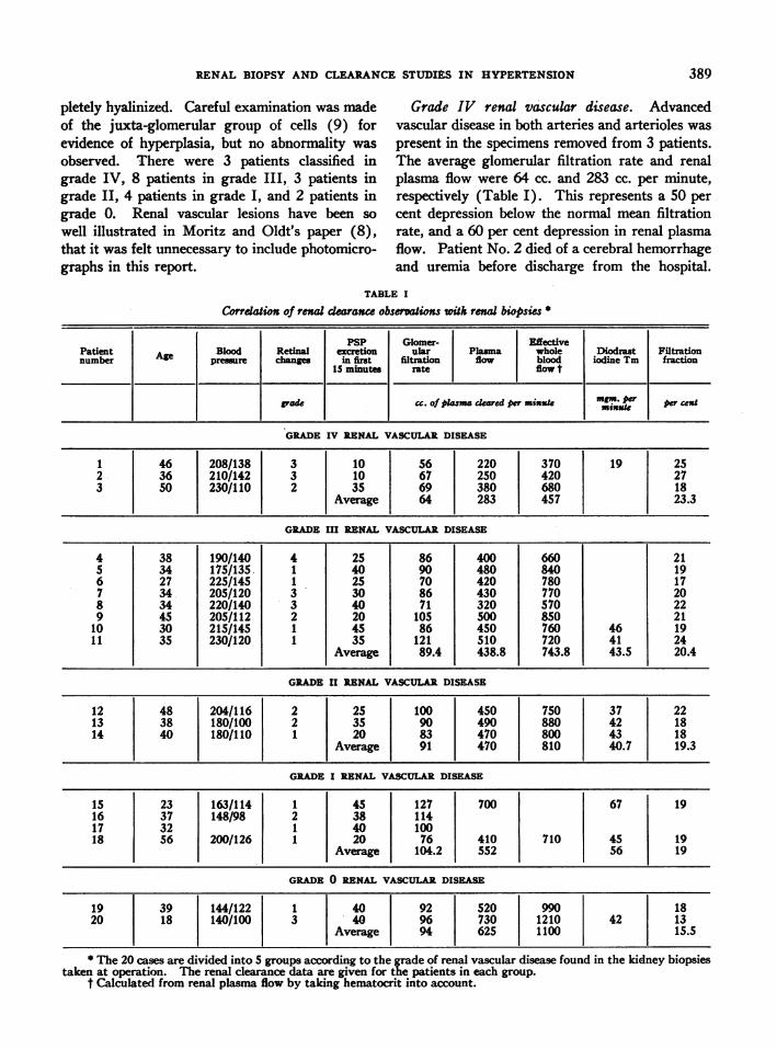
60 70 80 90 100 110Glomerulor Filtration Rate cc. per min.
FIG. 3In this figure, the glomerular filtration rate is plotted
against the grade of renal vascular disease. It indicatesthat the filtration rate decreases as the vascular diseaseincreases. It varies from the normal range to seriousreduction, the latter being found only in the grade IVbiopsy group.
392
RENAL BIOPSY AND CLEARANCESTUDIES IN HYPERTENSION
Grade4
:36
0
200 300 400 500 600 700Ronal Plosma Flow cc. per min.
FIG. 4
In thiis figure, the renal plasma flow is plotted againstrenal vascular disease as judged by the kidney biopsies.The renal blood flow decreases as the renal vascular dis-ease increases. The range is from the normal level to amarked reduction, the latter being found in the grade IVbiopsy group only.
ceptions, however, were noted. Thus, patient No.18 had grade I vascular disease but a marked di-minution in glomerular filtration, while pafientsNos. 9 and 11 had grade III vascular disease andnormal filtration rates. In the main, most dis-crepancies, when present, suggested a greaterdegree of vascular disease than was actually pres-ent. This is to be contrasted with ordinary func-tion tests which, in general, fail to indicate vascu-lar disease unfil it is present in its most advancedform.
The pafients in our biopsy group may be com-pared with a series of 60 pafients with essentialhypertension, reported by Goldring and associates(1), in whom only renal clearance studies wereperformed. These patients were not operatedupon and no renal biopsies were available. Exceptfor the 8 severe cases in Goldring's group, all but1 of whom died before his report was published,the patients in our series are believed to have suf-fered from a more advanced grade of hypertension.This statement is based principally upon thegreater observed depression in renal clearance. InGoldring's series, approximately 50 per cent ofthe patients had a glomerular filtration rate above
92 cc. per minute, while less than one-third of ourpatients showed as satisfactory a rate. This isemphasized because of the good clinical results thatfollowed sympathectomy in our series. Most ofthe patients experienced marked clinical improve-ment and a recent follow-up has shown no deathother than that of patient No. 2, who died in thehospital.
An increase in filtration fraction in certain hy-pertensive patients has been emphasized in theliterature as it indicates constriction of the efferentglomerular arterioles. The cause of this constric-tion is unknown, but it has been interpreted bysome to result from the action of a circulatinghumoral pressor substance. It is of interest tonote that in the early stages of renal arteriolardisease, biopsy groups 0 and I, the filtration frac-tion was normal in 4 of 4 cases. In grade II, itwas normal in 2 of 3 patients. In grades III andIV, it was above the normal level in 6 of 11 cases.This suggests that efferent constriction is not pres-ent in the earlier stages of renal arteriolar disease.
In order to reduce to a minimum other causesof modified renal blood flow, particularly that dueto vasoconstriction, the blood flow determinationsboth before and after operation were made withthe patient in the horizontal position in a com-fortable quiet environment. Studies made underother conditions, designed to bring out the effectof vasoconstriction, have been made in other pa-tients and will be reported separately.
There was no significant difference in filtrationrate or renal plasma flow following bilateral sym-pathectomy, after circulatory adjustments hadbeen made.
It is most important, however, to emphasize thefact that glomerular filtration rate tends to bemaintained soon after operation, as well as yearslater, and it may be assumed that renal damagemay not progress rapidly thereafter. If this bethe only effect of sympathectomy in essential hy-pertension, it is well worth the surgical risk andeffort involved. Patients with a moderate degreeof impairment of glomerular filtration (70 to 100cc. per minute) are not seriously handicapped andit is conceivable that if their kidneys are able toescape further damage, life may not be materiallyshortened by the vascular changes that have de-veloped up to the time of operation.
393

J. H. TALBOTT, B. CASTLEMAN, R. H. SMITHWICK, R. S. MELVILLE, L. J. PECORA
SUMMARY
(1) Renal clearance studies performed on 20patients with essential hypertension showed a sig-nificant correlation with the microscopic appear-
ance of their respective renal tissues which were
removed for biopsy at the time of sympathectomy,i.e., the more severe the renal vascular disease, themore reduced were the glomerular filtration rateand the renal blood flow. In the cases with grade0 and I renal vascular disease, the renal clearanceobservations were either normal or only very
slightly reduced. Only in grade IV renal vasculardisease was renal blood flow seriously reduced.
(2) The filtration fraction was normal in 7 outof 8 cases in biopsy groups 0, I, and II. It was
increased in 6 of 11 cases in biopsy groups III andIV. These findings indicate that constriction ofthe efferent glomerular arterioles was not presentin the early stages of renal vascular disease.
(3) Bilateral radical lumbo-dorsal splanchni-cectomy had relatively little effect on renal clear-ance, when measured in the horizontal position.Although glomerular filtration was reduced in theimmediate postoperative period about 20 per cent,within a year it returned to and continued to main-tain its preoperative level. Renal plasma flow was
essentially unchanged.
BIBLIOGRAPHY
1. Goldring, W., Chasis, H., Ranges, H. A., and Smith,H. W., Effective renal blood flow in subjects withessential hypertension. J. Clin. Invest., 1941, 20,637.
2. Foa, P. P., Woods, W. W., Peet, M. M., and Foa.N. L., Effective renal blood flow, glomerular filtra-tion rate and tubular excretory mass in arterialhypertension. Arch. Int. Med., 1942, 69, 822.
3. Chasis, H., and Redish, J., Effective renal blood flowin the separate kidneys of subjects with essentialhypertension. J. Clin. Invest., 1941, 20, 655.
4. Smith, H. W., The Physiology of the Kidney. Ox-ford University Press, New York, 1937.
5. Castleman, B., Smithwick, R. H., and Palmer, R. S.,Renal biopsies from hypertensive patients. Ab-stract from scientific proceedings of the 41st an-
nual meeting of the American Association of Pa-thologists and Bacteriologists. Am. J. Path., 1941,17, 617.
6. Smith, H. W., Goldring, W., and Chasis, H., Themeasurement of the tubular excretory mass, ef-fective blood flow and filtration rate in the normalhuman kidney. J. Clin. Invest., 1938, 17, 263.
7. Smithwick, R. H., A technique for splanchnic resec-
tion for hypertension. Surgery, 1940, 7, 1.8. Moritz, A. R., and Oldt, M. R., Arteriolar sclerosis
in hypertensive and non-hypertensive individuals.Am. J. Path., 1937, 13, 679.
9. Goormaghtigh, N., Histological changes in the is-chemic kidney with special reference to the juxta-glomerular apparatus. Am. J. Path., 1940, 16, 409.
10. Corcoran, A. C., and Page, I. H., Renal blood flowand sympathectomy in hypertension. Arch. Surg.,1941, 42, 1072.
11. Alving, A. S., Adams, W., Grimson, K. S., Scott, C.,and Sandiford, I., Effect of bilateral paravertebralsympathectomy on the cardiorenal system in essen-
tial hypertension. Proc. Inst. Med. Chicago, 1941,13, 306.
12. Hines, E. A., and Brown, G. E., Standard test formeasuring the variability of blood pressure: its sig-nificance as an index of the prehypertensive state.Ann. Int. Med., 1933, 7, 209.
394