bioorganic & medicinal chemistry...
TRANSCRIPT
Bioorganic & Medicinal Chemistry Letters 27 (2017) 1428–1436
Contents lists available at ScienceDirect
Bioorganic & Medicinal Chemistry Letters
journal homepage: www.elsevier .com/locate /bmcl
Design, synthesis and biological evaluation of nonsecosteroidal vitaminD3 receptor ligands as anti-tumor agents
http://dx.doi.org/10.1016/j.bmcl.2017.01.0840960-894X/� 2017 Elsevier Ltd. All rights reserved.
⇑ Corresponding author.E-mail address: [email protected] (C. Zhang).
Bin Wang, Meixi Hao, Can Zhang ⇑State Key Laboratory of Natural Medicines and Jiangsu Key Laboratory of Drug Discovery for Metabolic Diseases, Center of New Drug Discovery, China Pharmaceutical University,24 Tong Jia Xiang, Nanjing 210009, China
a r t i c l e i n f o a b s t r a c t
Article history:Received 19 December 2016Revised 17 January 2017Accepted 27 January 2017Available online 1 February 2017
Keywords:Vitamin D3
VDR ligandNonsecosteroidalPhenyl-pyrrolyl pentane skeletonBinding affinityAgonistic activityAnti-proliferative activity
1a,25-dihydroxyvitamin D3 (1,25-(OH)2D3, also known as calcitriol), the active form of vitamin D3, isbeing increasingly recognized for cancer therapy. Our previous work showed that phenyl-pyrrolyl pen-tane analogs, which mimicked anti-proliferative activities against several cancer cell lines of the naturalsecosteroidal ligand 1,25-(OH)2D3. Here, in order to optimize the structural features and discover morepotent derivative, a series of nonsecosteroidal vitamin D3 receptor (VDR) ligands bearing acetylene bondlinker was designed, synthesized and evaluated. Most of them showed moderate to good binding affini-ties and agonistic activities. Especially, compound 19f displayed the most anti-proliferative activitiesagainst MCF-7 and PC-3 cells with the IC50 values of 1.80 and 5.35 lM, respectively, which was compa-rable to positive control 1,25-(OH)2D3. Moreover, compound 19f exhibited reduced toxicity againsthuman normal liver cell line (L02) compared with the parental compound 7. Besides, the preliminarystructure–activity relationships (SARs) were also analyzed.
� 2017 Elsevier Ltd. All rights reserved.
The active form of vitamin D3, the hormone 1a,25-dihydroxyvi-tamin D3 (1,25-(OH)2D3, also known as calcitriol) participates innumerous biological processes.1–3 In addition to its classical rolein mineral homeostasis and bone mineralization,4 this hormoneregulates numerous cellular pathways that could have a role indetermining cancer risk and prognosis.5–9 Because of this ‘‘nonclas-sical” actions, 1,25(OH)2D3 has attracted considerable interest as apotential drug for the treatment of cancer disease. The effects of1,25(OH)2D3 are mediated through the vitamin D3 receptor (VDR)which is a ligand-dependent transcription factor belonging to thesuperfamily of nuclear hormone receptors.10–14
So far, more than 3000 secosteroid analogs have been synthe-sized,15 andmany of them exhibit efficient VDR activities and someanalogs have been successfully used in the treatment of bone, min-eral, and skin disorders (Fig. 1).16,17 However, adverse effects, par-ticularly hypercalcemia, limit the clinical application of secosteroidanalogs in the management of cancer disease.15 Recently, muchattention has been directed towards nonsecosteroidal VDR ligands,which mimic various activities in vitro and in vivo of the naturalligand 1,25(OH)2D3 without direct structural relationship to 1,25(OH)2D3, such as VDR binding and inhibition of proliferation ofcancer cells.18,19 Moreover, they have simpler structures comparedwith secosteroid analogs.20–27 Above all, some nonsecosteroidal
VDR ligands avoid hypercalcemia effect, such as LG190178 andits analogs. Therefore, the development of VDR ligands with non-secosteroidal skeleton is required to create novel therapy for theVDR-related cancer disease.
We previously reported novel phenyl-pyrrolyl pentane skeletonas a nonsecosteroidal VDR ligand skeleton which had anti-prolifer-ative effects against cancer cells and avoided hypercalcemia effectin vivo.28,29 While the VDR binding affinities of all the synthesizedcompounds suffered a remarkable decrease as compared with 1,25(OH)2D3, compound 7 had a moderate VDR binding affinity in vitro,which deserved further study. Here, we wish to report compound 7as a lead for further optimization to explore the structure-activityrelationships (SARs) and to discover more potent derivative withhigh VDR binding affinity and anti-proliferative effect against can-cer cells. As shown in Fig. 2, first, we attempted to introduce acet-ylene bond linker to minimize the flexibility and to fix thedirection of the side chain, and then designed 3-pentanolyl asthe terminal hydrophobic group of the side chain based on previ-ous SARs. Additionally, to explore the effects of substitutions ofpolarity and steric hindrance on the pyrrole ring, compounds17c–i and 18a–d were designed. Second, phenyl-pentane groupon C-4 position of pyrrole ring replaced C-5 position to investigatethe influence of the substitution positions of the terminal hydro-philic group of the pyrrole ring to give compounds 19a, 19f–jand 20a–e.

Fig. 1. Chemical structures of secosteroidal and nonsecosteroidal VDR ligands.
Fig. 2. Design of novel nonsecosteroidal vitamin D receptor ligands with phenyl-pyrrolyl pentane skeleton.
B. Wang et al. / Bioorganic & Medicinal Chemistry Letters 27 (2017) 1428–1436 1429
The synthetic pathway of target compounds 17c–i and 18a–d isoutlined in Scheme 1. Key intermediate 8 was readily preparedusing our previously reported approach,25 and then it reacted withethyl pyrrole-2-carboxylate in the presence of lewis acid BF3�Et2Oat 0 �C to give intermediate 9a, following the treatment withiodoethane in DMF to afford intermediate 10a. The intermediate11a was obtained by reduction reaction of intermediate 10a inthe presence of HCOONH4, which was acylated with trifluo-romethanesulfonic anhydride to give 12a in moderate yield. Inter-
mediate 12a subjected to sonogashira coupling reaction withtrimethylsilylacetylene in the presence of palladium catalystafforded 13a and subsequent removal of trimethylsilyl group usingtetrabutylammonium fluoride (TBAF) gave acetylene 14a. Hydroly-sis of intermediate 14a by KOH produced the key intermediate 15a,which was alkylated with n-butyl lithium and 3-pentanone at�78 �C to give key intermediate alcohol 16a. Interestingly,followed by moving to room temperature for 2 h, alcohol 16atransformed to compound 17i, which were obtained by one step

Scheme 1. Synthesis of target compounds 17c–i and 18a–d. Reagents and conditions: (a) Ethyl 1H-pyrrole-2-carboxylate, BF3�Et2O, 0 �C, 1 h, 53%; (b) C2H5I, NaH, DMF, 0–25 �C, 2 h, 82%; (c) Pd/C, HCOONH4, CH3OH/EtOAc (10:1), 25 �C, 1 h, 98%; (d) Tf2O, TEA, toluene, 0 �C, 2 h, 67%; (e) TMS acetylene, PdCl2(dppf)2, TEA, DMF, 100 �C, overnight,63%; (f) TBAF, THF, rt, 1 h, 95%; (g) 2 mol/L KOH, EtOH, 80 �C, 6 h, 95%; (h) 3-pentanone, n-BuLi, THF, �78 to 0 �C, 2 h, 75%; (i) EDCI, HOBt, TEA, RNH2, DCM, rt, overnight, 35–96%; (j) EDCI, DMAP, ROH, DCM, rt, overnight, 26–64%; (k) n-BuLi, THF, 25 �C, 2 h, 79%; (l) 2 mol/L KOH, EtOH, rt, 1 h, 83–94%; (m) LiAlH4, EtOAc, rt, 1 h, 86%.
1430 B. Wang et al. / Bioorganic & Medicinal Chemistry Letters 27 (2017) 1428–1436
from carboxylic acid group to ketone group. By reaction of inter-mediate 16a with the corresponding amines or alcohols, targetcompounds 17c–h and intermediates 17a–b were obtained.Finally, target compounds 18a–cwere obtained by hydrolysis reac-tions of intermediates 17a–c in the presence of KOH. On the other
hand, target compound 18d was obtained by reduction reaction ofcompound 17c.
The synthetic pathway of target compounds 19a, 19f–j and20a–e is outlined in Scheme 2. Intermediate 9b, which was theregioselectivity isomer of intermediate 9a, was produced by

Scheme 2. Synthesis of target compounds 19a, 19f–j and 20a–e. Reagents and conditions: (a) Ethyl 1H-pyrrole-2-carboxylate, BF3�Et2O, 25 �C, 1 h, 44%; (b) C2H5I, NaH, DMF,0–25 �C, 2 h, 85%; (c) Pd/C, HCOONH4, CH3OH/EtOAc (10:1), 25 �C, 1 h, 97%; (d) Tf2O, TEA, toluene, 0 �C, 2 h, 64%; (e) TMS acetylene, PdCl2(dppf)2, TEA, DMF, 100 �C, overnight,53%; (f) TBAF, THF, rt, 1 h, 95%; (g) 2 mol/L KOH, EtOH, 80 �C, 6 h, 96%; (h) 3-pentanone, n-BuLi, THF, �78 to 0 �C, 2 h, 54%; (i) EDCI, HOBt, TEA, RNH2, DCM, rt, overnight, 35–96%; (j) EDCI, DMAP, ROH, DCM, rt, overnight, 64%; (k) n-BuLi, THF, 25 �C, 2 h, 77%; (l) 2 mol/L KOH, EtOH, rt, 1 h, 83–94%; (m) LiAlH4, EtOAc, rt, 1 h, 89%.
B. Wang et al. / Bioorganic & Medicinal Chemistry Letters 27 (2017) 1428–1436 1431
reacting with ethyl pyrrole-2-carboxylate in the presence of lewisacid BF3�Et2O at 20 �C instead of at 0 �C. By the same manner asdescribed for the preparation of intermediates 10a–16a, interme-diates 10b–16b were obtained. As described before, compound19j were obtained by one step from carboxylic acid group toketone group. By reaction of intermediate 16b with the corre-sponding amines or alcohols, target compounds 19a, 19f–j andintermediates 19b–e were obtained. Finally, target compounds20a–d were obtained by hydrolysis reactions of intermediates19a–d in the presence of KOH. On the other hand, target compound20e was obtained by reduction reaction of compound 19e.
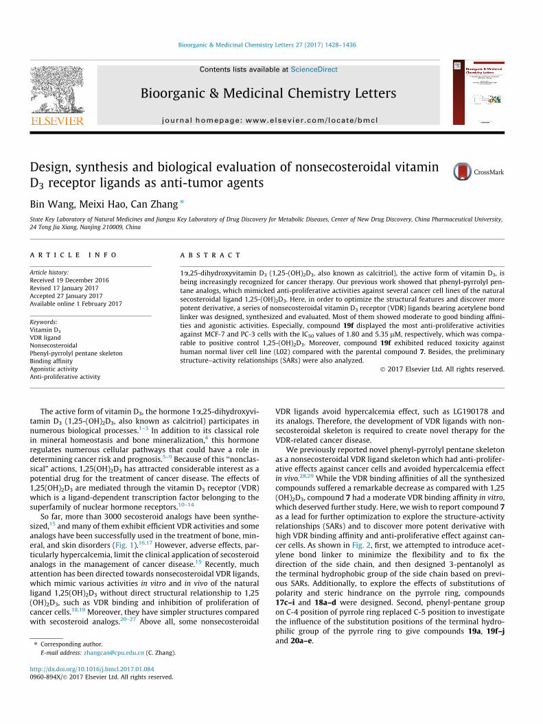
The receptor binding affinity assay of synthesized compoundswas initially performed at a concentration of 1 lM and the bindingaffinity was displayed by a relative value based on 1,25(OH)2D3
being assigned as 100%. As shown in Table 1. the results displayedthat although seven compounds (17d, 18a, 18d, and 19f–i) demon-strated more effective binding affinities than the parental com-pound 7 (25.5%), with binding values at the range of 29.7–62.2%,none compound displayed equivalent binding affinity comparedto 1,25(OH)2D3. Additionally, five compounds (17e, 18c, 20a, 20c,and 20e) showed moderate binding affinities with the percentageof binding ranging from 13.6% to 20% and other compounds hadno obvious binding affinities.
Our SARs analysis started with C-5 position of pyrrole ring bear-ing phenyl- pentane group and showed that compounds with theterminal hydrophilic groups in the A ring section, such as car-boxylic acid (18a), hydroxyl (18d), and amino groups (17d)showed significant binding affinities. However, by one-atomextension of the hydrophilic groups of compounds 17d and 18a,17e and 18c were synthesized and the binding affinities of themwere dramatically decreased. Additionally, introducing a large car-boxylic acid obtained compound 18b, which displayed poor bind-ing affinity, suggesting sterically hindered substitutes may not beaccepted in VDR ligand binding domains. In order to verify thehypothesis, benzene and aromatic heterocyclic groups were intro-duced to synthesize compounds 17f and 17h. As expected, the lowbinding affinities of these compounds testify our hypothesis.Besides, when carboxylic acid group was protected by methylgroup, the binding affinity of the resulting compound 17c weredramatically decreased compared with compound 18c, suggestingcarboxylic acid group plays an important role in the VDR bindingaffinity. Furthermore, a similar binding affinities were observedwhen replacing ester group with hydrophobic groups, such aspropargyl and n-butyl groups.
In order to explore the influence of substitution positions on thebinding affinities, compounds 19a, 19f–j and 20a–e were
Table 1Structures and binding abilities of target compounds.
Compd X Y R1 R2 R Relative VDRBinding ability (%)a
17c N C C2H5 H –
17d N C C2H5 H 62.2 ± 3.5
17e N C C2H5 H 18.8 ± 1.8
17f N C C2H5 H 4.0 ± 0.3
17g N C C2H5 H –
17h N C C2H5 H –
17i N C C2H5 H –
18a N C C2H5 H 55.4 ± 3.1
18b N C C2H5 H 5.9 ± 0.3
18c N C C2H5 H 13.6 ± 1.5
18d N C C2H5 H 59.7 ± 2.7
19a C N H C2H5 –
19f C N H C2H5 50.3 ± 3.1
19g C N H C2H5 55.3 ± 4.1
19h C N H C2H5 31.6 ± 1.7
19i C N H C2H5 29.7 ± 2.1
19j C N H C2H5 –
20a C N H C2H5 14.6 ± 1.1
1432 B. Wang et al. / Bioorganic & Medicinal Chemistry Letters 27 (2017) 1428–1436

Table 1 (continued)
Compd X Y R1 R2 R Relative VDRBinding ability (%)a
20b C N H C2H5 4.3 ± 0.1
20c C N H C2H5 20.0 ± 2.4
20d C N H C2H5 5.9 ± 0.3
20e C N H C2H5 19.8 ± 1.2
7 – – – – – 25.5 ± 1.41,25(OH)2D3 – – – – – 100.0 ± 2.3
a The values represent the mean ± SD of three independent experiments. 1,25(OH)2D3 is assigned as 100%.
B. Wang et al. / Bioorganic & Medicinal Chemistry Letters 27 (2017) 1428–1436 1433
synthesized by removing the substitution on the C-5 position ofpyrrole group to C-4 position. As shown in Table 1, similar SARswere observed. Although compound 19g showed the most potentaffinity among them, no better result was observed compared tocompound 17d. On the contrary, some compounds bearing samegroups showed decreased binding affinity, such as compounds20a, 20b, and 19f as compared with 18a, 18c, and 17d. The resultssuggest that the position of the substitute is important by affectingthe conformation of the terminal hydrophilic group which is essen-tial for binding, as in the case of the conformationally restricted Aring of secosteroid. To further explore the difference on the bindingaffinities between C-5 position of pyrrole group and C-4 position,
Fig. 3. (A) Docking structure of the complex VDR-compound 18a. (B) Docking structureCompound 18a is shown in stick representation with carbon and oxygen atoms in greenand oxygen atoms in wheat and red, respectively. The hydrogen bonds formed are show
we performed the docking analyses of compounds 18a and 20a,which had the same hydrophilic substitute, but displayed remark-able difference on binding affinities, based on crystallographicstructure of 1,25(OH)2D3 in complex with VDR (PDB code: 1DB1).As shown in Fig. 3C, docking analyses demonstrated that the sidechain of both compounds present similar conformations. However,substitution of pyrrole group from C-5 position to C-4 positionresulted in conformational change in the A ring section(Fig. 3A and B). In the docking, the introducing carboxylic acid ofcompound 18a was beautifully adjusted in VDR and formed twohydrogen bonds with Ser237 and Arg274. Unfortunately, thechange of substitution positions resulted in a longer distance
of the complex VDR-compound 20a. (C) Superposition of compounds 18a and 20a.and red, respectively. Compound 20a is shown in stick representation with carbonn as yellow dashed lines.
Table 2The binding affinities of selected compounds.
Compd VDR binding (IC50, nM)a Compd VDR binding (IC50, nM)a
17d 73.21 ± 3.68 19g 51.31 ± 3.5418a 85.66 ± 2.45 7 76.76 ± 5.1218d 123.43 ± 2.32 1,25(OH)2D3 1.13 ± 0.1119f 56.48 ± 2.17
a IC50: the concentration that causes 50% of cell proliferation inhibition. Data areexpressed as mean ± SD from three independent experiments.
1434 B. Wang et al. / Bioorganic & Medicinal Chemistry Letters 27 (2017) 1428–1436
between the introduced carboxylic acid of compound 20a andSer237, so only one hydrogen bond with Arg274 was formed,which maybe the important reason of different binding affinities.
Subsequently, we further evaluated the VDR binding affinities(IC50) of compounds 17d, 18a, 18d, and 19f–g with excellent inhi-bition at 1 lM. As shown in Table 2, introducing hydroxyl and car-boxylic acid groups as the terminal hydrophilic groups showeddecreased affinities compared with the parental compound 7.However, compounds 17d, 19f, and 19g, all of which were intro-duced amino groups instead displayed more potent than lead com-pound 7, which deserved further investigations.
It is proved that vitamin D3-agonistic activity is associated withHL-60 cell differentiation induction.30,31 Therefore, the vitaminD3-agonistic ability can be estimated as the potential to differenti-ate human promyelocytic leukemia cell line (HL-60) into macro-phages. All synthesized compounds were tested for HL-60 celldifferentiation using calcitriol as positive control, as shown inTable 3. Most compounds displayed good agonistic activities withEC50 values in the nanomole range. Among them, compounds 19fand 20e showed higher agonistic activities than positive control1,25(OH)2D3. As similar as the SARs of binding affinities, com-pounds with the terminal hydrophilic groups in the A ring section,such as carboxylic acid (18a) and anime groups (17d) also showed
Table 3The HL-60 differentiation-inducing activities and anti-proliferative activities ofsynthesized compounds in vitro.
Compd HL-60 differentiation -inducingactivity(EC50, nM)a
In vitro anti-proliferativeactivity (IC50, lM)b
MCF-7 PC-3
17c >50 >50 36.27 ± 0.7617d 10.12 ± 0.86 3.77 ± 0.26 5.85 ± 0.3717e 68.91 ± 2.29 17.80 ± 0.56 8.50 ± 0.6317f >50 >50 >5017g >50 >50 >5017h >50 >50 >5017i >50 >50 >5018a 9.36 ± 0.86 5.14 ± 0.22 2.25 ± 0.1418b 17.67 ± 1.15 13.00 ± 0.25 5.00 ± 0.2818c 293.44 ± 5.68 >50 5.13 ± 0.3518d 136.86 ± 7.43 5.95 ± 0.14 >5019a 287.65 ± 10.59 26.46 ± 0.44 17.45 ± 0.6619f 6.98 ± 0.36 1.80 ± 0.19 5.35 ± 0.3119g 19.29 ± 0.86 13.44 ± 0.12 10.07 ± 0.4719h 142.17 ± 7.13 20.17 ± 0.69 15.06 ± 0.5019i 12.56 ± 0.59 1.88 ± 0.17 11.40 ± 0.2119j >50 >50 48.12 ± 0.2220a 54.33 ± 4.33 17.54 ± 0.57 6.30 ± 0.4420b >50 30.55 ± 0.32 >5020c 12.68 ± 0.76 12.60 ± 0.31 3.75 ± 0.1420d 68.32 ± 4.13 18.02 ± 0.26 10.28 ± 0.3320e 7.33 ± 0.28 1.91 ± 0.16 6.05 ± 0.407 6.54 ± 0.07 1.37 ± 0.02 2.8 ± 0.581,25(OH)2D3 8.12 ± 0.03 11.10 ± 0.26 16.20 ± 0.29
a EC50: Vitamin D3-agonistic activity was estimated as HL-60 differentiationinducing ability. Data represent mean ± SD, n = 3, *P < 0.05.
b IC50: the concentration that causes 50% of cell proliferation inhibition. Data areexpressed as mean ± SD from three independent experiments.
significant agonistic activities. By introducing a large carboxylicacid substitute, compound 18b was synthesized and showed aslight decreased agonistic activity compared to compound 18a,although a dramatically decreased binding affinity was observed.This discrepancy between agonistic activity and binding affinitycould be explained by the interactions between the VDR ligandcomplex and other cofactors. For the transcriptional activation ofVDR, it is required that the AF-2 transactivation motif of VDR inter-acts with several types of cofactor such as VDR interacting proteins(DRIPs).32–35 Furthermore, introducing large sterically hinderedsubstitutes, such as 17f and 17h displayed remarkable decreasedagonistic activities. Additionally, when removing the substitutionon the C-5 position of pyrrole group to C-4 position, most com-pounds with the same hydrophilic groups showed decreased ago-nistic activities. However, compound 19f (EC50 = 6.98 nM)bearing N,N-diethyl-1,2-ethanediamine, which had less bindingaffinity than 17d, showed the best agonistic activity. Unfortu-nately, no improvement in agonistic activities of all the synthe-sized compounds was observed compared with the parentalcompound 7 (EC50 = 6.54 nM). We estimate that acetylene bondintroduced has weaker cell permeability than ether bond andresults in the reduced agonistic activity.
To evaluate the anti-proliferative activities of synthesized com-pounds, human breast cancer cell line (MCF-7)36,37 and humanprostate cancer cell line (PC-3),38 which over express VDR wereselected as cell models to test the anti-proliferative effects by thestandard MTT assay, with 1,25(OH)2D3 as positive control. Asshown in Table 3, the results displayed that most of the synthe-sized compounds showed moderate to good activities with IC50
values in the micromole range and in some cases better than thatof 1,25(OH)2D3, while compounds 17h–i, and 17k were proved tobe poor activities against both cell lines. Interestingly, compounds17d, 18a, 18d, 19f, 19i and 20e showed higher anti-proliferativeactivities than 1,25(OH)2D3 against MCF-7 cells. In addition, thir-teen compounds exhibited remarkable anti-proliferative activitieswith the IC50 values ranging from 2.2 to 15.1 lM, which were com-parable to that of 1,25(OH)2D3 (IC50 = 17.2 lM) on PC-3 cells. Nota-bly, compounds 17d, 18a, 19f, 19i and 20e displayed moreeffective anti-proliferative activities against both cell lines com-pared with 1,25(OH)2D3, although only compounds 19f and 20eshowed better agonistic activities than 1,25(OH)2D3, which sug-gests that they may work though the multiple molecular mecha-nisms. This primary screening results revealed that phenyl-pyrrolyl pentane derivatives with acetylene bond linker exhibitedstrong anti-proliferative activities. As similar as the SARs of thebinding affinity and agonistic activity, compounds bearing hydro-philic groups, such as 17d, 18a, 19f, and 19i, showed better agonis-tic activities than that of hydrophobic groups, such as 17c, 17i and19j, which suggested that it is necessary to introduce hydrophilicgroups into the A ring section. Compound 18d bearing hydroxylgroup displayed poor anti-proliferative activity against PC-3 cells,although it showed significant binding affinity and moderate ago-nistic activity, which suggests that chemical modification of com-pound 18d may induce AF2 conformations and cofactorinteractions distinct from those of natural ligands and can resultin cell type-selective modulation of target gene expression.39
Besides, compound 18b also showed a slight decreased anti-prolif-erative activity compared to compound 18a alike that of agonisticactivities. Furthermore, compound 17f–i bearing sterically hin-dered or hydrophobic substitutes also exhibited poor anti-prolifer-ative activities against both cell lines as similar as the bindingaffinities and agonistic activities. Additionally, as similar as theSARs of agonistic activities, most synthesized compounds exhib-ited decreased anti-proliferative activities compared with that ofthe same A ring section, such as 18a, 18b and 18c–20a, 20c and20b, respectively. In this regard, it could be proved that the

Table 4The anti-proliferative activities of selected compounds and 1,25(OH)2D3 over L02 normal cell line and selectivities for both cancer cells.
Compd In vitro anti-proliferative activities (IC50, lM)a Selectivities
L02 MCF-7 PC-3 L02/MCF-7 L02/PC-3
17d 20.52 ± 0.12 3.77 ± 0.26 5.85 ± 0.37 5.4 3.519f 42.14 ± 0.46 1.80 ± 0.19 5.35 ± 0.31 23.4 7.920e 35.43 ± 0.34 1.91 ± 0.16 6.05 ± 0.40 18.5 5.97 21.24 ± 0.32 1.37 ± 0.02 2.8 ± 0.58 15.5 7.61,25(OH)2D3 >50 11.10 ± 0.26 16.20 ± 0.29 >4.5 >3.1
a IC50: the concentration that causes 50% of cell proliferation inhibition. Data are expressed as mean ± SD from three independent experiments.
Fig. 4. Superposition of compounds 19f and 1a,25-(OH)2-D3. Compound 19f isdepicted in magenta and 1a,25-(OH)2-D3 is depicted in cyan.
B. Wang et al. / Bioorganic & Medicinal Chemistry Letters 27 (2017) 1428–1436 1435
anti-proliferative activities of synthesized compounds are posi-tively correlative with VDR agonistic activities. Moreover, com-pound 19f also showed the most anti-proliferative activities
Fig. 5. (A) Structure of the complex VDR-1a,25-(OH)2-D3. (B) Docking structure of thecarbon and oxygen atoms in cyan and red, respectively. The hydrogen bonds formed ar
Fig. 6. Schematic diagram of stru
against MCF-7 and PC-3 cells with the IC50 values of 1.80 and5.35 lM, respectively, which was comparable to positive control1,25-(OH)2D3.
As further evaluation for the selective cytotoxicities of thepromising compounds 17d, 19f, and 20e, they were tested overhuman normal liver cell line (L02) using MTT assay. As illustratedin Table 4, all compounds displayed moderate cytotoxicitiesagainst L02 cells. Notably, compound 19f (IC50 = 42.14 lM) hadremarkable decreased cytotoxicity compared with the parentalcompound 7 (IC50 = 21.24 lM) and showed better selectivities forboth cancer cells (L02/MCF-7 = 23.4, L02/PC-3 = 7.9). Finally, aninvestigation on the binding affinities and anti-proliferative activ-ities for cancer and normal cells of these derivatives showed thatcompound 19f exhibited desirable results.
To confirm the detailed interactions of the most promising com-pound 19f, molecular docking study was conducted based on crys-tallographic structure of 1,25(OH)2D3 in complex with VDR (PDBcode: 1DB1). Compound 19f was manually docked into the crystalstructure of VDR using software Discovery Studio 3.0. Fig. 4 showsthe superposition of the conformations of compound 19f and the
complex VDR-compound 19f. The ligands are shown in stick representation withe shown as red dashed lines.
cture-activity relationships.

1436 B. Wang et al. / Bioorganic & Medicinal Chemistry Letters 27 (2017) 1428–1436
natural ligand 1,25(OH)2D3. Docking analyses demonstrated thatthe side chain and A ring part of compound 19f present similarconformations to those observed in the presence of 1,25(OH)2D3.As shown in Fig. 5, the hydroxyl group in the side chain was ableto form the same hydrogen-bonding interactions with His 305and His 397 as the hVDR LBD bound to 1a,25-(OH)2-D3 complex.However, the A ring part of compound 19f form hydrogen-bondinginteraction only with Ser 237 by amide bond, while 1a,25-(OH)2-D3 binded with Ser 237, Arg 274, Tyr 143, and Ser 278. In addition,the N,N-diethyl ethyl amine group introduced made longer A ringpart than that of 1a,25-(OH)2-D3. These factors might play impor-tant roles in reducing the binding affinity of compound 19f to VDR(see Fig. 6).
According to the above results (Tables 1 and 3), we can drawsome conclusions: (1) introducing the hydrophilic moieties at theR group is important to improve the VDR binding affinities andanti-proliferative activities, and introduction of the hydrophobicsegments may lead to a remarkable decrease or even loss of bind-ing affinities and anti-proliferative activities. (2) Introduction oflarge groups such as benzene ring cannot be tolerated, nearly lead-ing to a loss of affinities and activities. (3) C-5 position of pyrrolering bearing phenyl-pentane group is not absolutely required butmay increase the binding affinity.
In summary, we synthesized and evaluated a series of novelphenyl-pyrrolyl pentane derivatives with acetylene bond linkeras VDR ligands. Structural optimization of the parental compound7 led to the synthesis of 22 derivatives. Seven analogs (17d, 18a,18d, and 19f–i) demonstrated more effective binding affinitiesthan the parental compound 7. Moreover, compound 19f not onlyshowed excellent agonistic activity to VDR but also displayed moreanti-proliferative effect against MCF-7 and PC-3 cells with the IC50
values of 1.80 and 5.35 lM, respectively. Besides, compound 19fexhibited reduced toxicity against human normal liver cell line(L02) compared with the parental compound 7. In conclusion, onthe basis of the abilities of these compounds, introducing acetylenebond linker for phenyl-pyrrolyl pentane derivatives may be a strat-egy for the discovery of new drugs for the treatment of cancerdiseases.
Based on the preliminary investigation results, our efforts arenow focused on the modification and understanding the mode ofaction of these novel molecules. It is expected that the biologicalresults described and further modification studies will expeditethe development of new chemotherapeutic agents for the clinicalintervention of cancer disease.
Acknowledgments
This work was supported by the National Natural Science Foun-dation of China (81273468, 81473153), National Basic ResearchProgram of China (2015CB755500), Natural Science Foundation
of Jiangsu Province of China (BK20140672), and 111 Project fromthe Ministry of Education of China and the State Administrationof Foreign Expert Affairs of China (No. 111-2-07).
A. Supplementary material
Supplementary data associated with this article can be found, inthe online version, at http://dx.doi.org/10.1016/j.bmcl.2017.01.084.
References
1. Bouillon R, Okamura WH, Norman AW. Endocr Rev. 2000;16:200.2. Omdahl JL, Morris HA, May BK. Annu Rev Nutr. 2002;22:139.3. Reichel H, Koeffler HP, Norman AW. N Engl J Med. 1989;320:980.4. De L. Am J Clin Nutr. 2004;80:1689.5. Byers T. Am J Epidemiol. 2010;172:1.6. Ma Y, Zhang P, Wang F, Yang J, Liu Z, Qin H. J Clin Oncol. 2011;29:3775.7. Feldman D, Krishnan AV, Swami S, Giovannucci E, Feldman BJ. Nat Rev Cancer.
2014;14:342.8. Li M, Chen P, Li J, Chu R, Xie D, Wang HJ. Clin Endocrinol Metab. 2014;7:2327.9. Krishnan AV. Endocrinology. 2010;151:32.10. Dusso AS, Brown AJ, Slatopolsky E. Am J Physiol Renal Physiol. 2005;289:8.11. Lemon BD, Fondell JD, Freedman LP. Mol Cell Biol. 1997;17:1923.12. Blanco JCG, Wang IM, Tsai SY, et al. Proc Natl Acad Sci USA. 1995;92:1535.13. Masuyama H, Brownfield CM, Arnaud R, MacDonald PN. Mol Endocrinol.
1997;11:1507.14. Mengus G, May M, Carre L, Chambon P, Davidson I. Genes Dev. 1997;11:1381.15. Plum LA, DeLuca HF. Nat Rev Drug Discov. 2010;9:941.16. Kubodera N. Heterocycles. 2010;80:83.17. Binderup L, Binderup E, Godtfredsen WO. Development of new vitamin D
analogs. In: Feldman D, Pike JW, Glorieux FH, eds. Vitamin D. 2nded. Amsterdam: Elsevier Academic Press; 2005:1027–1043.
18. Boehm MF, Fitzgerald P, Zou A, et al. Chem Biol. 1999;6:265.19. Yamada Sachiko, Makishima Makoto. Trends Pharmacol Sci. 2014;35:7.20. Swann SL, Bergh J, Farach-Carson MC, Ocasio CA, Koh JT. J Am Chem Soc.
2002;124:13795.21. Perakyla M, Malinen M, Herzig KH, Carlberg C. Mol Endocrinol. 2005;19:2060.22. Hosoda S, Tanatani A, Wakabayashi K, et al. Bioorg Med Chem Lett.
2005;15:4327.23. Ma Y, Khalifa B, Yee YK, et al. J Clin Invest. 2006;116:892.24. Shinya F, Yoshiyuki T, Atsushi K, et al. J Am Chem Soc. 2011;133:20933.25. Fabrice C, Yoshiteru S, Yassmine C, Dino M, Annick D, Natacha R. J Med Chem.
2012;55:8440.26. Fujii S, Sekine R, Kano A, et al. Bioorg Med Chem. 2014;22:5891.27. Sachiko Y, Makoto M. Trends Pharmacol Sci. 2014;35:324.28. Shen W, Xue JW, Zhao ZK, Zhang C. Eur J Med Chem. 2013;69:768.29. Ge ZX, Hao MX, Xu M, et al. Eur J Med Chem. 2016;107:48.30. Hosoda S, Tanatani A, Wakabayashi KI, et al. Bioorg Med Chem Lett.
2005;15:4327.31. Thomas E, Brion JD, Peyrat JF. Eur J Med Chem. 2014;36:381.32. Freedman LP, Reszka AA. Vitamin D, 2nd ed., vol. 1. Elsevier; 2005, p. 263.33. Watarai Y, Ishizawa M, Ikura T, et al. J Med Chem. 2015;58:9510.34. Kudo T, Ishizawa M, Maekawa K, et al. J Med Chem. 2014;57:4073.35. Liu C, Zhao GD, Mao XL, et al. Eur J Med Chem. 2014;85:569.36. Perez R, Seoane S, Garcia T, Segura C, Macia M. Curr Med Chem. 2007;14:3051.37. Carballa DM, Seoane S, Zacconi F, et al. J Med Chem. 2012;55:8642.38. Uskokovic MR, Manchand P, Koike M, et al. 220th Am Chem Soc (ACS) Natl
Meet 2000, Abst MEDI 194.39. Inaba Y, Yamamoto K, Yoshimoto N, et al. Mol Pharmacol. 2007;71:298.