biomphalaria pfeifferi snails and intestinal
TRANSCRIPT
9. Griffith DE, Aksamit T, Brown-Elliott BA, Catanzaro A, Daley C, Gordin F, et al.; ATS Mycobacterial Diseases Subcommittee; American Thoracic Society; Infectious Disease Society of America. An official ATS/IDSA statement: diagnosis, treatment, and prevention of nontuberculous mycobacterial diseases. Am J Respir Crit Care Med. 2007;175:367–416. http://dx.doi.org/10.1164/rccm.200604-571ST
Address for correspondence: Romaric Larcher, Intensive Care Medicine Department, Lapeyronie Hospital, Montpellier University Hospital, 371 Ave Doyen Gaston Giraud, Montpellier 34295, France; email: [email protected]
Biomphalaria pfeifferi Snails and Intestinal Schistosomiasis, Lake Malawi, Africa, 2017–2018
Mohammad H. Alharbi, Charlotte Condemine, Rosie Christiansen, E. James LaCourse, Peter Makaula, Michelle C. Stanton, Lazarus Juziwelo, Seke Kayuni, J. Russell StothardAuthor affiliations: Ministry of Health, Qassim, Saudi Arabia (M.H. Alharbi); Liverpool School of Tropical Medicine, Liverpool, UK (M.H. Alharbi, C. Condemine, R. Christiansen, E.J. LaCourse, S. Kayuni, J.R. Stothard); Research for Health Environment and Development, Mangochi, Malawi (P. Makaula); Lancaster University Medical School, Lancaster, UK (M.C. Stanton); Ministry of Health, Lilongwe, Malawi (L. Juziwelo); Medical Aid Society of Malawi, Blantyre, Malawi (S. Kayuni)
DOI: https://doi.org/10.3201/eid2503.181601
Two surveys conducted in 2017 and 2018 demonstrated Biomphalaria pfeifferi snails in Lake Malawi in Africa. Epi-demiologic examination of 175 local children at 3 primary schools confirmed emergence of intestinal schistosomia-sis. These findings highlight autochthonous transmission of Schistosoma mansoni flukes in Lake Malawi and the need to revise international travel advice.
Throughout sub-Saharan Africa, Biomphalaria pfeifferi snails are freshwater intermediate hosts for Schistosoma
mansoni blood flukes, which cause intestinal schistosomiasis (1). Geographic distribution of B. pfeifferi snails delineates
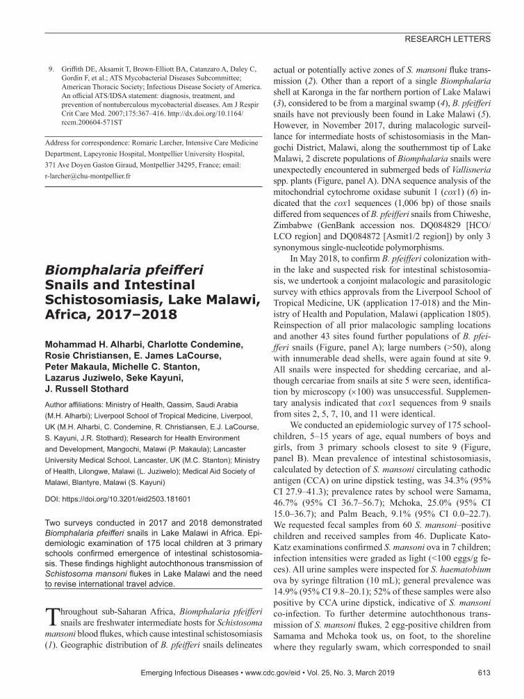
actual or potentially active zones of S. mansoni fluke trans-mission (2). Other than a report of a single Biomphalaria shell at Karonga in the far northern portion of Lake Malawi (3), considered to be from a marginal swamp (4), B. pfeifferi snails have not previously been found in Lake Malawi (5). However, in November 2017, during malacologic surveil-lance for intermediate hosts of schistosomiasis in the Man-gochi District, Malawi, along the southernmost tip of Lake Malawi, 2 discrete populations of Biomphalaria snails were unexpectedly encountered in submerged beds of Vallisneria spp. plants (Figure, panel A). DNA sequence analysis of the mitochondrial cytochrome oxidase subunit 1 (cox1) (6) in-dicated that the cox1 sequences (1,006 bp) of those snails differed from sequences of B. pfeifferi snails from Chiweshe, Zimbabwe (GenBank accession nos. DQ084829 [HCO/LCO region] and DQ084872 [Asmit1/2 region]) by only 3 synonymous single-nucleotide polymorphisms.
In May 2018, to confirm B. pfeifferi colonization with-in the lake and suspected risk for intestinal schistosomia-sis, we undertook a conjoint malacologic and parasitologic survey with ethics approvals from the Liverpool School of Tropical Medicine, UK (application 17-018) and the Min-istry of Health and Population, Malawi (application 1805). Reinspection of all prior malacologic sampling locations and another 43 sites found further populations of B. pfei-fferi snails (Figure, panel A); large numbers (>50), along with innumerable dead shells, were again found at site 9. All snails were inspected for shedding cercariae, and al-though cercariae from snails at site 5 were seen, identifica-tion by microscopy (×100) was unsuccessful. Supplemen-tary analysis indicated that cox1 sequences from 9 snails from sites 2, 5, 7, 10, and 11 were identical.
We conducted an epidemiologic survey of 175 school-children, 5–15 years of age, equal numbers of boys and girls, from 3 primary schools closest to site 9 (Figure, panel B). Mean prevalence of intestinal schistosomiasis, calculated by detection of S. mansoni circulating cathodic antigen (CCA) on urine dipstick testing, was 34.3% (95% CI 27.9–41.3); prevalence rates by school were Samama, 46.7% (95% CI 36.7–56.7); Mchoka, 25.0% (95% CI 15.0–36.7); and Palm Beach, 9.1% (95% CI 0.0–22.7). We requested fecal samples from 60 S. mansoni–positive children and received samples from 46. Duplicate Kato-Katz examinations confirmed S. mansoni ova in 7 children; infection intensities were graded as light (<100 eggs/g fe-ces). All urine samples were inspected for S. haematobium ova by syringe filtration (10 mL); general prevalence was 14.9% (95% CI 9.8–20.1); 52% of these samples were also positive by CCA urine dipstick, indicative of S. mansoni co-infection. To further determine autochthonous trans-mission of S. mansoni flukes, 2 egg-positive children from Samama and Mchoka took us, on foot, to the shoreline where they regularly swam, which corresponded to snail
Emerging Infectious Diseases • www.cdc.gov/eid • Vol. 25, No. 3, March 2019 613
RESEARCH LETTERS
collection sites 10 and 11 (Figure, panel B). Children who were positive for either S. mansoni CCA or S. haemato-bium eggs received praziquantel (40 mg/kg).
Colonization of B. pfeifferi snails in Lake Malawi and surrounding water is of concern, especially because active S. mansoni infections were found in local children. This finding highlights emergence of intestinal schistosomia-sis, not previously documented here (5,7,8) or detected in this region by the most recent national survey (F. Fleming, Schistosomiasis Control Initiative, Imperial College Lon-don; 2017 Dec 20; pers. comm).
Intestinal schistosomiasis has been detected in children ≈150 km away, along the shoreline of the Lower Shire River (9). Finding snails and infected children in Mangochi Dis-trict suggests recent ecologic and epidemiologic change. In May 2018, the lake was ≈75–80 cm higher than in November 2017, which perhaps favored detection of B. pfeifferi snails in the previously more accessible Vallisneria plant beds. Seasonal dynamics, such as lake level fluctuations, are well known, along with longer duration perturbations of the lake biota, either induced by climate change or mediated by an-thropogenic activities. These changes have altered transmis-sion of urogenital schistosomiasis (10); overfishing, particu-larly of the molluscivorous fish Trematocranus placodon, is changing the distribution of many freshwater snails (5).
Local aquaculture of fish (e.g., Oreochromis spp., called chambo) through use of water pumped inland from the lake has created novel, permanent water bodies colo-nized by B. pfeifferi snails (e.g., sites 2–7), which may now (re)seed snails into the lake for further establishment. Absence of cox1 genetic diversity in the B. pfeifferi snails
we sampled implies a limited number or even a single founder event, but as conditions for autochthonous trans-mission became favorable, after introduction of S. mansoni flukes, intestinal schistosomiasis in local schoolchildren has emerged. This finding is of substantial public health concern in light of current control efforts, which consist only of annual praziquantel distribution in schools (7,8). We recommend increased surveillance of snails and char-acterization of schistosomes, along with intensified control interventions to arrest further spread of intestinal schisto-somiasis. We also recommend revising and updating health and travel advice given to shoreline community residents and tourists who use the lake.
AcknowledgmentsWe thank Alexandra Shaw and Joanna Fawcett for assistance during the epidemiologic survey in the Mangochi District. We are grateful to the local health and education authorities of Malawi; district teachers; local community health workers Flora Jumbe, Caroline Nthubula, Angelina Mwenyewe, and Witness Mapira; and hosting communities for their enthusiasm and support. We are also indebted to Danie and Hazel Britz for assistance at Palm Beach School, to Paul and Stacey Kennedy for local boat hire, and to Anthony Butterworth and Liz Corbett for their kind hospitality in Blantyre.
M.H.A. was funded by a PhD scholarship from the Ministry of Health, Kingdom of Saudi Arabia, and S.K. by the Commonwealth Scholarship Commission. CCA urine dipsticks were supplied by Rapid Medical Diagnostics (http://www.rapid-diagnostics.com), South Africa (lot no. 171103130).
614 Emerging Infectious Diseases • www.cdc.gov/eid • Vol. 25, No. 3, March 2019
RESEARCH LETTERS
Figure. Locations sampled for Biomphalaria pfeifferi snails and of 3 primary schools where children were tested for intestinal schistosomiasis in the region of Lake Malawi, Africa. A) Locations sampled for B. pfeifferi snails in November 2017 (gray dots) and May 2018 (black dots), Lake Malawi, Africa. + indicates snails present, – indicates snails absent, and ● indicates site not sampled; symbol position indicates year of sampling (left, 2017; right, 2018). Numbers within circles indicate site numbers. Collected snail numbers are indicated by circle size. In 2017, snails were collected at 2 sites and not collected at 12 sites; in 2018, snails were collected at 10 sites and not collected at 47 sites. On each sampling occasion, >50 B. pfeifferi snails were collected at site 9. Coordinates of B. pfeifferi–positive sites: site 1, 14.27752°S, 35.10419°E; site 2, 14.31371°S, 35.14174°E; site 3, 14.31424°S, 35.14383°E; site 4, 14.31354°S, 35.14424°E; site 5, 14.31568°S, 35.14030°E; site 6, 14.32033°S, 35.13613°E; site 7, 14.32100°S, 35.13072°E; site 8, 14.36919°S, 35.17629°E; site 9, 14.39363°S, 35.22104°E; site 10, 14.42708°S, 35.23349°E; and site 11, 14.44928°S, 35.23890°E. B) Location of the 3 sampled primary schools (Palm Beach, 14.391346°S, 35.215137°E; Samama 14.417465°S, 35.217580°E; Mchoka 14.439481°S, 35.220644°E) showing local prevalence (% [95% CI]) of intestinal schistosomiasis indicated by Schistosoma mansoni circulating cathodic antigen (CCA) detected by urine dipstick. Water collection sites pinpointed by 2 Schistosoma egg–positive children from Samama and Mchoka Schools are indicated.
About the AuthorMr. Alharbi is a PhD student under the supervision of L.J. and J.R.S. He has specific interests in medical malacology and molecular epidemiology of schistosomiasis in Africa and the Kingdom of Saudi Arabia.
References 1. Brown DS. Freshwater snails of Africa and their medical
importance, 2nd ed. London: Taylor & Francis; 1994. p. 321. 2. Stensgaard AS, Utzinger J, Vounatsou P, Hürlimann E, Schur N,
Saarnak CF, et al. Large-scale determinants of intestinal schistosomiasis and intermediate host snail distribution across Africa: does climate matter? Acta Trop. 2013;128:378–90. http://dx.doi.org/10.1016/j.actatropica.2011.11.010
3. Smith HE. On a collection of land and freshwater snails transmitted by Mr H.H. Johnston C.B. from British Central Africa. Proc Zoo Soc (London). 1893;632–641.
4. Mandahl-Barth G. The freshwater Mollusca of Lake Malawi. Rev Zool Bot Afr. 1972;86:129–60.
5. Madsen H, Bloch P, Makaula P, Phiri H, Furu P, Stauffer JR Jr. Schistosomiasis in Lake Malaŵi villages. EcoHealth. 2011;8:163–76. http://dx.doi.org/10.1007/ s10393-011-0687-9
6. Jørgensen A, Kristensen TK, Stothard JR. Phylogeny and biogeography of African Biomphalaria (Gastropoda: Planorbidae), with emphasis on endemic species of the great East African lakes. Zool J Linn Soc. 2007;151:337–49. http://dx.doi.org/10.1111/j.1096-3642.2007.00330.x
7. Mtethiwa AH, Nkwengulila G, Bakuza J, Sikawa D, Kazembe A. Extent of morbidity associated with schistosomiasis infection in Malawi: a review paper. Infect Dis Poverty. 2015;4:25. http://dx.doi.org/10.1186/s40249-015-0053-1
8. Makaula P, Sadalaki JR, Muula AS, Kayuni S, Jemu S, Bloch P. Schistosomiasis in Malawi: a systematic review. Parasit Vectors. 2014;7:570. http://dx.doi.org/10.1186/s13071-014-0570-y
9. Poole H, Terlouw DJ, Naunje A, Mzembe K, Stanton M, Betson M, et al. Schistosomiasis in pre-school-age children and their mothers in Chikhwawa district, Malawi with notes on characterization of schistosomes and snails. Parasit Vectors. 2014;7:153. http://dx.doi.org/10.1186/1756-3305-7-153
10. Van Bocxlaer B, Albrecht C, Stauffer JR Jr. Growing population and ecosystem change increase human schistosomiasis around Lake Malaŵi. Trends Parasitol. 2014;30:217–20. http://dx.doi.org/ 10.1016/j.pt.2014.02.006
Address for correspondence: J. Russell Stothard, Liverpool School of Tropical Medicine, Parasitology Department, Pembroke Place, Liverpool Merseyside L3 5QA, UK; email: [email protected]
Emerging Infectious Diseases • www.cdc.gov/eid • Vol. 25, No. 3, March 2019 615
RESEARCH LETTERS
Originally published in October 2007
https://wwwnc.cdc.gov/eid/article/13/10/e1-1310_article
etymologia revisitedschistosomiasis [shis”-, skis” to-so-mi’ə-sis], from the Greek—skhistos (split) and soma (body)
Infection of the blood with a parasite of the genus Schistosoma. Originally thought a single organism with a split body, the parasite was eventually recognized as having male and female forms. Three main species cause human infection: S. haematobium, S. mansoni, and S. japonicum. Each species has its own range of host snails. The parasite releases eggs containing larvae through feces or urine; if the eggs reach water, the larvae are released and may penetrate a snail. A very large number of larvae are then produced inside the snail and released back into the water. Infection is acquired through skin contact with contaminated water.
Schistosomiasis, which leads to chronic hepatic and intestinal fibrosis of the urinary tract, was first identified in Egypt in 1851 by German pathologist Theodor Bilharz and is also called bilharzia. Approximately 160 million persons throughout the world are infected, particularly in Africa, the Middle East, South America, and Southeast Asia.
Source: Institute of Tropical Medicine of Antwerp: www.itg.be