biomimetic artificial organelles with in vitro and in vivo
TRANSCRIPT

ARTICLE
Biomimetic artificial organelles with in vitro andin vivo activity triggered by reduction inmicroenvironmentT. Einfalt1,2, D. Witzigmann2, C. Edlinger1, S. Sieber2, R. Goers1,3, A. Najer1, M. Spulber1, O. Onaca-Fischer1,
J. Huwyler2 & C.G. Palivan 1
Despite tremendous efforts to develop stimuli-responsive enzyme delivery systems, their
efficacy has been mostly limited to in vitro applications. Here we introduce, by using an
approach of combining biomolecules with artificial compartments, a biomimetic strategy to
create artificial organelles (AOs) as cellular implants, with endogenous stimuli-triggered
enzymatic activity. AOs are produced by inserting protein gates in the membrane of poly-
mersomes containing horseradish peroxidase enzymes selected as a model for natures own
enzymes involved in the redox homoeostasis. The inserted protein gates are engineered by
attaching molecular caps to genetically modified channel porins in order to induce redox-
responsive control of the molecular flow through the membrane. AOs preserve their struc-
ture and are activated by intracellular glutathione levels in vitro. Importantly, our biomimetic
AOs are functional in vivo in zebrafish embryos, which demonstrates the feasibility of using
AOs as cellular implants in living organisms. This opens new perspectives for patient-
oriented protein therapy.
DOI: 10.1038/s41467-018-03560-x OPEN
1 Department of Chemistry, University of Basel, Klingelbergstrasse 80, CH-4056 Basel, Switzerland. 2 Department of Pharmaceutical Sciences, Division ofPharmaceutical Technology, University of Basel, Klingelbergstrasse 50, CH-4056 Basel, Switzerland. 3 Department of Biosystems Science and Engineering,ETH Zürich, Mattenstrasse 26, CH-4058 Basel, Switzerland. Correspondence and requests for materials should be addressed toC.G.P. (email: [email protected])
NATURE COMMUNICATIONS | (2018) 9:1127 | DOI: 10.1038/s41467-018-03560-x | www.nature.com/naturecommunications 1
1234
5678
90():,;

M imicking biological processes by engineering biomi-metic nanostructures represents an elegant strategyfor addressing problems in various scientific fields,
including materials science, chemistry, electronics and medi-cine1–3. By applying a bottom-up biomimetic design (i.e.arranging molecules at the nanoscale via self-assembly), it ispossible to combine individual biological units, known fortheir sophisticated structure and activity (e.g. proteins, lipids,DNA), with robust synthetic materials (e.g. polymers, poroussilica surfaces, nanoparticles). This serves to develop nanoscalebiomimics with enhanced properties and functionalities2,4–8
with potential for a wide range of applications (sensitive bio-sensing, patient tailored therapeutics, detoxification of envir-onmental pollutants, etc.)6,9–12.
Of particular interest are two different concepts that are cur-rently the main focus in this research field: (i) artificial organelles(AOs) based on an essential need to offer efficient solutions forimproved therapy and diagnostics13 and (ii) protocell systemsintended to provide simple models of cells for understandingvarious internal processes2,14. These concepts are complementary,one is essential for advancing medical applications (AOs) whereasthe second concept mimics cell behaviour based on very simplesystems (protocells). Similarly, as in nature, the sizes of thecompartments are completely different: while AOs have nano-metre range sizes, protocells reach the micrometre range. Eventhough protocells represent the first archetypes of an artificial cell,they still inherently lack the material variety and responsivenessfound in the most basic cellular structures, and have not yet beeninvestigated in vivo to determine whether they preserve theirfunctionality. AOs are particularly attractive nanoscale biomimicsbecause they can provide a required compound/signal, detoxifyharmful compounds, or change cellular conditions and reactions.AOs are based on compartmentalisation of active compounds(enzymes, proteins, catalysts, mimics) within artificial nano-assemblies that reach and function in the intracellular environ-ment, and thus serve as simplified mimics of nature’s ownorganelles. Various examples of systems with potential to act asAOs have been developed based on liposomes, porous silicananoparticles and polymer compartments (polymersomes) incombination with biomacromolecules13–16. However, very fewhave been evaluated in vitro to assess their in situ cellular func-tionality6,15–18, and to the best of our knowledge, none has beenassessed in vivo. In vivo functionality of such AOs is a crucialfactor that is necessary to demonstrate that the concept of AOs isfeasible in living organisms, and thus AOs can act as cellularimplants.
Notably, natural organelles have membranes, since inside cellscompartmentalisation is essential to provide confined reactionspaces for complex metabolic reactions. Therefore, an AO shouldpreserve the compartmentalisation as a key factor in mimickingnatural organelles. In this respect, polymer compartments, namedpolymersomes, are ideal candidates for the creation of AOs,because of their hollow spherical structure with a membraneserving as a border for an inner cavity and their greatermechanical stability than lipid-based compartments, i.e. lipo-somes19,20. In addition, the chemical nature of the copolymersprovides the possibility of controlling their properties (e.g. size,stability biocompatibility, flexibility, stimuli-responsiveness)2,21.Polymersomes have been shown to serve either as carriers forbiomolecules and mimics1,2,21,22, or more recently for develop-ment of nanoreactors and even the generation of AOs2,6,11,23. Akey factor for supporting in situ reactions20,24 is to render thepolymersome membranes permeable for substrates and products.An elegant approach bioinspired from the cell membrane is toincorporate biopores and membrane proteins25–27. Selectivemembrane permeability towards protons and ions is achieved by
inserting small pore forming peptides27, while membrane pro-teins induce size-dependent cut-off permeability26,28–30 or evenmediate the diffusion of specific molecules10,31. The few reportedAOs exhibit enzymatic reactions either inside porous polymer-somes6,16,32 or inside polymersomes equipped with channelporins17, with the aim of emulating cellular pathways (e.g. reac-tive oxygen species detoxification or glucose oxidation).
Another essential factor for tuning AO functionality is a trig-gered response to its environment, as, for example, the redox stateof the cell, which regulates various processes involved in cellularsignalling pathways33,34. While there are a few reported examplesof polymersomes with a stimuli-responsive permeable membranebased on the incorporation of genetically or chemically modifiedmembrane proteins35, only two of them have served for thedesign of catalytic nanocompartments36,37, and none has beenused to control reactions inside AOs. Activation of the AO by aspecific endogenous stimulus inside cells represents a challengingstep in development of functional AOs in vivo. The design of AOswith triggered activity and the demonstration of their in vivofunctionality represent necessary steps towards the creation of cellimplants, and the provision of smart solutions for personalisedmedicine by a straightforward change of the biomolecules insidethe AOs.
Here, we present a strategy for designing AOs with an in situenzymatic reaction that is triggered by the presence of an intra-cellular stimulus, and demonstrate in vitro and in vivo func-tionality. Genetically modified outer membrane protein F(OmpF) porins were incorporated into polymersomes to induceredox responsiveness to the membrane, and horseradish perox-idase (HRP) simultaneously encapsulated inside their cavity toprovide a source of the AO functionality. Such AOs with func-tionality triggered by intracellular changes represent an advancein mimicking that of nature’s own organelles, especially those thatare involved in the redox equilibrium of the cellular homo-eostasis. Amphiphilic block copolymers poly(2-methyloxazoline)-block-poly(dimethylsiloxane)-block-poly(2-methyloxazoline)(PMOXAm-PDMSn-PMOXAm) were used to self-assemble intopolymersomes, because such copolymers have already beenshown to form membranes in which biopores and membraneproteins can be successfully inserted36–38, and to be taken up andto be non-toxic to various cell lines17. Once inserted in thepolymersome membrane, the modified OmpF porins act asprotein gates independent of the insertion direction, i.e. orien-tation in the membrane36,37,39,40, and trigger the in situ HRPenzymatic reaction when a stimulus is present in the cellularenvironment. HRP was selected as model enzyme, because per-oxidases play a significant role in the redox homoeostasis of cellsand cell apoptosis41. This strategy of providing stimuli-responsiveness to polymersome membranes neither affects themembrane integrity, as for stimuli-responsive synthetic mem-branes of compartments42, nor the size and structure of thepolymersomes. Crucial steps were the evaluation of AO toxicityand functionality in human epithelial tumour cells (HeLa cells),and once these were established in vivo tolerability, preservationof the AO structure, and in situ regulation of the activity of theencapsulated enzyme in the vertebrate zebrafish embryo (ZFE)model.
ResultsBioengineering protein gates by modification of channel por-ins. The key factors in the design of AOs with activity triggeredby changes in environmental conditions are on-demand perme-ability of the compartment towards enzymatic substrates/pro-ducts and structural integrity of the polymersome, which mimicsthat of natural organelles. Therefore, our biomimetic strategy
ARTICLE NATURE COMMUNICATIONS | DOI: 10.1038/s41467-018-03560-x
2 NATURE COMMUNICATIONS | (2018) 9:1127 | DOI: 10.1038/s41467-018-03560-x | www.nature.com/naturecommunications

aimed to equip PMOXA6-PDMS44-PMOXA6 polymersomemembranes with protein gates that are responsive to changes inglutathione (GSH) concentrations in intracellular environ-ments, while preserving the structure of the nanocompartment(Fig. 1a).
It has been shown very recently that chemical modificationsof amino acid residues at key locations of the OmpF porinbackbone influence the translocation of substrates through thepore in a pH-responsive manner36. Here we go one step furtherby using a double mutant of OmpF37 to attach molecular capsto genetically introduced cysteine residues that serve to block/unblock the OmpF pore upon changes in redox potential, whichoccur when the system enters the intracellular microenviron-ment (Fig. 1b). In contrast to polymersomes with membranescontaining OmpF genetically modified to release a payload inreductive conditions35, our system controls the overall func-tionality of the AOs. We chose a cysteine double mutant ofOmpF (OmpF-M) because cysteine residues, replacing theamino acids K89 and R270, were expected to form reduction-sensitive disulphide bonds with molecules selected to serve asmolecular caps. These molecular caps remain attached in mildlyoxidising environments and block substrate diffusion throughthe pore, whereas in the presence of reducing agents, such asintracellular GSH, their cleavage restores normal passage ofsmall molecular weight molecules (<600 Da) through the OmpFpores. This approach mimics pathways of metabolism regula-tion, where proteins within the membranes of natural cellorganelles are irreversibly activated or deactivated ondemand43,44. In addition, we were interested in developing anirreversible protein gate in order to be able to rapidly evaluatethe functionality of the organelle in vivo.
The ability of the cysteine residues of OmpF-M to formdisulphide bonds with thiol groups of small molecular weightmolecules was examined by two complementary assays, one usinga suitable spin probe (bis-(2,2,5,5-tetramethyl-3-imidazoline-1-
oxyl-4-yl) disulphide) and the second using the fluorescent dyeSAMSA fluorescein (SAMSA-CF) (Fig. 1c, d).
Coupling reaction of the molecular caps with the cysteineresidues of OmpF-M resulted in the formation of OmpFconjugates (OmpF-S-S-CF for OmpF conjugated with SAMSA-CF, and OmpF-S-S-NO for OmpF conjugated with bis-(2,2,5,5-tetramethyl-3-imidazoline-1-oxyl-4-yl) disulphide), respectively.
Binding of the thiol reactive spin probe to the protein wasevaluated by a combination of LC-MS-MS and electronparamagnetic resonance (EPR). Upon in-gel digestion of theporin45, LC-MS-MS analysis of the peptide fragments indicated avery high labelling efficiency of the spin probe to cysteine residuesof the OmpF-M (96 ± 4%). Standard deviation is based on threemeasurements. The EPR spectrum of the bis-(2,2,5,5-tetramethyl-3-imidazoline-1-oxyl-4-yl) disulphide in phosphate-buffered sal-ine (PBS) at 298 K consists of an isotropic triplet pattern(Supplementary Figure 1) with a hyperfine coupling aN value of15.8 G that is similar to reported values for analogous nitroxideprobes where no aggregation was present46,47. In contrast,OmpF-S-S-NO gave a broad anisotropic EPR spectrum with noisotropic component, and is similar to that reported for 5-DSA inlipid bilayers or cholesterol aqueous solutions48. This EPRspectrum indicates hindered rotation of the nitroxide probe49
after binding to the OmpF mutant (OmpF-S-S-NO), anddemonstrates successful binding of the bis-(2,2,5,5-tetramethyl-3-imidazoline-1-oxyl-4-yl) disulphide to the modified OmpFmutant (Fig. 2a).
After exposure of OmpF-S-S-NO to 10 mM DTT an isotropicEPR spectrum (aN value of 15.9 G) characteristic of the freelyrotating spin probe was observed (Fig. 2b). This clearlydemonstrates that the nitroxide spin probe that is bound to thiolgroups of the OmpF-M under oxidative conditions is cleaved in areductive environment.
SAMSA-CF (Thermo Fischer Scientific) was selected as amolecular cap because its size (molecular weight 521.49 Da) wasexpected to block the OmpF-M pore, and because of its ability
Inactivea b
c d
Active
Cysteine residues
OmpF-SH
OmpF-SH
OmpFSAMSA CF
SAMSA CF
SAMSA CF
OmpF-SH OmpF-SS
=
S
O
HO O O
COOH
HN
O
X
COOH
++ OO RT, 48 h aq, NaOH, RT, 48 h
NN
NNS
SS
S
=
NN O
Molecular cap
Enzymatic substrate
Fig. 1 Engineering stimuli-responsive OmpF. a Schematic representation of modified OmpF acting as a gate in catalytic nanocompartments. b Molecularrepresentation of the OmpF-M cysteine mutant37. c Chemical modification of OmpF-M cysteine mutant with the spin probe bis-(2,2,5,5-tetramethyl-3-imidazoline-1-oxyl-4-yl) disulphide. d Chemical modification of OmpF-M cysteine mutant with the fluorophore SAMSA-CF
NATURE COMMUNICATIONS | DOI: 10.1038/s41467-018-03560-x ARTICLE
NATURE COMMUNICATIONS | (2018) 9:1127 | DOI: 10.1038/s41467-018-03560-x | www.nature.com/naturecommunications 3

to form cleavable disulphide bonds50. Thus, attachment ofSAMSA-CF to OmpF-M introduces a stimuli-responsiveness tothe pore, and therefore to the polymersome membrane whenOmpF-S-S-CF is inserted. In addition, the fluorescent proper-ties of SAMSA-CF allow pore modification to be analysed by acombination of sodium dodecyl sulfate polyacrylamide gelelectrophoresis (SDS-PAGE) and fluorescence correlationspectroscopy (FCS).
LC-MS-MS analysis of the peptide fragments indicated a highlabelling degree of OmpF-M (81 ± 31%). In addition, afluorescent band appeared in the SDS-PAGE gel whenSAMSA-CF was conjugated to OmpF-M, whereas the OmpFwild type did not interact with the fluorophore; this fluorescentband supports the formation of OmpF-S-S-CF (SupplementaryFigure 2). To mimic the intracellular reductive environment,where the GSH concentration is kept at a constantly high level(10 mM GSH) by cytosolic enzymes51, such as glutathionereductase, we studied the behaviour of the reduction-responsivemolecular caps in a similar environment. Because of theabsence of a steady-state concentration and constant regenera-tion of GSH, we used 30 mM GSH to mimic the intracellularsteady state of GSH. In SDS-PAGE the fluorescent banddisappeared when the OmpF-S-S-CF was mixed with GSH,
indicating successful cleavage of the molecular cap underreductive conditions (Supplementary Figure 2).
The binding of SAMSA-CF to OmpF-M cysteine residues wasalso evaluated by FCS, because it allows the determination ofdiffusion coefficients, which are correlated to possible interactionsof the fluorescent molecules with supramolecular assemblies, suchas polymersomes, liposomes and nanoparticles in the pico- tonanomolar concentration region9,21,47–52. We compared themolecular brightness and diffusion times of SAMSA-CF in PBS(pH 7.4), SAMSA-CF in 1% OG PBS (pH 7.4) and SAMSA-CFbound to OmpF (OmpF-S-S-CF) in 1% OG PBS (pH 7.4)(Fig. 2c). A labelling efficiency of an average of two SAMSA-CFmolecules per monomer was calculated by comparing themolecular brightness (counts per molecule, CPM in kHz) ofSAMSA-CF (2.2 ± 0.7 kHz) with that of protein bound toSAMSA-CF (4.8 ± 0.6 kHz) (Fig. 2c). Standard deviations inmolecular brightness are based on individual measurements ofthe same probe (n= 60). In contrast, wild-type OmpF treatedsimilarly to the cysteine mutant OmpF-M, did not present anyfluorescence after purification, and there was therefore no bindingof SAMSA-CF to OmpF-WT. To determine the kinetics of OmpFpore opening, we used FCS to evaluate the cleavage of SAMSA-CF from labelled OmpF-M upon addition of 30 mM GSH at pH
10 mM DTT
Experimental
a b
c d
Simulation
3330 3340 3350
1.0 100
75
50
Fre
e dy
e fr
actio
n (%
)
25
00 15 30
Time (min)45 60 75
0.5
Free SAMSA-CF
OmpF-S-S-CF
Nor
mal
ised
(G
τ)
0.0101 102 103
τ (μs)
104 105 106
3360 3370
Magnetic field / G
3380 3390 3400 3410 3330 3340 3350 3360 3370
Magnetic field / G
3380 3390 3400
Experimental
Simulation
a = 15.9 G
NO
NO
NO
NO
Fig. 2 Characterisation of stimuli-responsive OmpF Panel. a EPR spectra of bis-(2,2,5,5-tetramethyl-3-imidazoline-1-oxyl-4-yl) disulphide-labelledOmpF-M experimental (black) and simulated (blue) and b bis-(2,2,5,5-tetramethyl-3-imidazoline-1-oxyl-4-yl) disulphide-labelled OmpF-M in 1% OGincubated with 10 mM DTT experimental (black) and simulated (blue). c Normalised FCS autocorrelation curves for SAMSA-CF in PBS (black),SAMSA-CF in 1% OG (blue) and OmpF-S-S-CF in 1% OG (Red). Dotted line—experimental autocorrelation curves, full line—fit. d SAMSA-CF releasekinetics from OmpF-M in 30 mM GSH, 1% OG, as measured by FCS and analysed with a two-component fit. Error bars show standard deviations from60 measurements
ARTICLE NATURE COMMUNICATIONS | DOI: 10.1038/s41467-018-03560-x
4 NATURE COMMUNICATIONS | (2018) 9:1127 | DOI: 10.1038/s41467-018-03560-x | www.nature.com/naturecommunications
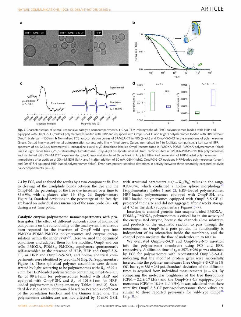
7.4 by FCS, and analysed the results by a two-component fit. Dueto cleavage of the disulphide bonds between the dye and theOmpF-M, the percentage of the free dye increased over time to85 ± 9%, with a plateau after 1 h (Fig. 2d, SupplementaryFigure 3). Standard deviations in the percentage of the free dyeare based on individual measurements of the same probe (n= 60)during a set time point.
Catalytic enzyme-polymersome nanocompartments with pro-tein gates. The effect of different concentrations of individualcomponents on the functionality of the final system has alreadybeen reported for the insertion of OmpF wild type intoPMOXA-PDMS-PMOXA polymersomes and enzyme encap-sulation within the inner cavity53. Here we used the optimisedconditions and adapted them for the modified OmpF and ourAOs. PMOXA6-PDMS44-PMOXA6 copolymers spontaneouslyself-assembled in the presence of HRP, HRP and OmpF-S-S-CF, or HRP and OmpF-S-S-NO, and hollow spherical com-partments were identified by cryo-TEM (Fig. 3a, SupplementaryFigure 4). These spherical polymer assemblies were demon-strated by light scattering to be polymersomes with: RH of 99 ±2 nm for HRP-loaded polymersomes containing OmpF-S-S-CF,RH of 89 ± 4 nm for polymersomes loaded with HRP andequipped with OmpF-SH, and RH of 101 ± 1 nm for HRP-loaded polymersomes (Supplementary Tables 1 and 2). Stan-dard deviations were determined based on Pearson’s coefficientof the correlation function and the Guinier fitted one. Thepolymersome architecture was not affected by 30 mM GSH,
with structural parameters ρ (ρ= RG/RH) values in the range0.90–0.96, which confirmed a hollow sphere morphology54
(Supplementary Tables 1 and 2). HRP-loaded polymersomes,HRP-loaded polymersomes equipped with OmpF-SH, andHRP-loaded polymersomes equipped with OmpF-S-S-CF allpreserved their size and did not aggregate after 2 weeks storageat 4 °C in the dark (Supplementary Figures 5–7).
Insertion of channel proteins into enzyme-loaded PMOXA6-PDMS44-PMOXA6 polymersomes is critical for in situ activity ofthe encapsulated enzyme, because the channels allow substratesand products of the enzymatic reaction to pass through themembrane. As OmpF is a pore protein, its functionality isindependent of its orientation inside the membrane, and thechannel porin mediates the flow of molecules up to 600 Da.
We evaluated OmpF-S-S-CF and OmpF-S-S-NO insertioninto the polymersome membrane using FCS and EPR,respectively. A diffusion time of τd= 2573 ± 960 µs was obtainedby FCS for polymersomes with reconstituted OmpF-S-S-CF,indicating that the modified protein gates were successfullyinserted into the polymer membranes (free OmpF-S-S-CF in 1%OG has τd= 588 ± 261 µs). Standard deviation of the diffusiontimes is acquired from individual measurements (n= 60). Bycomparing the molecular brightness of the free fluorophore(CPM= 2.2 ± 0.7 kHz) and the OmpF-S-S-CF equipped poly-mersomes (CPM= 18.9 ± 11.1 kHz), it was calculated that therewere five OmpF-S-S-CF porins/polymersome; these values aresimilar to those reported previously for wild-type OmpF36
(Fig. 3b).
HRP + OmpF-SH HRP + OmpF-S-S-CF HRP1.0
0.5
Nor
mal
ised
- G
(τ)
0.0
3330
3340
3350
3360
3370
Magnetic field (G)
g = 20.0065
a b
c d
Experimental
Experimental0.025 0.035
0.030
0.025
0.020
0.015
0.010
0.020
0.015
Inte
nsity
(a.
u. s
–1)
Inte
nsity
(a.
u. s
–1)
0.010
0.0051 2 1 2
n.s.
t = 0 h t = 1 h *
Simulation
aN = 15.9 G
ΔH = 10 G
3380
3390
3400
3330
3340
3350
3360
3370
Magnetic field (G)
3380
3390
3400
3410
101 102 103
τ (μs)
104 105 106
Fig. 3 Characterisation of stimuli-responsive catalytic nanocompartments. a Cryo-TEM micrographs of: (left) polymersomes loaded with HRP andequipped with OmpF-SH, (middle) polymersomes loaded with HRP and equipped with OmpF-S-S-CF, and (right) polymersomes loaded with HRP withoutOmpF. Scale bar= 100 nm. b Normalised FCS autocorrelation curves of SAMSA-CF in PBS (black) and OmpF-S-S-CF in the membrane of polymersomes(blue). Dotted line= experimental autocorrelation curves, solid line= fitted curve. Curves normalised to 1 to facilitate comparison. c Left panel: EPRspectrum of bis-(2,2,5,5-tetramethyl-3-imidazoline-1-oxyl-4-yl) disulphide-labelled OmpF reconstituted in PMOXA-PDMS-PMOXA polymersomes (blackline). c Right panel: bis-(2,2,5,5-tetramethyl-3-imidazoline-1-oxyl-4-yl) disulphide-labelled OmpF reconstituted in PMOXA-PDMS-PMOXA polymersomesand incubated with 10 mM DTT experimental (black line) and simulated (blue line). d Amplex Ultra Red conversion of HRP-loaded polymersomes:immediately after addition of 30mM GSH (left), and 1 h after addition of 30mM GSH (right). OmpF-S-S-CF equipped HRP-loaded polymersomes (green)and OmpF-SH equipped HRP-loaded polymersomes (blue). Error bars present standard deviations in activity between three separately prepared catalyticnanocompartments (n= 3)
NATURE COMMUNICATIONS | DOI: 10.1038/s41467-018-03560-x ARTICLE
NATURE COMMUNICATIONS | (2018) 9:1127 | DOI: 10.1038/s41467-018-03560-x | www.nature.com/naturecommunications 5

HRP-loaded polymersomes containing OmpF-S-S-NO pro-duced a broad EPR spectrum (Fig. 3c), indicative of low mobility,a result similar to that reported for 5-DSA and 16-DSA insertedin polymersomes membranes55. However, when these HRP-loaded polymersomes containing OmpF-S-S-NO were exposed toreductive conditions (10 mM DTT), an isotropic EPR spectrum(aN= 15.9 G) was observed superimposed on the broad peak,indicating successful cleavage of some of the nitroxide spin probefrom the OmpF (Fig. 3c).
Stimuli-responsiveness of the catalytic nanocompartments. Theeffect of an external stimulus on the functionality of the HRP-loaded polymersomes equipped with OmpF-S-S-CF was eval-uated by their response to the addition of 30 mM GSH. Thefluorescent signal associated with formation of a resorufin-likeproduct (RLP) during the in situ enzymatic reaction in the pre-sence of Amplex Ultra Red (AR) as a substrate for HRP wasmeasured spectroscopically56. Enzymatic turnover of the ARsubstrate was significantly lower with HRP-loaded polymersomesequipped with OmpF-S-S-CF (by up to 36±4%) compared toHRP-loaded polymersomes equipped with OmpF-SH, suggestingthat the molecular cap is sufficient to reduce the passage of smallmolecules through the pore. Note that the very low activity ofHRP-loaded polymersomes without inserted OmpF was takeninto account for background correction. Standard deviation isbased on three measurements of separately prepared catalyticnanocompartments. Addition of 30 mM GSH to the systemincreased the activity of HRP-loaded polymersomes equippedwith OmpF-S-S-CF up to that of HRP-loaded polymersomesequipped with OmpF-SH. This indicates that reduction of thedisulphide bridge between the attached SAMSA-CF cap andcysteine residues of the OmpF-M successfully restored theOmpF-M pore permeability for the substrate of the enzyme byreleasing the molecular cap (Fig. 3d, Supplementary Figures 8and 9).
Nanocompartments as stimuli-responsive AOs. Here we havegone a step further by developing stimulus-triggered AOs, whosefunctionality is modulated by the responsiveness of modifiedOmpF porins inserted in the membrane of the catalytic nano-compartments. Previously designed AOs successfully overcamethe first barrier of cell membranes and escaped from endo-somes17. As PMOXA-PDMS-PMOXA polymersomes are stableat acidic pH27,36, we consider that this will favour a successfullysosomal/endosomal escape during the recycling of lysosomesand endosomes.
Possible internalisation mechanisms of various PMOXA-PDMS-PMOXA-based polymersomes and their high cytocompatibility invarious cell lines have already been reported17,57–59. Here, weevaluated the cytocompatibility of the biomimetic AOs by testingtheir cellular toxicity using the 3-(4,5-dimethylthiazol-2-yl)-5-(3-carboxymethoxyphenyl)-2-(4-sulphophenyl)-2H-tetrazolium (MTS)assay before studying their intracellular activation and enzymaticactivity. Notably, their biocompatibility at the cellular level wasshown by the absence of any decrease in viability in HeLa cells evenafter 48 h (i.e. polymer concentration ranging from 0.25 to 0.75mgml−1) (Supplementary Figure 10).
In order to study cellular internalisation and intracellularlocalisation, we first conjugated HRP with Atto488 (HRP-Atto488) and Atto647 (HRP-Atto647), respectively (Supplemen-tary Figure 11). Then we encapsulated labelled-HRP inside thecavity of polymersomes, polymersomes equipped with OmpF-S-S-CF, and polymersomes equipped with OmpF-SH. Cellularuptake assays in HeLa cells indicated successful internalisationresulting in a particulate intracellular staining pattern withincreasing intensity in a time-dependent manner from 8 to 24 h(Fig. 4a, Supplementary Figures 12 and 13). The quantitativeanalysis indicates that after 24 h AOs did not co-localise withearly endosomes or lysosomes, confirming successful intracellularendosomal escape (Supplementary Figure 14)17. Localised HRP-Atto488 signals confirmed the intracellular integrity of thepolymersomes. In sharp contrast, if cells were treated with a
Control Hela cells
a
b
HoechstHRP-Atto488Cell mask
HoechstRLPCell mask
Unpermiabilised HRP-Atto488 polymersomes
OmpF-SH equipped HRP-Atto488 polymersomes
OmpF-S-S-CF equipped HRP-Atto488 polymersomes
= HRP-Atto488 loaded AO
= AR= H2O2= RLP= AO
Cellular u
ptake
100 nm
Control Hela cellsUnpermiabilised HRP-Atto488 polymersomes
OmpF-SH equippedHRP polymersomes
OmpF-S-S-CF equippedHRP polymersomes
Fig. 4 Cellular uptake and intracellular activation of AOs. a Confocal fluorescence micrographs of HeLa cells showing cellular uptake of fluorescentlylabelled HRP-loaded polymersomes and AOs loaded with fluorescently labelled HRP. Scale bar: 10 µm. b Cellular uptake and intracellular activation offluorescently labelled HRP-loaded polymersomes and fluorescently labelled HRP-loaded AOs. Blue signal: Hoechst 33342 nucleus stain. Grey signal:CellMask Deep Red Plasma membrane stain. Green signal: Atto488 HRP. Red signal: resorufin-like product (RLP). Scale bar 20 µm
ARTICLE NATURE COMMUNICATIONS | DOI: 10.1038/s41467-018-03560-x
6 NATURE COMMUNICATIONS | (2018) 9:1127 | DOI: 10.1038/s41467-018-03560-x | www.nature.com/naturecommunications
membrane disrupting agent (i.e. 0.1% saponin) (SupplementaryFigure 15), polymersome membranes were affected and resultedin an intracellular cytoplasmic distribution of HRP-Atto488.
The capacity of the AOs to act within target cells in a stimuli-responsive manner was investigated by using a combination ofconfocal laser scanning microscopy (CLSM) and flow cytometryto evaluate their potential to respond to increased intracellularGSH levels. HeLa cells were incubated with HRP-loadedpolymersomes without OmpF or with HRP-loaded polymer-somes equipped with either OmpF-S-S-CF (AOs) or withOmpF-SH. Extracellular polymersomes were removed bywashing before imaging the intracellular activity of AOs. Cellswere incubated with a 1:1 substrate mixture of H2O2 and AR toallow the intracellular deposition, and finally conversion of ARinto its RLP by AOs. Note that both hydrogen peroxide and ARpass through the cellular membrane via passive partitioning,while they do not penetrate the membrane of polymersomes(Supplementary Figure 9). In contrast to untreated cells, orthose incubated with HRP-loaded polymersomes withoutOmpF, a significant increase of intracellular fluorescence wasobserved with AOs equipped with OmpF-S-S-CF or OmpF-SH(Fig. 4b, Supplementary Figure 16). A similar trend wasobserved when AR turnover was quantified by flow cytometry(Supplementary Figure 17). The strong fluorescent signal forAOs based on HRP-loaded polymersomes equipped withOmpF-S-S-CF confirmed successful intracellular cleavage ofthe molecular cap attached to OmpF-M, and subsequentactivation of the AOs within the intracellular environment ofthe HeLa cells (Supplementary Figure 17).
In vivo activity of stimuli-responsive AOs. As a step further toobtaining insight into their safety, tolerability and performancein vivo, AOs were studied in a ZFE model. ZFEs were selected,because of their recognition as a complementary vertebrateanimal model for applications, such as compound screening indrug discovery, toxicological studies and recombinant diseasemodels60–62. Compared to rodent in vivo models, the ZFEoffers unique advantages: (i) high reproducibility, (ii) low costs,(iii) high level of genetic homology to humans, (iv) availabilityof transgenic lines and (v) most importantly for the evaluationof AO, optical transparency. Due to their optical transparency,ZFE provide the possibility of imaging fluorescently-taggedobjects and fluorescent processes in vivo at a high resolutionover time63 (Supplementary Figure 18). Our approach offersthe possibility of gaining detailed insight into the circulationbehaviour of AOs and subsequent enzymatic reactions as wereported recently for nano-particulate drug delivery systemsin vivo64. In order to follow the biodistribution of AOs, weinjected intravenously via the duct of Cuvier HRP-Atto488-loaded polymersomes with membranes equipped with OmpF-S-S-CF or with OmpF-SH, respectively. No acute toxicity, suchas change in behaviour i.e. mobility, seizures, heart failure orother toxic effects such as malformations, denaturation of tissuefluids or yolk mass was observed in ZFE injected with AOs after24 h. ZFE analysed 2 h post intravenous injection of all types ofAOs containing Atto488 conjugated HRP showed a distinctfluorescent staining pattern (Supplementary Figure 19) in theposterior cardinal vein region, and we hypothesise that poly-mersomes are recognised by the ZFE early immune system and
100
80
60
40% o
f max
20
Control ZF embryo
a b c
d Unpermeabelised HRP-Atto488polymersomes
OmpF-SH equipped HRP-Atto488polymersomes
OmpF-S-S-CF equipped HRP-Atto488 polymersomes
0
100 + Cyto B
+ Poly(I:C)
+ Colehicine
+ NaN3
+ Cyto B
AOs
Ctrl
80
60
40% o
f max
20
0101 102
FL1 Log: Atto488-HRP
103 104 101 102
FL1 Log: Atto488-HRP
103 104101 102
FL1 Log: Atto488-HRP
Zebrafish embryo
= Resazurin
Macrophage
103 104
ZF MelanocytesGFP MacrophagesHRP-Atto647
ZF MelanocytesHRP-Atto488RZLP
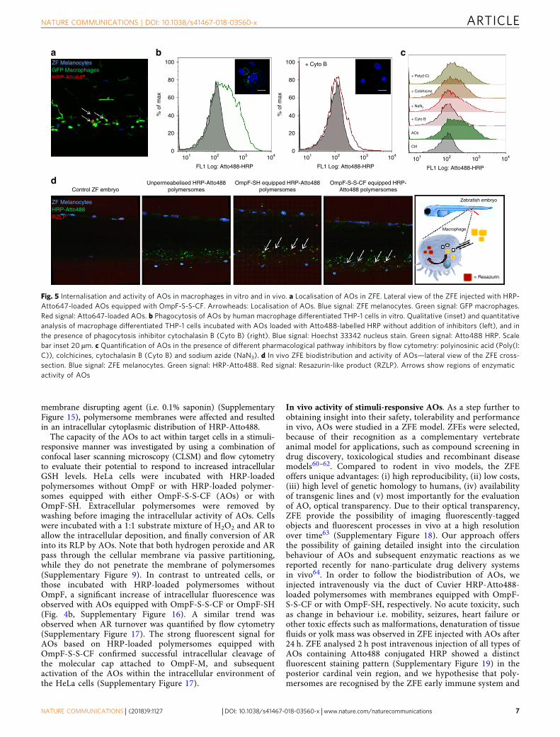
Fig. 5 Internalisation and activity of AOs in macrophages in vitro and in vivo. a Localisation of AOs in ZFE. Lateral view of the ZFE injected with HRP-Atto647-loaded AOs equipped with OmpF-S-S-CF. Arrowheads: Localisation of AOs. Blue signal: ZFE melanocytes. Green signal: GFP macrophages.Red signal: Atto647-loaded AOs. b Phagocytosis of AOs by human macrophage differentiated THP-1 cells in vitro. Qualitative (inset) and quantitativeanalysis of macrophage differentiated THP-1 cells incubated with AOs loaded with Atto488-labelled HRP without addition of inhibitors (left), and inthe presence of phagocytosis inhibitor cytochalasin B (Cyto B) (right). Blue signal: Hoechst 33342 nucleus stain. Green signal: Atto488 HRP. Scalebar inset 20 µm. c Quantification of AOs in the presence of different pharmacological pathway inhibitors by flow cytometry: polyinosinic acid (Poly(I:C)), colchicines, cytochalasin B (Cyto B) and sodium azide (NaN3). d In vivo ZFE biodistribution and activity of AOs—lateral view of the ZFE cross-section. Blue signal: ZFE melanocytes. Green signal: HRP-Atto488. Red signal: Resazurin-like product (RZLP). Arrows show regions of enzymaticactivity of AOs
NATURE COMMUNICATIONS | DOI: 10.1038/s41467-018-03560-x ARTICLE
NATURE COMMUNICATIONS | (2018) 9:1127 | DOI: 10.1038/s41467-018-03560-x | www.nature.com/naturecommunications 7

are subsequently taken up by macrophages65. The remarkablerecognition of polymersome-based AOs by the ZFE immunesystem was confirmed by the colocalisation of AOs loaded withAtto647-conjugated HRP (Atto647-HRP) injected into trans-genic ZFE specifically expressing eGFP in macrophages (Fig. 5a,Supplementary Figure 20). In strong contrast to AOs loadedwith Atto647-HRP, the free Atto647-HRP enzyme did not showsignificant macrophage colocalisation after 24 h, even whenAtto647-HRP was injected at concentrations of 0.2 mg ml−1
(Supplementary Figure 21). Notably, only macrophages in cir-culation were targeted and not tissue resident macrophages (i.e.star shaped).
Once cellular internalisation of AOs by the early immunesystem of ZFE was successful in vivo, we explored the uptakerate, exact intracellular localisation and internalisation mechan-isms of AOs in immune cells in vitro by using humanmacrophage differentiated THP-1 cells. AOs internalisationstarted as early as 30 min, and a strong internalisation byimmune cells was achieved after 3 h (Supplementary Figure 22),with increasing uptake rates at higher time points. As THP-1cells are immature macrophages with reduced phagocytoticcapacity, a higher uptake rate of AOs is possible for mature(primary) macrophages in vitro and in vivo.66 Importantly, allmacrophage uptake studies were performed in the presence ofserum proteins to mimic physiological conditions in vivobecause opsonisation of nanoparticles by serum proteins canhighly influence their interaction with cells.67 To obtain amechanistic understanding of the internalisation process, THP-1 macrophages were pre-treated with different pharmacologicalpathway inhibitors.67 We used inhibitors with specific inhibi-tion profiles: (i) polyinosinic acid to block scavenger receptors,(ii) colchicine to inhibit pinocytosis, (iii) cytochalasin B asphagocytosis inhibitor and (iv) sodium azide to inhibit allenergy-dependent uptake processes. Cells not incubated withAtto488 HRP-loaded AOs served as a control. A 1.28-foldincrease in the mean fluorescence intensity (MFI) was observedby flow cytometry analysis of the cells incubated with Atto488HRP-loaded AOs for 6 h, which indicates internalisation ofAOs by THP-1 macrophages. The uptake of AOs by macro-phages was significantly inhibited by cytochalasin B (a 0.13-foldincrease in MFI) and in a lower degree by sodium azide (0.43-fold increase in MFI), which indicates an energy-dependentphagocytotic internalisation process (Fig. 5b, c). On thecontrary, polyinosinic acid did not inhibit the AOs uptake,suggesting little or no involvement of the scavenger receptor inthe internalisation mechanism of AOs (Fig. 5c).
The internalisation process analysed by CLSM using Lyso-Tracker™ Red DND-99 as a reporter for the lysosomal compart-ments indicates that AOs co-localise with lysosomalcompartments during their internalisation process (Supplemen-tary Figure 23). Interestingly, we could not detect a lysosomesignal (lysotracker) 24 h after incubation of macrophages withAOs, suggesting the presence of an intracellular lysosomal escapemechanism once the AOs are taken-up by macrophages(Supplementary Figure 23). After internalisation in macrophages,the signals associated with Atto488-HRP-loaded AOs in lysoso-mal compartments changed to larger intracellular vesicularsignals. This suggests an expansion of the AO-bearing lysosomalcompartments before the AOs are released into the cytosol. Foran exact mechanism by which AOs escape the lysosomalcompartment and interact with cellular membranes, furtherinvestigations are planned but they are beyond the scope of thisstudy.
In order to assess in vivo stability, integrity and functionality ofAOs when exposed to the conditions in the macrophagemicroenvironment, we performed a second injection of AOs
together with the enzyme substrate AR. Injection of the co-substrate H2O2 in combination with AR was not necessary, sincemacrophages have the ability to produce H2O2. In addition, co-injection of H2O2 resulted in a red colouring of the whole bloodvolume, presumably due to haemolysis and thus interaction ofAR with erythrocyte enzymes or haemoglobin66 (SupplementaryFigure 24). Distinct colocalisation within macrophages of theconverted AR oxidation product was found only for HRP-Atto488-loaded AOs equipped with either OmpF-SH or OmpF-S-S-CF: the molecular cap of OmpF-S-S-CF was cleaved in vivoleading to activation of the AOs. In sharp contrast, HRP-Atto488-loaded polymersomes without OmpF remained inactive, demon-strating that the polymersome membrane is sufficiently robust toremain intact in ZFE macrophages (Fig. 5d).
DiscussionDesign and development of AOs able to function inside cells andsupport the natural organelles is a necessary step towards thecreation of cellular implants. Complementary as a concept to thatof protocells, AOs respond to an essential need to offer efficientsolutions for improved therapeutic and diagnostic options. Pre-viously reported examples of AOs were based on confined spacesfor reactions by compartmentalisation of enzymes inside nanos-cale assemblies, but were not able to function in a stimuli-responsive manner. Here, we introduce a strategy to develop AOswith functionality that can be switched on by changes in thecellular microenvironment. These stimuli-responsive AOs arecreated by simultaneous encapsulation of an enzyme involved inthe cellular redox homoeostasis and insertion of a geneticallyengineered channel porin to serve as a protein gate that triggersthe enzymatic activity inside AOs.
Our AOs preserved their architecture and were activated afterreaching the cellular microenvironment. More exciting, they arefunctional in a vertebrate ZFE model, which proves that theconcept of AOs as cellular implants is feasible in vivo. Further-more, stability, biocompatibility and low toxicity of AOs repre-sent real advantages for medical applications compared toexisting solutions for enzyme replacement, such as direct enzymedelivery and transfection67.
We believe that in the future, the high versatility of our strategywill allow straightforward development of a large variety of AOsfor specific medical applications by changing the encapsulatedenzymes and/or of the stimuli-responsive property of the proteingates. However, a careful selection of substrates is required toovercome the limited ability to transit through the plasmamembrane of specific substrates, which are commonly used inbulk enzymatic reactions.
This example of AOs activated by changes in cellular micro-environment and that remains functional in vivo, opens theperspective of complex in situ reactions inside AOs, and repre-sents an important advance towards the generation of multi-functional systems that will support the development ofpersonalised medicine.
MethodsOmpF expression and extraction. The OmpF K89 R270 cysteine mutant and theOmpF wild type were expressed in BL21 (DE3) Omp8 Escherichia coli cells;detailed procedure is described in Supplementary Methods.42 The extracted frac-tion was analysed by a 4–15% Mini-PROTEAN® TGX™ Precast SDS (Bio-RadLaboratories, USA) gel to confirm the protein purity and the protein concentrationwas determined using a BCA assay kit (Pierce Chemical Co, Rockford, USA).OmpF was stored at 4 °C in 3% OG at a concentration of 1.2 mgml−1 for severalweeks.
OmpF modification with SAMSA fluorescein (OmpF-S-S-CF). The OmpF K89R270 double cysteine mutant was modified by disulphide binding of SAMSAfluorescein to the free cysteine residues. The same reaction was also performed in
ARTICLE NATURE COMMUNICATIONS | DOI: 10.1038/s41467-018-03560-x
8 NATURE COMMUNICATIONS | (2018) 9:1127 | DOI: 10.1038/s41467-018-03560-x | www.nature.com/naturecommunications

the presence of OmpF wild type in 3% octyl-glucopyranoside (OG) (Anatrace,USA) and 3% OG in order to serve as controls. Twenty microlitres of 959 µMSAMSA-CF (5-((2-(and-3)-S-(acetylmercapto) succinoyl) amino) fluorescein)(Thermo Fischer Scientific) dissolved in 5% DMSO, 1% OG in PBS buffer wasadded to 400 µl of 0.4 mg ml−1 OmpF. The mixture was shaken in dark conditionsfor 30 min, when deprotection of SAMSA-CF was initiated by adjusting the pH ofthe solution to 8.5 with 0.5 M NaOH. The reaction mixture was incubated andshaken in the absence of light for 24 h at room temperature, after which another 5µl of 959 µM SAMA fluorescein was added. Twenty-four hours after the secondaddition of SAMSA fluorescein the protein was purified from the reaction mixtureby washing 25 times with 1% OG in PBS pH 7.4 in Amicon Ultra-0.5 ml centrifugalfilters for protein purification and concentration, molecular cut-off: 30 kDA(Millipore) (10 min at 13,000 RPM). The volume was adjusted to 475 µl with PBSpH 7.4, and the protein concentration was determined by UV-Vis spectroscopy.Forty microlitres of the purified protein fraction was taken for FCS analysis andSDS gel electrophoresis. The volume was adjusted to 500 µl and the protein wasdialysed against 1 l of 0.05 % OG in PBS for 16 h and twice against PBS for 2 husing 14 kDa Membra-CelTM (Carl Roth, Germany) dialysis membranes. Theprotein concentration was verified by UV-VIS (A280) (Thermo Fischer Scientific,Switzerland).
OmpF modification with (bis-(2,2,5,5-tetramethyl-3-imidazoline-1-oxyl-4-yl)disulphide) (OmpF-S-S-NO*). The OmpF K89 R270 double cysteine mutant wasmodified by disulphide binding of (bis-(2,2,5,5-tetramethyl-3-imidazoline-1-oxyl-4-yl) disulphide) (Noxygen, Germany) to the free cysteine residues. The samereaction was also done in presence of OmpF wild type in 3% OG PBS and 3% OGPBS in order to serve as controls for unspecific binding of (bis-(2,2,5,5-tetramethyl-3-imidazoline-1-oxyl-4-yl) disulphide) to wild-type OmpF and unspecific inter-actions with OG micelles. Twenty microlitres of dissolved (bis-(2,2,5,5-tetramethyl-3-imidazoline-1-oxyl-4-yl) disulphide) (1.4 mM) in 4% DMSO, 1% OG in PBSbuffer were added to 400 µl of 0.4 mg ml−1 OmpF and mixed. The reaction wasperformed as described above for OmpF-S-S-CF. Twenty-four hours after thesecond addition of (bis-(2,2,5,5-tetramethyl-3-imidazoline-1-oxyl-4-yl) disulphide)the conjugated protein was purified from the reaction mixture by washing it 25times with 1% OG in PBS at pH 7.4 using Amicon Ultra-0.5 ml centrifugal filtersfor protein purification and concentration; molecular cut-off: 30kDA (Millipore).The volume was adjusted to 475 µl using PBS at pH 7.4, and the protein con-centration was determined by UV-Vis spectroscopy. Forty microlitres of the pur-ified protein fraction was taken for EPR analysis. The volume was adjusted to 500µl and the protein dialysed against 1 l of 0.05 % OG in PBS for 16 h and twiceagainst PBS for 2 h using 14 kDa Membra-CelTM (Carl Roth, Germany) dialysismembranes. The protein concentration was verified by UV–VIS (A280) (ThermoFischer Scientific, Switzerland).
Characterisation of SAMSA fluorescein conjugated OmpF. A 4–15% Mini-PROTEAN® TGX™ Precast SDS (Bio-Rad Laboratories, USA) gel polyacrylamidegel was used, then samples were mixed with BN-PAGE loading buffer and 15 µl ofthe final OmpF solution was added to the gel. To show the effect of GSH, separateprobes were incubated with the loading buffer supplemented with 30 mM GSH.The gels were run at 200 V for 45 min. (Supplementary Figure 2), and scannedunstained and stained with Coomasie blue.
Fluorescence correlation spectroscopy. All FCS measurements were performedusing a Zeiss LSM 510-META/Confocor2 (Carl Zeiss, Jena, Germany) with anargon laser (488 nm), and ×40 water immersion C-Apochromat Objective lens.Measurements were performed at room temperature using a sample volume of 20µl on a covered Lab-Tek Nunc® Lab-Tek® II chambered cover glass (Nalage NuncInternational). Measurements were recorded over 3 s, and each measurement wasrepeated 60 times. The structural parameter and the diffusion time of the free dyein PBS pH 7.4 (SAMSA-CF) were determined independently. The autocorrelationfunction was calculated using a software correlator, and fitted with a one com-ponent fit (LSM 510 META-ConfoCor2 System). Detailed description of the FCSmeasurements and calculations are described in the Supplementary Methods.
Preparation of reduction-triggered catalytic polymersomes. To producereduction-triggered catalytic PMOXA-b-PDMS-b-PMOXA nanocompartments,triblock copolymer films with different subsets of Outer membrane protein F(OmpF) were rehydrated with HRP. The detailed preparation technique andcontrol experiments are described in the Supplementary Methods.
Characterisation of catalytic nanocompartments. The size and morphology ofthe stimuli-responsive catalytic nanocompartments were characterised by a com-bination of light scattering (SLS, DLS) and cryogenic transmission electronmicroscopy (Cryo-TEM). Detailed procedures are described in detail in the Sup-plementary Methods.
Enzymatic assay. The emission fluorescence intensity was determined using a LS55 Fluorescence Spectrometer (Perkin Elmer). Samples were incubated with a final
concentration of 30 mM GSH in PBS at pH 7.4, and the pH was kept at this value.For the measurement, 10 µl of the samples mixed with GSH were transferred to220 µl of the reaction mixture (4.5 µM H2O2 and 3.4 µM AR) in PBS at pH 7.4. Thereaction mixture was excited at 530 nm and the emission intensity was monitoredat 590 nm. Fluorescence was expressed as relative fluorescence units and wasmeasured at the same instrument setting in all experiments. The detailed procedureis described in the Supplementary Methods.
Cell toxicity assay. The [3-(4,5-dimethyl-2-yl)-5-(3-carboxymethoxyphenyl)-2-(4-sulphophenyl)-2H-tetrazolium (MTS) assay (Promega) was used to determinecell viability. HeLa cells were seeded in a triplicate at a density of 2.5 × 10³ cells perwell in a 96-well plate. Cells were cultured for 24 h in Dulbecco’s modified Eagle’smedium (DMEM) growth medium (supplemented with 10% foetal calf serum,penicillin (100 units ml−1) and streptomycin (100 µg ml−1)). After 24 h, themedium 100 μl aliquots containing the corresponding concentration of samples[0.25, 0.5 and 0.75 mgml−1] were added to the cell medium. Cells incubated onlyin medium served as control (100%). After 24 h of incubation 20 µl of MTSsolution was added to each well. The plates were incubated for 1 h at 37 °C, andabsorption was measured at λ= 490 nm. The quantity of formazan product asmeasured by absorbance at 490 nm is directly proportional to the number of livingcells in the culture. Absorption of cells where no nanoparticles were added servedas 100%.
24 h uptake of catalytic nanocompartments. HeLa (epitheloid cervix carcinoma,human; ATCC, CCL-2) cells were cultured at a density of 3 × 10⁴ cells per well inan eight-well Lab-Tek (NalgeNunc International, USA) for 24 h in DMEM growthmedium supplemented with 10% foetal calf serum, penicillin (100 units ml−1) andstreptomycin (100 µg ml−1) to allow attachment to the surface. After attachment,the medium was removed and catalytic nanocompartments were added to a finalpolymer concentration of 0.5 mg ml−1. Cells were washed twice before beingimaged at the respective time points.
Flow cytometry analysis of AO activity. HeLa (epitheloid cervix carcinoma,human; ATCC, CCL-2) cells were seeded in a well of a 24-well plate (8 × 10⁴ cellsper well) and cultured in DMEM containing 10% foetal calf serum, penicillin (100units ml−1) and streptomycin (100 µg ml−1)) for 24 h at 37 °C in a humidified CO2
incubator. Then the medium was exchanged and polymersome solution was addedto a final concentration of 0.5 mg ml−1 for another 24 h. Cells were washed threetimes with PBS, trypsinised, centrifuged, washed, centrifuged and then suspendedin 1 ml PBS. AR/H2O2 was added to a final concentration of 10 µM, and after 2 h,flow cytometry analysis was performed using a BD FACSCanto II flow cytometer(BD Bioscience, USA). Doublets were excluded using FSC and SSC detectors, singlecells were excited at 561 nm and the emission was detected in FL5 (586/15;Resorufin Channel). A total of 10,000 single cells for each sample were analysed,and data processed using Flow Jo VX software (TreeStar, Ashland, OR).
Intracellular stability of AO. HeLa (epitheloid cervix carcinoma, human; ATCC,CCL-2) cells were seeded at a density of 3 × 10⁴ cells ml−1 onto poly-D-lysine-coated glass coverslips. Cells were cultured for 24 h in DMEM growth medium(supplemented with 10% foetal calf serum, penicillin (100 units ml−1) and strep-tomycin (100 µg ml−1)). After attachment to the surface, the medium was removedand catalytic nanocompartments were added to a final polymer concentration of0.5 mg ml−1. Cells were incubated for an additional 24 h in the medium, thenwashed three times with PBS and fixed with 4% PFA for 15 min. After a neu-tralisation step using 50 mM NH4Cl, cells were either treated with PBS (control) or0.1% saponin for 10 min at room temperature. After additional washing steps, cellnuclei were counterstained for 10 min using Hoechst 33342 (0.5 µg ml−1). Finallycells were embedded in Vectashield antifade mounting media. CLSM was per-formed using an Olympus FV‑1000 inverted microscope (Olympus Ltd, Tokyo,Japan) equipped with a ×60 UPlanFL N oil‑immersion objective (numericalaperture 1.40). Cells were excited at 405 nm (Hoechst 33342) and 488 nm(Atto488-HRP), and the fluorescence signal was collected using Kalman modusbetween 425 and 475 nm and 500 and 600 nm, respectively. To minimise spectralcross talk, the samples were scanned using sequential mode. The laser settings wereadjusted depending on the treatment. Images were processed using the Fiji opensource image processing package of ImageJ.
Intracellular localisation of AO. HeLa (epitheloid cervix carcinoma, human;ATCC, CCL-2) cells were seeded at a density of 3 × 10⁴ cells per well onto poly-D-lysine-coated glass coverslips. Cells were cultured for 24 h in DMEM growthmedium (supplemented with 10% foetal calf serum, penicillin (100 units ml−1) andstreptomycin (100 µg ml−1)) to allow attachment to the surface. After attachment,the medium was removed and catalytic nanocompartments were added to a finalpolymer concentration of 0.5 mg ml−1. After 24 h cells were washed three timeswith PBS and then fixed for 15 min using 4% PFA. After a neutralisation step using50 mM NH4Cl, cells were incubated with either rabbit polyclonal anti-EEA1(ab2900, Abcam) (1:1000) or anti-LAMP1 (ab24170, Abcam) antibody (1:1000) forearly endosome or lysosome staining, respectively58. The cells were washed withPBS, followed by staining with the secondary goat anti-rabbit polyclonal
NATURE COMMUNICATIONS | DOI: 10.1038/s41467-018-03560-x ARTICLE
NATURE COMMUNICATIONS | (2018) 9:1127 | DOI: 10.1038/s41467-018-03560-x | www.nature.com/naturecommunications 9
Dylight633-labelled antibody (1:1000; #35562, Thermo Fisher Scientific). Cellnuclei were counterstained for 10 min using Hoechst 33342 (0.5 µg ml−1). Finally,cells were embedded in Vectashield antifade mounting media. CLSM was per-formed using an Olympus FV‑1000 inverted microscope described above. Z-stackswere taken using Kalman modus and a step size of 450 nm. Cells were excited at405 nm (Hoechst 33342), 488 nm (Atto488-HRP), and 633 nm (Early Endosome/Lysosome). The fluorescence signal was collected between 425 and 475, 500 and600, and 655 and 755 nm, respectively, by scanning in the sequential mode, andprocessed as described above.
Colocalisation of polymersomes with markers of early endosome (EEA1) orlysosome (LAMP1) were carried out using the JaCoP plug-in in the Fiji software.Pearson’s correlation coefficient (PCC), Mander’s coefficients (M1/M2, usingthresholds of A= 200 and B= 180), and Costes’ randomisation-basedcolocalisation (200 randomisation rounds) were used to assess the extent ofcolocalisation.
In cellulo activity of AOs by CLSM. HeLa cells (epitheloid cervix carcinoma,human; ATCC, CCL-2) were cultured at a density of 3 × 10⁴ cells per well in aneight-well Lab-Tek (NalgeNunc International, USA) for 24 h in DMEM growthmedium (supplemented with 10% foetal calf serum, penicillin (100 units ml−1)and streptomycin (100 µg ml−1) to allow attachment to the surface. Afterattachment, the medium was removed and catalytic nanocompartments wereadded to a final polymer concentration of 0.25 mg ml−1. Cells were then incu-bated for an additional 24 h in medium, washed twice with PBS and AR/H2O2
added in the ratio of 1:1 to a final concentration of 10 µM in DMEM-basedgrowth medium. After 30 min, cells were washed three times with PBS and theirnuclei counterstained for 10 min using Hoechst 33342 (0.5 µg ml−1). Cells werewashed twice with D-PBS and cultured in DMEM. CellMask Deep Red Plasmamembrane stain (0.5 µl ml−1) was added and cells were analysed after 5 min.CLSM was performed as described in the previous section. The laser settings forRLP, the photomultiplier tube gain and the pinhole settings were kept constantduring the analysis. Images were processed using Olympus FluoView software(v3.1, Olympus).
In vivo activity of AOs. Standard ZFE culture medium at pH 7.4 was prepared atfinal concentrations of 5 mM sodium chloride, 0.25 mM potassium chloride, 0.5mM magnesium sulphate, 0.15 mM potassium dihydrogen phosphate, 0.05 mMsodium phosphate dibasic, 0.5 mM calcium chloride, 0.71 mM sodium bicarbonateand 0.001% (w/v) methylene blue.
Collected eggs from adult ABC/TU ZFE (wild type) and EGFPs843 ZFE (GFP-macrophage line) were kept in ZFE culture medium at 28 °C. PTU (0.03 mgml−1)was added 1-day post fertilisation (dpf) in order to avoid pigment cell formation.Three different enzyme-loaded polymersomes were injected into 2-dpf ZFEaccording to an adapted protocol originally designed for microangiography. ZFEwere anaesthetised using 0.01% tricaine (w/v) and cast into 0.3% (w/v) agarosecontaining the same amount of tricaine. Immobilised ZFE were injected with eitherwith 3 nl of 0.2 mg ml−1 free HRP or 3 nl AO solution (5 mgml−1), removed fromthe agarose and kept in ZFE culture medium containing PTU for 24 h. Then, asecond injection of 1 nl AR (78 µM) was performed following the same procedure.As control experiments, ZFE were injected with the enzymatic substrate AR andAR mixed with H2O2 without previous AO injection. Fluorescence imaging ofinjected ZFE was performed using an Olympus FV1000 confocal microscope(Olympus Schweiz AG, Volketswil, Switzerland). ZFE were excited at 488 nm(Atto488 HRP), 559 nm (Melanocytes) and 635 nm (Resazurin-like product) andthe fluorescence signal was collected between 500 and 530, 575 and 620, and 655and 755 nm, respectively.
Monocyte cell culture and differentiation to macrophages. THP-1 cells (ATCC,TIB 202) were cultured at a starting density of 2 × 10⁵ cells ml−1 in Roswell ParkMemorial Institute (RPMI-1640) medium containing 10% FCS, penicillin (100units ml−1)/streptomycin (100 µg ml−1), 10 mM HEPES, 1% sodium pyruvate and0.05 mM mercaptoethanol. For uptake studies, THP-1 cells were seeded at adensity of 5.5 × 10⁴ cells per well onto poly-D-lysine-coated Ibidi 8-Well µ-Slides or2 × 105 cells per ml into a 12-well plate (TPP, Switzerland) for CLSM or flowcytometry, respectively. Differentiation of human monocytic cell line THP-1 tomacrophages was induced 24 h after seeding using 200 nM phorbol 12-myristate13-acetate for 72 h.
Uptake study using pathway inhibitors. The uptake mechanism of Atto488-HRP-loaded polymersomes equipped with OmpF-S-S-CF into THP-1 macro-phages was investigated using different pharmacological pathway inhibitors67. Cellswere pre-incubated using 10 µg ml−1 cytochalasin B (phagocytosis) for 2 h, 0.1%sodium azide (energy-dependent uptake process) for 30 min, 100 µg ml−1 colchi-cine (pinocytosis) for 2 h and 2.5 µg ml−1 polyinosinic acid (scavenger receptor) for30 min, and then treated with Atto488-HRP-loaded polymersomes.
Qualitative uptake of AOs observed by CLSM. Macrophage differentiated THP-1 cells were incubated with Atto488-HRP-loaded polymersomes equipped withOmpF-S-S-CF at a final polymer concentration of 0.25 mgml−1 for specific time
points as indicated. LysoTracker Red DND-99 (Invitrogen) was added to cells 1 hbefore imaging at a concentration of 50 nM when indicated. Cell nuclei werecounterstained using Hoechst 33342 (2.5 µg ml−1). Cell membranes were stainedusing CellMask Deep Red Plasma membrane stain (0.5 µl ml−1) when indicateddirectly before imaging. Live cell imaging was performed as described in the pre-vious section using an Olympus FV‑1000 inverted microscope (Olympus Ltd,Tokyo, Japan) equipped with a ×60 UPlanFL N oil‑immersion objective (numericalaperture 1.40). Orange colour indicated colocalisation of polymersomes withlysosomes (LysoTracker Red DND-99).
Quantitative uptake studies by flow cytometry. Differentiated THP-1 cells wereincubated with Atto488-HRP-loaded polymersomes equipped with OmpF-S-S-CFat a final polymer concentration of 0.25 mgml−1 for specific time points as indi-cated, or in the presence of different pharmacological pathway inhibitors for 6 h.Flow cytometry analysis was performed using a BD FACSCanto II flow cytometer(BD Bioscience, USA) as described in the previous section.
Ethical regulations. All procedures on live zebrafish embryos (Danio rerio) werecarried out following the Swiss legislation on animal welfare.
Data availability. The data that support the findings of this study are included inthe Supplementary Information; the remaining data are available from the corre-sponding author upon reasonable request.
Received: 25 May 2017 Accepted: 23 February 2018
References1. Küchler, A., Yoshimoto, M., Luginbühl, S., Mavelli, F. & Walde, P. Enzymatic
reactions in confined environments. Nat. Nanotechnol. 11, 409–420 (2016).2. Palivan, C. G. et al. Bioinspired polymer vesicles and membranes for biological
and medical applications. Chem. Soc. Rev. 45, 377–411 (2016).3. Grzybowski, B. A. & Huck, W. T. S. The nanotechnology of life-inspired
systems. Nat. Nanotechnol. 11, 585–592 (2016).4. Xiao, K., Wen, L. & Jiang, L. Biomimetic solid‐state nanochannels: from
fundamental research to practical applications. Small 12, 2810–2831 (2016).5. Yoo, J.-W., Irvine, D. J., Discher, D. E. & Mitragotri, S. Bio-inspired,
bioengineered and biomimetic drug delivery carriers. Nat. Rev. Drug Discov.10, 521–535 (2011).
6. Balasubramanian, V. et al. Biomimetic Engineering Using Cancer CellMembranes for Designing Compartmentalised Nanoreactors with Organelle‐Like Functions. Adv. Mater. 29, doi: https://dx.doi.org/10.1002/adma.201605375 (2017)
7. Parodi, A. et al. Synthetic nanoparticles functionalised with biomimeticleukocyte membranes possess cell-like functions. Nat. Nanotechnol. 8, 61–68(2013).
8. Brea, R. J., Rudd, A. K. & Devaraj, N. K. Nonenzymatic biomimeticremodeling of phospholipids in synthetic liposomes. Proc. Natl. Acad. Sci.USA 113, 8589–8594 (2016).
9. Najer, A. et al. Nanomimics of host cell membranes block invasion and exposeinvasive malaria parasites. ACS Nano 8, 12560–12571 (2014).
10. Zhang, X. et al. Active surfaces engineered by immobilizing protein-polymernanoreactors for selectively detecting sugar alcohols. Biomaterials 89, 79–88(2016).
11. Baumann, P., Balasubramanian, V., Onaca-Fischer, O., Sienkiewicz, A. &Palivan, C. G. Light-responsive polymer nanoreactors: a source of reactiveoxygen species on demand. Nanoscale 5, 217–224 (2013).
12. Burns, J. R., Seifert, A., Fertig, N. & Howorka, S. A biomimetic DNA-basedchannel for the ligand-controlled transport of charged molecular cargo acrossa biological membrane. Nat. Nanotechnol. 11, 152–156 (2016).
13. Palivan, C. G., Fischer-Onaca, O., Delcea, M., Itel, F. & Meier, W.Protein–polymer nanoreactors for medical applications. Chem. Soc. Rev. 41,2800 (2012).
14. Hammer, D. A. & Kamat, N. P. Towards an artificial cell. FEBS Lett. 586,2882–2890 (2012).
15. Thingholm, B., Schattling, P., Zhang, Y. & Städler, B. Subcompartmentalisednanoreactors as artificial organelle with intracellular activity. Small 12,1806–1814 (2016).
16. Godoy Gallardo, M., Labay, C., Jansman, M. M. T., Ek, P. K. & Hosta-Rigau, L.Intracellular microreactors as artificial organelles to conduct multipleenzymatic reactions simultaneously. Adv. Health. Mater. 6, doi: https://dx.doi.org/10.1002/adhm.201601190 (2017).
17. Tanner, P., Balasubramanian, V. & Palivan, C. G. Aiding nature’s organelles:artificial peroxisomes play their role. Nano Lett. 13, 2875–2883 (2013).
ARTICLE NATURE COMMUNICATIONS | DOI: 10.1038/s41467-018-03560-x
10 NATURE COMMUNICATIONS | (2018) 9:1127 | DOI: 10.1038/s41467-018-03560-x | www.nature.com/naturecommunications

18. Zhang, Y., Baekgaard Laursen, M. & Städler, B. Small subcompartmentalisedmicroreactors as support for hepatocytes. Adv. Healthc. Mater. 6, 1601141(2017).
19. Discher, B. M., Hammer, D. A., Bates, F. S. & Discher, D. E. Polymer vesiclesin various media. Curr. Opin. Colloid Interface Sci. 5, 125–131 (2000).
20. Discher, B. M. et al. Polymerosome: though vesicles made from diblockcopolymers. Science 284, 1143–1146 (1999).
21. Gunkel-Grabole, G. et al. Polymeric 3D nano-architectures for transport anddelivery of therapeutically relevant biomacromolecules. Biomater. Sci. 3, 25–40(2015).
22. Itel, F. et al. Molecular organisation and dynamics in polymersomemembranes: a lateral diffusion study. Macromolecules 47, 7588–7596 (2014).
23. Baumann, P., Spulber, M., Fischer, O., Car, A. & Meier, W. Investigation ofhorseradish peroxidase kinetics in an ‘organelle‐like’ environment. Small 13,doi: https://doi.org/10.1002/smll.201603943 (2017).
24. Dzieciol, A. J. & Mann, S. Designs for life: protocell models in the laboratory.Chem. Soc. Rev. 41, 79–85 (2012).
25. Garni, M., Thamboo, S., Schoenenberger, C.-A. & Palivan, C. G. Biopores/membrane proteins in synthetic polymer membranes. Biochim. Biophys. Acta1859, 619–638 (2017).
26. Nardin, C., Widmer, J., Winterhalter, M. & Meier, W. Amphiphilic blockcopolymer nanocontainers as bioreactors. Eur. Phys. J. E 4, 403–410 (2001).
27. Lomora, M. et al. Polymersomes with engineered ion selective permeability asstimuli-responsive nanocompartments with preserved architecture.Biomaterials 53, 406–414 (2015).
28. Graff, A., Sauer, M., Van Gelder, P. & Meier, W. Virus-assistedloading of polymer nanocontainer. Proc. Natl. Acad. Sci. USA 99, 5064–5068(2002).
29. Kumar, M., Habel, J. E. O., Shen, Y.-X., Meier, W. P. & Walz, T. High-densityreconstitution of functional water channels into vesicular and planar blockcopolymer membranes. J. Am. Chem. Soc. 134, 18631–18637 (2012).
30. Muhammad, N., Dworeck, T., Fioroni, M. & Schwaneberg, U.Engineering of the E. coli Outer Membrane Protein FhuA to overcome thehydrophobic mismatch in thick polymeric membranes. J. Nanobiotechnol. 9, 8(2011).
31. Kumar, M., Grzelakowski, M., Zilles, J., Clark, M. & Meier, W. Highlypermeable polymeric membranes based on the incorporation of the functionalwater channel protein Aquaporin Z. Proc. Natl. Acad. Sci. USA 104,20719–20724 (2007).
32. Peters, R. J. R. W. et al. Cascade reactions in multicompartmentalisedpolymersomes. Angew. Chem. Int. Ed. 53, 146–150 (2013).
33. Hatori, Y. et al. Neuronal differentiation is associated with a redox-regulatedincrease of copper flow to the secretory pathway. Nat. Commun. 7, 10640(2016).
34. Dunnill, C. J., Ibraheem, K., Mohamed, A., Southgate, J. & Georgopoulos, N.T. A redox state-dictated signalling pathway deciphers the malignant cellspecificity of CD40-mediated apoptosis. Oncogene 36, 2515–2528 (2016).
35. Onaca, O. et al. Functionalised nanocompartments (Synthosomes) with areduction-triggered release system. Angew. Chem. Int. Ed. 47, 7029–7031(2008).
36. Einfalt, T. et al. Stimuli-triggered activity of nanoreactors by biomimeticengineering polymer membranes. Nano Lett. 15, 7596–7603 (2015).
37. Edlinger, C. et al. Biomimetic strategy to reversibly trigger functionality ofcatalytic nanocompartments by the insertion of pH-responsive biovalves.Nano Lett. 17, 5790–5798 (2017).
38. Itel, F., Najer, A., Palivan, C. G. & Meier, W. Dynamics of membrane proteinswithin synthetic polymer membranes with large hydrophobic mismatch. NanoLett. 15, 3871–3878 (2015).
39. Nardin, C., Thoeni, S., Widmer, J., Winterhalter, M. & Meier, W.Nanoreactors based on (polymerised) ABA-triblock copolymer vesicles.Chem. Commun. 15, 1433–1434 (2000).
40. Xiao, Q. et al. Bioactive cell-like hybrids coassembled from (glyco)dendrimersomes with bacterial membranes. Proc. Natl. Acad. Sci. USA 113,E1134–E1141 (2016).
41. Mason, R. P. et al. Glutathione peroxidase activity is neuroprotective inmodels of Huntington’s disease. Nat. Genet. 45, 1249–1254 (2013).
42. Sun, H., Meng, F., Cheng, R., Deng, C. & Zhong, Z. Reduction-responsivepolymeric micelles and vesicles for triggered intracellular drug release. Antiox.Redox Signal 21, 755–767 (2014).
43. Egan, R. W., Paxton, J. & Kuehl, F. A. Mechanism for irreversible self-deactivation of prostaglandin synthetase. J. Biol. Chem. 251, 7329–7335(1976).
44. Mailloux, R. J., Jin, X. & Willmore, W. G. Redox regulation of mitochondrialfunction with emphasis on cysteine oxidation reactions. Redox Biol. 2,123–139 (2014).
45. Shevchenko, A., Tomas, H., Havlis, J., Olsen, J. V. & Mann, M. In-gel digestionfor mass spectrometric characterisation of proteins and proteomes. Nat.Protoc. 1, 2856–2860 (2006).
46. Zhou, L. & Schlick, S. Electron spin resonance (ESR) spectra of amphiphilicspin probes in the triblock copolymer EO13PO30EO13 (Pluronic L64):hydration, dynamics and order in the polymer aggregates. Polymer 41,4679–4689 (2000).
47. Beghein, N. et al. Characterisation of self-assembling copolymers in aqueoussolutions using electron paramagnetic resonance and fluorescencespectroscopy. J. Control Release 117, 196–203 (2007).
48. Deo, N., Somasundaran, P., Subramanyan, K. & Ananthapadmanabhan, K. P.Electron paramagnetic resonance study of the structure of lipid bilayers in thepresence of sodium dodecyl sulfate. J. Colloid Interface Sci. 256, 100–105(2002).
49. Nakagawa, K. Spin-Probe investigations of head group behavior inaqueous dispersions of a nonionic amphiphilic compound. Lipids 42, 457–462(2007).
50. Serban, M. A., Yang, G. & Prestwich, G. D. Synthesis, characterisation andchondroprotective properties of a hyaluronan thioethyl ether derivative.Biomaterials 29, 1388–1399 (2008).
51. Circu, M. L. & Aw, T. Y. Reactive oxygen species, cellular redox systems, andapoptosis. Free Radic. Biol. Med. 48, 749–762 (2010).
52. Rigler, R., Mets, Ü., Widengren, J. & Kask, P. Fluorescence correlationspectroscopy with high count rate and low background: analysis oftranslational diffusion. Eur. Biophys. J. 22, 169–175 (1993).
53. Ranquin, A., Versées, W., Meier, W., Steyaert, J. & Van Gelder, P. Therapeuticnanoreactors: combining chemistry and biology in a novel triblock copolymerdrug delivery system. Nano Lett. 5, 2220–2224 (2005).
54. Stauch, O., Schubert, R., Savin, G. & Burchard, W. Structure of artificialcytoskeleton containing liposomes in aqueous solution studied by static anddynamic light scattering. Biomacromolecules 3, 565–578 (2002).
55. Wu, D. et al. Effect of molecular parameters on the architecture andmembrane properties of 3D assemblies of amphiphilic copolymers.Macromolecules 47, 5060–5069 (2014).
56. Siti, W. et al. An intercompartmental enzymatic cascade reaction in channel-equipped polymersome-in-polymersome architectures. J. Mater. Chem. B 2,2733–2737 (2014).
57. Kiene, K. et al. PDMS-b-PMOXA polymersomes for hepatocytetargeting and assessment of toxicity. Eur. J. Pharm. Biopharm. 119, 322–332(2017).
58. Dieu, L.-H., Wu, D., Palivan, C. G., Balasubramanian, V. & Huwyler, J.Polymersomes conjugated to 83-14 monoclonal antibodies: in vitro targetingof brain capillary endothelial cells. Eur. J. Pharm. Biopharm. 88, 316–324(2014).
59. Camblin, M. et al. Polymersomes containing quantum dots for cellularimaging. Int. J. Nanomed. 9, 2287 (2014).
60. Ali, S., Champagne, D. L., Spaink, H. P. & Richardson, M. K. Zebrafishembryos and larvae: a new generation of disease models and drug screens.Birth Defects Res. C Embryo Today 93, 115–133 (2011).
61. Rizzo, L. Y. et al. In vivo nanotoxicity testing using the Zebrafish embryoassay. J. Mater. Chem. B Mater. Biol. Med. 1, 3918–3925 (2013).
62. MacRae, C. A. & Peterson, R. T. Zebrafish as tools for drug discovery. Nat.Rev. Drug Discov. 14, 721–731 (2015).
63. Fenaroli, F. et al. Nanoparticles as drug delivery system against tuberculosis inzebrafish embryos: direct visualisation and treatment. ACS Nano 8, 7014–7026(2014).
64. Sieber, S. et al. Zebrafish as an early stage screening tool to study the systemiccirculation of nanoparticulate drug delivery systems in vivo. J. Control. Release264, 180–191 (2017).
65. Torraca, V., Masud, S., Spaink, H. P. & Meijer, A. H. Macrophage-pathogeninteractions in infectious diseases: new therapeutic insights from the zebrafishhost model. Dis. Model Mech. 7, 785–797 (2014).
66. Daigneault, M., Preston, J. A., Marriott, H. M., Whyte, M. K. B. & Dockrell, D.H. The identification of markers of macrophage differentiation in PMA-stimulated THP-1 cells and monocyte-derived macrophages. PLoS ONE 5,e8668 (2010).
67. Lunov, O. et al. Differential uptake of functionalised polystyrene nanoparticlesby human macrophages and a monocytic cell line. ACS Nano 5, 1657–1669(2011).
AcknowledgementsWe gratefully acknowledge the financial support provided by the Swiss Nanoscience Institute,the Swiss National Science Foundation and the National Centre of Competence in Research—Molecular Systems Engineering. J.H., D.W. and S.S. acknowledge financial support from theNovartis University Basel Excellence Scholarship for Life Sciences, the NanoReg II researchprogram of the European Union, the FAG Basel and Stiftung zur Förderung des Pharma-zeutischen Nachwuchses in Basel. The authors thank Prof. W. Meier (University of Basel) forproviding the polymer and constructive discussions, and Dr. I. A. Dinu (University of Basel)for synthesising the polymer. “Somersault18:24” is acknowledged for illustration templates. T.E. thanks M. Garni (University of Basel), Dr. M. Lomora (University of Basel), Dr. G.Quebatte (University of Basel) for fruitful discussions, and G. Persy (University of Basel) for
NATURE COMMUNICATIONS | DOI: 10.1038/s41467-018-03560-x ARTICLE
NATURE COMMUNICATIONS | (2018) 9:1127 | DOI: 10.1038/s41467-018-03560-x | www.nature.com/naturecommunications 11
TEM-measurements. T.E. and D.W. thank Dr. S. Schenk (University of Basel) for providingvaluable scientific advice in cell culture and support during their studies. We acknowledge Dr.M. Chami from the University of Basel, C-CINA, for the cryo-TEM experiments and Dr. B.A.Goodman for editing the manuscript. In addition, the authors thank Prof. Dr. M. Affolter(University of Basel) for supporting zebrafish breeding.
Author contributionsT.E. contributed to the OmpF modification and characterisation, AOs production andcharacterisation, in vitro and in vivo assays, and the writing of the manuscript; C.E.contributed to the double mutant of OmpF and characterisation of the OmpF mod-ification; D.W. contributed to the in vitro and in vivo assays; S.S. contributed to thein vivo assays; R.G. contributed to characterising the enzymatic reactions; A.N. con-tributed to the FCS experiments; M.S. contributed to the EPR experiments; O.O.-F.contributed to the OmpF modification and characterisation; J.H. contributed to thein vitro and in vivo experiments and writing of the manuscript; and C.G.P. contributed tothe AOs concept and writing of the manuscript.
Additional informationSupplementary Information accompanies this paper at https://doi.org/10.1038/s41467-018-03560-x.
Competing interests: The authors declare no competing interests.
Reprints and permission information is available online at http://npg.nature.com/reprintsandpermissions/
Publisher's note: Springer Nature remains neutral with regard to jurisdictional claims inpublished maps and institutional affiliations.
Open Access This article is licensed under a Creative CommonsAttribution 4.0 International License, which permits use, sharing,
adaptation, distribution and reproduction in any medium or format, as long as you giveappropriate credit to the original author(s) and the source, provide a link to the CreativeCommons license, and indicate if changes were made. The images or other third partymaterial in this article are included in the article’s Creative Commons license, unlessindicated otherwise in a credit line to the material. If material is not included in thearticle’s Creative Commons license and your intended use is not permitted by statutoryregulation or exceeds the permitted use, you will need to obtain permission directly fromthe copyright holder. To view a copy of this license, visit http://creativecommons.org/licenses/by/4.0/.
© The Author(s) 2018
ARTICLE NATURE COMMUNICATIONS | DOI: 10.1038/s41467-018-03560-x
12 NATURE COMMUNICATIONS | (2018) 9:1127 | DOI: 10.1038/s41467-018-03560-x | www.nature.com/naturecommunications