biometra - unimi.it · giovanni francesco castino 1*, nina cortese 1, fabio grizzi , marco erreni ,...
TRANSCRIPT

workshop
BIOMETRA
September 26th, 2016
Aula Magna - LITA, Segrate
Book of Abstracts

Workshop BIOMETRA 2016 Program
2
Program
9.30-9.45 Welcome and introduction
9.45-10.45
SESSION 1: Experimental Oncology & Genomics
G. Castino
"PD1+ Tumor infiltrating T lymphocytes accumulate in highly metabolic tumors
in pancreatic adenocarcinoma"
C. Vigo
"Feasibility of lifestyle interventions in breast cancer survivors"
L. Abdel Hadi
"Multicellular origin and cancer-promoting effects of Sphingosine-1-phosphate in
human glioblastoma"
L. Ferrari
"Molecular mechanisms involved in variable expressivity of RASopathies:
dissecting functional and genetic features of Noonan syndrome and
Neurofibromatosis type 1"
E. Palagano
"Atypical mutations in the diagnosis of human osteopetrosis: an additional layer
of complexity"
10.45-11.30
SESSION 2: Cell Biology
D. Schiumarini
"Role of the plasma membrane sphingolipid composition in inflammatory response
to Pseudomonas Aeruginosa infection in Cystic Fibrosis"
E. Borroni
"New perspectives on the biological function of the Atypical Chemokine Receptor
2 revealed by SILAC-based phosphoproteomic mapping of its signaling
properties"
C. Vanetti
"Sex-dependent properties of male and female human umbilical vein endothelial
cells (HUVECs): focus on eNOS"
M. Samarani
"Lysosomal dysfunction leads to cell damage altering the plasma membrane
sphingolipid composition"

Workshop BIOMETRA 2016 Program
3
11.30-11.45 Coffee break
11.45-12.45
SESSION 3: Methods and Technologies for Biomedical Science
F. Giavazzi
"Endocytic re-awakening of motility of jammed epithelial"
R. Lanfranco
"Rapid analytical methods for environmental monitoring based on invisible
plastics"
A. Gamba
"Why the same disease can be originated by different genes? Protein complexes as
cause of locus heterogeneity"
L. Ponzoni
"Chronic Nicotine exposure through electronic or standard cigarettes affects the
rewarding properties of Δ-9-tetrahydrocannabinol (THC)"
F. Calcaterra
"MSF (Migration Stimulating Factor)-driven lymphatic differentiation of
endothelial colony-forming cells isolated from adult peripheral blood"
12.45-14.00 Lunch
14.00-15.00
INVITED LECTURE
Matteo Caleo (CNR Neuroscience Institute, Pisa)
"Plasticizing the injured brain: new approaches to stimulate functional recovery
after stroke"
15.00-16.15
SESSION 4: Neuroscience
S. Grassi
"Identification of the antigen recognized by rHIgM22, a remyelination-promoting
human monoclonal antibody"
A. Frasca
"Neural stem cell-based therapy for Rett syndrome"
S. Di Lascio
"Study of the role of lncRNAs in the pathogenesis of Congenital Central
Hypoventilation Syndrome"
Workshop BIOMETRA 2016 Program
4
E. Vezzoli
"Effects of lactate deficiency in the formation of long-term memory and on the
structure of excitatory synapses in mice hippocampus"
L. Pizzamiglio
"New role of ATM in hippocampal neurons during development"
M. Busnelli
"Bivalent ligands boost G-protein coupling of dimeric Oxytocin receptors and
promote social behaviors"
16.15-16.30 Coffee break
16.30-17.30
SESSION 5: Immunology and Immunobiology
E. Rottoli
"Characterization of signaling and metabolic reprogramming in T cells by
purinergic P2X7 receptor"
R. Parente
"Crosstalk between the long pentraxin PTX3 and the complement system in the
immune response to Aspergillus Fumigatus"
E. Pontarini
"NK cells recruitment to the salivary glands regulates early viral control but is
dispensable for the formation of inducible tertiary lymphoid structures"
L. Drufuca
"Multistep regulation of Toll Like Receptor 4 signaling by IL-10-dependent
microRNAs"
E. Focchi
"Prenatal exposure to Poly I:C increases susceptibility to epilepsy in the adult
offspring"
17.30-18:00 Best talk award

Workshop BIOMETRA 2016
5
Oral presentations

Workshop BIOMETRA 2016 Invited lecture
6
INVITED LECTURE
PLASTICIZING THE INJURED BRAIN: NEW APPROACHES TO STIMULATE
FUNCTIONAL RECOVERY AFTER STROKE
Matteo Caleo
CNR Neuroscience Institute, Pisa; email: [email protected]
Ischemic injuries within the motor cortex results in functional deficits that profoundly impact
activities of daily living in patients. Current rehabilitation protocols achieve only limited
recovery of motor abilities. The brain reorganizes spontaneously after injury, and it is
believed that appropriately boosting these neuroplastic processes may restore function via
recruitment of spared areas and pathways. In this presentation, I will describe our recent work
on robot-assisted motor training to rehabilitate motor function in a mouse model of focal
stroke. Second, I will report on the role of GABAergic inhibition in limiting motor
improvements after cortical stroke. Finally, I will present experimental approaches in which
robotic therapy is coupled with delivery of plasticising drugs that render the remaining,
undamaged pathways more sensitive to experience-dependent modifications. These
combinatorial strategies hold promise for the definition of more effective rehabilitation
paradigms that can be translated into clinical practice.

Workshop BIOMETRA 2016 Experimental oncology & Genomics
7
PD1+ TUMOR INFILTRATING T LYMPHOCYTES ACCUMULATE IN HIGHLY
METABOLIC TUMORS IN PANCREATIC ADENOCARCINOMA
Giovanni Francesco Castino1*
, Nina Cortese1, Fabio Grizzi
1, Marco Erreni
1, Daoud Rahal
2,
Giovanni Capretti3, Cristina Ridolfi
3, Francesca Gavazzi
3, Paola Spaggiari
2, Massimo
Roncalli2, Alessandro Zerbi
3, Massimo Locati
1,4, Federica Marchesi
1,4
1Dipartimento di Immunologia e Infiammazione, Humanitas Clinical and Research Center,
Rozzano, Italy 2Dipartimento di Anatomia Patologica, Humanitas Clinical and Research Center, Rozzano,
Italy 3Sezione di Chirurgia Pancreatica, Dipartimento di Chirurgia, Humanitas Clinical and
Research Center, Rozzano, Italy 4BIOMETRA, Università degli Studi di Milano, Milan, Italy
*e-mail: [email protected]
The pathways that regulate immune cell function and metabolism are tightly linked and in
many pathologic states, including cancer, metabolic dysfunction can severely impact on the
efficacy of the immune response. On these premises, here we investigated the association of
tumor metabolic activity and immune infiltration in Pancreatic Ductal Adenocarcinoma
(PDAC), a microenvironment characterized by a strong immunosuppression.
We performed immunohistochemical evaluation of metabolic and immune infiltrate markers
in paraffin-embedded tissue specimens from 40 PDAC patients, surgically operated at
Humanitas Clinical and Research Center. The growth and metabolic activity of PDAC cells
in vivo was analyzed by optical 3D tomography and the immune infiltrate by multicolor flow
cytometry.
In the cohort of patients analyzed, PDAC tumors with high density of GLUT-1 displayed a
significantly higher density of PD-1 positive T cells (PD1-TILs). A similar association
between the tumor metabolic state and PD1-TIL infiltration was confirmed in preclinical
models of PDAC, in which highly glycolytic PDAC cells injected orthotopically in the
pancreas recruited a higher amount of PD1+ effector cells compared to low glycolytic PDAC
cells.
Both in human and preclinical models of PDAC, GLUT-1 expression closely associated to
the density of PD1-TILs, suggesting a reciprocal regulation of glucose metabolism, T cell
recruitment and activation.
Keywords: immunology, cancer, metabolism

Workshop BIOMETRA 2016 Experimental oncology & Genomics
8
FEASIBILITY OF LIFESTYLE INTERVENTIONS IN BREAST CANCER
SURVIVORS
Chiara Vigo1,2
, Mara Malacarne1,2
, Fabio Tosi1,2
, Roberto Sala1,2
, Daniela Lucini1,2
1BIOMETRA, Università degli Studi di Milano, Milan, Italy
2Exercise Medicine and Functional Conditions Section, Humanitas Clinical and Research
Center, Rozzano, Italy
Ample epidemiological data show that behavioral cardiovascular risk factors, such as
sedentariness and overweight or obesity are higher in Breast Cancer Survivors (hereupon,
Patients) as compared to the normal population. Numerous investigations show also that
changing lifestyle might reduce cardiometabolic risk profile and, concurrently, recurrences in
Patients. In this investigation we evaluated the feasibility of an individual dietary and
physical activity educational intervention program performed during standard medical
examination in Patients.
Methods
In this feasibility study we examined 72 Patients (all females, age 51± 9 yrs) attending our
Exercise Medicine unit. Patients at the initial medical examination received an individual
dietary and physical activity program, inclusive of a follow up appointment. The first
encounter lasted nominally 45 min, while follow ups lasted 30 min. At every encounter
patients received a physical examination and were interviewed on adherence to the program
(outcomes bodyweight, waist circumference and self reported METs/min/week).
Results
Weight loss was observed in 77.8% at the first follow-up (after about 40 days). Average
peak reduction was 4.2 ± 4.1 Kg ( corresponding to Δ% of 5.8 ±5.5, p < 0.001). Their waist
circumference was reduced by 3.9 ± 5.7 cm (p < 0.001). Simultaneously physical activity
increased from 230.56 ± 420.34 to 732.86 ± 657.67 METS/min/week (p<0.001). At the end
of the ongoing intervention (after 4±2 encounters in 7±6 months) 67.9 % of patients
maintained a significant reduction in weight (Δ% 6.9±4.8).
Conclusions
Our data show the feasibility of introducing an individual dietary and physical activity
educational intervention program into the routine of an outpatient life style clinic. This
intervention obtained significant improvements in elements of cardiometabolic risk and could
represent a simple and low cost approach to optimize cardiometabolic risk profile in breast
cancer survivors.
Keywords: breast cancer survivor, cardiometabolic risk, lifestyle program, exercise
medicine, weight management

Workshop BIOMETRA 2016 Experimental oncology & Genomics
9
MULTICELLULAR ORIGIN AND CANCER-PROMOTING EFFECTS OF
SPHINGOSINE-1-PHOSPHATE IN HUMAN GLIOBLASTOMA
Loubna Abdel Hadi
1*, Cristina Tringali
1 and Laura Riboni
1
1BIOMETRA, Università degli Studi di Milano, Milan, Italy
*e-mail: [email protected]
Glioblastoma (GBM) is the most frequent and lethal brain tumor, and is characterized by not
only the presence of cancer cells but also a considerable amount of parenchymal cells.
Among them, microglia and endothelial cells are recognized as crucial for both tumor growth
and spread. However, the signals regulating the interplay between different cells in the GBM
niche are little known. The sphingolipid metabolite sphingosine-1-phosphate (S1P) has
emerged as a crucial factor in promoting GBM growth, invasion, and drug-resistance,
through interaction with its specific receptors. Notwithstanding, its cellular origin in the
GBM niche remains only partially known. In this study we investigated the capacity of
microglia and endothelial cells of the GBM niche to act as source and/or target of S1P. We
found that different cells of the GBM microenvironment, including GBM-derived tumor
cells, stem cells, and endothelial cells, as well as microglia are all able to rapidly synthesize
and secrete S1P. Among different cell types, GBM stem cells and GBM-derived endothelial
cells were found to be particularly effective in releasing newly synthesized S1P
extracellularly. Further experiments revealed that after co-culture, GBM and parenchymal
cells exhibit enhanced expression of S1P receptors, and of sphingosine kinase (leading to
increased S1P secretion), respectively. In addition, we found that extracellular S1P is able to
induce multiple effects on different cells, by promoting growth, stemness and survival of
tumor cells, migration and vasculogenesis of endothelial cells, and inflammatory properties
of microglia. In conclusion, our data demonstrate that different cell types of the GBM niche
and their cross-talk contribute to the S1P enrichment of the GBM microenvironment, where
S1P prompts multiple processes which favor GBM progression and malignancy.
Keywords: GBM, GBM niche, sphingolipid metabolism, S1P

Workshop BIOMETRA 2016 Experimental oncology & Genomics
10
MOLECULAR MECHANISMS INVOLVED IN VARIABLE EXPRESSIVITY OF
RASOPATHIES: DISSECTING FUNCTIONAL AND GENETIC FEATURES OF
NOONAN SYNDROME AND NEUROFIBROMATOSIS TYPE 1
Luca Ferrari1*
, Cristina Battaglia1 and Paola Riva
1
1BIOMETRA, Università degli Studi di Milano, Milan, Italy
*e-mail: [email protected]
Introduction: RASopathies are a class of developmental disorders caused by germline
mutation in several genes encoding proteins for Ras/MAPK pathway. They share phenotypic
features that include postnatal reduced growth, facial dysmorphisms, cardiac defects, mental
retardation, skin defects, musculo-skeletal defects, short stature and cryptorchidism. Among
them, the two most frequent syndromes are Neurofibromatosis type 1 (NF1; 1:2000-5000)
and Noonan Syndrome (NS; 1:1000-2500). Despite the allele and/or locus heterogeneity and
a certain genotype/phenotype correlation, the RASopathies are characterized by substantial
variable expressivity and incomplete penetrance and 10-20% of patients remains without
molecular diagnosis.
Aims: the long lasting interest in this research field has allowed us to develop suitable
approaches for genetic diagnosis of RASopathies with the aim of identifying both new
pathogenic variants and new causative genes. Moreover, we aim to identify the molecular
mechanisms affecting penetrance and expressivity of specific traits. At this purpose we are
investigating the presence of Expression Quantitative Trait Loci elements and the effects of
Differential Allelic Expression of causative genes in NS and the presence of possible
modifier genes and/or genomic rearrangements in a selected cohort of NF1 patients with
peculiar phenotype.
Methods: both diagnosis and research projects are developed by using conventional
molecular genetics methods such as PCR and Sanger sequencing, by Next Generation
Targeted Resequencing, by RNA sequencing and by CGH+SNP array.
Results: during these years we have identified several new mutations affecting known
Rasopathies genes and we recently identified also new genes possibly implicated in NS by
taking advantage of NGS. The study of the mechanisms implicated in NS and NF1 genetic
heterogeneity will address a genotype-phenotype correlation and provide new insights on
pathogenesis of these syndromes.
Keywords: RASopathies, variable expressivity, Next Generation Sequencing

Workshop BIOMETRA 2016 Experimental oncology & Genomics
11
ATYPICAL MUTATIONS IN THE DIAGNOSIS OF HUMAN OSTEOPETROSIS:
AN ADDITIONAL LAYER OF COMPLEXITY
Eleonora Palagano1,2*
, Lucia Susani2,3
, Ciro Menale
2,3, Paolo Uva
4, Domenico Mavilio
1,2,
Paolo Vezzoni2,3
, Anna Villa2,3
, Cristina Sobacchi2,3
1BIOMETRA, Università degli Studi di Milano, Milan, Italy
2Humanitas Clinical and Research Institute, Rozzano, Italy
3CNR-IRGB, Milan Unit, Milan, Italy
4CRS4, Science and Technology Park Polaris, Pula, Italy
*e-mail: [email protected]
Human Autosomal Recessive Osteopetrosis (ARO) is a rare skeletal disease characterized by
increased bone density. ARO is genetically and phenotypically heterogeneous, with
mutations identified in at least 7 genes and a range of severity and clinical manifestations. In
the most severe forms, ARO is often lethal if left untreated, and a precise molecular
classification is relevant for the patients’ management. Here we describe the identification of
atypical mutations in two well-known mutated genes in ARO, TCIRG1 and CLCN7.
The first mutations were found in a deep intronic region in the TCIRG1 gene. More in detail,
we identified 4 different intronic single nucleotide changes in intron 15 in 5 patients from 4
unrelated families. These novel mutations were found exactly in the middle of a long intron
(more than 300 bp), far from the canonical splice sites; for this reason, they were missed by a
standard protocol for gene amplification and sequencing, focused on exons and exon-intron
boundaries, and went ignored by exome sequencing. Despite their position, these mutations
were predicted to impact on the splicing process of the neighboring exons. Indeed for two
variants, by cloning and sequencing a number of independent cDNA clones covering exons
14 to 17, we demonstrated a greatly reduced splicing efficiency.
The second particular type of mutations is represented by two synonymous variants in the
TCIRG1 and CLCN7 genes, which we found in two unrelated ARO patients, respectively. In
silico analysis predicted an impact on the splicing process in both cases, and indeed we
confirmed a disruptive effect exploiting the minigene technology, for what pertains to the
variant in the TCIRG1 gene, and through cloning of the RT-PCR product and sequencing of
independent clones, for what pertains to the variant in the CLCN7 gene . By these means we
provided evidence that the synonymous changes investigated were not silent and were
responsible for the disease in these affected individuals. Thus, we highlight the possibility
that at least in some cases ARO is due to synonymous changes, erroneously considered silent.
Keywords: genetics, bone, Next Generation Sequencing, molecular biology, cellular biology

Workshop BIOMETRA 2016 Cell biology
12
ROLE OF THE PLASMA MEMBRANE SPHINGOLIPID COMPOSITION IN
INFLAMMATORY RESPONSE TO PSEUDOMONAS AERUGINOSA INFECTION
IN CYSTIC FIBROSIS
Domitilla Schiumarini1*
, Massimo
Aureli1, Nicoletta Loberto
1, Rosaria Bassi
1, Giulia
Mancini1, Maria Cristina Dechecchi
2, Sandro Sonnino
1
1BIOMETRA, Università degli Studi di Milano, Milan, Italy
2Laboratory of Molecular Pathology, University Hospital of Verona, Verona, Italy
*e-mail: [email protected]
Cystic fibrosis (CF) is caused by mutations in the Cystic Fibrosis Transmembrane
Conductance Regulator gene and is characterized by progressive chronic infection of
airways. Sphingolipids play a regulatory role in the airways inflammation in CF. In
particular, the modulation of enzymes involved in their catabolism such as the plasma
membrane (PM) glucocerebrosidase-GBA2 induces a reduction of IL-8 secretion after P.
aeruginosa (PAO-1) infection. However, it is accepted that, in addition to GBA2, several
enzymes involved in sphingolipids catabolism are associated with PM. These enzymes could
be directly involved in in-situ sphingolipids PM modifications resulting in the activation of
inflammatory response occurring in CF, even if their possible involvement is still unknown.
To address this issue, we investigated the effect of PAO-1 on the sphingolipid composition
and glycohydrolases associated with specialized regions of the cell plasma membrane
involved in the regulation of the signal transduction called lipids rafts.
We isolated lipid rafts from non CF (NuLi-1) and CF (CuFi-1) human bronchial epithelial
cells subjected or not to PAO-1 infection. In CuFi-1 cells PAO1 infection causes an increase
in glucosylceramide and ceramide, and a reduction in GM3. We found also an increased
activity of β-glucocerebrosidase (GBA1), β-galactosidase (β-gal) and β-hexosaminidase (β-
hex). Conversely, no changes were found in the sphingolipid composition of lipid rafts of
NuLi-1 cells. These data suggest that, in CF cells, PAO infection causes a recruitment of PM
glycohydrolases into lipid rafts. The concomitant presence of the enzymes and their
substrates induces local changes of sphingolipids composition and PM conformation that,
together with ceramide formation, is responsible for the activation of the inflammatory
response.
Supported by: Italian Cystic Fibrosis Research Foundation (grant FFC #24/2014)
Keywords: sphingolipids analysis, Cystic Fibrosis, cell signaling, inflammation

Workshop BIOMETRA 2016 Cell biology
13
NEW PERSPECTIVES ON THE BIOLOGICAL FUNCTION OF THE ATYPICAL
CHEMOKINE RECEPTOR 2 REVEALED BY SILAC-BASED
PHOSPHOPROTEOMIC MAPPING OF ITS SIGNALLING PROPERTIES
Alessandro Vacchini1,2
, Cinzia Cancellieri1,2
, Andrea Ferraro2, Armando Negri
3,4, Gabriella
Tedeschi3,4
, Massimo Locati1,2
, Elena Borroni1,2
1BIOMETRA, Università degli Studi di Milano, Milan, Italy
2Humanitas Clinical and Research Center, Rozzano, Italy
3Department of Veterinary Science and Public Health, University of Milan, Milan, Italy
4Fondazione Filarete, Milan, Italy.
ACKR2 is a prototypic atypical chemokine receptor with an essential role in the control of
inflammation and in the proper development of the adaptive immune responses. As all
ACKRs, it is structurally uncoupled from G proteins and therefore does not activate the
conventional signaling pathways leading to directional migration of expressing cells.
Conversely, we and other have demonstrated that ACKR2 plays a key role in generating
chemokine gradients in tissues by means of its ability to scavenge a broad range of
inflammatory chemokines. We have previously demonstrated that ACKR2 activates β-
arrestin1-dependent signaling pathways which reorganize the actin cytoskeleton and support
the receptor scavenger function. To define the signalling pathways activated downstream
ACKR2, both in constitutive and ligand-stimulated conditions, we carried out a large-scale
mass spec-based quantitative phosphoproteomic analysis by SILAC technique. ACKR2 and
its conventional counterpart CCR5 have been expressed in HEK293T cells using a
tetracycline-inducible system and stimulated with the CCL3L1, an inflammatory chemokine
recognized by both receptors. Using computational approaches, we performed a comparative
system-based analysis of the ACKR2- and CCR5-mediated phosphoproteomes. Gene
Ontology analysis revealed a complex cytoskeletal reorganization signaling network based on
the dynamic interplay between actin and microtubules for both receptors. The analysis also
revealed a unique phosphorylation signature for the two receptors. In particular, ACKR2-
responsive phosphosites were enriched for signaling pathways involved in mRNA
transcription and protein translation networks and in components of the mTOR signaling
cascade, signatures not observed in the CCR5 phosphoproteome. In conclusion, our study
provides the first system-wide mapping of phosphorylation events downstream conventional
and atypical chemokine receptors and suggests the involvement of ACKR2 in unpredicted
complex biologic events.
Keywords: chemokine receptor, signalling

Workshop BIOMETRA 2016 Cell biology
14
SEX-DEPENDENT PROPERTIES OF MALE AND FEMALE HUMAN UMBILICAL
VEIN ENDOTHELIAL CELLS (HUVECs): FOCUS ON eNOS
Claudia Vanetti1*
, Francesco Bifari1, Lucia M. Vicentini
1, Maria Grazia Cattaneo
1
1BIOMETRA, Università degli Studi di Milano, Milan, Italy
*e-mail: [email protected]
Atherosclerosis and cardiovascular diseases (CVDs) are classical examples of diseases where
sex/gender differences have been described. A significant body of evidence suggests that
CVDs are less prevalent in women than men until midlife, and the female advantage has been
attributed to estrogens, which are lost with menopause.
Since the earliest event in the onset of atherosclerosis and CVDs is endothelial dysfunction
(ED) - a reduced release of nitric oxide (NO) coupled with an increase in Reactive Oxygen
Species (ROS) in the vascular wall - many in vitro studies have been focused on endothelial
cells (ECs). However, the sex of ECs has not been consistently reported in these studies.
To investigate inborn sex differences in ECs, we focused on the role of eNOS and of its
product NO since this gaseous mediator plays a key role not only in CVD onset and
development, but also in angiogenesis, by stimulating EC proliferation, migration and
differentiation. We found that female HUVECs constitutively expressed an higher amount
of eNOS both at mRNA and protein level. Moreover, female HUVECs possess greater
migratory and 3-D spheroid sprouting properties in comparison to male cells. The increased
migratory and angiogenic capabilities observed in female HUVECs were counteracted by the
pretreatment with the NO synthesis inhibitor L-NAME.
These results suggest that the constitutive higher expression of eNOS observed in female
HUVECs might contribute to the protection against CVDs characteristic of the younger
female population. We will carry out further studies on ECs from different sources and ages
to determine if the increase in eNOS expression observed in female HUVECs is preserved
during lifetime and in ECs obtained from different vascular bed.

Workshop BIOMETRA 2016 Cell biology
15
LYSOSOMAL DYSFUNCTION LEADS TO CELL DAMAGE ALTERING THE
PLASMA MEMBRANE SPHINGOLIPID COMPOSITION
Maura Samarani1*
, Nicoletta Loberto1, Simona Prioni
1, Paola Giussani
1, Alessandro Prinetti
1,
Rosaria Bassi1, Massimo Aureli
1, Sandro Sonnino
1
1BIOMETRA, Università degli Studi di Milano, Milan, Italy
*e-mail: [email protected]
Lysosomal homeostasis is fundamental for cell viability. Alteration in the endolysosomal
function together with the accumulation of uncatabolized molecules are common features of
several diseases. Nevertheless, the molecular mechanism linking lysosomal dysfunction to
the onset of cell damage is still unknown.
To address this issue, we generated an artificial model of lysosomal impairment represented
by cell cultures of human fibroblasts subjected to sucrose loading. We found that sucrose
accumulation within the lysosomes causes the nuclear translocation of the Transcription
Factor EB, a master regulator of lysosomal function, which in turn leads to an increased
lysosomal biogenesis and the consequent accumulation of impaired lysosomes. Since the
sphingolipid catabolism occurs mainly in lysosomes, in sucrose-loaded cells the lysosomal
impairment is associated with an increased content of complex sphingolipids not correctly
hydrolyzed. In addition, we found an augmented fusion of impaired lysosomes with the cell
plasma membrane. This event is responsible for: i) release of undegraded molecules in the
extracellular milieu, ii) increase of sphingolipid-hydrolases at the cell surface, and iii)
enrichment of complex sphingolipids uncatabolized within the lysosomes. In this way, the
coexistence of sphingolipids (substrates) and their catabolic enzymes at the plasma
membrane level results in the ectopic production of ceramide, which in turn leads to the
activation of cell death pathways such as apoptosis and autophagy. These events unveil a new
molecular pathway in which sphingolipids and lysosomes are the main players in the onset of
cell damage.
Keywords: lysosomal dysfunction, sphingolipid metabolism, cell damage, plasma
membrane, neuronal differentiation and senescence

Workshop BIOMETRA 2016 Methods and technologies for biomedical science
16
ENDOCYTIC RE-AWAKENING OF MOTILITY OF JAMMED EPITHELIAL
Fabio Giavazzi1*
, Chiara Malinverno3, Salvatore Corallino
3, Martin Bergert
2, Aldo Ferrari
2,
Giorgio Scita3,4
, Roberto Cerbino1
1BIOMETRA, Università degli Studi di Milano, Milan, Italy
2Laboratory of Thermodynamics in Emerging Technologies, Zurich, Switzerland
3IFOM-FIRC Institute of Molecular Oncology, Milan, Italy
4Dipartimento di Oncologia e Emato-Oncologia, Università degli Studi di Milano, Milan,
Italy.
*e-mail: [email protected]
Dynamics of epithelial monolayers has recently been interpreted in terms of a jamming or
rigidity transition. How cells control the proximity to that transition is, however, unknown.
Here we show that elevation of RAB5A, a member of a three-genes family that is frequently
hijacked by different epithelial-like tumors to promote their dissemination, is sufficient to
induce large-scale, coordinated motility, ballistic motion and large cell volume fluctuations in
otherwise kinetically-arrested monolayers. Our findings suggest that the increased
fluctuations and reawakening of motility are the result of globally enhanced endosomal
trafficking and macropinocytic internalization. These variations lead to an increase in
junctional tension and traction forces exerted on the substrate and promote the extension of
persistent cell protrusions which align with local velocity. To rationalize our findings, we
propose a simple model where, in addition to cell-cell adhesion and cortical tension, we
consider an active reorientation mechanism for the velocity of self-propelled cells. The model
explains the observed reawakening of RAB5A motility in terms of a combination of large-
scale directed migration and a local unjamming. These changes in multicellular dynamics
allow collectives to migrate under physical constrains and may be exploited by tumors for
interstitial dissemination.
Keywords: endocytosis, collective cell migration, jamming transition, cell-cell adhesion,
active matter

Workshop BIOMETRA 2016 Methods and technologies for biomedical science
17
RAPID ANALYTICAL METHODS FOR ENVIRONMENTAL MONITORING
BASED ON INVISIBLE PLASTICS
Roberta Lanfranco1*
, Fabio Giavazzi1, Matteo Salina
2, Marco Buscaglia
1
1BIOMETRA, Università degli Studi di Milano, Milan, Italy
2Proxentia S.r.l., Segrate (MI), Italy
*e-mail: [email protected]
Techniques that enable the identification and recognition of various molecules in a liquid
sample usually require complex instrumentations and qualified personnel to conduct the
experiments and are time consuming. Examples of common approaches are Liquid
Chromatography or Mass Spectrometry. An effective device, easy-to-use and possibly
autonomous, that can discriminate different classes of molecule in real time is of great
interest in various fields, and in particular in environmental monitoring. In this framework,
we propose a novel method to discriminate harmful molecules based on the different
characteristic of adsorption on an isorefractive-to-water fluorinated plastic. This kind of
material was already exploited in the shape of planar surface for the study of various bio-
molecular interactions, as the antibody-antigen binding [1] or DNA hybridization [2]. In this
work, we use the same planar material without any surface treatment to detect polluting
molecules in water, as different surfactants, paraffin and bio-molecules. In this case, the
selectivity is possible because the adsorption process strongly depends on the hydrophobic
moiety of the molecules: both the equilibrium and the kinetics constants are order of
magnitude different for these molecular classes [3]. In order to realize a portable, disposable
and autonomous sensor, we realized different invisible micro-porous plastic media, as micro-
porous membrane and chromatography column, integrated into microfluidic devices. We
tested these substrates for molecular adsorption proving their real applicability.
[1] F. Giavazzi, M. Salina et al, PNAS USA, 110, 15633 (2013)
[2] G. Nava, E. Ceccarello et al. Phys. Chem. Chem. Phys,18, 13395-13402 (2016)
[3] R. Lanfranco et al. PRApplied 5, 054012 (2016)
Keywords: molecular detection, fluorinated materials, environmental monitoring.

Workshop BIOMETRA 2016 Methods and technologies for biomedical science
18
WHY THE SAME DISEASE CAN BE ORIGINATED BY DIFFERENT GENES?
PROTEIN COMPLEXES AS CAUSE OF LOCUS HETEROGENEITY
Alessio Gamba1*
, Laura Cantù1, Mario Salmona
2, Gianfranco Bazzoni
2
1BIOMETRA, Università degli Studi di Milano, Milan, Italy
2IRCCS Istituto di Ricerche Farmacologiche Mario Negri, Milan, Italy
*e-mail: [email protected]
Complex diseases, including neurodegenerative disorder, are due to mutations in more than
one gene. Hence, it can be useful to study the interactions among the corresponding proteins,
such as those occurring within Protein Complexes (PC). In particular, we hypothesize that
membership of more disease gene proteins (DGP) in the same PC might account for “Locus
Heterogeneity” (LH), a condition whereby different genes (or loci) cause the same
pathology. The aim of this study is to analyze to which extent PC are related to LH.
We derived our data from manually curated databases: OMIM reports diseases, genes and
Phenotypic Series (PS, a list of DGP causing the same disease); HPRD, CORUM and
CHPC2012 databases report PC characterized in human experimental systems.
The first step aims at relating one PS with its associated DGPs and with one or more PC and
consists of generating a set of triplets, each composed of one PS, one DGP and one PC (see
figure). The second step aims at subdividing (by means of a Python script) the triplets into
the following four categories, or outputs. Output 1 is the condition whereby two (or more)
different DGPs (black dots) cause similar diseases (i.e., belonging to the same PS) and are
found in the same PC. The resulting PS-to-PC relations are then prioritized using the Jaccard
index. In addition, outputs 2 and 3 are the conditions whereby different DGPs are found in
the same PS (but in a different PC) and in the same PC (but in a different PS), respectively.
The first output is key to proving our hypothesis of a direct correlation between PS with PC,
whereas outputs 2 and 3 allow integrating our results with information coming from
additional databases (BioGrid and HPO) to look for PC-independent causes of LH.
In conclusion, we have found several instances of LH due to a shared membership of the
relevant DGP into the same PC. Detailed results and representative examples will be
provided in the presentation.
Keywords: protein complexes, protein interaction, molecular medicine, systems
biochemistry

Workshop BIOMETRA 2016 Methods and technologies for biomedical science
19
CHRONIC NICOTINE EXPOSURE THROUGH ELECTRONIC OR STANDARD
CIGARETTES AFFECTS THE REWARDING PROPERTIES OF Δ9-
TETRAHYDROCANNABINOL (THC)
L. Ponzoni1*
, M. Moretti1,2
, G. Cannazza3, M. Zoli
4, M. Sala
1,2, C. Gotti
2, D. Braida
1
1BIOMETRA, Università degli Studi di Milano, Milan, Italy
2CNR, Institute of Neuroscience, Milan, Italy
3Department of Life Sciences, University of Modena and Reggio Emilia, Modena, Italy
4Section of Physiology and Neurosciences, Department of Biomedical, Metabolic and Neural
Sciences, University of Modena and Reggio Emilia, Modena, Italy
*e-mail: [email protected]
Among adolescent tobacco
smokers who also smoke
marijuana, the frequency of
marijuana use is associated
with greater levels of nicotine
addiction (1). Many smokers
have recently switched from
standard to electronic cigarettes
(e-cig) as an alternative to
tobacco despite the contrary
recommendation of the World
Health Organization (2008).
Recently we have validated a
new mouse model of smoke
exposure (2) and the results show that chronic intermittent exposure to e-cig vapour or
tobacco smoke (cig) for 7 weeks has the same effects on nAChR up-regulation, brain nicotine
and cotinine levels. Tobacco smoke led to more severe mecamylamine-precipitated
withdrawal (WDW) and more evident cognitive deficit 24 hours after cig cessation whereas
e-cig vapour elicits more severe anxiety. Based on these results, the aim of the present work
was to test whether e-cig or standard cig exposure could reinforce the subsequent effects of
Δ9-Tetrahydrocannabinol (THC). Thus, 2 or 60 days after exposure, animals were injected
i.p. with a low dose (0.01 mg/kg) of THC or vehicle and submitted to Conditioned Place
Preference task. The second aim was to to correlate the behavioural findings with possible
neurochemical and neurobiological changes (variations in nicotinic and cannabinoid
receptors level or subtypes and changes in lipid neurotransmitters/neuromodulators). Mice
exposed to either e-cig or cig showed a higher sensitivity to THC, compared to control group,
in terms of increased time spent in the drug-associated compartment. The increased
sensitivity persists up to 60 days from nicotine withdrawal. Preliminary results indicate that
CB1 cannabinoid receptor function is not affected at least in the Nucleus Accumbens.
In conclusion, our results show that standard tobacco cigarette and e-cigarette exposure
induce altered response to THC-induced CPP probably through multiple neurotransmitters
involvement.
1. Rubinstein et al. 2014 Drug and Alcohol Dependence 141:159–162
2. Ponzoni et al. 2015 Eur Neuropsycopharmacol 25(10):1775-86
The study was supported by Fondazione Umberto Veronesi and Zardi Gori Foundation
Keywords: behaviour, addiction, electronic cigarette, reward

Workshop BIOMETRA 2016 Methods and technologies for biomedical science
20
MSF (MIGRATION STIMULATING FACTOR)-DRIVEN LYMPHATIC
DIFFERENTIATION OF ENDOTHELIAL COLONY-FORMING CELLS
ISOLATED FROM ADULT PERIPHERAL BLOOD
Francesca Calcaterra1,2*
, Claudia Carenza1, Barbara Bottazzi
3, Ilaria Laface
3, Domenico
Mavilio1,2
, Silvia Della Bella1,2
1BIOMETRA, Università degli Studi di Milano, Milan, Italy
2Laboratory of Clinical and Experimental Immunology, Humanitas Clinical and Research
Center, Rozzano (MI), Italy 3Laboratory of Immunopharmacology, Humanitas Clinical and Research Center, Rozzano
(MI), Italy
*e-mail: [email protected]
Background: Endothelial colony-forming cells (ECFCs) are endothelial progenitor cells
(EPCs) endowed with the unique ability to sustain adult blood vasculogenesis. Lymphatic
EPCs (LEPCs) may similarly sustain lymphovasculogenesis, and their identification would
be relevant to the pathogenesis of pathological lymphangiogenesis, as occurring in cancer
microenvironment. In this context, peritumoral expression of fibronectins is associated with
increased lymphangiogenesis in several human cancers. MSF is an oncofetal fibronectin
overexpressed in the tumor microenvironment, whose possible role in promoting
lymphangiogenesis has never been investigated, so far.
Aims: to investigate 1) the lymphatic differentiative potential of ECFCs; 2) the ability of
MSF to promote ECFC lymphatic differentiation.
Methods: ECFCs were isolated using a protocol we previously optimized. The expression of
lymphatic markers (PROX-1, podoplanin, LYVE-1 and VEGFR-3) and the production of
lymphatic-associated molecules (CCL21 and PTX3) were analyzed in ECFCs either
unstimulated or stimulated with VEGF-C and MSF.
Results: The expression of all the lymphatic markers was detected in basal conditions and
upregulated by VEGF-C and MSF. CCL21 in the supernatants was increased by VEGF-C
and reduced by MSF.
Conclusions: ECFCs may act as putative LEPCs and this finding should be considered when
using ECFCs in regenerative medicine. ECFCs may represent a useful tool for the study of
lymphangiogenesis. The ability of MSF to promote ECFC lymphatic differentiation may
suggest that in the tumor microenvironment MSF may act as a lymphangiogenic factor.
Further studies investigating the mechanisms involved in this effect of MSF may provide
novel insights into the comprehension of tumor lymphangiogenesis.
Keywords: endothelial progenitor cells, lymphangiogenesis, tumor immunology, extra-
cellular matrix

Workshop BIOMETRA 2016 Neuroscience
21
IDENTIFICATION OF THE ANTIGEN RECOGNIZED BY rHIgM22, A
REMYELINATION-PROMOTING HUMAN MONOCLONAL ANTIBODY
Sara Grassi1*, Simona Prioni
1, Yana Zorina
2, Livia Cabitta
1, Sandro Sonnino
1, Alessandro
Prinetti1
1BIOMETRA, Università degli Studi di Milano, Milan, Italy
2Acorda Therapeutics, Inc., Ardsley, NY, USA
*e-mail: [email protected]
Recombinant human IgM22 (rHIgM22) binds to myelin and to oligodendrocytes, and
promotes remyelination in a mouse model of multiple sclerosis. rHIgM22 preferentially
reacts with sulfatide-positive (O4-positive) oligodendrocytes. Moreover, binding of rHIgM22
is abolished in CNS tissue slices from Cst(-/-) mice, suggesting that its binding to myelin
requires the presence of a product of cerebroside sulfotransferase, possibly sulfatide,
abundantly expressed in oligodendrocytes and myelin. However the exact identity of the
antigen recognized by this antibody remains to be elucidated.
We have tested the binding of rHIgM22 to purified lipids and to lipid extracts prepared from
mouse brain, brain myelin, mixed glial cultures, and O4-positive oligodendrocytes using TLC
immunostaining and SPR using liposomes and lipid monolayers with different composition.
Our preliminary results show that rHIgM22 binds to sulfatide in vitro, while it does not bind
to other myelin sphingolipids, including galactosylceramide and sphingomyelin, suggesting
that sulfatide at the oligodendrocyte surface might be important for the binding of rHIgM22
to the surface of these cells and to myelin. However, rHIgM22 does not bind structures
expressing sulfatide outside the nervous system, thus additional factors are likely relevant for
the immunoreactivity of rHIgM22 in CNS. Indeed, we have observed in lipid extracts from
different sources another lipid molecule selectively recognized by rHIgM22, whose identity
is still under investigation. Remarkably, this lipid is also present in the extracts from mixed
glial cultures, which do not contain mature O4-positive oligodendrocytes, suggesting that
other glial cells in addition to oligodendrocytes might be important in the response to
rHIgM22.
Keywords: Multiple Sclerosis, sulfatide, sphingolipids
Workshop BIOMETRA 2016 Neuroscience
22
NEURAL STEM CELL-BASED THERAPY FOR RETT SYNDROME
Angelisa Frasca1*
, Silvia Ferrari1, Francesco Bedogni
2, Nicoletta Landsberger
1
1BIOMETRA, Università degli Studi di Milano, Milan, Italy
2San Raffaele Rett Research Unit, Division of Neuroscience, San Raffaele Scientific Institute,
Milan, Italy
*e-mail: [email protected]
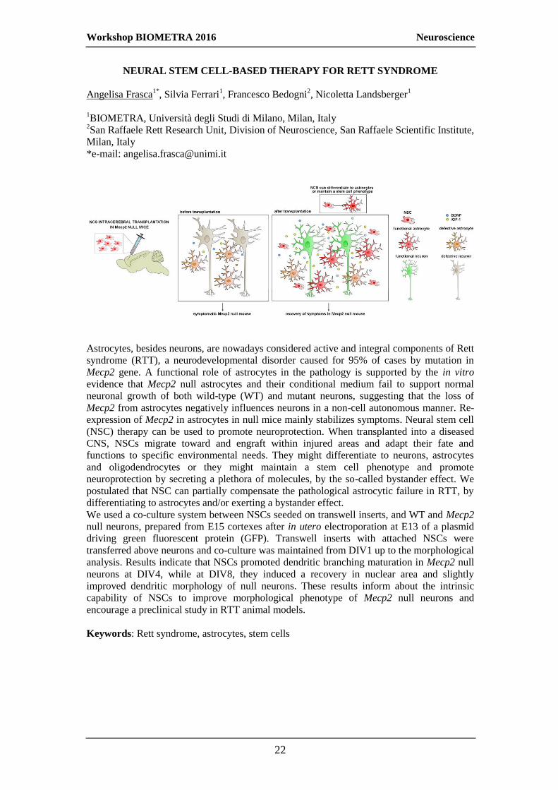
Astrocytes, besides neurons, are nowadays considered active and integral components of Rett
syndrome (RTT), a neurodevelopmental disorder caused for 95% of cases by mutation in
Mecp2 gene. A functional role of astrocytes in the pathology is supported by the in vitro
evidence that Mecp2 null astrocytes and their conditional medium fail to support normal
neuronal growth of both wild-type (WT) and mutant neurons, suggesting that the loss of
Mecp2 from astrocytes negatively influences neurons in a non-cell autonomous manner. Re-
expression of Mecp2 in astrocytes in null mice mainly stabilizes symptoms. Neural stem cell
(NSC) therapy can be used to promote neuroprotection. When transplanted into a diseased
CNS, NSCs migrate toward and engraft within injured areas and adapt their fate and
functions to specific environmental needs. They might differentiate to neurons, astrocytes
and oligodendrocytes or they might maintain a stem cell phenotype and promote
neuroprotection by secreting a plethora of molecules, by the so-called bystander effect. We
postulated that NSC can partially compensate the pathological astrocytic failure in RTT, by
differentiating to astrocytes and/or exerting a bystander effect.
We used a co-culture system between NSCs seeded on transwell inserts, and WT and Mecp2
null neurons, prepared from E15 cortexes after in utero electroporation at E13 of a plasmid
driving green fluorescent protein (GFP). Transwell inserts with attached NSCs were
transferred above neurons and co-culture was maintained from DIV1 up to the morphological
analysis. Results indicate that NSCs promoted dendritic branching maturation in Mecp2 null
neurons at DIV4, while at DIV8, they induced a recovery in nuclear area and slightly
improved dendritic morphology of null neurons. These results inform about the intrinsic
capability of NSCs to improve morphological phenotype of Mecp2 null neurons and
encourage a preclinical study in RTT animal models.
Keywords: Rett syndrome, astrocytes, stem cells

Workshop BIOMETRA 2016 Neuroscience
23
STUDY OF THE ROLE OF lncRNAs IN THE PATHOGENESIS OF CONGENITAL
CENTRAL HYPOVENTILATION SYNDROME
Simona Di Lascio1*, Roberta Benfante
1,2, Diego Fornasari
1
1BIOMETRA, Università degli Studi di Milano, Milan, Italy
2CNR, Neuroscience Institute, Milan, Italy
*e-mail: [email protected]
Heterozygous mutations in the coding region of PHOX2B gene are associated with
Congenital Central Hypoventilation Syndrome (CCHS), a rare disorder characterized by a
broad variety of symptoms of autonomic nervous system dysfunction including inadequate
control of breathing. PHOX2B is a transcription factor that plays a crucial role in autonomic
nervous system development. In vivo and in vitro studies suggest that a loss of function
mechanism, combined with a dominant-negative effect and/or toxic gain of function of the
mutated proteins, is responsible for the entire disease spectrum. We have recently reported
that several mutant proteins interfere with the transcriptional activity of the wild-type protein
in a promoter-specific manner. Of note, we showed that PHOX2B is capable of controlling
its own transcription by binding its own promoter, and PHOX2B mutants can negatively
interfere with the expression of the normal allele, thus further reducing the amount of normal
PHOX2B protein. Nevertheless, so far, very few studies aimed at understanding how
PHOX2B gene expression is regulated. Many studies are showing that long noncoding RNAs
(lncRNAs) play important and diverse roles in gene expression regulation and several
lncRNAs have been implicated in the molecular pathogenesis of some neuro-developmental
disorders. Bioinformatics analyses of the PHOX2B locus region predict an antisense
transcript that partially overlaps the first exon of PHOX2B. Natural antisense transcripts
(NATs), a class of >200 nucleotide long non-coding RNAs (lncRNAs), are RNA molecules
that are transcribed from the opposite DNA strand with respect to sense transcripts, partially
overlap sense RNAs, and positively or negatively regulate gene expression at different levels
by means of multiple mechanisms. Our data provide evidence of the expression of an
antisense transcript that negatively influences PHOX2B gene expression, and suggest that
PHOX2B mutations alter antisense transcription.
Keywords: molecular biology, transcriptional regulation, long noncoding RNAs, autonomic
nervous system, sympathetic neuron

Workshop BIOMETRA 2016 Neuroscience
24
EFFECTS OF LACTATE DEFICENCY IN THE FORMATION OF LONG-TERM
MEMORY AND ON THE STRUCTURE OF EXCITATORY SYNAPSES IN MICE
HIPPOCAMPUS
Elena Vezzoli1,2*
, Luisa Ponzoni1, Norma Lattuada
1, Corrado Calì
3, Daniela Braida
1, Paola
Viani1, Pierre J. Magistretti
3, Andrea Falqui
3, Mariaelvina Sala
4, Maura Francolini
1
1BIOMETRA, Università degli Studi di Milano, Milan, Italy
2Department of Pharmacological and Biomolecular Sciences, Università degli Studi di
Milano, Milan, Italy 3King Abdullah University of Science and Technology (KAUST), Biological and
Environmental Sciences and Engineering Division (BESE), Thuwal, Kingdom of Saudi
Arabia 4National Research Council (CNR), Institute of Neuroscience, Milan, Italy
*e-mail: [email protected]
The inactivation of
glycogen metabolism,
and the consequent
decrease of lactate
production in
hippocampal astrocytes,
lead to an impairment of
long-term memory and
defects in long-term
potentiation (LTP). These
effects were rescued by
L-lactate, that is an
energy source for
neurons and it is crucial
to mediate astrocyte-
neuron functional
connections (Suzuki et
al., 2011). As LTP
defects are associated with alterations in dendritic spines morphology and density, we studied
if anatomical differences in hippocampal excitatory synapses were associated to defects in
long term memory in mice treated with the inhibitor of glycogen phosphorylase, 1,4-dideoxy-
1,4-imino-D-arabinitol (DAB) and if these defects were rescued by L-lactate administration.
We evaluated the effect of DAB administration on short and long-term memory, with two
memory paradigms (novel object recognition (NOR) and passive avoidance (PA)) and we
demonstrated that while DAB did not affect long-term memory evaluated with the PA test,
the treatment impaired both short and long-term episodic memory evaluated with the NOR.
L-lactate treatment reversed DAB-induced memory impairment. Regardless of the memory
paradigm used, 24 hours after treatment and training, hippocampal neurons from mice treated
with DAB, showed a marked reduction in dendritic spine density compared to mice injected
with vehicle. Through Serial-Block Face Scanning Electron Microscopy (SBFSEM) and
volume reconstruction, powerful techniques to study large volume of tissue at ultrastructural
level, we studied the role of the astrocyte-neuron lactate shuttle on the 3D architecture and
number of excitatory synapses through its inhibition with DAB and the combined effects
DAB and Lactate.
Keywords: lactate, short-long term memory, serial-block face scanning electron microscopy

Workshop BIOMETRA 2016 Neuroscience
25
NEW ROLE OF ATM IN HIPPOCAMPAL NEURONS DURING DEVELOPMENT
Lara Pizzamiglio1*
, Elisa Focchi1,3
, Luca Murru
2, Matteo Tamborini
3, Maria Passafaro
2,
Elisabetta Menna2,3
, Michela Matteoli2,3
and Flavia Antonucci1,2
1BIOMETRA, Università degli Studi di Milano, Milan, Italy
2Institute of Neuroscience, C.N.R., Milan, Italy
3Humanitas Clinical and Research Center, IRCCS Rozzano, Italy
*e-mail: [email protected]
ATM (Ataxia Telangiectasia mutated) is a serine/threonine protein kinase linked to DNA
damage response. Despite its activity in dividing cell has been largely investigated, a high
number of evidences are demonstrating new roles of ATM in adult neurons, such as its
involvement in adult neurogenesis, its crucial function in oxidative stress response and its
interaction with two synaptic vesicle proteins, VAMP2 and synapsin-I. Coherently, the
neurodegenerative condition associated to genetic mutations in Atm gene, the Ataxia
Telangiectasia (A-T), exhibits a variable phenotype with impairment in cognition. Thus,
ATM may impact brain functions through distinct mechanisms, in different brain regions and
cell populations. To directly address these new and unclear aspects of ATM in neurons, we
prepared hippocampal neuronal cultures starting from ATM heterozygous mice. Here we
found a significant excitatory/inhibitory unbalance toward inhibition as indicated by the
higher frequency of miniature inhibitory postsynaptic current events, an increased number of
GABAergic synapses and a more precocious development of inhibitory system (GABA
switch). In vivo, the enhanced inhibition still persists and, even if a higher excitation is also
present, a reduced neuronal excitability is shown as indicated by the lower action potential
frequency generated in response to high-current intensity stimuli. Finally, we found an
elevated extracellular signal-regulated kinase 1/2 (ERK1/2) phosphorylation in heterozygous
hippocampi associated with lower expression levels of PP1 phosphatase. These data unveils
an unexpected role of ATM in the maintenance of the appropriate GABAergic development
and transmission in hippocampal formation, laying the basis for a more clear comprehension
of cognitive defects occurring in A-T and opening to novel therapeutic strategies.
Keywords: electrophysiology, live imaging, biochemistry, cellular biology
Workshop BIOMETRA 2016 Neuroscience
26
BIVALENT LIGANDS BOOST G-PROTEIN COUPLING OF DIMERIC OXYTOCIN
RECEPTORS AND PROMOTE SOCIAL BEHAVIORS
Marta Busnelli1,2*
, Gunnar Kleinau3, Markus Muttenthaler
4,5, Stoytcho Stoev
6, Maurice
Manning6, Lucka Bibic
7, Lesley A. Howell
7, Peter J. McCormick
7 , Simona Di Lascio
2,
Daniela Braida2, Mariaelvina Sala
1,2, G.Enrico Rovati
8, Tommaso Bellini
2, Bice Chini
1
1CNR, Institute of Neuroscience, Milan, Italy
2BIOMETRA, Università degli Studi di Milano, Milan, Italy
3Institute of Experimental Pediatric Endocrinology, Charité-Universitätsmedizin Berlin,
Germany 4Institute for Molecular Bioscience, The University of Queensland, Brisbane, Australia
5Department of Biochemistry and Cancer Biology, University of Toledo, Toledo, OH USA
6School of Pharmacy, University of East Anglia, Norwich Research Park, Norwich, UK
7Department of Pharmacological and Biomolecular Sciences, Università degli Studi di
Milano, Milan, Italy
*e-mail: [email protected]
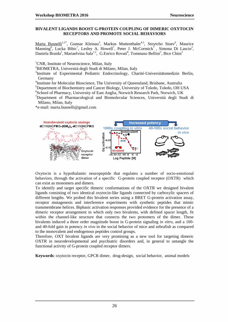
Oxytocin is a hypothalamic neuropeptide that regulates a number of socio-emotional
behaviors, through the activation of a specific G-protein coupled receptor (OXTR) which
can exist as monomers and dimers.
To identify and target specific dimeric conformations of the OXTR we designed bivalent
ligands consisting of two identical oxytocin-like ligands connected by carboxylic spacers of
different lengths. We probed this bivalent series using a BRET G-protein activation assay,
receptor mutagenesis and interference experiments with synthetic peptides that mimic
transmembrane helices. Biphasic activation responses provided evidence for the presence of a
dimeric receptor arrangement in which only two bivalents, with defined spacer length, fit
within the channel-like structure that connects the two protomers of the dimer. These
bivalents induced a three order magnitude boost in G-protein signaling in vitro, and a 100-
and 40-fold gain in potency in vivo in the social behavior of mice and zebrafish as compared
to the monovalent and endogenous peptides control groups.
Therefore, OXT bivalent ligands are very promising as a new tool for targeting dimeric
OXTR in neurodevelopmental and psychiatric disorders and, in general to untangle the
functional activity of G-protein coupled receptor dimers.
Keywords: oxytocin receptor, GPCR dimer, drug-design, social behavior, animal models

Workshop BIOMETRA 2016 Immunology and immunobiology
27
CHARACTERIZATION OF SIGNALING AND METABOLIC REPROGRAMMING
IN T CELLS BY PURINERGIC P2X7 RECEPTOR
Elsa Rottoli1*
, Fabio Grassi1
1BIOMETRA, Università degli Studi di Milano, Milan, Italy
*e-mail: [email protected]
Adenosine triphosphate is an ubiquitous extracellular messenger, which activates purinergic
receptors in the plasma membrane of eukaryotic cells termed P2 receptors. We showed that T
effector/memory (TEM) cells express high levels of P2rx7 encoding for the ATP-gated
ionotropic P2X7 receptor subtype. The deletion of the gene in these cells results in increased
survival and proliferation rate both in vitro and in in vivo. P2rx7-/-
TEM cells are
characterized by a bioenergetic advantage with respect to wild-type (WT) cells.
Morphometric analysis of mitochondria revealed altered cristae and increased mitochondrial
mass in P2rx7-/-
TEM cells. Consistent with these morphometric changes, Western Blot
analysis showed an impairment of the autophagic flux in p2rx7-/-
TEM cells by a decrease in
both lipidated form of LC3 protein, namely LC3-II and LC3-I and accumulation of p62
protein. Microarray gene expression analysis showed that P2rx7-/-
TEM cells clustered
together and separately from WT cells. Among differentially expressed genes we identified
cyclin-dependent kinase inhibitor 1A (Cdkn1a), encoding for p21Waf1/Cip1
, as a transcript
upregulated in WT cells. P21 regulates progression through G1 to S phase in mammalian
cells. To address whether P2X7 signaling directly regulated Cdkn1a expression we
stimulated WT TEM cells with BzATP as a selective P2X7 agonist. This resulted in
significant increase in Cdkn1a transcripts with respect to unstimulated cells and this increase
was abrogated by the selective P2X7 antagonist A-438079. These results suggest that P2X7
activity limits expansion of TEM cells in ATP-rich microenvironment (e.g. during
inflammation), thereby controlling potential T cell mediated tissue damage.
Keywords: purinergic signaling, T cell activation, autoimmunity, cell death, senescence

Workshop BIOMETRA 2016 Immunology and immunobiology
28
CROSSTALK BETWEEN THE LONG PENTRAXIN PTX3 AND THE
COMPLEMENT SYSTEM IN THE IMMUNE RESPONSE TO ASPERGILLUS
FUMIGATUS
Raffaella Parente1*
, Francesca Petroni1, Marina Sironi
1, Sonia Valentino
1, Barbara Bottazzi
1,
Alberto Mantovani1,2
, Antonio Inforzato1,3
1Humanitas Clinical and Research Center, Rozzano, Italy
2Humanitas University, Rozzano, Italy
3BIOMETRA, Università degli Studi di Milano, Milan, Italy
*e-mail: [email protected]
Aspergillus fumigatus (AF) is the major etiologic agent of Invasive aspergillosis, a life-
threatening infection amongst immunocompromised individuals. The innate immune system
plays a key role in the host resistance to AF. In this regard, the Alternative Pathway (AP) of
Complement has been described to cooperate with the long pentraxin PTX3 in the
complement-mediated reaction to AF. PTX3 exerts an opsono-phagocytic activity towards
AF: it enhances recognition, phagocytosis and killing of fungal conidia by immune cells via
Complement and Fc receptors pathways. Based on this, we hypothesized that PTX3 can
establish multiple interactions with conidia, Complement and effector cells, thus acting as a
molecular bridge in bringing together target microbes and immune components.
In this study, we found that PTX3 inhibits the interaction of AF conidia with factor H (FH,
the major soluble inhibitor of AP), which is a critical immune evasion strategy adopted by
AF. This is due to competition for common binding sites on the conidial wall and is
recapitulated by the N-terminal domain of PTX3 (N_PTX3) and a Cys/Ser substitution
mutant of this domain (N_PTX3C47S/C49S/C103S) that forms dimers, as opposed to
N_PTX3 that assembles into tetramers. This indicates that the minimal quaternary structure
that is required for PTX3 to bind AF conidia and displace FH is the dimer. Consistent with
these findings, the cofactor activity of FH on AF conidia (i.e., the factor I-mediated cleavage
of C3b to iC3b) is inhibited by PTX3, N_PTX3 and N_PTX3_3S. Moreover, we observed
that PTX3 forms a stable complex with C3b in an FH-dependent manner, and recruits C3b
onto AF conidia.
We propose that PTX3 enhances C3b deposition onto AF conidia via downregulation of the
FH cofactor activity and a novel FH-dependent mechanism that leads to C3b recruitment
onto the fungal wall. Given that C3b is the prototypic opsonin of Complement (i.e., that is
recognized by the phagocytic receptor CR1), our findings might explain the PTX3-dependent
enhancement of AF phagocytosis and, possibly, killing by innate immune cells.
Keywords: innate immunity, pentraxins, complement, invasive aspergillosis

Workshop BIOMETRA 2016 Immunology and immunobiology
29
NK CELLS RECRUITMENT TO THE SALIVARY GLANDS REGULATES EARLY
VIRAL CONTROL BUT IS DISPENSABLE FOR THE FORMATION OF
INDUCIBLE TERTIARY LYMPHOID STRUCTURES
Elena Pontarini1, Davide Lucchesi
2, Liliane Fossati-Jimack
2, Cristina Croia
2, Michele
Bombardieri2, Domenico Mavilio
1,3
1Clinical and Experimental Immunology, Humanitas Clinical and Research Centre, Milan,
Italy 2Experimental Medicine and Rheumatology, Queen Mary University of London, London,
UK 3BIOMETRA, Università degli Studi di Milano, Milan, Italy
Natural Killer (NK) cells are a central component of the innate immune system and are
crucial in containing and eliminating viral infections. The salivary glands (SG) represent a
permissive site for the persistence of several viruses that show specific sialotropism. We
investigated the relevance of NK cells in the early phases of the immune response upon
Adenovirus (AdV) infection within the SG. In our murine model, intra-SG AdV delivery
triggers the formation of inflammatory infiltrates that progressively acquire features of
tertiary lymphoid structures (TLS). Herein, we characterized the NK cell response to AdV at
glandular mucosal sites in order to investigate whether NK cell-mediated viral control might
affect the virus-induced TLS formation. We found that in response to AdV delivery, NK cells
were enriched within the SG, displaying an activated phenotype and a functional cytotoxic
potential. Peripheral NK cells are recruited within AdV infected SG, undergo proliferation
and contribute to the clearance of AdV-infected cells in the early phases of the immune
response. Nonetheless, selective depletion of NK cells did not affect the number,
organization and functionality of TLS in SG. We conclude that, in the present model NK
cells are able to exert an early control of viral infection but are dispensable for the formation
of inflammatory aggregates. Moreover, our AdV-induced SG inflammation model is an ideal
platform to study the immune response to viral infections within the SG and might lead to a
better understanding of the mechanisms underlying virus-induced formation of TLS in this
organ.
Keywords: NK cells, Sjogren’s Syndrome, autoimmunity, ectopic lymphoid structures, viral
Workshop BIOMETRA 2016 Immunology and immunobiology
30
MULTISTEP REGULATION OF TOLL LIKE RECEPTOR 4 SIGNALING BY IL-10-
DEPENDENT MICRORNAS
Graziella Curtale1,2
, Tiziana Renzi1,2
, Lorenzo Drufuca1,2
, Flavia Bazzoni3, Marzia Rossato
3,
Massimo Locati1,2
1BIOMETRA, Università degli Studi di Milano, Milan, Italy
2Humanitas Clinical and Research Center, Rozzano, Italy
3Department of Pathology, University of Verona, Verona, Italy
MicroRNAs (miRs) are small non-coding RNAs with a post-transcriptional effect with an
emerging role in several biological functions. To identify miRs potentially involved in the
regulation of the inflammatory response, we analyzed the miR expression profile regulated
by the Toll Like Receptor 4 agonist LPS in human monocytes. We confirmed induction of
know pro-inflammatory miRs (miR-155, miR-146a) and identified a set of previously
unknown miRs, including the cluster miR-125a~99b~let-7e. As expression of this miR
cluster was significantly higher at late time points, when the IL-10-dependent anti-
inflammatory loop is usually detected, we measured the expression of these miRs in
monocytes stimulated with IL-10, in the presence or not of LPS, showing a potentiating
effect of IL-10 on LPS-induced cluster miR-125a~99b~let-7e expression. We found a
STAT3 consensus site and demonstrated the binding of STAT3 to the miR-cluster promoter
in condition of IL-10 stimulation. These data were consistent with the decrease of cluster
expression levels in the presence of blocking antibody against IL-10R and JAK-STAT
inhibitors. An in silico analysis was performed to assess the potential role of cluster miR-
125a~99b~let-7e in the context of inflammation; notably we found that the TLR pathway was
potentially regulated by these miRs at multiple steps of the signaling cascade. We validated
TLR4 and CD14 receptors, the adaptor proteins MyD88 and IRAK1 and a set of pro-
inflammatory cytokines as direct targets. Furthermore, the enforced expression of miR-
cluster in LPS stimulated monocytes led to an extensive down-regulation of pro-
inflammatory cytokines. In conclusion, we have identified an IL-10-responsive miR cluster
with anti-inflammatory activity in monocytes, able to target multiple components of the
TLR4 pathway, candidating miRs as new feedback modulators of LPS response involved in
the resolution of inflammation. A cluster miR-125a~99b~let-7e floxed animal is at present
being crossed with a LyM-CRE animal for selective deletion of this miR cluster in monocyte-
macrophages.
Keywords: chemokine, chemokine receptor, leukocyte mobilization

Workshop BIOMETRA 2016 Immunology and immunobiology
31
PRENATAL EXPOSURE TO POLY I:C INCREASES SUSCEPTIBILITY TO
EPILEPSY IN THE ADULT OFFSPRING
Elisa Focchi1,2*
, Marco Rasile3, Flavia Antonucci
1, Raffaella Morini
3, Elisabetta Menna
2,3,
Davide Pozzi3, Irene Corradini
2,3, Michela Matteoli
2,3
1BIOMETRA, Università degli Studi di Milano, Milan, Italy
2CNR, Istituto di neuroscienze, Milan, Italy
3Humanitas Clinical and Research Center, Rozzano, Italy
*e-mail: [email protected]
In the last years, evidences have accumulated showing a direct connection between epilepsy
and brain inflammation. Indeed, epilepsy is associated to enhanced inflammation, while
activation of the immune response consequent to infections strongly increases the risk of
seizures.
By using the Poly I:C (polyinosinic-polycytidylic acid) mouse model of inflammation, we
demonstrated that a single Poly I:C injection at gestation day 9 (GD9) is able to increase
susceptibility to kainate-induced seizures in the offspring at postnatal day 90. As a control,
mice injected with Poly I:C in the adult life show no increased susceptibility to kainate-
induced seizures, thus providing the evidence that the higher susceptibility to seizures
consequent to prenatal Poly I:C exposure is the consequence of a neurodevelopmental
process.
We also evaluated the inflammatory profile of the Poly I:C-prenatally treated mice and we
found that a single Poly I:C injection in early/mid gestation is able to induce an inflammatory
response in the embryos 6 hours after the injection, as demonstrated by the significant
increase of IL6 and IL1-beta mRNA levels. On the other hand, no significant signs of
inflammation were found in adult mice, not even changes in synaptic protein levels and
synaptic morphology indicating that these features, usually related to epilepsy, are not
directly involved in the susceptibility to seizures in our model. Interestingly, we have found,
both in vitro and in vivo, a higher network hyperexcitability. Although the mechanisms
underlying this phenotype are still unknown, these results highlight the possibility that a
single inflammatory event during pregnancy may alter neurodevelopment, leading to
neurologic disorders in the adult offspring.
Keywords: biochemistry, cellular biology, molecular biology, animal behavior

Workshop BIOMETRA 2016
32
Other abstracts

Workshop BIOMETRA 2016 Other abstracts
33
DOWN-REGULATION OF ATYPICAL CHEMOKINE RECEPTOR ACKR2
EXPRESSION BY HEMATOPOIETIC PROGENITORS PROMOTES MYELOID
CELL MOBILIZATION AND DIFFERENTIATION
Ornella Bonavita1,2
, Nicoletta Caronni1,2
, Benedetta Savino1,2
, Matteo Massara1,2
, Laura
Crisafulli2,3
, Francesca Ficara2,3
, Alberto Mantovani2,4
, Massimo Locati1,2
, Raffaella
Bonecchi2,4
1BIOMETRA, Università degli Studi di Milano, Milan, Italy
2Humanitas Clinical and Research Center, Rozzano, Italy
3Istituto di Ricerca Genetica e Biomedica, Consiglio Nazionale delle Ricerche, Milan, Italy
4Humanitas University, Rozzano, Italy
The atypical receptor ACKR2 is a scavenger receptor for many inflammatory CC
chemokines and it has been shown to prevent the development of exacerbated inflammatory
reactions. ACKR2 is expressed either by non-hematopoietic cell compartment or by
hematopoietic cells. The aim of this investigation was to gain inside into the role of ACKR2
in myeloid cells trafficking and differentiation. To address the role of ACKR2 in the
proposed research we took advantage from ACKR2-gene targeted mice, FACS analysis,
primary cell cultures and cell line. Monocytes and neutrophils from ACKR2-/-
mice had
increased chemokine-induced mobilization and pharmacological blocking experiments
revealed that it was CXCR4-independent. ACKR2-/-
mice showed increased number of
monocytes confined to bone marrow sinusoids compared to wild type. Bone marrow chimera
experiments showed that the increased mobilization was due to the hematopoietic
compartment. The analysis of hematopoietic progenitors revealed that ACKR2 is expressed
by Lin−Sca-1
+c-Kit
+ cells (LSK) to faint thereafter in more mature myeloid progenitors in
contrast with canonical chemokine receptor CCR2. Moreover myeloid committed progenitors
from ACKR2-/-
mice expressed high levels of CCR1, CCR2 and CCR5, but not of CXCR4
and they had higher proliferation and differentiation rate compared to ACKR2 sufficient
LSK. These data suggest that ACKR2 is not only involved in regulating chemokine gradient
and leukocytes recruitment, but can directly regulate cell activity through the inhibition of
chemokine receptor expression by a mechanism still unclear. Collectively these results
demonstrate that ACKR2 is differentially expressed by hematopoietic progenitors during
myeloid cell maturation and its expression on LSK is involved in controlling their
localization and mobilization.
Keywords: chemokine, chemokine receptor, leukocyte mobilization

Workshop BIOMETRA 2016 Other abstracts
34
ROLE OF SEROTONIN 5HT2-TYPE RECEPTORS ON REWARDING AND
BEHAVIOURAL EFFECTS OF METHYLENEDIOXYMETHAMPHETAMINE
(MDMA) AND ITS DERIVATIVES IN ZEBRAFISH
D. Braida1*
, L. Ponzoni1, M. Sala
1,2
1BIOMETRA, Università degli Studi di Milano, Milan, Italy
2CNR, Institute of Neuroscience, Milan, Italy
*e-mail: [email protected]
The synthetic phenethylamines are recreational drugs known to produce psychostimulant
effects (1). However, their abuse potential and pharmacological effects have not been widely
studied.
In the present study, the effect on reward, social preference and anxiety-like behaviour of
2,5-dimetoxy-4-bromo-amphetamine hydrobromide (DOB) and para-methoxyamphetamine
(PMA), in comparison with MDMA was investigated in zebrafish, an emerging model to
study emotional behaviour in an inexpensive and quick manner. In addition, the role of
serotonin 5-HT2 like- receptors on the above mentioned effects was evaluated.
Zebrafish were treated i.m. with a wide range of doses of DOB (0.05-20 mg/kg), PMA
(0.0005-2 mg/kg) or MDMA (0.25-160 mg/kg). Animals were submitted to a conditioned
place preference (CPP) task for the rewarding properties, to a social preference task to
evaluate social behaviour, and to the novel tank diving and light-dark tests to study emotional
behaviour. Hallucinatory behaviour was evaluated in terms of appearance of a trance-like
effect. The serotonin 5-HT2 subtype receptor antagonist ritanserin (0.025-2.5 mg/kg/i.m.) was
given in association with the maximal effective dose of MDMA, DOB and PMA. MDMA
and its derivatives exhibited dose-dependent CPP, anxiolytic effect and increase in social
preference in a biphasic fashion, being PMA the most potent. These effects were
accompanied, for DOB (2 mg/kg) and PMA (0.1 mg/kg), by a trance-like hallucinatory
behaviour. MDMA, at a high dose as 160 mg/kg, did not induce any hallucinatory behaviour.
Ritanserin significantly blocked all the effects, suggesting the involvement of serotonin
5HT2-like subtype receptors.
Collectively, these findings demonstrate for the first time the rewarding, prosocial and
anxiolytic properties of DOB and PMA accompanied by hallucinatory behaviour and focus
on the mechanisms of their action through the serotonergic-like system. Our results reinforce
zebrafish as an emerging experimental model for screening new hallucinogens.
1. Hill SL, Thomas SH (2011) Clin Toxicol (Phila) 49:705-719
The study was supported by Zardi Gori Foundation
Keywords: zebrafish, hallucinogens, addiction, anxiety, sociability
SOCIAL PREFERENCE ANXIETY
ADDICTION HALLUCINATIONS
MDMA
DOB
PMA
BLOCKED BY
RITANSERIN

Workshop BIOMETRA 2016 Other abstracts
35
EVIDENCE FOR A ROLE OF A NEW ANTIOXIDANT COMPOUND IN THE
PREVENTION OF AGE RELATED OSTEOBLAST DYSFUNCTION
Lavinia Casati1*
, Francesca Pagani 1, Valeria Sibilia
1
1BIOMETRA, Università degli Studi di Milano, Milan, Italy
*e-mail address: [email protected]
Aim & Results: increasing evidence suggests a role for oxidative stress in age-related
decrease both in osteoblast number and function suggesting the potential use of antioxidant
compounds in the prevention/treatment of osteoporosis. This study was undertaken to
investigate whether ghrelin, previously reported to stimulate osteoblast proliferation,
counteracts tert-butyl hydroperoxide (t-BHP)-induced oxidative damage in osteoblastic cells.
For this purpose, MC3T3-E1 cells and rat calvariae osteoblasts were exposed to t-BHP,
250M, for 3 h and a pre-treated with increasing ghrelin concentrations (10-9/ -11
M, 24 h
before). We found that ghrelin (10-9
M) significantly prevented t-BHP-induced osteoblastic
dysfunction and changes in the cytoskeleton organization evidenced by the staining of the
actin fibres with Phalloidin-FITC in MC3T3 cells.
As far as the molecular pathway involved in such ghrelin protective action, we investigated
Wnt/beta catenin pathway. We have shown that ghrelin increases GSK-3β phosphorylation
and β-catenin accumulation in the nucleus (by Western blot). Moreover cAMP/PKA pathway
is activated by ghrelin, since H89, an inhibitor of PKA, is able to abolish the ghrelin effect.
In addition to its anabolic action, ghrelin reduces the osteoclastogenesis, thus we analysed
the transcriptomic and epigenetic profile. When we analysed the OPG/RANKL
transcriptomic profile, we found that Ghrelin (10-9
M) is able to induce OPG expression and
to modify RANKL/OPG ratio, probably by changing the level of the histone modification
H3K4me3 and of its related histone post-translational enzyme, Jarid1b. Moreover we found
that the ghrelin increase the global DNA methylation level, promoting an “anti-aging” action
in osteoblasts.
Conclusions: these findings suggest that ghrelin could be considered a promising compound
to re-equilibrate bone cell activities in condition of impaired bone remodelling, and may have
therapeutic potential in age related osteoblast dysfunction.
Keywords: bone remodelling, oxidative stress, ghrelin, wnt/beta catenin pathway,
epigenetics

Workshop BIOMETRA 2016 Other abstracts
36
PTC SUPPRESSION STRATEGY AS “PERSONALIZED MEDICINE” APPROACH
FOR CDKL5 RELATED PATHOLOGIES
Maria Fazzari1*
, Charlotte Kilstrup-Nielsen2, Nicoletta Landsberger
1
1BIOMETRA, Università degli Studi di Milano, Milan, Italy
2Department of Biotechnology and Life Sciences, University of Insubria, Varese, Italy
*e-mail: [email protected]
Rett syndrome (RTT) is a childhood neurological disorder and it is considered one of the
main causes of severe intellectual disability in girls. Classic RTT results from mutations in
MECP2 gene, but atypical forms exist where alterations occur at CDKL5 or FOXG1 loci.
CDKL5 is a serine/threonine kinase characterized by a N-terminal catalytic domain and a
long C-terminus unique to CDKL5.
Alterations of CDKL5 gene comprise a wide range of variants including non sense mutations,
which give rise to premature termination codons (PTCs). PTCs may cause the production of a
truncated protein or the activation of non sense-mediated decay (NMD). However, during
translation, it is possible that a near-cognate aminoacyl-tRNA binds the PTC. This mispairing
may result in the incorporation of amino acid attached to near-cognate aminoacyl-tRNA and
then in the production of full-length protein. This process is known as read-through and can
be facilitated using a wide range of drugs like aminoglycoside and non aminoglycoside
compounds.
In order to test PTC suppression, we have chosen some human CDKL5 non sense mutations
located either in the catalytic N-terminus (R59X, R134X) or in the C-terminus (R550X,
S855X) domain; the latter could be more suitable for this study because read-through might
produce a protein maintaining most of its functions. We introduced the selected mutations in
the pEGFPC1-hCdkl5107 construct and evaluated read-through using aminoglycoside drugs
after transient transfection: full-length proteins were detectable by WB and IF for all mutants
tested. Moreover, while truncated proteins showed an altered subcellular
localisation/organisation, after read-through the cellular phenotype was more similar to wild
type meaning that a rescue could be possible.
In the future, since our PTCs might activate the NMD mechanism, the read-through
efficiency will be tested in presence of NMD inhibitors in patients-derived fibroblasts.
Keywords: Rett syndrome, non sense mutations, read-through

Workshop BIOMETRA 2016 Other abstracts
37
HUMAN LIVER-RESIDENT CD56(BRIGHT)/CD16(NEG) NK CELLS ARE
RETAINED WITHIN HEPATIC SINUSOIDS VIA THE ENGAGEMENT OF CCR5
AND CXCR6 PATHWAYS
Hudspeth K1, Di Vito C
1, Zaghi E
1, Donadon M
2, Cimino M
2, Pontarini E
1, Tentorio P
1, Preti
M1, Hong M
3, Bertoletti A
3, Bicciato S
4, Invernizzi P
5, Lugli E
1, Torzilli G
2, Gershwin ME
6,
Mavilio D1,7
1Unit of Clinical and Experimental Immunology, Humanitas Clinical and Research Center,
Rozzano, Italy 2Department of Hepatobiliary & General Surgery, Humanitas Clinical and Research Center,
Rozzano, Italy 3Viral Hepatitis Laboratory, Singapore Institute for Clinical Sciences, Agency of Science,
Technology and Research (A*STAR), Singapore 4Department of Life Sciences, Center for Genome Research, University of Modena and
Reggio Emilia, Modena, Italy 5Liver Unit and Center for Autoimmune Liver Diseases, Humanitas Clinical and Research
Center, Rozzano, Italy 6Division of Rheumatology, Allergy, and Clinical Immunology, University of California
Davis, Davis, CA, USA 7BIOMETRA, Università degli Studi di Milano, Milan, Italy
The liver-specific natural killer (NK) cell population is critical for local innate immune
responses, but the mechanisms that lead to their selective homing and the definition of their
functionally relevance remain enigmatic.
We took advantage of the availability of healthy human liver to rigorously define the
mechanisms regulating the homing of NK cells to liver and the repertoire of receptors that
distinguish liver-resident NK (lr-NK) cells from circulating counterparts.
Nearly 50% of the entire liver NK cell population is composed of functionally relevant
CD56(bright) lr-NK cells that localize within hepatic sinusoids. CD56(bright) lr-NK cells
express CD69, CCR5 and CXCR6 and this unique repertoire of chemokine receptors is
functionally critical as it determines selective migration in response to the chemotactic
stimuli exerted by CCL3, CCL5 and CXCL16. Here, we also show that hepatic sinusoids
express CCL3(pos) Kupffer cells, CXCL16(pos) endothelial cells and CCL5(pos) T and NK
lymphocytes. The selective presence of these chemokines in sinusoidal spaces creates a
unique tissue niche for lr-CD56(bright) NK cells that constitutively express CCR5 and
CXCR6. CD56(bright) lr-NK cells co-exist with CD56(dim) conventional NK (c-NK) cells
that are, interestingly, transcriptionally and phenotypically similar to their peripheral
circulating counterparts. Indeed, CD56(dim) c-NK cells lack expression of CD69, CCR5, and
CXCR6 but express selectins, integrins and CX3CR1.
Our findings disclosing the phenotypic and functional differences between lr-NK cells and c-
NK cells are critical to distinguish liver-specific innate immune responses. Hence, any
therapeutic attempts at modifying the large population of CD56(bright) lr-NK cells will
require modification of hepatic CCR5 and CXCR6.
Keywords: natural killer cells, liver, chemokine receptors, innate immunity.

Workshop BIOMETRA 2016 Other abstracts
38
SPHINGOLIPIDS AS ACTORS IN THE STABILIZATION OF CFTR AT PLASMA
MEMBRANE
G. Mancini1*
, N. Loberto1, R. Bassi
1, D. Schiumarini
1, S. Munari
1, M.C. Dechecchi
1, G.
Lippi1, G. Cabrini
1, M.
Aureli
1, A. Tamanini
1, S. Sonnino
1
1BIOMETRA, Università degli Studi di Milano, Milan, Italy
*e-mail: [email protected]
Cystic Fibrosis Transmembrane conductance Regulator (CFTR) is a chloride channel
expressed at the plasma membrane (PM) of epithelial cells. Mutations in CFTR gene cause
Cystic Fibrosis (CF), an autosomal recessive disease characterized by severe lung disease due
to the loss of CFTR at the cell PM. Many pharmacological agents have been designed to
increase the surface level of mutated CFTR (correctors), as well as its PM stability and
activity (potentiators), even if their efficacy seems to be time-limited, particularly for the
most common CF-causing mutation F508del. Several factors influence the PM CFTR
stability, such as its compartmentalization in the sphingolipid(SL)-enriched lipid rafts and its
interaction with the a component of lipid rafts: the scaffolding protein ezrin, namely its
phosphorylated form. However is known that the phosphorylation state of ezrin depends on
PM levels of ceramide.
Based on these findings, we investigated the effects of corrector (VX-809) and potentiator
(VX-770) on CFTR PM microenvironment.
We analysed the SL composition and the phosphorylation state of ezrin in CF (CuFi-1) and
non-CF (NuLi-1) bronchial epithelial cell lines, treated or not with VX-809 and VX-770.
Moreover, we evaluated the SL pattern of lipid rafts.
In both cell lines we observed a marked reduction of phosphorylated ezrin, after the
combined treatment with corrector and potentiator. Interestingly, even if the cell SL total
content did not change, in treated cells we found a marked increase of all lipid rafts-
associated SL species, in particular ceramide, glucosylceramide and ganglioside GM3, which
could be responsible for the ezrin dephosphorylation.
These preliminary data suggest that the combined treatment induces modification in lipid
rafts organization in terms of proteins and lipids that could limit the stability of mutated
CFTR at PM level. These results could permit the development of new therapeutic strategies
for CF treatment.
Supported by Italian Cystic Fibrosis Research Foundation grant FFC#09/2015
Keywords: biochemistry, sphingolipids, cystic fibrosis, primary cell culture

Workshop BIOMETRA 2016 Other abstracts
39
THE HUMAN-RESTRICTED DUPLICATED FORM OF THE α7 NEURONAL
NICOTINIC RECEPTOR, CHRFAM7A: EXPRESSION, TRANSCRIPTIONAL
REGULATION AND ROLE IN THE INFLAMMATORY PROCESS
Maroli A.1*
, Fornasari D.1,2
, Benfante R1,2
1BIOMETRA, Università degli Studi di Milano, Milan, Italy
2CNR, Neuroscience Institute, Milan, Italy
*e-mail: [email protected]
In the last years, increasing evidence have linked several neurological disorders to
inflammation. Controlling neuro-inflammation has indeed become a promising approach to
treat neuro-degenerative diseases. The Central Nervous System exerts a control on the
peripheral inflammation, through the “cholinergic anti-inflammatory pathway”, in which the
splenic terminals of the Vagus nerve induce an anti-inflammatory response by stimulating the
release of acetylcholine (ACh). The ACh interacts with the α7 Nicotine Receptor (CHRNA7)
expressed by the macrophages, thus inducing down-regulation of pro-inflammatory
cytokines. Recently, the CHRFAM7A gene has been discovered. It is the human restricted
product of a partial duplication and fusion of exons 5-10 of CHRNA7 gene with the novel
FAM7A gene and maps 1.6 Mb apart from CHRNA7 in inverted orientation. The
CHRFAM7A gene undergo alternative splicing, giving rise to two proteins of 46 and 35
KDa, which differ from the α7 conventional subunit for the N-terminus domain. These
proteins seem to have a dominant negative effect on the conventional α7 function. Moreover,
we have previously shown that CHRFAM7A is down-regulated after LPS treatment in THP-
1 cell model and primary monocytes and macrophages by a transcriptional mechanism reliant
on NF-κB. Our purpose is to define the transcriptional regulation of CHRFAM7A to better
understand its role in peripheral and central inflammation. In this study, we characterize the
gene’s regulatory region and its isoforms’ expression in neuronal and immune cell models.
Moreover, we investigate the responsiveness of CHRFAM7A gene to pro- and anti-
inflammatory stimuli, defining a human mechanism modulating the inflammatory response.
Keywords: CHRNA7, CHRFAM7A, inflammation, transcriptional regulation.

Workshop BIOMETRA 2016 Other abstracts
40
ATYPICAL CHEMOKINE RECEPTOR 2 PROMOTES TUMOR
METASTATIZATION BY INHIBITING RECRUITMENT AND ACTIVATION OF
ANTI-METASTATIC NEUTROPHILS
Matteo Massara1,2
, Ornella Bonavita1,2
, Benedetta Savino1,2
, Valeria Mollica Poeta1,2
, Elisa
Setten1,2
, Nicoletta Caronni1,2
, Camilla Recordati3, Marina Sironi
2, Alberto Mantovani
1,4,
Massimo Locati1,2
, Raffaella Bonecchi2,4
1BIOMETRA, Università degli Studi di Milano, Milan, Italy
2Humanitas Clinical and Research Center, Rozzano, Italy
3Fondazione Filarete, Milan, Italy
4Department of Biomedical Sciences, Humanitas University, Rozzano, Italy
The atypical chemokine receptor 2 (ACKR2) is a scavenger receptor for most inflammatory
CC chemokines, with a protective role in chronic inflammation, autoimmunity and
inflammation-related cancer. The aim of this project is to investigate the role of ACKR2 in
primary tumor development and metastatization in different mouse models. Tumor arising in
ACKR2-/-
NeuT mice, that develop spontaneous breast adenocarcinoma, showed a more
aggressive phenotype when compared to WT NeuT mice. On the contrary, ACKR2-/-
NeuT
mice were protected from spontaneous lung metastasis. Metastasis protection was registered
also using the 4T1 orthotopic model of breast cancer. FACS analysis indicated an increase of
Ly6G+ polymorphonuclear cells and Ly6C
high monocytes in the blood and in the pre-
metastatic lungs of ACKR2-/-
mice. Depletion experiments demonstrated that neutrophils are
implied in metastasis protection in the B16F10 melanoma metastasis models. Moreover,
ACKR2-/-
neutrophils express increased levels of the chemokine receptor CCR2 and have
increased CCR2-dependent cytotoxic activity. These results indicate that ACKR2 expression
has a dual and opposite role in tumor biology, inhibiting primary tumor growth but
promoting lung metastatization trough the inhibition of anti-metastatic neutrophils.
Keywords: chemokine receptor, cancer, metastasis, neutrophil

Workshop BIOMETRA 2016 Other abstracts
41
CHARACTERIZATION OF A NOVEL NKp46POS
/V1-RESTRICTED TCR
LYMPHOCYTES RESIDENT WITHIN THE HUMAN INTESTINE
Joanna Mikulak1,2*
, Ferdinando Oriolo1,5,
Kelly L. Hudspeth1, Alessandra Roberto
1, Elena
Bruni1, Paolo Tentorio
1, Federico Colombo
3, Anna Villa
1,2,4 and DomenicoMavilio
1,5
1Unit of Clinical and Experimental Immunology, Humanitas Clinical and Research Center,
Rozzano, Milan, Italy 2Institute of Genetic and Biomedical Research (IRGB), CNR, Milan, Italy
3Flow Cytometry and Cell Sorting Unit, Humanitas Clinical and Research Center, Rozzano,
Milan, Italy 4Telethon Institute for Gene Therapy, Division of Regenerative Medicine, Stem Cells and
Gene Therapy, Istituto di Ricovero e Cura a Carattere Scientifico (IRCCS) San Raffaele
Scientific Institute, Milan, Italy
5BIOMETRA, Università degli Studi di Milano, Milan, Italy
*e-mail: [email protected]
NKp46 is a member of Natural Cytotoxic Receptors (NCRs) triggering Natural Killer (NK)
cell cytotoxicity and production of inflammatory cytokine against harmful cells. Although
NKp46 had been first identified as a specific NK cell receptors, recent studies revealed that it
is also expressed on other immune cells such as Innate Lymphoid cells, T and NKT
lymphocytes. We previously reported that NKp46 expression is inducible upon in vitro
activation on a subset of human peripheral blood VT cells. Here, we identify a novel
NKp46pos
/ T lymphocytes subset resident in human intestine under homeostatic conditions
within the intraepithelial (IEL) compartment (average of 50% of all T IELs) and, to a
lower degree, in lamina propria (LP) (average of 10% of all T LPLs). This NKp46pos
/ T
IEL subset is characterized by a cytotoxic phenotype expressing CD56, CD8, NKG2D and
NKG2C. Moreover, we found that the expression of NKp46 in intestinal IELs is restricted
to V1 TCR chain repertoire that indeed distinguish their tissue-like phenotype from
peripheral blood T cells which mainly exhibited a V2 TCR distribution. We also
observed that IL-2 and IL-15-stimulation induce a de-novo expression of NKp46 in human
thymic T cell precursors, but not in circulating T cells. Their co-culture with intestinal
epithelial cells, IL-10 and TGF- can modulate the NKp46 expression according to the
activation steady state of these cells. These results suggest an early extra-thymic and gut-
conditioned maturation of NKp46pos
/ T cells likely relevant both in GALT homeostasis and
physiopathology of the gut.
Keywords: T lymphocytes, NKp46, cytotoxicity, gut, homeostasis

Workshop BIOMETRA 2016 Other abstracts
42
MECHANISMS OF GENERATION OF LONG-LIVED T MEMORY STEM CELLS
DURING LYMPHOPENIA
Alessandra Roberto1,3*
, Veronica Zanon1, Luca Castagna
2, Stefania Bramanti
2, Roberto
Crocchiolo2, Paolo Tentorio
3, Armando Santoro
2, Emma Gostick
4, Kristin Ladell
4, James
McLaren4, David A. Price
4, Mario Roederer
5, Domenico Mavilio
3,6, Enrico Lugli
1
1Laboratory of Translational Immunology, Humanitas Clinical and Research Center,
Rozzano, Milan, Italy 2Hematology and Bone Marrow Transplant Unit, Humanitas Cancer Center, Rozzano, Milan,
Italy 3Unit of Clinical and Experimental Immunology, Humanitas Clinical and Research Center,
Rozzano, Milan, Italy 4Institute of Infection and Immunity, Cardiff University School of Medicine, Cardiff, UK
5Center for Cancer Research, National Cancer Institute, NIH, Bethesda, Maryland, USA
6BIOMETRA, Università degli Studi di Milano, Milan, Italy
*e-mail: [email protected]
Recent evidence indicated that T memory stem cells (TSCM), the earliest differentiated
memory T cell subset in humans, display superior persistence in vivo as well as effector
functions; however, their mechanisms of generation in vivo remain elusive. In the context of
T-replete haploidentical bone marrow transplantation, we show that TSCM cells of donor
origin dominate the T cell compartment of lymphopenic recipients early after T cell transfer
and precede the expansion of effector cells. Importantly, transferred naïve T cells (TN), but
not TSCM or memory cells (TMEM), preferentially survive post ‐transplant cyclophosphamide,
used as GVHD prophylaxis, thus suggesting that post-transplant TSCM originate from TN cells.
In this regard, TN-derived TSCM cells specific for exogenous and self/tumor antigens
contribute to peripheral reconstitution of lymphopenic patients by differentiating into effector
T cells. Likewise, pathogen-specific TMEM cells generate detectable recall responses, but only
in the presence of the cognate antigen. The genome-wide expression analysis on sorted TN,
TSCM and TMEM cells under different stimulatory conditions revealed gene products
specifically expressed by self-renewing TSCM cells that are shared with somatic stem cells.
These genes are currently being used to induce stem cell-like properties in tumor-specific T
cells to be used in adoptive transfer experiments.
Keywords: Immune-reconstitution, T-cells, bone marrow transplantation, adoptive T cell
transfer

Workshop BIOMETRA 2016 Other abstracts
43
STRUCTURAL STUDIES AT THE PROTEOLIPIDOMICS HEDGE:
THE K+-CHANNEL KCV IN BIOMIMETIC MEMBRANES
Valeria Rondelli1*
, Elena Del Favero1, Paola Brocca
1, Anna Moroni
2, Laura Cantù
1
1BIOMETRA, Università degli Studi di Milano, Milan, Italy
2Department of Biosciences, Università degli Studi di Milano, Milan, Italy
*e-mail: [email protected]
The activities of integral membrane proteins are often affected by the surrounding molecules
within the membrane. An important parameter is the hydrophobic thickness of the lipid
bilayer. In fact, both membrane and proteins may deform to ensure good hydrophobic
matching. Another important parameter for the regulation of membrane proteins functions is
the specific composition of the membrane portion where they are hosted. K+-channels are
transmembrane proteins abundant in virtually all biological systems. We studied a viral K+-
channel, Kcv (1), in interaction with different model membranes. Biochemical and functional
evidences indicate that K+-channels localize in lipid raft microdomains and that changes in
the membrane cholesterol content can directly modulate the ion channel functionality.
Calorimetry and X-ray spectroscopy revealed that the thermotropic and structural properties
of model biomimetic membranes are affected by the presence of Kcv, depending on lipid
composition and on the amount of cholesterol. Results show that the channel protein dictates
its preferred environment, inducing a lateral phase separation in membranes where the
average cholesterol content is different from the favored. Neutron reflectometry (2)(3) has
also been applied to study the structural changes induced by Kcv on single, extended, model
membranes, both in adhering bilayers and in floating membranes prepared by the Langmuir-
Blodgett/Langmuir-Schaefer technique. It evidences that Kcv inserts in the membranes with a
preferential orientation. Moreover, the protein induces cross asymmetric rearrangement of
cholesterol in the two leaflets of the hosting membranes.
(1) C. Pagliuca et al., Biochemistry 46 (2007) 1079-1090.
(2) V. Rondelli et al., Biochimica et Biophysica Acta 1818 (2012) 2860–2867.
(3) V. Rondelli et al., Nature Scientific Reports 6 (2016) 20997.
Keywords: model membranes, potassium channels, protein-lipid interaction, cholesterol

Workshop BIOMETRA 2016 Other abstracts
44
LSD1 AND neuroLSD1: THE YIN AND YANG OF EXPERIENCE-DRIVEN
NEURONAL PLASTICITY
Francesco Rusconi
1*, Barbara Grillo
1, Emanuela Toffolo
1, Federica Giona
1, Alessandra
Longaretti1, Elena Battaglioli
1
1BIOMETRA, Università degli Studi di Milano, Milan, Italy
*e-mail: [email protected]
In mammals, different forms of stress including psychosocial stress can impact on several
aspects of human health, fostering mood and anxiety disorders. However, very little is known
about the mechanisms underlying brain physiology of stress response, hindering development
of new therapeutic strategies. Here we indicate a role for transcriptional corepressor Lysine-
specific demethylase 1 (LSD1) and its dominant-negative splicing isoform neuroLSD1, in the
modulation of emotional behavior. In mouse hippocampus, LSD1 and neuroLSD1 interact
with transcription factor SRF setting chromatin state of SRF-targeted genes egr1 and c-fos.
Deletion or reduction of neuroLSD1 in mutant mice induce decreased levels of activating
histone marks at egr1 and c-fos promoters, which dampen their psychosocial stress-induced
transcription, and result in low anxiety-like behavior. SAHA administration to
neuroLSD1KO mice restores egr1 and c-fos promoter chromatin structure, and behavioral
phenotype. These findings indicate LSD1 as a molecular transducer of stress stimuli as well
as a stress-response modifier. Indeed, LSD1 expression itself is acutely increased by
psychosocial stress at transcriptional and splicing levels suggesting involvement of LSD1 in
adaptive response to stress.
Keywords: epigenetics, neuroscience, anxiety, transcription, memory
Workshop BIOMETRA 2016 Other abstracts
45
A COMPOSITE AUTONOMIC INDEX AS UNITARY METRIC FOR HEART RATE
VARIABILITY: A TRANSLATIONAL APPROACH
Roberto Sala1,2
, Mara Malacarne1,2
, Fabio Tosi1,2
, Chiara Vigo1,2
, Daniela Lucini1,2
1BIOMETRA, Università degli Studi di Milano, Milan, Italy
2Exercise Medicine and Functional Conditions Section, Humanitas Clinical and Research
Center, Rozzano, Italy
Introduction: Here we assess whether a unitary cardiac autonomic nervous system index
(ANSI), obtained combining multiple metrics from Heart Rate Variability (HRV) into a radar
plot could provide an easy appreciation of autonomic performance in a clinical setting.
Methods: We standardize data using percentile ranking of autonomic proxies from a
relatively large reference population (n= 1593, age 39±13 years ) composed of individuals
that are either healthy, or with various levels of putative autonomic dysregulation as
consequence of increased arterial pressure, Body Mass Index or smoking.
Autonomic indices are obtained from autoregressive spectral analysis of (ECG derived) HRV
at rest and during standing up. An ordinal scale is developed in order to define a gradual
level of cardiometabolic risk.
Results: With growing risk level there is a significant negative trend of various, HRV
derived, autonomic indices towards lower ranks (test for linearity p0.001 for all indices, but
LFnu p=0.005). Furthermore, the rank value of ANSI, quantified individually by the radar
plot area, is markedly reduced in moving from low to high risk level (linearity test, p<0.001).
A similar information is provided also by ANSI indices obtained from a reduced set of only 3
autonomic proxies.
Conclusions: Our observational study shows the feasibility of testing simpler metrics of
cardiac autonomic regulation based on a multivariate unitary index in a preventive setting.
Keywords: autonomic nervous system, personal prevention, cardiometabolic risk,
hypertension, BMI, smoke

Workshop BIOMETRA 2016 Other abstracts
46
MICRORNA miR-135b CONTROLS MACROPHAGE POLARIZED ACTIVATION
Grazia Naths Sukubo1,2
, Matthieu Pesant1,2
, Massimo Locati1,2
1BIOMETRA, Università degli Studi di Milano, Milan, Italy
2Humanitas Clinical and Research Center, Rozzano, Italy
MicroRNAs (miRs) are small non-coding RNAs with a post-transcriptional effect. Recent
evidence indicate that miRNAs are involved in the inflammatory response, but their role in
the activation of macrophages in response to different stimuli is not well defined. The aim of
this work was to typify the miRNome of human polarized macrophages and to define its
relevance for the polarization process. Monocyte-derived macrophages were polarized to
classic (M1; LPS + INFγ) or alternative (M2; IL-4) cells and 732 miRNAs were profiled
using TaqMan MicroRNA Arrays. Out of 732 miRNAs investigated, 74 resulted differentially
expressed among the polarized macrophages. Among these, miR-135b was strongly
associated with M1 polarization. Although miR-135b expression profile was intimately
linked to macrophage polarity, as its expression levels were decreased in M1 re-polarized to
M2 and enhanced in M2 re-polarized to M1, its overexpression had no effect neither on
resting nor in M1 macrophages. Conversely, in silico analysis using the Ingenuity Pathway
Analysis software identified miR-135b as a potential modulator of the alternative activation
pathway through direct targeting of the M2-related key transcriptional factors c-Myc and
KLF4. These targets were validated by luciferase assay and Western blot analysis. Consistent
with these findings, miR-135b overexpression led to a significant reduction in the production
of M2-associated chemokines (CCL18 and CCL22) when its function was investigated in M2
cells. Finally, miR-135b proved to be responsible for preventing the expression of a set of M2
genes in M1 macrophages tested under repolarization settings. These results indicate that
macrophage phenotypes are characterized by differential expression of miRNA profiles and
identified miR-135b as an M1-associated miRNA targeting the transcription factors c-Myc
and KLF4, thus limiting macrophage plasticity. Altogether these data indicate miR-135b as a
pro-inflammatory miRNA possibly involved in chronicization of the inflammatory response.
Keywords: macrophage, microRNA

Workshop BIOMETRA 2016 Other abstracts
47
AUTONOMIC CARDIAC REGULATION IN ELITE ATHLETES.PRELIMINARY
OBSERVATIONS WITH A UNITARY AUTONOMIC INDEX ON THE NATIONAL
ITALIAN OLYMPIC COMMITTEE TEAM
Fabio Tosi1,2
, Roberto Sala1,2
, Mara Malacarne1,2
, Chiara Vigo1,2
, Daniela Lucini1,2
1BIOMETRA, Università degli Studi di Milano, Milan, Italy
2Exercise Medicine and Functional Conditions Section, Humanitas Clinical and Research
Center, Rozzano, Italy
Introduction: Long term endurance training, as occurring in elite athletes, is associated to
cardiac neural remodeling in favor of cardio-protective vagal mechanisms, resulting in
bradycardia at rest and an augmented contribution of cardiac parasympathetic nerve activity.
Hence we hypothesize that intensity of individual work might be significantly correlated to
autonomic regulation.
Methods: We studied 711 (23.6 ± 6.2 years) elite athletes that took part to the selection
procedure for the 2016 Rio Olympic Games for the National Italian Olympic Committee
(CONI). Indices from Heart Rate Variability HRV obtained at rest, during standing up and
during recovery from exercise test were used to compute a percent ranked unitary autonomic
index for sport (ANSIs), taken as proxy of quality of autonomic regulation.
Results: Within the observed wide range of energy expenditure, ANSIs appears significantly
correlated to individual and discipline specific training work-loads (r=0.25, p<0.001 and
r=0.78, p<0.001, respectively). ANSIs is also positively linked to metabolic profile.
Conclusions: Estimated intensity of physical activity correlates with quality of cardiac
autonomic regulation, as expressed by a novel unitary index of cardiac autonomic regulation:
ANSIs.
Keywords: exercise, elite sports, competition, vagal drive, Heart Rate Variability

Workshop BIOMETRA 2016 Author index
48
AUTHOR INDEX
A
Abdel Hadi L., p. 9
B
Bonavita O., p. 33
Borroni E., p. 13
Braida D., p. 34
Busnelli M., p. 26
C
Calcaterra F., p. 20
Caleo M., p. 6
Casati L., p. 35
Castino G.F., p. 7
D
Di Lascio S., p. 23
Drufuca L., p. 30
F
Fazzari M., p. 36
Ferrari L., p. 10
Focchi E., p. 31
Frasca A., p. 22
G
Gamba A., p. 18
Giavazzi F., p. 16
Grassi S., p. 21
H
Hudspeth K., p. 37
L
Lanfranco R., p. 17
M
Mancini G., p. 38
Maroli A., p. 39
Massara M., p. 40
Mikulak J., p. 41
P
Palagano E., p. 11
Parente R., p. 28
Pizzamiglio L., p. 25
Pontarini E., p. 29
Ponzoni L., p. 19
R
Roberto A., p. 42
Rondelli V., p. 43
Rottoli E., p. 27
Rusconi F., p. 44
S
Sala R., p. 45
Samarani M., p. 15
Schiumarini D., p. 12
Sukubo N.G., p. 46
T
Tosi F., p. 47
V
Vanetti C., p. 14
Vezzoli E., p. 24
Vigo C., p. 8