biomedical applications of materials processed in glow...
TRANSCRIPT

Chapter 1
Biomedical Applications ofMaterials Processed in Glow Discharge Plasma
V. Tereshko, A. Gorchakov, I. Tereshko,V. Abidzina and V. Red’ko
Additional information is available at the end of the chapter
http://dx.doi.org/10.5772/55548
1. Introduction
There is exhaustive literature about interactions of charged particles with solid surfaces [l, 2].For a long period only high energies were assumed to cause any significant modifications.However, low-energy ion bombardments (up to 5 keV) of metal and alloy samples were shownto be very efficient too: the increase of dislocation density (up to 10 mm in depth from theirradiated surface) was detected [3–7]. In fact, a bulk long-range modification of materials inthe glow discharge plasma (GDP) took place. The above results were obtained by the use oftransmission electron microscopy for well annealed samples with initially small dislocationdensity (armco-Fe, Ni3Fe, etc.) [4, 6]. For materials with initially increased dislocation density(unannealed copper, M2 high-speed steel, titanium alloys) reorganization of dislocationstructure is the most considerable: either intensive formation of the dislocation fragments orgrinding of the fragments with corresponding increase in their disorientation is observed.These reorganizations also take place well below the irradiated surface. When the ion energydecreases by 1 keV, the modified layer became even deeper [7].
The above results can only be explained by taking the nonlinear nature of atom interactionsinto account. The ion bombardment is assumed to induce nonlinear oscillations in crystallattices leading to self-organization of the latter. Modelling shows the formation of newcollective atom states. The observed phenomena include the redistribution of energy, cluste‐rization, structure formation when the atoms stabilizes in new non-equilibrium positions,localized structures, auto-oscillations, and travelling waves and pulses [3–7].
The next step was to look at the influence of low-energy GDP on liquids. Water thatoccupies up 70 percent of the Earth's surface and is the main component of all living things
© 2013 Tereshko et al.; licensee InTech. This is an open access article distributed under the terms of theCreative Commons Attribution License (http://creativecommons.org/licenses/by/3.0), which permitsunrestricted use, distribution, and reproduction in any medium, provided the original work is properly cited.

was taken for investigation. Water molecules are able to create molecular associates usingVan der Waals forces as well as labile hydrogen interactions [8–11]. Owing to hydrogenbonds molecules of water are capable to form not only random associates (one having noordered structure) but clusters, i.e. associates having some ordered structure [9–11]. Thenetwork of hydrogen bonds and the high order of intermolecular cooperativity facilitatelong-range propagation of molecular excitations [12, 13]. This allows, in principle, toconsider water and water-based solutions as systems sensitive to weak external forces.Indeed, the study of luminescence at long time scale shows that the structural equilibri‐um in water is not stable: it changes after dissolution of small portions of added substan‐ces and after exposition of aqueous samples to UV and mild X-ray irradiation [14].
The results obtained by Lobyshev, et al opened up the new avenues to water and aqueoussolutions as non-equilibrium systems capable of self-organization [14]. The key property ofself-organization is, however, nonlinearity to which, in models of water, hasn’t paid therequired attention yet. The present paper is aimed to cover this flaw. Basic models of nonlinearchains that can be related to water structure were investigated. We observed self-organizationprocesses resulting in the displacement of atoms and their stabilization in new positions, whichcan be viewed as the formation of water clusters.
In experiments, we exposed crop seeds, baking yeast and water to GDP. The results were verypromising: the seed sprouts showed greater growth and the yeast showed greater metabolicactivity compared to the control samples. The results on volunteers with different diseases,who either drunk the processed water or was injected intravenously with the processedphysiological solution, were encouraging too. The diagnostics of volunteers’ blood immunecells (lymphocytes and leukocytes) showed significant normalization of their state towardhomeostasis.
Next part of this paper is devoted to the study of properties of implants processed in GDP.The modern medicine is characterized by active introduction of high technologies to clinicalpractice. It requires sufficient biocompatibility of implanted mechanical, electromechani‐cal and electronic devices with natural tissues. The properties of materials are crucial, sinceinsufficient biocompatibility can lead to the negative reactions to the implant from the sideof surrounding tissues causing inflammatory processes, dysfunction of the endothelium,disturbance of homeostasis, destruction and the necrosis of bone tissue and so forth [15,16]. The formation of hydrophilic coatings and the modification of chemical compositionand topography of the implant surface make it possible to reduce the frequency of thedevelopment of negative processes. The bone, fibrous and endothelial tissues are unique‐ly structured, and the attempts to design the next generations of implants are focused onthe development of unique nanotopography of the surface of implants based on theimitation of nature. Our and other studies showed the effectiveness of vacuum-plasmatechnology for improving biocompatibility and durability (mechanical and chemical) ofimplanted materials [17–19]. New avenues in the application of above technology to thetitanium implants and their influence to surrounding tissues are explored in this paper.
Advances in Biomaterials Science and Biomedical Applications4

2. Modelling atomic and molecular chains
Molecular dynamics were used to develop the model. To describe the atomic and molecularinteractions, Morse (1) and Born-Mayer (2) potentials were chosen [2].
Morse potential takes the form
( ) ( )0 0( ) exp 2 2expU r J r r r ra aé ù é ù= - - - - -ë û ë û (1)
where J and α are the parameters of dissociation energy and anharmonicity respectively;Δr =(r − r0) is the displacement from an equilibrium.
Born-Mayer potential takes the form
( )r
aU r T e-
= × (2)
where T, a, and r are the energy constant, the shielding and atomic lengths respectively.
We assume the existence of multiple equilibria corresponding to thermodynamic as well non-thermodynamic branches. Expanding the potentials in a Taylor series (up to the fifth orderterm), find the interaction force
( ) 2 3 4 5dU rF K r A r B r C r D r
dr= - = - D + D - D + D - D (3)
For the Morse potential
2 3 4
5 6
2 , 3 , 2.3 ,1.25 , 1.1
K J A J B JC J D J
a a a
a a
= = =
= =(4)
where K, A, B, C, D are the coefficients of elasticity, quadratic cubic, fourth and fifth ordersnonlinearities respectively.
For the Born-Mayer potential
2 2 4 5 6, , , , .2 6 24 120
T T T T TK A B C Da a a a a
= = = = = (5)
The coefficient values are presented in Table 1.
Biomedical Applications of Materials Processed in Glow Discharge Plasmahttp://dx.doi.org/10.5772/55548
5

Coefficient Born-Mayer potential Morse potential
K, N/m 9,341∙104 1,140∙104
A, N/m2 1,951∙1015 8,244∙1014
B, N/m3 2,716∙1025 3,046∙1025
C, N/m4 2,836∙1035 7,980∙1035
D, N/m5 2,370∙10445 3,385∙10446
Table 1. Coefficients for Morse and Born-Mayer potentials.
There are many models that describe water molecules [13]. The molecular structure of wateris presented in Figure 1. The covalent and hydrogen bonds are marked by the grey springsand the bold lines respectively. For simplicity, in our simulations we consider a chain, i.e. 1Dlattice, of water molecules (see the marked area of Figure 1).
Figure 1. Molecular structure of water in a solid phase. The ellipse marks a piece of 1D chain used in simulations.
Considering only single component of r = (x, y, z), say x, and viewing the atom as interactingnonlinear oscillators, the system equations take the form:
( )
( ) ( ) ( ) ( )
( ) ( ) ( ) ( )
( ) ( ) ( ) ( ) ( )
( )
22 3 4 51
1 1 1 1 1 2 12
2 3 4 5 12 1 2 1 2 1 2 1
22 3 4
1 1 1 12
5 2 3 41 1 1 1 1
51
'
' ,
,
ii i i i i i i i
i i i i i i i i i i
ii i
d xm K x Ax Bx Cx Dx K x x
dtdx
A x x B x x C x x D x xdt
d xm K x x A x x B x x C x x
dtD x x K x x A x x B x x C x x
dxD x x
dt
b
b
- - - -
- + + + +
+
= - + - + - + - -
- - + - - - + - -
= - - + - - - + - -
- - + - - - + - - - +
+ - -
( ) ( ) ( ) ( )
( )
22 3 4
1 1 1 12
5 2 3 4 51 ' ' ,
nn n n n n n n n
nn n n n n n n
d xm K x x A x x B x x C x x
dtdx
D x x K x Ax Bx Cx Dxdt
b
- - - -
-
= - - + - - - + - -
- - - + - + - -
(6)
Advances in Biomaterials Science and Biomedical Applications6

where xi, i = 1,…, n is displacement of i-th oscillator from the its equilibrium position, K’ is thecoefficient of elasticity on the chain borders, and ß and ß’ are the damping factors inside thechain and on its borders respectively. The system (6) was solved by the Runge–Kutta method.
Relaxation processes of atoms after stopping the external influence were under investigation.Sources that gave impulses to atoms of the chains were both direct ion impact on the first atomof the chain (single impact) and random impacts on randomly chosen atoms of the chain(plasma treatment). In practice the atom bonds are important to keep unbroken, so all typesof influences were low-energy ones.
2.1. Hydrogen atom chain
We carried out the simulations for chain consisting of 50 hydrogen atoms (Figure 2). Morsepotential was chosen; Ni defines the number of i-th atoms. For single impact the first atom ofthe chain was displaced with velocity V = 500 m/s, which corresponds to 10-3 eV of the exposedenergy. In case of plasma treatment the following atoms were exposed to low-energy impacts:atom N1 (V = 538 m/s), atom N10 (V = 1682 m/s) and atom N30 (V = 1237 m/s).
Figure 2. Chain of hydrogen atoms.
Figure 3 illustrates the atom displacements after the plasma treatment. The atoms are stabilizedin the new positions that can be described as (nano)clusters (atoms N1--29 and N30--50). After atomrelaxation the simulations were continued fourth times longer, and the persistent stabilizationwas always observed.
Biomedical Applications of Materials Processed in Glow Discharge Plasmahttp://dx.doi.org/10.5772/55548
7
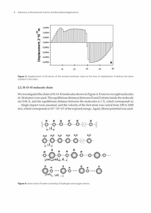
Figure 3. Displacement of 50 atoms of the excited nonlinear chain at the time of stabilization. N defines the atomnumber in the chain.
2.2. H–O–H molecule chain
We investigated the chain of H–O–H molecules shown in Figure 4. From two to eight molecules(6–24 atoms) were used. The equilibrium distances between H and O atoms inside the moleculeare 0.96 Å, and the equilibrium distance between the molecules is 1 Å, which corresponds to… Single impact were assumed, and the velocity of the first atom was varied from 100 to 1600m/s, which corresponds to 10-5–10-2 eV of the exposed energy. Again, Morse potential was used.
Figure 4. Atom chain of water consisting of hydrogen and oxygen atoms.
Advances in Biomaterials Science and Biomedical Applications8
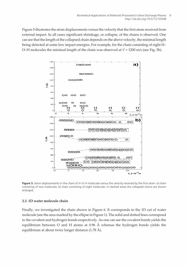
Figure 5 illustrates the atom displacements versus the velocity that the first atom received fromexternal impact. In all cases significant shrinkage, or collapse, of the chains is observed. Onecan see that the length of the collapsed chain depends on the above velocity, the minimal lengthbeing detected at some low impact energies. For example, for the chain consisting of eight H–O–H molecules the minimal length of the chain was observed at V = 1200 m/s (see Fig. 5b).
Figure 5. Atom displacements in the chain of H–O–H molecules versus the velocity received by the first atom: a) chainconsisting of two molecules, b) chain consisting of eight molecules. In dashed areas the collapsed chains are shownenlarged.
2.3. 1D water molecule chain
Finally, we investigated the chain shown in Figure 6. It corresponds to the 1D cut of watermolecule (see the area marked by the ellipse in Figure 1). The solid and dotted lines correspondto the covalent and hydrogen bonds respectively. As one can see the covalent bonds yields theequilibrium between O and H atoms at 0.96 Å whereas the hydrogen bonds yields theequilibrium at about twice longer distance (1.78 Å).
Biomedical Applications of Materials Processed in Glow Discharge Plasmahttp://dx.doi.org/10.5772/55548
9

Figure 6. chain of water consisting of hydrogen and oxygen atoms. The solid and dotted lines mark the covalent andhydrogen bonds respectively.
In simulations we considered simple chain consisting of two 1D water molecules. The initialvelocity of the first atom was taken at V = 500 m/s. The Morse and Born-Mayer potentials wereused for this investigation.
Figure 7 represents the initial and final stabilized conditions of atom chains (after direct low-energy ion impact to the first atom of the chain) calculated with Born-Mayer (Figure 7a) andMorse (Figure 7b) potentials
Figure 7. Initial and final positions of atoms of 1D water molecule after direct low-energy impact to the first atom(oxygen): a) Born-Mayer potential, b) Morse potential. The right figures show the enlarged final chains.
Advances in Biomaterials Science and Biomedical Applications10

3. Biomedical applications
Biological objects are known for their high sensitivity to weak external fields. The evidencethat electromagnetic fields can have “non-thermal” biological effects is now overwhelming.When the production of heat shock proteins is triggered electromagnetically it needs 100million times less energy than when triggered by heat [20]. Low-frequency weak magneticfields may lead to the resonant change of the rate of biochemical reactions although the impactenergy is by ten orders of magnitude less than kBT where kB is the Boltzmann constant and Tis the temperature of the medium [21].
The therapeutic ability of the low intensity electromagnetic radiations is actively discussed[22]. The low-power millimeter wave irradiation and magnetic-resonance therapy are used inpractical medicine already, which differ significantly from the drug treatment by the fact thatthey do not clog organism with the undesirable chemical compounds, i.e. xenobiotics. In thischapter we discuss the biomedical application of vacuum-plasma technologies.
3.1. Activating and therapeutic properties of water processed in GDP
To understand the above extreme sensitivity of living objects, investigations in influences ofweak fields on water appear to be essential. Indeed, water plays a major role in biologicalprocesses. A man consumes about 2 l of drinking water a day. Water is the main componentof human, animal, plant and generally every living being body. A new-born child bodycontains 97% of water, decreasing to 70–75% with aging. In particular, human brain consistsof about 85% of water.
So, we performed experiments with water, crop seeds and baking yeast S. cerevisiae. The cropseeds and yeast were processed directly in GDP. Also, the untreated crop seeds and yeast werepoured with the water processed in GDP. In all cases practically the same biotrophic effectswere observed. Namely, the seed sprouts showed the growth in 3–4 times higher than thecontrol samples. Both the processed yeast and the unprocessed one that immersed in theprocessed (by GDP) water showed greater metabolic activity compared to the control samples.
The obtained results allow suggesting that the discovered phenomena can be used for directcorrection of pathological states. Therefore we processed water and physiological solution.The samples were exposed to low-energy ion irradiation in GDP of residual gases. The ionenergy depends on the voltage in the plasma generator. The latter was kept at 1.2 keV whilethe current in the plasma generator was maintained at 70 mA. The temperature in the chamberwas controlled during the irradiation process and did not exceed 298 K (25° C). The irradiationtime was 60 minutes.
In test experiments, volunteers with different diseases either drunk the processed water orthey were injected intravenously with the similarly processed physiological solution. Thecourse of treatment included 3–5 sessions of 0.5 l physiological solution transfusion. Thepreliminary results appeared to be very promising. We were most interested in the therapeutictreatment of the global inflammatory processes such as cardio-vascular diseases and pancre‐
Biomedical Applications of Materials Processed in Glow Discharge Plasmahttp://dx.doi.org/10.5772/55548
11
atic (insular) diabetes complicated by the acute and chronic forms of atherosclerosis. Also,different types of oncology, say, leukemia, etc., were under investigation.
The blood immune cells were taken for diagnostics. The immune system is known as one ofthe leading homeostatic systems in the organisms. It may serve as a mirror that reflectspractically all adaptations and pathological rearrangements. The immunocompetent cells,lymphocytes and leukocytes, have a set of properties that may be used as an indicator of theorganism state. In addition, the structural organization of blood lymphocytes and leukocytesmakes possible a most efficient use of microspectral analysis and different fluorescent probesfor their studies [23].
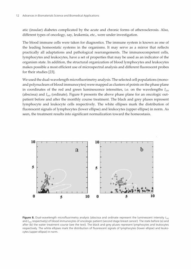
We used the dual-wavelength microfluorimetry analysis. The selected cell populations (mono-and polynuclears of blood immunocytes) were mapped as clusters of points on the phase planein coordinates of the red and green luminescence intensities, i.e. on the wavelengths I530
(abscissa) and I640 (ordinate). Figure 8 presents the above phase plane for an oncologic out-patient before and after the monthly course treatment. The black and grey pluses representlymphocyte and leukocyte cells respectively. The white ellipses mark the distribution offluorescent signals of lymphocytes (lower ellipse) and leukocytes (upper ellipse) in norm. Asseen, the treatment results into significant normalization toward the homeostasis.
Figure 8. Dual-wavelength microfluorimetry analysis (abscissa and ordinate represent the luminescent intensity I530and I640 respectively) of blood immunocytes of oncologic patient (second stage breast cancer). The state before (a) andafter (b) the water treatment course (see the text). The black and grey pluses represent lymphocytes and leukocytesrespectively. The white ellipses mark the distribution of fluorescent signals of lymphocytes (lower ellipse) and leuko‐cytes (upper ellipse) in norm.
Advances in Biomaterials Science and Biomedical Applications12

3.2. Biocompatibility of titanium alloys and stainless steel processed in GDP
Stainless steel, titanium and its alloys are among the most utilized biomaterials and are stillthe materials of choice for many structural implantable device applications [24, 25]. Weprocessed both the titanium and stainless steel samples and investigated changing in theirproperties caused by GDP.
Current titanium implants face long-term failure problems due to poor bonding to juxtaposedbone, severe stress shielding and generation of debris that may lead to bone cell death andperhaps eventual necrotic bone [26–28]. Improving the bioactivity of titanium implants,especially with respect to cells, is a major concern in the near and intermediate future. Surfaceproperties such as wettability, chemical composition and topography govern the biocompat‐ibility of titanium. Conventionally processed titanium currently used in the orthopedic anddental applications exhibits a micro-rough surface and is smooth at the nanoscale. Surfacesmoothness on the nanoscale has been shown to favor fibrous tissue encapsulation [27–29]. Anapproach to design the next-generation of implants has recently focused on creating uniquenanotopography (or roughness) on the implant surface, considering that natural bone consistsof nanostructured materials like collagen and hydroxyapatite. Some researchers have achievednano-roughness in titanium substrates by compacting small (nanometer) constituent particlesand/or fibers [30]. However, nanometer metal particles can be expensive and unsafe tofabricate. For this reason, alternative methods of titanium surface treatment are desirable.
For the investigation of biocompatibility of implanted materials the tests in vitro with thecultures of different cells (fibroblasts, lymphocytes, macrophages, epithelial cells, etc.) are used.The influence of material is typically evaluated according to such indicators as adhesion,change in the morphological properties, inhibition of an increase in the cellular population,oppression of metabolic activity and others.
The adhesion of cells, as is known, plays exceptionally important role in the biological process‐es, such as formation of tissues and organs during embryogenesis, reparative processes, immuneand inflammatory reactions, etc. Capability for movement is the characteristic property offibroblasts, cells of immune system and cells, which participate in the inflammation. More‐over, in immunocytes and leukocytes it consists not only in the free recirculation in the bloodstream or lymph but also in the penetration into vascular walls and active migration into thesurrounding tissues. Adhesion and flattening of cells to the base layer always precede theirlocomotion. The degree of flattening is important preparatory step to the cell amoeboid mobility.We concentrate our attention on the above components in experiments with titanium alloys.
Titanium samples were cut into pieces (1 cm × 0.5 cm) and placed in a specially constructedplasma generator. They were exposed to glow discharge plasma by ions of the residual gases ofthe vacuum. The ion energy depended on the voltage in the plasmatron and did not exceed 1–10 keV. Irradiated fluence was 1017 ion∙cm-2. The temperature of the specimens was controlledduring the irradiation process and did not exceed 343 K while the irradiation time varied from5 to 60 minutes. Rutherford Backscattering Spectrometry (RBS) was used to study the changesafter the irradiation. Cell adhesion to titanium samples was tested with L929 mouse connec‐tive tissue (fibroblasts-like cells). L929 cells were cultured in Dulbecco’s modified Eagle’s
Biomedical Applications of Materials Processed in Glow Discharge Plasmahttp://dx.doi.org/10.5772/55548
13

medium with 10% fetal bovine serum. Initial cell density was 5∙105 cells/ml. The samples wereplaced into the sterile disposable 9 cm diameter tissue culture Petri dishes. 2 ml growth mediumwith cells were distributed into each Petri then incubated in the 5% CO2 at 37º C for 2 hours. Afterthat period, cultures were prepared for scanning electron microscopy (SEM).
RBS data for the irradiated sample show the presence of iron on the surface that occurred fromhigh-carbon steel cathode as a result of secondary emission process (Figure. 9). Percentage ofiron and thickness of the layer were calculated using RUMP, the program for simulation andanalysis based on RBS and Elastic Recoil Detection techniques.
Figure 9. RBS spectrum of the titanium sample irradiated for 5 min at 10 kV.
Advances in Biomaterials Science and Biomedical Applications14

The obtained data for different voltage and time of the irradiation are presented in Table 2.
Voltage, kVTime of
irradiation, min
Fe:Ti atomic
ratio
Density of
flattened cells
per μm2
Percentage of
flattened cells
Increase factor
in amount of all
cells in
comparison with
control sample
0.4 60 0.0277:1 534±20 50.2±2.0 1.78
1.2 30 0.0560:1 413±9 43.7±0.9 1.63
10 5 0.0549:1 381±15 42.5±1.7 1.53
Control 0 0:1 26±8 4.4±1.5 1
Table 2. Data obtained from the experiments with titanium samples exposed to GDP.
Calculated data indicate an increase in the density of flattened cells as well as in the cell amountin comparison with the control sample. According to Table 2 one conclude that best adhesion(column 4) and most prolific cell attachment (column 5) correspond to the samples that wereexposed to GDP for maximum time at minimum voltage. For this sample we observed lesspercentage of iron and thickness of the iron layer in comparison with others that were exposedto higher voltage plasma irradiation.
Figure 10 demonstrates SEM images of control and irradiated samples. In comparison withthe control sample, analysis of cell attachment for the irradiated samples shows high conflu‐ence (attachment ratio) and better spreading.
We also performed experiments on the adhesion of immune-competent cells of human bloodto the stainless steel samples. Figure 11 represents the microphotography of the healthy personlymphocytes and leukocytes adhered to the irradiated and non-irradiated plates. As can beseen from photographs, cells, which are located on the different samples, are essentiallydifferent. The morphology of leukocytes and lymphocytes, which were adhered to theirradiated material, indicates the expressed amoeboid mobility.
In the majority of the cases endoprosthetics is conducted not in the healthiest people. This factis very important and it must be considered. Figure 12 displays the results of similar study ofthe blood nucleus of person who suffers from second stage hypertonia, coronary artery diseaseand atherosclerosis. From the above data one can conclude that the nature of adhesion of cellsto the base layer depends on both the physico-chemical state of this base layer and the state oforganism, the owner of cells.
Biomedical Applications of Materials Processed in Glow Discharge Plasmahttp://dx.doi.org/10.5772/55548
15

(a) (b)
Figure 10. SEM images of cell attachment on (a) the control sample and (b) the titanium sample that was irradiatedfor 5 min at 10 kV.
Advances in Biomaterials Science and Biomedical Applications16

Figure 11. Luminescent microscopy (1000×) of lymphocytes and granulocytes of the blood of healthy donor adheredto (a) non-irradiated and (b) irradiated in GDP surface of the stainless steel samples. The cell nucleus fluorochromiza‐tion is performed by propidium iodide (λfl = 615 nm).
Figure 12. Luminescent microscopy (1000×) of lymphocytes and granulocytes of the blood of donor suffering fromsecond stage hypertonia, coronary artery disease and atherosclerosis. The cell nuclei are adhered to (a) non-irradiatedand (b) irradiated in GDP surface of the stainless steel samples. The fluorochromization is performed by propidiumiodide (λfl = 615 nm).
Biomedical Applications of Materials Processed in Glow Discharge Plasmahttp://dx.doi.org/10.5772/55548
17

4. Discussion and conclusions
Studying the homogeneous chains, like the hydrogen atom chain, exposed to low energies weobserved clusterization. It is important to stress that this is truly self-organization phenomenoninduced by an external excitation. The chains utilize the excitation energy to initialize nonlinearoscillations and redistribute the energy throughout the chain, which leads to the patternformation. In the case of multiple impacts on randomly chosen atoms (so-called plasmaprocessing) the atom displacements are by an order higher than in the case of single impact.Thus, the plasma treatment leads to more active self-organization processes and atomrearrangements.
In the cases of inhomogeneous chains containing H and O atoms another type of structures isdeveloped. The shrinkage of chains is so significant that we can say about the collapsedstructures. This collapse is observed irrespective of the choice of the atom interaction poten‐tials, whereas the collapsed chain patterns are found to depend on the latter.
To conclude, the performed simulations demonstrated that the system nonlinearity is, in fact,the main reason for the development of self-organization processes leading to significantmodifications even in case of low-energy impacts.
In experiments with water and biological objects processed in GDP significant biotrophiceffects were detected. The crop seeds and yeast processed directly or indirectly (beingimmersed in the water processed in GDP) showed markedly greater metabolic activitycompared to the control samples. Using the water and the physiological solutions processedin GDP we observed significant therapeutic effects in the test treatments of cardiovascular,oncologic and other diseases. The obtained results suggest the use of discovered phenomenafor direct corrections of pathological states by shifting a body state towards its homeostasis.Understanding the mechanisms of the latter will be our next priority.
Next part of this study is devoted to experiments with the titanium alloys and stainlesssteel exposed to GDP. The experiments with titanium samples reveal an increase in thedensity of flattened (to the sample surface) cells as well as in the cell amount in compari‐son with the control sample. These are nothing but preparatory step to the cell amoe‐boid mobility. Indeed, adhesion and flattening of cells to the base layer always precedetheir locomotion. According to the results, best adhesion and most prolific cell attach‐ment correspond to the samples that were exposed to GDP for maximum time at mini‐mum voltage. Similar results were obtained in the experiments with stainless steel samples:the morphology of leukocytes and lymphocytes, which were adhered to the irradiatedmaterial, indicated the expressed amoeboid mobility. The results with the blood nucleusof person who suffers from several diseases revealed some deviations in the morphologyof adhered cells compared to the healthy blood. Thus, the nature of adhesion of cells tothe base layer depends on both the physico-chemical state of this base layer and the stateof organism, the owner of cells. This circumstance determines even more stringentrequirements for the material of the implants.
Advances in Biomaterials Science and Biomedical Applications18

Author details
V. Tereshko1*, A. Gorchakov2, I. Tereshko3, V. Abidzina3 and V. Red’ko4
*Address all correspondence to: [email protected]
1 School of Computing, University of the West of Scotland, Paisley, UK
2 MISTEM, Mogilev, Belarus
3 Department of Physics, Belarusian-Russian University, Mogilev, Belarus
4 Department of Physical Methods of Control, Belarusian-Russian University, Mogilev, Belarus
References
[1] Ziegler, J. F, Biersack, J. P, & Littmark, U. The Stopping and Range of Ions in Solids.New York: Pergamon; (1985).
[2] Eckstein, W. Computer Simulation of Ion-Solids Interaction. Berlin: Springer; (1991).
[3] Tereshko, I. V, Khodyrev, V. I, Tereshko, V. M, Lipsky, E. A, Goncharenya, A. V, &Ofori-sey, S. Self-organizing processes in metals by low-energy ion beams. Nucl.Instr. and Meth. B (1993). , 80, 115-119.
[4] Tereshko, I. V, Khodyrev, V. I, Lipsky, E. A, Goncharenya, A. V, & Tereshko, A. M.Materials modification by low-energy ion irradiation. Nucl. Instr. and Meth. B(1997). , 128, 861-864.
[5] Tereshko, I. V, Glushchenko, V. V, & Tereshko, A. M. Computer simulation of the de‐fect structure formation in crystal lattices by low-energy ion irradiation. Comput.Mater. Sci. (2002). , 24, 139-143.
[6] Tereshko, I, Abidzina, V, Tereshko, A, & Elkin, I. Nanostructural evolution of steeland titanium alloys exposed to glow discharge plasma. Nucl. Instr. and Meth. B(2007). , 261, 678-681.
[7] Tereshko, I. V, Abidzina, V. V, Elkin, I. E, Tereshko, A. M, Glushchenko, V. V, &Stoye, S. Formation of nanostructures in metals by low-energy ion irradiation. Surf.& Coat. Tech. (2007). , 201, 8552-8556.
[8] Stillinger, F. N. Water revisited. Science (1980). , 209(4455), 451-457.
[9] Liu, K, Cruzan, J. D, & Saykally, R. J. Water clusters. Science (1996). , 271(5251),929-933.
Biomedical Applications of Materials Processed in Glow Discharge Plasmahttp://dx.doi.org/10.5772/55548
19

[10] Keutsch, F. N, & Saykally, R. J. Water clusters: untangling the mysteries of the liquid,one molecule at a time. PNAS (2001). , 98(19), 10533-10540.
[11] Galamba, N. Cabral BJC. The changing hydrogen-bond network of water from thebulk to the surface of a cluster: a Born-Oppenheimer molecular dynamics study. J.Am. Chem. Soc. (2008). , 130, 17955-17960.
[12] Luck, W. A. The importance of cooperativity for the properties of liquid water. J.Mol. Struct. (1998).
[13] Shelton, D. P. Collective molecular rotation in water and other simple liquids. Chem.Phys. Lett. (2000).
[14] Lobyshev, V. I, Shikhlinskaya, R. E, & Ryzhikov, B. D. Experimental evidence for in‐trinsic luminescence of water. J. Mol. Liquids (1999).
[15] Park, J. B, & Lakes, R. S. Biomaterials: An Introduction. New York: Plenum; (1992).
[16] Ratner, B. D, Hoffman, A. S, Schoen, F. J, & Lemons, J. E. editors. Biomaterials Sci‐ence: Introduction to Materials in Medicine. New York: Academic; (1996).
[17] Abidzina, V, Deliloglu-gurhan, I, Ozdal-kurt, F, Sen, B. H, Tereshko, I, Elkin, I, Bu‐dak, S, Muntele, C, & Ila, D. Cell adhesion study of the titanium alloys exposed toglow discharge. Nucl. Instr. and Meth. B. (2007). , 261, 624-626.
[18] Mandl, S, & Rauschenbach, B. Improving the biocompatibility of medical implantswith plasma immersion ion implantation. Surf. Coat. Technol. (2002).
[19] Lopez-heredia, M. A, Legeay, G, Gaillard, C, & Layrolle, P. Radio frequency plasmatreatments on titanium for enhancement of bioactivity. Acta biomater. (2008). , 4,1953-1962.
[20] Blank, M, & Goodman, R. Stimulation of stress response by low frequency electro‐magnetic fields: possibility of direct interaction with DNA. IEEE Trans. Plasma Sci.(2000). , 28, 168-172.
[21] Binhi, V. N, & Savin, A. V. Effects of weak magnetic fields on biological systems:physical aspects. Physics- Uspekhi (2003). , 46(3), 259-291.
[22] Betskii, O. V, Devyatkov, N. D, & Kislov, V. V. Low intensity millimeter waves inmedicine and biology. Crit. Rev. Biomed. Eng. (2000).
[23] Gorchakov, A. M, & Karnaukhov, V. N. Melenets YuV, and Gorchakova FT. Identifi‐cation of pathological conditions by luminescence analysis of immunocompetentblood cells. Biophysics (1999). , 44(3), 550-555.
[24] Hermawan, H, & Ramdan, D. Djuansjah JRP. Metals for biomedical applications. In:Fazel R, editor. Biomedical Engineering- From Theory to Applications. Rijeka: In‐Tech; (2011). , 411-430.
Advances in Biomaterials Science and Biomedical Applications20

[25] Williams, D. F. Titanium for medical applications. In: Brunette DM, Tengvall P, Tex‐tor M, Thomsen P, editors. Titanium in Medicine. Berlin: Springer; (2001). , 13-24.
[26] Buser, D, Nydegger, T, Oxland, T, Cochran, D. L, Schenk, R. K, Hirt, H. P, Snétivy, D,& Nolte, L. P. Interface shear strength of titanium implants with a sandblasted andacid-etched surface: a biomechanical study in the maxilla of miniature pigs. J. Bi‐omed. Mater. Res. (1999). , 45(2), 75-83.
[27] Kaplan, F. S, Hayes, W. C, Keaveny, T. M, Boskey, A, & Einhorn, T. A. Biomaterials.In: Simon SP, editor. Orthopedic Basic Science. Columbus: American Academy of Or‐thopedic Surgeons; (1994). , 460-478.
[28] Kaplan, F. S, Hayes, W. C, Keaveny, T. M, Boskey, A, Einhorn, T. A, & Iannotti, J. P.Form and function of bone. In: Simon SP, editor. Orthopedic Basic Science. Colum‐bus: American Academy of Orthopedic Surgeons; (1994). , 127-185.
[29] Boyan, B. D, Dean, D. D, Lohmann, C. H, Cochran, D. L, Sylvia, V. L, & Schwartz, Z.The titanium-bone cell interface in vitro: the role of the surface in promoting osteoin‐tegration. In: Brunette DM, Tengvall P, Textor M, Thomsen P, editors. Titanium inMedicine. Berlin: Springer; (2001). , 561-586.
[30] Webster, T. J, & Ejiofor, J. U. Increased osteoblast adhesion on nanophase metals: Ti,Ti6Al4V, and CoCrMo. Biomaterials (2004). , 25, 4731-4739.
Biomedical Applications of Materials Processed in Glow Discharge Plasmahttp://dx.doi.org/10.5772/55548
21
