biomechanicsoflateralinterbodyspacers:goingwiderfor ... each specimen using standard lumbar dual...
TRANSCRIPT

The Scientific World JournalVolume 2012, Article ID 381814, 6 pagesdoi:10.1100/2012/381814
The cientificWorldJOURNAL
Research Article
Biomechanics of Lateral Interbody Spacers: Going Wider forGoing Stiffer
Luiz Pimenta,1, 2 Alexander W. L. Turner,3 Zachary A. Dooley,3
Rachit D. Parikh,3 and Mark D. Peterson4
1 Instituto de Patologia da Coluna, 04101-000 Sao Paulo, SP, Brazil2 Department of Neurosurgery, University of San Diego, San Diego, CA 92103, USA3 NuVasive, Inc., San Diego, CA 92121, USA4 Southern Oregon Orthopedics, Medford, OR 97504, USA
Correspondence should be addressed to Luiz Pimenta, [email protected]
Received 3 August 2012; Accepted 23 September 2012
Academic Editors: G. J. Hooper, A. V. Korompilias, and C. C. Niu
Copyright © 2012 Luiz Pimenta et al. This is an open access article distributed under the Creative Commons Attribution License,which permits unrestricted use, distribution, and reproduction in any medium, provided the original work is properly cited.
This study investigates the biomechanical stability of a large interbody spacer inserted by a lateral approach and compares thebiomechanical differences with the more conventional transforaminal interbody fusion (TLIF), with and without supplementalpedicle screw (PS) fixation. Twenty-four L2-L3 functional spinal units (FSUs) were tested with three interbody cage options: (i)18 mm XLIF cage, (ii) 26 mm XLIF cage, and (iii) 11 mm TLIF cage. Each spacer was tested without supplemental fixation, and withunilateral and bilateral PS fixation. Specimens were subjected to multidirectional nondestructive flexibility tests to 7.5 N·m. Therange of motion (ROM) differences were first examined within the same group (per cage) using repeated-measures ANOVA, andthen compared between cage groups. The 26 mm XLIF cage provided greater stability than the 18 mm XLIF cage with unilateral PSand 11 mm TLIF cage with bilateral PS. The 18 mm XLIF cage with unilateral PS provided greater stability than the 11 mm TLIFcage with bilateral PS. This study suggests that wider lateral spacers are biomechanically stable and offer the option to be used withless or even no supplemental fixation for interbody lumbar fusion.
1. Introduction
The lateral transpsoas approach for lumbar interbody spinalfusion has gained popularity in recent years for a varietyof indications [1–8]. The approach provides wide access tothe lateral aspect of the disc allowing extensive discectomy,preservation of the anterior and posterior longitudinalligaments, annulus and posterior elements, and placement ofa large interbody spacer [9].
The biomechanical stability of a lumbar fusion constructis determined by the extent of resection of local bone and lig-ament, implant size and positioning, and the type of supple-mental internal fixation used. Previous biomechanical assess-ment has demonstrated the stability of an 18 mm anterior-posterior (A-P) width extreme lateral interbody fusion(XLIF) interbody cage [10]. XLIF cages with larger anterior-posterior widths (22 mm and 26 mm) have been developedin order to reduce the risk of subsidence in osteoporotic
patients by distributing load over a greater area of theendplate. These larger cages potentially provide additionalstability over standard 18 mm spacers by blocking motion.
The objective of this cadaveric study was to comparethe stability of different A-P width XLIF cages with andwithout supplemental pedicle screw (PS) fixation. A moreconventionally used transforaminal interbody fusion (TLIF)group was included for reference purposes.
2. Material and Methods
Twenty-four L2-L3 functional spinal units (FSUs) were dis-sected from fresh-frozen human spines (average age: 50.1,range 21–76 years; 22 male, 2 female). A-P and lateral radio-graphs were used to exclude deformity and degeneration.Bone mineral density (BMD) was assessed prior to dissectionof each specimen using standard lumbar dual energy X-ray absorptiometry (DEXA) scans (Discovery C, Hologic

2 The Scientific World Journal
(a) (b) (c)
Figure 1: Axial view of cages used in testing (CoRoent, NuVasive, Inc, San Diego, CA): (a) 26 mm XLIF cage, (b) 18 mm XLIF Cage, and (c)11 mm TLIF cage.
(a) (b)
(c)
Figure 2: Lateral fluoroscopy images during testing of (a) 18 mm XLIF cage, (b) 26 mm XLIF cage, and (c) 11 mm TLIF cage, implanted atL2-L3 intervertebral space.
Inc., Bedford, MA). The FSUs were divided into 3 BMD-matched subgroups of 8 FSUs, each with an average BMD of0.89 g/cm2. The caudal and cephalad ends of each specimenwere mounted in polyurethane casting resin (Smooth-Cast300; Smooth-On, Inc., Easton, PA), with the disc space posi-tioned horizontally. Each group was tested with a differentinterbody spacer (Figures 1 and 2): (i) 18 mm A-P widthXLIF cage (CoRoent XL, NuVasive, Inc, San Diego, CA), (ii)26 mm A-P width XLIF cage (CoRoent XL-XW; NuVasive,Inc.), or (iii) 11 mm A-P width TLIF cage (CoRoent LC;NuVasive, Inc.). Discectomy, endplate preparation, and cageinsertion were performed following usual XLIF [2] and TLIF[3] techniques.
Each FSU was subjected to multidirectional nonde-structive flexibility testing using a custom 6 degree-of-freedom spine test system controlled by LabVIEW (National
Instruments, Austin, TX). Specimens were subjected to astandard protocol consisting of 3 fully reversed cycles offlexion extension, lateral bending, and axial rotation to7.5 N·m without an axial load or follower load [11, 12],under the following conditions: (i) intact, (ii) intact disc withbilateral pedicle screw (PS) fixation, (iii) interbody cagealone, (iv) cage + unilateral PS fixation, and (v) cage +bilateral PS fixation.
Infrared light-emitting diode marker arrays were fixed tothe L2 and L3 vertebral bodies. Intervertebral (L2-L3) rangeof motion (ROM) was measured using an Optotrak Certussystem (Northern Digital Inc., Waterloo, ON, Canada).Data from the third motion cycle was analyzed. ROM wasnormalized to the intact condition (percent intact ROM).ROM differences were first examined within groups (percage) using repeated-measures ANOVA and Holm-Sidak
The Scientific World Journal 3
Bila
t P
S
Cag
e al
one
Cag
e+
un
ilat
PS
Cag
e+
bila
t P
S
Flexion extension125
100
75
50
25
0RO
M (
% o
f in
tact
)
(a)
Lateral bending
Bila
t P
S
Cag
e al
one
Cag
e+
un
ilat
PS
Cag
e+
bila
t P
S
150
125
100
75
50
25
0
RO
M (
% o
f in
tact
)
(b)
Axial rotationB
ilat
PS
Cag
e al
one
Cag
e+
un
ilat
PS
Cag
e+
bila
t P
S
250
200
150
100
50
0
TLIF18 mm XLIF26 mm XLIF
RO
M (
% o
f in
tact
)
(c)
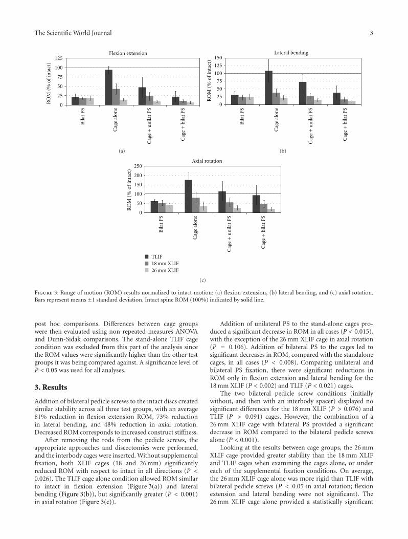
Figure 3: Range of motion (ROM) results normalized to intact motion: (a) flexion extension, (b) lateral bending, and (c) axial rotation.Bars represent means ±1 standard deviation. Intact spine ROM (100%) indicated by solid line.
post hoc comparisons. Differences between cage groupswere then evaluated using non-repeated-measures ANOVAand Dunn-Sidak comparisons. The stand-alone TLIF cagecondition was excluded from this part of the analysis sincethe ROM values were significantly higher than the other testgroups it was being compared against. A significance level ofP < 0.05 was used for all analyses.
3. Results
Addition of bilateral pedicle screws to the intact discs createdsimilar stability across all three test groups, with an average81% reduction in flexion extension ROM, 73% reductionin lateral bending, and 48% reduction in axial rotation.Decreased ROM corresponds to increased construct stiffness.
After removing the rods from the pedicle screws, theappropriate approaches and discectomies were performed,and the interbody cages were inserted. Without supplementalfixation, both XLIF cages (18 and 26 mm) significantlyreduced ROM with respect to intact in all directions (P <0.026). The TLIF cage alone condition allowed ROM similarto intact in flexion extension (Figure 3(a)) and lateralbending (Figure 3(b)), but significantly greater (P < 0.001)in axial rotation (Figure 3(c)).
Addition of unilateral PS to the stand-alone cages pro-duced a significant decrease in ROM in all cases (P < 0.015),with the exception of the 26 mm XLIF cage in axial rotation(P = 0.106). Addition of bilateral PS to the cages led tosignificant decreases in ROM, compared with the standalonecages, in all cases (P < 0.008). Comparing unilateral andbilateral PS fixation, there were significant reductions inROM only in flexion extension and lateral bending for the18 mm XLIF (P < 0.002) and TLIF (P < 0.021) cages.
The two bilateral pedicle screw conditions (initiallywithout, and then with an interbody spacer) displayed nosignificant differences for the 18 mm XLIF (P > 0.076) andTLIF (P > 0.091) cages. However, the combination of a26 mm XLIF cage with bilateral PS provided a significantdecrease in ROM compared to the bilateral pedicle screwsalone (P < 0.001).
Looking at the results between cage groups, the 26 mmXLIF cage provided greater stability than the 18 mm XLIFand TLIF cages when examining the cages alone, or undereach of the supplemental fixation conditions. On average,the 26 mm XLIF cage alone was more rigid than TLIF withbilateral pedicle screws (P < 0.05 in axial rotation; flexionextension and lateral bending were not significant). The26 mm XLIF cage alone provided a statistically significant

4 The Scientific World Journal
reduction in ROM compared with TLIF with unilateral screwin all directions (P < 0.05). The 26 mm XLIF spacer withunilateral PS was significantly more rigid (P < 0.05) thanTLIF with unilateral screws in all directions and bilateralPS in lateral bending and axial rotation. The 26 mm XLIFcage with bilateral pedicle fixation was also significantly morerigid (P < 0.05) than TLIF with unilateral screws in alldirections and TLIF with bilateral PS in lateral bending andaxial rotation.
The 26 mm XLIF cage in the stand-alone condition wassignificantly more rigid in flexion extension (P < 0.05) thanthe 18 mm XLIF cage. On average, the 26 mm XLIF cagealone was more rigid than the 18 mm XLIF spacer withunilateral pedicle screws, in all directions tested, althoughstatistically significant differences were not detected. The26 mm XLIF cage with both unilateral and bilateral PS wasmore rigid than the 18 mm XLIF cage alone (P < 0.05).
Finally, the 18 mm XLIF cage in the stand-alone con-dition provided greater stability than TLIF with unilateralPS in all directions tested (P < 0.05 in lateral bending;flexion extension and axial rotation were not significant).The 18 mm XLIF spacer with both unilateral and bilateralpedicle screws provided significant reductions in ROM overTLIF with the unilateral PS in all directions (P < 0.05). XLIFwith the 18 mm cage and bilateral PS was more rigid thanTLIF with the same fixation (P < 0.05) in axial rotation. TLIFwith bilateral pedicle screw was more rigid than the 18 mmXLIF cage alone in flexion extension (P < 0.05).
4. Discussion
This biomechanical study analyzed the stability of differentlateral constructions for lumbar interbody fusion. TLIFconstructs were tested to establish a baseline for a commonlyused technique. Both XLIF interbody spacers, with andwithout pedicle screw fixation, provided improved stabilityover the TLIF constructs. Additionally, the 26 mm XLIFcage also reduced ROM to a greater extent than the 18 mmXLIF cage. These biomechanical results suggest that thestability provided by the XLIF spacers, with adequate cageheight sizing and good bone quality, may allow for lesssupplemental fixation than a more destabilizing approachsuch as TLIF thus avoiding posterior muscle dissection andadjacent facet joint injury.
The reduced stability observed with the TLIF approachcompared to XLIF in the current study is likely due toresection of stabilizing structures such as the facet joint,ligamentum flavum, and posterior longitudinal ligament inorder to insert the interbody spacer. These structures areall retained during XLIF. Additionally, XLIF allows tallerinterbody implants to be placed across the disc space sinceTLIF cage sizing is typically limited by the approach, whichis constrained by the proximity of the nerve roots andthe often smaller intervertebral space posteriorly. The XLIFcages distract the disc space and generate tension in theretained ligaments, which contributes to stability. Potentially,improved stability over the results obtained here using a TLIFapproach may be possible using different cage designs orinsertion techniques.
Previously, two groups studied the biomechanical sta-bility of XLIF constructs [10, 13]. Despite some differencesin testing methodology (e.g., the tested lumbar level), theROM results with the 18 mm XLIF cage obtained in thepresent study were similar to those previously observed. Besset al. [13] investigated 18 mm XLIF cages as a stand-aloneconstruct and with various instrumented constructs (lateralplate, unilateral or bilateral PS). They observed that the XLIFimplant, with or without supplemental fixation, providedsignificantly decreased ROM in all loading modes comparedwith intact. Cappuccino et al.[10] and the current studyconfirmed these findings.
Laws et al. [14] compared direct lateral interbody fusion(DLIF), similar to XLIF, with anterior lumbar interbodyfusion (ALIF). Cage width was not provided; however, stand-alone DLIF was shown to demonstrate greater stability thanstand-alone ALIF in all directions tested. In a historicalliterature comparison [10], Cappuccino et al. also noted sub-stantially less motion with XLIF over ALIF [15] with thegreatest differences in flexion extension and lateral bending.Minimal differences were demonstrated between the groupsif supplemental fixation was added to ALIF, TLIF, or an18 mm XLIF cage. In the present study, we demonstratedthat 26 mm XLIF interbody spacers can potentially provide1.5 (flexion extension) to 2.7 (axial rotation) times as muchstability as a TLIF construct with bilateral pedicle screws.
Stand-alone fusion constructs have historically been seento be biomechanically insufficient to provide stabilization inall directions [16], whether the technique is ALIF [15, 17],TLIF [17], or PLIF [18], or even in lateral approach (usinga cylindrical threaded cage) [19]. As previously discussed,TLIF involves removal of posterior anatomic structures,while ALIF requires removal of the anterior longitudinalligament (ALL). The importance of ALL retention in inter-body fusion construct stability was seen after its resectionfollowing a laterally inserted ALIF cage, which led toincreased ROM by 59% and 142% in axial rotation andflexion extension, respectively [17].
Unlike ALIF and TLIF, stand-alone lateral interbodyfusion has been performed in an off-label fashion withsuccess for cases without instability [3–5, 20–22]. Despitethe greater stability over other approaches, and the abilityto insert a long cage that spans the strongest lateral boneof the ring apophysis [23], subsidence of stand-alone 18 mmXLIF cages has been observed, which can impair disc spacedistraction and indirect decompression [5]. With the wider22 mm and 26 mm XLIF spacers, greater endplate areais covered which decreases the pressure on the vertebralendplate and should increase the load required to causesubsidence. The result of this was seen as a lower rate ofsubsidence comparing 18 and 22 mm XLIF cages in someclinical studies [24, 25].
Cage shape also appears to play an important biome-chanical role in stability of the fusion construct. In the studyby Le Huec et al. [19], a stand-alone construct with a cylin-drical laterally placed interbody cage was not able to provideas much stability as the intact spine. Only after the additionof a lateral plate was the stability sufficiently improved, withstiffness increased by 3.1 times relative to the intact spine.

The Scientific World Journal 5
Cylindrical cages likely provide limited stability since thereis limited implant-endplate contact area to resist motion. Incontrast, the rectangular XLIF spacers provide much greaterimplant-endplate contact area, which blocks motion andhence gives greater stability. This was further demonstratedby the increased stability provided by the 26 mm XLIF spacercompared with the 18 mm.
Although our cadaveric study design provided well-controlled biomechanical results, there are some inherentlimitations associated with the study design. For the currentstudy, L2-L3 lumbar levels were used. This may bias stabilityresults towards the larger XLIF cages compared with testingat more caudal vertebral levels, since the vertebrae are smallerat L2-L3 and the same size interbody implants will occupy agreater proportion of the endplate area. Each interbody cagetype was studied independently in three different groups,which will introduce additional specimen variability. Effectsof this were minimized by creating groups with similarBMD and selecting specimens with good bone quality andminimal degeneration or deformity. The pure momentloading applied to the specimens in order to measure ROM istypical of physiologic levels; however, it does not investigatethe stabilizing effect of surrounding musculature seen invivo, which may alter the results. In addition, the currentstudy demonstrates immediate postoperative stability of theconstruct and does not take into consideration the long-termimpact of cage settling, bone ingrowth, or cyclic loading.
The transpsoas lateral approach is an evolving techniquein minimally invasive spine surgery. Studies presenting initialresults with 22 mm XLIF cages have been reported [3,24, 25]. Development of new implants for specific patientgroups and/or indications can be very useful and shouldfirst be evaluated experimentally to ensure intended benefits,such as biomechanical stabilization, are realized. Thesefindings should be confirmed in clinical studies. Clinicalconsiderations for using larger 26 mm XLIF cages over the18 mm devices include larger psoas exposure and need forappropriate neuro-monitoring [26].
With the results found in this work and in the literature,it can be suggested that the stability of a lumbar interbodyfusion construct can be modified to a lesser or greaterextent by: (1) removal of bone/ligament structures, (2)cage positioning, (3) cage design/size, and (4) supplementalfixation options. In the lateral XLIF approach, maintenanceof the ALL and a stand-alone wide cage is sufficient tosignificantly reduce intervertebral motion. In some cases, thismay be sufficient to allow bone growth and fusion to takeplace; however, other factors such as existing instability, bonequality, and patient activity level should first be evaluatedwhen considering fixation options.
Disclosure
CoRoent PEEK intervertebral fusion cages are not FDA-cleared for use without supplemental fixation.
Conflict of Interests
L. Pimenta and M. D. Peterson have a stock and stock optionownership and are both Consultants at NuVasive, Inc. A. W.
L. Turner, Z. A. Dooley, and R. D. Parikh are all salariedemployees at NuVasive, Inc, and also possess a stock andstock option ownership at the same company.
Acknowledgment
Study materials were provided by NuVasive, Inc.
References
[1] E. H. Elowitz, D. S. Yanni, M. Chwajol, R. M. Starke, and N.I. Perin, “Evaluation of indirect decompression of the lumbarspinal canal following minimally invasive lateral transpsoasinterbody fusion: radiographic and outcome analysis,” Min-imally Invasive Neurosurgery, vol. 54, no. 5-6, pp. 201–206,2011.
[2] R. E. Isaacs, J. Hyde, J. A. Goodrich, W. B. Rodgers, and F.M. Phillips, “A prospective, nonrandomized, multicenter eval-uation of extreme lateral interbody fusion for the treatmentof adult degenerative scoliosis: perioperative outcomes andcomplications,” Spine, vol. 35, supplement 26, pp. S322–S330,2010.
[3] L. Marchi, N. Abdala, L. Oliveira, R. Amaral, E. Coutinho, andL. Pimenta, “Stand-alone lateral interbody fusion for the treat-ment of low-grade degenerative spondylolisthesis,” The Scien-tific World Journal, vol. 2012, Article ID 456346, 2012.
[4] L. Marchi, L. Oliveira, R. Amaral et al., “Lateral interbodyfusion for treatment of discogenic low back pain: minimallyinvasive surgical techniques,” Advanced Orthopaedics, vol.2012, Article ID 282068, 7 pages, 2012.
[5] L. Oliveira, L. Marchi, E. Coutinho, and L. Pimenta, “A radio-graphic assessment of the ability of the extreme lateral inter-body fusion procedure to indirectly decompress the neuralelements,” Spine, vol. 35, supplement 26, pp. S331–S337, 2010.
[6] W. B. Rodgers, E. J. Gerber, and J. A. Rodgers, “Lumbar fusionin octogenarians: the promise of minimally invasive surgery,”Spine, vol. 35, supplement 26, pp. S355–S360, 2010.
[7] A. K. Sharma, C. K. Kepler, F. P. Girardi, F. P. Cammisa, R. C.Huang, and A. A. Sama, “Lateral lumbar interbody fusion:clinical and radiographic outcomes at 1 year: a preliminaryreport,” Journal of Spinal Disorders and Techniques, vol. 24, no.4, pp. 242–250, 2011.
[8] J. A. Youssef, P. C. McAfee, C. A. Patty et al., “Minimally inva-sive surgery: lateral approach interbody fusion: results andreview,” Spine, vol. 35, supplement 26, pp. S302–S311, 2010.
[9] B. M. Ozgur, H. E. Aryan, L. Pimenta, and W. R. Taylor,“Extreme Lateral Interbody Fusion (XLIF): a novel surgicaltechnique for anterior lumbar interbody fusion,” Spine Jour-nal, vol. 6, no. 4, pp. 435–443, 2006.
[10] A. Cappuccino, G. B. Cornwall, A. W. L. Turner et al., “Biome-chanical analysis and review of lateral lumbar fusion con-structs,” Spine, vol. 35, supplement 26, pp. S361–S367, 2010.
[11] M. M. Panjabi, “Biomechanical evaluation of spinal fixationdevices: I. A conceptual framework,” Spine, vol. 13, no. 10, pp.1129–1134, 1988.
[12] H. J. Wilke, K. Wenger, and L. Claes, “Testing criteria for spinalimplants: recommendations for the standardization of in vitrostability testing of spinal implants,” European Spine Journal,vol. 7, no. 2, pp. 148–154, 1998.
[13] R. S. Bess, G. B. Cornwall, R. E. Vance K. N. Bachus, and D.S. Brodke, “Biomechanics of lateral arthrodesis,” in ExtremeLateral Interbody Fusion (XLIF), J. A. Goodrich and I. J. Volcan,

6 The Scientific World Journal
Eds., pp. 31–40, Quality Medical Publishing, St. Louis, Mo,USA, 2008.
[14] C. J. Laws, D. G. Coughlin, J. C. Lotz, H. A. Serhan, and S.S. Hu, “Direct lateral approach to lumbar fusion is a biome-chanically equivalent alternative to the anterior approach: anin vitro study,” Spine, vol. 37, no. 10, pp. 819–825, 2012.
[15] B. P. Beaubien, A. Derincek, W. D. Lew, and K. B. Wood,“In vitro, biomechanical comparison of an anterior lumbarinterbody fusion with an anteriorly placed, low-profile lumbarplate and posteriorly placed pedicle screws or translaminarscrews,” Spine, vol. 30, no. 16, pp. 1846–1851, 2005.
[16] P. C. Mcafee, “Interbody fusion cages in reconstructive oper-ations on the spine,” Journal of Bone and Joint Surgery A, vol.81, no. 6, pp. 859–880, 1999.
[17] A. Ploumis, C. Wu, G. Fischer et al., “Biomechanical compari-son of anterior lumbar interbody fusion and transforaminallumbar interbody fusion,” Journal of Spinal Disorders andTechniques, vol. 21, no. 2, pp. 120–125, 2008.
[18] T. Lund, T. R. Oxland, B. Jost et al., “Interbody cage stabilisa-tion in the lumbar spine,” Journal of Bone and Joint Surgery B,vol. 80, no. 2, pp. 351–359, 1998.
[19] J. Le Huec, M. Liu, W. Skalli, and L. Josse, “Lumbar lateralinterbody cage with plate augmentation: in vitro biomechan-ical analysis,” European Spine Journal, vol. 11, no. 2, pp. 130–136, 2002.
[20] L. Oliveira, L. Marchi, E. Coutinho, N. Abdala, and L.Pimenta, “The use of rh-BMP2 in standalone eXtreme LateralInterbody Fusion (XLIF): clinical and radiological results after24 months follow-up,” World Spinal Column Journal, vol. 1,no. 1, pp. 19–25, 2010.
[21] W. B. Rodgers, C. S. Cox, and E. J. Gerber, “Experience andearly results with a minimally invasive technique for anteriorcolumn support through eXtreme Lateral Interbody Fusion(XLIF),” US Musculoskeletal Review, vol. 2, no. 1, pp. 28–32,2007.
[22] R. Amaral, L. Marchi, L. Oliveira et al., “Opcao minimamenteinvasiva lateral para artrodese intersomatica toraco-lombar,”Coluna/Columna, vol. 10, no. 3, pp. 239–243, 2011.
[23] J. P. Grant, T. R. Oxland, and M. F. Dvorak, “Mapping thestructural properties of the lumbosacral vertebral endplates,”Spine, vol. 26, no. 8, pp. 889–896, 2001.
[24] T. V. Le, A. A. Baaj, E. Dakwar et al., “Subsidence of polyether-etherketone intervertebral cages in minimally invasive lateralretroperitoneal transpsoas lumbar interbody fusion,” Spine,vol. 37, no. 14, pp. 1268–1273, 2012.
[25] L. Pimenta, L. Marchi, E. Coutinho, and L. Oliveira, “A com-parative study on cage subsidence following standalone lateralinterbody fusion,” in Proceedings of the 4th Annual SOLASResearch Meeting, San Diego, Calif, USA, 2011.
[26] A. G. Tohmeh, W. B. Rodgers, and M. D. Peterson, “Dynam-ically evoked, discrete-threshold electromyography in theextreme lateral interbody fusion approach: clinical article,”Journal of Neurosurgery, vol. 14, no. 1, pp. 31–37, 2011.

Submit your manuscripts athttp://www.hindawi.com
Stem CellsInternational
Hindawi Publishing Corporationhttp://www.hindawi.com Volume 2014
Hindawi Publishing Corporationhttp://www.hindawi.com Volume 2014
MEDIATORSINFLAMMATION
of
Hindawi Publishing Corporationhttp://www.hindawi.com Volume 2014
Behavioural Neurology
EndocrinologyInternational Journal of
Hindawi Publishing Corporationhttp://www.hindawi.com Volume 2014
Hindawi Publishing Corporationhttp://www.hindawi.com Volume 2014
Disease Markers
Hindawi Publishing Corporationhttp://www.hindawi.com Volume 2014
BioMed Research International
OncologyJournal of
Hindawi Publishing Corporationhttp://www.hindawi.com Volume 2014
Hindawi Publishing Corporationhttp://www.hindawi.com Volume 2014
Oxidative Medicine and Cellular Longevity
Hindawi Publishing Corporationhttp://www.hindawi.com Volume 2014
PPAR Research
The Scientific World JournalHindawi Publishing Corporation http://www.hindawi.com Volume 2014
Immunology ResearchHindawi Publishing Corporationhttp://www.hindawi.com Volume 2014
Journal of
ObesityJournal of
Hindawi Publishing Corporationhttp://www.hindawi.com Volume 2014
Hindawi Publishing Corporationhttp://www.hindawi.com Volume 2014
Computational and Mathematical Methods in Medicine
OphthalmologyJournal of
Hindawi Publishing Corporationhttp://www.hindawi.com Volume 2014
Diabetes ResearchJournal of
Hindawi Publishing Corporationhttp://www.hindawi.com Volume 2014
Hindawi Publishing Corporationhttp://www.hindawi.com Volume 2014
Research and TreatmentAIDS
Hindawi Publishing Corporationhttp://www.hindawi.com Volume 2014
Gastroenterology Research and Practice
Hindawi Publishing Corporationhttp://www.hindawi.com Volume 2014
Parkinson’s Disease
Evidence-Based Complementary and Alternative Medicine
Volume 2014Hindawi Publishing Corporationhttp://www.hindawi.com